INDEX
Introduction : ………... 4
1. Dopaminergic System ………... 5
2. Dopaminergic Pathways ………... 8
3. Seven-Transmembrane domain receptors ……….. 11
4. Dopamine Receptors ………. 13
4.1. D1-like receptor subfamily ………... 15
4.1.1 Dopamine D1 receptor ……… 15
4.1.2 Dopamine D5 receptor ………..… 17
4.2. D2-like receptors subfamily ……… 17
4.1.2 Dopamine D2 receptor ……… 17
4.2.2 Dopamine D3 receptor ………..…… 20
4.1.3 Dopamine D4 receptor ……… 21
5. Dopamine Transporter ……...……… 23
6. Distribution of dopamine receptors in SNC ………... 25
7. Basal Ganglia ……...………..…… 29
8. D2R functions in the CNS ………..…... 31
9. D2R signal transduction pathways ………..……... 36
10. D2R isoforms and G protein-coupling ………..……... 44
11. Gene targeting: D2R-deficient mice ………...…... 48
Results and Discussion:……… 55
Chapter 1 ………... 64
Manuscript N°1: Impaired light masking in dopamine D2 receptor–null mice ... 65
Chapter 2 ………...… 68
Manuscript N°2: Regulation of Akt signaling by D2 and D3 dopamine receptors in vivo ………... 69
Chapter 3 ………... 71
Dopamine D2 receptors-mediated signaling and neuronal aging: implications with Parkinson's disease ……… 72
• D2R and Parkinson’s disease ……….. 72
• Dopamine dependent neurotoxicity of α-syn ……….. 78
• Specific aim ………. 81
• Materials and Methods ……… 84
• Construction of Mutant Forms of the D2R Receptor ………… 84
• Construction of Mutant Forms of the D2L Receptor ………… 84
• Membrane preparations ………. 85
• Fractionation of Mice Brain Tissue ……….. 86
• Western blotting and immunoprecipitation analysis …………. 87
• Immunohistochemistry ……….. 88
• Results ………. 90
• Pathological Inclusions of α-syn in Different Brain regions … 90 • Detergent Insoluble α-syn in D2R-KO Brains ……….. 93
• Identification of an interaction between the D2R, DAT and α-syn ………. 97
• Conclusion ………. 103
References………... 109
Scientific research performed during the PhD program……… 124 Impaired light masking in dopamine D2 receptor–null mice
1. Dopaminergic System
The neurotransmitter, dopamine (DA), is the most abundant catecholamine in the central nervous system (CNS) where it is involved in a variety of physiological functions including motor control, sexual behaviour, cognition (Blackburn et al., 1992; Jackson and Westlind-Danielsson, 1994; Missale et al., 1998) and control of hormone synthesis and release (Vallone et al., 2000).
The etiology of several diseases has been linked to defective DA neurotransmission. In 1966, Oleh Hornykiewicz described that the content of DA, norepinephrine (NE), and serotonin (SER) was altered in post-mortem brains of patients with Parkinson's disease. In particular DA was the most drastically reduced (Hornykiewicz, 1966). This was the first observation that showed an association between a deficiency in a neurotransmitter and a neurological disease. Parkinson’s disease is characterised by akinesia, rigidity and resting tremors. This neuropathology can be partially alleviated by the administration of L-dopa the biosynthetic precursor of DA, as well as, by DA receptor agonists such as bromocryptine, lergotrile and apomorphine {Hornykiewicz, 1966 #6; Calne, 1978 #7}.
Alterations of the dopaminergic system (Figure1) have also been associated with other dysfunctions in CNS, such as Tourette's syndrome and schizophrenia. According to hypothesis that schizophrenia might directly result from an hyper-activity of the dopaminergic system (Carlsson, 1977;
Bowers et al., 1980; Seeman and Kapur, 2000), the administration of DA antagonists, which decrease the dopaminargic neurotra nsmission, by blocking the activity of DA receptors, are commonly used to hasten remission of acute psychotic illnesses and prevent later exacerbation of chronic psychotic symptoms (Creese et al., 1976).
In addiction, dopaminergic neurotransmission has a key role in the control of lactotrop cell proliferation, indeed loss of D2R mediated signalling leads to the formation of prolactinoma, the most frequent pituitary tumour, in young woman (Iaccarino et al., 2002). In particular Dopamine D2 receptors (D2R) agonists, such as bromocriptine and pergolide, are routinely admistrated in human patients in case of prolactinomas diagnosis.
2. Dopaminergic Pathways
Although it was established in the 1950s that dopamine was a neurotrasmitter in the CNS, the mechanisms by which DA alteres neuronal signalling in the CNS are still not well understoond (Carlsson et al., 1958). The development of methods for measuring and localizing DA preceded by two decades the development of methods for assaying receptors that responded to DA. One of the most important advances in the ability to define dopaminergic pathways in the brain came with the improvements in fluorescence histochemistry: these techniques have played a crucial role in the modern delineation of the DA circuit in the CNS (Dahlstrom and Fuxe, 1964).
Immunocytochemical studies, together with the application of autoradiographic techniques for visualizing and quantifying the dopaminergic innervation of brain regions have provided new details to assist in the analysis of the distribution of DAergic system (Hokfelt et al., 1984; Joyce et al., 1991).
The dopaminergic (DA) neurons represent a relatively rare neurotransmitter phenotype in the CNS. The majority of dopaminergic neurons in the mammalian CNS are located in the midbrain, where they form the substantia nigra (SN), the ventral tegmental area (VTA) and the retrorubral nuclei. Midbrain DA neurons project to the striatum (or
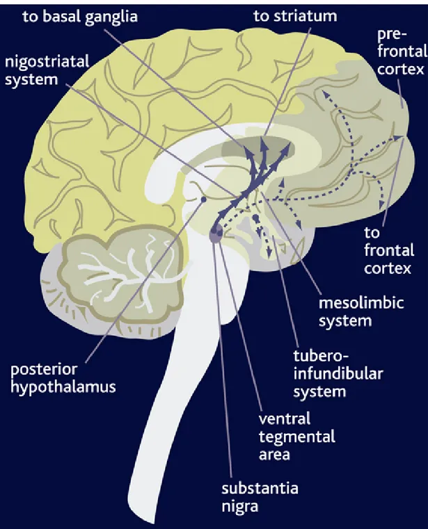
caudate-from multiple structures in the diencephalon and telencephalon. Projections originating from brain areas that synthesize this neurotransmitter give rise to four axonal pathways (Figure 2): (1) projections constituting the Nigrostriatal pathway arise from dopamine-synthesizing neurons of the midbrain nucleus, the substantia nigra compacta (SNc) which innervates the dorsal striatum (caudate-putamen). The nigrostriatal pathway is involved in the control of movement, regulates the extrapyramidal motor system and its degeneration causes Parkinson's disease. 2) The Mesolimbic pathway originates from the midbrain VTA and innervates the ventral striatum (nucleus accumbens), the olfactory tubercle (OT) and parts of the limbic system. It has been demonstrated to influence motivated behavior, such us emotional balance and reward. 3) The Mesocortical pathway arises from the ventral tegmental area (VTA) and projects to neocortex and prefrontal cortex. This pathway seems involved in some aspects of learning and memory; some examples are motivation, attention, planning and social behavior. Hyperactivity of this pathway has been associated with schizophrenia and hallucination. 4) The Tuberoinfundibular pathway arises from the hypothalamus. Projections of this pathway reach the median eminence of the hypothalamus where they release DA into the perivascular spaces of the hypophysial portal vessels. Thus, DA is transported to the anterior pituitary where it acts on the lactotrophs to inhibit prolactin (PRL) synthesis and release as well as plays an antiproliferative action on normal
and PRL-secreting tumors in experimental animals and humans. This hormone stimulates milk production from mammary glands and stimulates lactotroph proliferation by an autocrine mechanism in the pituitary gland. The first three pathways do not exhibit very clear anatomical boundaries between the neurones of the different cell groups which have been described to appear at the same time during development. These observations, coupled with the overlap in some projection fields of the A8, A10 (ventral tegmental area) and A9 (substantia nigra) cell groups, have led to the suggestion that these neurones might be collectively indicated as the mesotelencephalic DA systems (Moore and Bloom, 1978).
Fig 2: Mouse brain schematic diagram illustrating the ascending dopaminergic pathways
arising within the substantia nigra (pars compacta, SNc, A9) and the ventral tegmental area (VTA, A10) depicting main projection areas.
3. Seven-Transmembrane domain receptors
The Seven-Transmembrane domain receptors (7TM) are commonly referred to as G-protein-coupled receptors (GPCR), because they signal through activation of heterotrimeric G-Proteins (Lefkowitz, 2000), also defined as Guanidine-nucleotide regulatory protein complex and composed of α and βγ subunits.
In the absence of agonist, 7TM receptors, such as the β-2 adrenergic receptor β2-AR, are in the low-affinity state. After agonist binding, a transient high-affinity complex of agonist, activated receptor, and G-protein is formed. GDP is released from the G-protein and is replaced by GTP. This leads to dissociation of the G-protein complex into, an α subunits and, a βγ dimer or complex, which both activate several effectors. Gs, for instance, activates adenylyl cyclase, which leads to an increase in cyclic AMP (cAMP). This increase in cAMP in turn activates protein kinase A (PKA), which is a serine/threonine kinase that phosphorylates many different substrates, including 7TM. Actually, each of these subunits is known to be a member of a gene family; 16 α, 5 β and 12 γ proteins have been cloned to date.
G-proteins are generally referred to by their α-subunits. So, the Gs heterotrimeric complex contains α-Gs, α-Gq contains Gq, Gi contains α-Gi and so on. Four distinct subunit subfamilies are recognized: Gs proteins couple to stimulation of adenylyl cyclase; Gi proteins couple to inhibition of adenylyl cyclase as well as to activation of G-protein-coupled inwardly
rectifying potassium (GIRK) channels; Gq proteins couple to the activation of phospholipase C; and G12 proteins couple to the activation of Rho Guanine-nucleotide Exchange Factors (GEFs). However, the combinatorial complexity of αβγ heterotrimers that might form is obviously great and, as yet, relatively little is understood of the specific subunit composition of the G proteins that function in specific pathways (Pierce et al., 2002).
Both, the α subunit and, the βγ dimer signal through the activation, or inhibition, of an ever-expanding list of effectors. Agonist activation of the receptors induces conformational changes which are, yet, poorly understood, but which seem to involve, at minimum, rearrangements of membrane helices 6 and 3. This “activated receptor” can interact with the heterotrimeric G protein, and serves as a guanidine exchange factors (GEF) to promote GDP dissociation, and GTP binding and activation. Receptors vary in their degree of agonist-independent or constitutive activity to couple to G-proteins; receptor mutants resulting in augmented activity have been found in numerous disease. In the current model, the activated heterotrimer dissociates into an α subunit and a βγ dimer, both of which have an independent capacity to regulate separate effectors. Hydrolysis of GTP to GDP, a process that is known to be regulated by regulator of G-protein signalling (RGS) proteins, leads to reassociation of the heterotrimer and termination of the activation cycle (Ross and Wilkie, 2000).
4. Dopamine Receptors
The first evidence for the existence of DA receptors in the CNS came in the early 70s from biochemical studies showing that DA was able to stimulate adenylyl cyclase (AC) (Kebabian and Calne, 1979). Indeed, the DA receptors belong to a large superfamily of neurotrasmitters and hormone receptors which are coupled to their specific effector functions via heterotrimeric GTP-binding proteins (Hepler and Gilman, 1992).
In 1979, Kebabian and Calne found that DA exerts its effects by binding to two receptors types, which were defined as D1 and D2 receptors based on pharmacological and biochemical properties (Kebabian and Calne, 1979). Although, for some time, ample behavioural, biochemical and pharmacological observations had pointed to DA receptor heterogeneity, it was only when it became possible to discriminate between agonist and antagonist action at the level of the cyclic adenosine monophosphate-(c-AMP)-second messenger generating enzyme, adenylyl cyclase (AC), that the dual DA receptor hypothesis became strongly well-established.
Pharmacologically, the hallmark of the D1 receptor is it’s high affinity for benzazepine compounds such as SCH 23390, while D2 receptors are recognised for the high affinity for butyrophenones such as spiperone and haloperidol (Seeman and Van Tol, 1994). D1 and D2 receptors exert their biological actions by coupling and activating different G-protein complexes. The D1 receptors interact with the stimulatory G-proteins (Gs) and activate
adenylyl cyclase, whereas the D2R interacts with pertussis toxin sensitive G proteins (Go/i) to inhibit cAMP production (Gilman, 1984; Liu et al., 1994; Picetti et al., 1997).
With the advent of molecular cloning techniques and their application to the neurobiology, it has become clear that the original D1R/D2R classification scheme was too much restrictive (Gingrich and Caron, 1993).
Thus far, five distinct genes encoding for different DA receptor proteins have been defined through molecular cloning. Two of these cloned receptors exhibit the functional and pharmacological properties expected for classical D1 receptors, while the other receptors show the pharmacological characteristics of D2 receptors (Jackson and Westlind-Danielsson, 1994; Missale et al., 1998).
It is now generally recognised that the family of D1R-like and D2R-like receptors exist rather than singular receptor subtypes (fig.3 and fig.4).
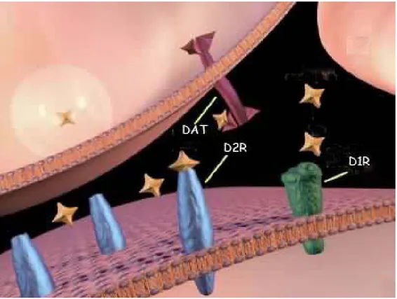
Figure 3: Dopaminergic Synapse and Synaptic Neurotransmission: DA binds D1 and D2R
receptors. DAT is implicated in the DA reuptake. Dopamine is synthesized in the nerve terminal and packaged in vesicles. The vesicle fuses with the membrane and releases dopamine. The dopamine molecules can then bind to a dopamine receptor. After the dopamine binds, it comes off the receptor and is removed from the synaptic cleft by uptake pumps (DAT) that reside on the terminal. This process is important so that not too much dopamine is left in the synaptic cleft at any one time.
4.1 D1-like receptor subfamily 4.1.1 Dopamine D1 receptor
The D1 receptor linked to the activation of adenylyl cyclase activity was first cloned in 1990 (Dearry et al., 1990; Monsma et al., 1990; Sunahara et al., 1990; Zhou et al., 1990). Overall, both the human and the rat receptors
are 446 residues in length, exhibiting 91% amino acid sequence conserved identity.
Hydropathy analysis of this protein predicts the presence of seven transmembrane domains where the N-terminus is localised to the extracellular surface and the C-terminus projects into the cytosol. This overall topography has been suggested for all of the G protein-linked receptors that have been cloned thus far. Several other structural features of the D1 receptor are particularly noteworthy. First, the D1 receptor genes appear to lack intones within the coding regions, there are two potential sites for N-linked glycosylation, one on the N-terminus and another on the second extracellular loop (Sunahara et al., 1990). Moreover, the D1 receptor has a small third cytoplasmic loop and a long C-terminus. These features are characteristic of Gs coupled receptors, and activate adenylyl cyclase such as the β-adrenergic receptor. In addition, there is one consensus site for cAMP-dependent phosphorylation in the third cytoplasmic loop and a conserved Cys at the C-terminus, the latter of which may serve as a site for palmitoylation.
When expressed in various mammalian cells, the cloned rat and human D1R have shown to exhibit the pharmacological and functional characteristics expected for a D1 receptor subtype. Indeed, pharmacologically specific binding for SCH23390 a D1-antagonist ligand, as well as the increase of
transfected cells (Dearry et al., 1990; Monsma et al., 1990; Sunahara et al., 1990; Zhou et al., 1990).
4.1.2 Dopamine D5 receptor
The rat D5 DA receptor is 475 amino acids in length and presents overall 83% identity in comparison with the human D5R, but 95% identical residues in the transmembrane regions (Grandy et al., 1991; Sunahara et al., 1991; Weinshank et al., 1991). So far, it has not been possible to differentiate pharmacologically D1 and D5R receptors. The sensitivities of these two receptor subtypes to antagonists are similar. Nevertheless, these compounds generally show a slightly higher affinity for the D1R than D5R, with (+) butaclamol as the most discriminating. The affinity of agonists at D1R and D5R receptors is similar. The most consistent difference is represented by DA itself, which has around 10 times higher affinity for the D5R than D1R (Grandy et al., 1991; Sunahara et al., 1991; Weinshank et al., 1991).
4.2 D2-like receptors subfamily 4.2.1 Dopamine D2 receptor
The first member of the DA receptor family to be cloned was the D2R. The cDNA encoded a protein of 415 amino acids which, when expressed in
cells exhibited appropriate radioligand binding activity with the pharmacological characteristics expected for a D2 receptor (Bunzow et al., 1988).
Biochemical analysis indicated that the N-terminus contained consensus sequences for three potential N-linked glycosylation. In contrast to the D1 and D5 receptors, the size of the C-terminus of the D2R receptor is rather small while the third intra-cytoplasmic loop appears larger (Bunzow et al., 1988). This feature of having a large third cytoplasmic loop and a short C-terminus is a characteristic of many receptors, which inhibit adenylyl cyclase activity (Dohlman et al., 1991). The human homologue of the rat D2 receptor has been subsequently cloned and shown to be 96% identical to the rat receptor with one amino acid deletion (Grandy et al., 1989).
After the initial cloning of the D2 receptor cDNA a second very similar cDNA was isolated. It was then determined that the D2 receptor exists in two isoforms that differ in length by 87 nucleotides (29 amino acids) and are derived from the same gene by a mechanism of alternative splicing (Dal Toso et al., 1989; Giros et al., 1989; Monsma et al., 1989; O'Malley et al., 1990). The location of this splice variant occurs within the putative third intra-cytoplasmic loop (IL3) of the D2 receptor. Detailed investigation of the D2 receptor gene has revealed the presence of eight exons, seven of which are coding. The alternative splicing of the sixth exon, which encodes a 29
(444aa) and D2S (415aa) isoforms in rodents (Dal Toso et al., 1989; Giros et al., 1989; Monsma et al., 1989; O'Malley et al., 1990).
The two isoform of the D2R have been studied most extensively: both D2 receptor isoforms, D2RL and D2RS, are present in human, rat, bovine and mouse tissues. In situ hybridization and RNAse protection studies, in vivo, suggest that both mRNAs are co-expressed in the same cells and regions where D2R are expressed (Montmayeur et al., 1991; Montmayeur et al., 1993; Guiramand et al., 1995). Interestingly, the D2RL isoform appears to be predominantly expressed in all brain regions and tissue examined with respect to D2S (Montmayeur et al., 1991). The two cloned D2R isoforms from rat and humans expressed in different cell lines reveal have virtually indistinguishable binding characteristics, corresponding to those of the native striatal DA D2 receptor. The similar expression patterns and pharmacology have led for long time to the conclusion that D2L and D2S had redundant functions in vivo (Jackson and Westlind-Danielsson, 1994). The recent availability of the antibody for both the isoforms has confirmed,
in vivo, a different ratio in D2R isoforms expression throughout the
forebrain (Khan et al., 1998). These evidences, taken together, have led to the suggestion that cellular mechanisms regulating the rate of splicing and the final ratios of the D2R isoform products may have a physiological and clinical relevance.
4.2.2 Dopamine D3 receptor
The second DA receptor within the D2R-like family cloned and characterised has been the DA D3 receptor (Sokoloff et al., 1990).
Overall, the D3 receptor is 52% homologous with the D2 receptor; however, this homology increases to about 75% within the transmembrane regions. As with the D2 receptor, the D3 receptor contains consensus sequences for N-linked glycosylation as well as a cAMP-dependent phosphorylation site in the third cytoplasmic loop (Sokoloff et al., 1990).
The human gene for D3R receptor has also been characterised (Giros et al., 1990). Surprisingly, the human receptor has 46 fewer amino acids in the third cytoplasmic loop, resulting in a protein of 400 residues in length. Excluding this deletion, the human receptor is 88% homologous overall and 97% homologous in the transmembrane domains when compared with the rat receptor.
Expression of the D3 receptor in CHO cells indicates that its pharmacology is similar to that of the D2 receptor. Few antagonists have been described to be slightly selective for D3 receptor, relatively to D2 receptor. Other antagonists examined were between 2 and 30 fold D2R selective. Conversely, agonist ligands demonstrated either equal or greater affinity for the D3 receptor (Sokoloff et al., 1995).
4.3.3 Dopamine D4 receptor
The most recent member in the D2R-like family identified and cloned is D4R receptor (Van Tol et al., 1991). This receptor consists of a protein of 387 residues in length with seven membrane-spanning domains. The membrane topography is similar to that for the D2 and D3 receptors, including the existence of a large third cytoplasmic loop and short C-terminus. The homology of the D4 receptor to the D2 and D3 receptors is 41% and 39% overall, respectively, and about 56% for both D2 and D3 receptors within the membrane-spanning domains. There is one potential site for N-linked glycosilation in the N-terminus and one consensus cAMP-dependent phosphorylation site in the third cytoplasmic loop of the D4 receptor (Van Tol et al., 1991). Characterization of the binding activity revealed a pharmacological profile, which was also similar to those of the D2 and D3 receptor. The D4 receptor display similar or lower affinities for both DAergic antagonists and agonists compared to the D2 receptor (Van Tol et al., 1991).
Importantly, however, the atypical antipsychotic clozapine, exhibit about 10-fold higher affinity for the D4 receptor than for D2 receptor. This has led to the suggestion that clozapine might exert its antipsychotic activity primarily through blocking the D4 receptor (Seeman et al., 1993). Several polymorphic variants of the D4 receptor have been found in humans, which
differ by the number of repeated units (composed of 16 aa), located in the third cytoplasmic loop region of the receptor (Van Tol et al., 1991). In line with the concept of a role of this domain in the activity of 7TM-receptors, the human D4 receptor polymorphic variants seem to have different impact in controlling behavioural properties in the human population (Benjamin et al., 1996; Ebstein et al., 1996). Thus, this region represents a key functional domain of D4 receptor activity. Furthermore, an increased number of D4 receptors have been reported in schizophrenic patients, suggesting a role for these receptors in human psychosis (Seeman et al., 1993).
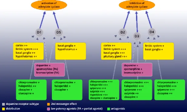
Figure 4. Dopamine receptor subtypes. There are two types of dopamine receptor,
D1-like and D2-D1-like receptors. The D1-D1-like receptors comprise D1- and D5-receptor subtypes that are associated with stimulation of adenylate cyclase. The D2-like receptors comprise D2-, D3- and D4-receptor subtypes and these are associated with inhibition of adenylate
receptors. All dopamine receptor subtypes are expressed in the brain in distinct but overlapping areas. D1 receptors are the most abundant and widespread in areas receiving dopaminergic innervation (the striatum, limbic system, thalamus and hypothalamus); D2 receptors are widespread in these areas, as well as the pituitary gland. D3 and D4 receptors are present in the limbic system. Schizophrenia is associated with dopaminergic hyperactivity. Dopamine antagonists used as antipsychotic drugs (eg chlorpromazine, haloperidol, risperidone) exert their effects mainly by blocking D2-like receptors. Dopamine agonists, such as apomorphine and bromocriptine, also have greater potency at D2-like receptors. Bromocriptine is used clinically to suppress prolactin secretion arising from tumours of the pituitary gland.
5. Dopamine Transporter
Dopaminergic neurons express the DA Transporter (DAT), a protein involved in the membrane reuptake process (Fig. 4). This protein together with DA autoreceptor is a key element in the control of DA signaling by regulating extracellular DA levels. DAT is a member of substrate-specific, high affinity, Na-dependent membrane transporters family (Amara and Kuhar, 1993). DAT mediates recapture of released dopamine; this event is generally thought to be the primary mechanism for limiting the extent, duration, and area of dopamine receptor activation. Furthermore, DAT represents a crucial target for a variety of psychostimulants, for
catecholamines, and selective neurotoxins (Javitch et al., 1985; Ritz et al., 1987).
The anatomical characterization of DAT expression has been greatly facilitated by radiolabeled ligands, including cocaine, mazindol, methylphenidate, GBR-12935 and nomifensine (Berger et al., 1985; Dubocovich and Zahniser, 1985; Janowsky et al., 1985; Javitch et al., 1985). An oversimplified scheme of dopamine transporter function would predict that 2Na+, 1Cl-, and 1dopamine molecule bind sequentially to the dopamine transporter, and are transferred across the plasma membrane.
The cloning of DAT cDNA, has been made possible by the high sequence homology with previously cloned transporters (i.e. GABA transporter). Indeed, using a PCR strategy has been possible to obtained cDNA clones of DAT from rat, bovine and human cDNA libraries (Kilty et al., 1991; Usdin et al., 1991; Bannon et al., 1992).
The distribution of DAT mRNA determined by in situ hybridization is consistent with its exclusive expression in dopamine cell dendrites and soma. The highest DAT mRNA levels, as expected have been observed in the substantia nigra and in the ventral tegmental area. In contrast, DAT mRNA is not detected in DA terminal fields or other brain regions (e.g. striatum, nucleus accumbens and cortex).
and Kuhar, 1993). In fact, several studies have shown that lesions of the nigrostriatal pathway produce parallel losses of dopaminergic neurons and DAT expression (Seeman and Niznik, 1990). Nevertheless, it has been observed that the level of DAT expression varies significantly among different dopaminergic cell populations. In addition even in the same cell group, the level of dopamine transporter expression may be subject to regulation.
6. Distribution of dopamine receptors in SNC
In the last decade, the cloning and the identification of multiple DA receptors (DA-Rs) have profoundly changed the understanding of DA-Rs anatomy and pharmacology (Gingrich and Caron, 1993). At present, the function of DA-Rs in different brain regions has not been clearly established and still represents a matter of debate.
Since the cloning of DA-Rs pharmacological, immunocytochemical, in situ hybridization and in situ binding approach have been extensively used to define the function and the distribution of each receptor subtype in the brain areas. This, together with the knockout approach for the different DA-Rs is starting to give a better idea of the possible function of each DA-Rs (Altar
and Marien, 1987; Mansour et al., 1990; Mengod et al., 1992; Surmeier et al., 1996; Aizman et al., 2000; Diaz et al., 2000).
The distribution and abundance of the five DA-Rs is very different depending on which subtype is analyzed. Among the DA-Rs, D1- and D2-R are by far the most robust and widely expressed. Conversely, D3-, D4- and D5-R have a more restricted pattern of expression. Whether there is a physiological significance for the expression of these receptors in particular areas is still under investigation. However, the restricted expression of some DA-Rs in the limbic system as well as the higher affinity for anti-psychotic drugs has suggested a potential role of these receptors in neurological diseases.
D1-R is mainly expressed in the caudate putamen (CP), nucleus accubens (Acb), olfactory tubercle (OT), cerebral cortex (Cx) and amygdala. A high abundance of D1-R has been shown also in the island of Calleja and in the subthalamic nucleus. The binding of D1-R specific ligands could be observed in the substantia nigra (SN), in spite of the lack of mRNA expression. This seems to suggest that D1-R is synthetised in striatal neurons that send their projections to the SN via the direct striato-nigral pathway. D1-R and D2-R mRNA expression is high within the striatum and nucleus accumbens.
D2-R has a very similar distribution with respect to D1-R, especially in areas like the CP, Acb, OT and SN. Low quantities of D2-R are also present in Cx and in the ventral tegmental area (VTA).
D1-R and D2-R mRNA expression is high within the striatum and nucleus accumbens. Cells expressing D2-R mRNA are a more widely distribution in the midbrain and hindbrain, and may be involved in a host of autonomic functions, as well as in the regulation of dopamine release. Cells expressing D2-R mRNA are abundant in the dopaminergic cells of the substantia nigra and ventral tegmental area. Within the substantia nigra the D2-R mRNA is primarily expressed in pars compacta, with a few cells in the pars reticulata. In contrast, while there are high levels of D1 receptor binding in the substantia nigra, pars reticulata,no cells expressing D1 receptor mRNA could be detected in the substantia nigra and ventral tegmental area (Mansour et al., 1990; Meador-Woodruff et al., 1992).
Cells expressing D2 receptor mRNA are also observed in the superior and inferior colliculus , and in the periacqueductal gray.
In the hypothalamus, cells expressing D1 receptor mRNA have a more limited distribution and are localised in the supraoptic, suprachiasmatic, paraventricular, and rostral arcuate nuclei. In contrast, cells expressing D2R receptor mRNA are more widely scattered in the hypothalamus and are found in the lateral preoptic area, anterior hypothalamic area, lateral hypothalamus, arcuate nucleus and dorsal mammillary nuclei. In the
anterior and intermediate lobe of the pituitary gland only the D2 receptor mRNA is detected at high levels (Mansour et al., 1990; Meador-Woodruff et al., 1992). D2-R is also localized in the retina (Doi et al., 2006), kidney and vascular system.
Examination of the neuro-anatomic distribution of D3-R mRNA in rat brain indicates that it is distinct from that of D2-R mRNA and restricted to few brain regions such as the islands of Calleja, a few septal nuclei, hypothalamus, and distinct regions of the thalamus and cerebellum. Both mRNAs and proteins of D2- and D3-R are expressed by both dopaminergic and dopaminoceptive cells.
Northern blot analysis revealed the presence of D4-R mRNA in the olfactory bulb, frontal cortex and hypothalamus in both rat and monkey brain.
In rat brain, the expression of D5-R mRNA is very restricted to the hippocampus and thalamus and does not seem to overlap significantly with the distribution of mRNA of D1-R. Interestingly, D5-R mRNA seems to be much more widely distributed in the primate brain as compared to rodents and in particular in the Cx where it overlaps with the mRNA of D1- and D2-R. However, the D5-R protein seems to be present in the Cx, CP, OT, VTA, and SN in rat brain.
7. Basal Ganglia
The basal ganglia represent key neural substrates through which the cerebral cortex affects the sensory-motor systems. The basal ganglia network is composed of five large subcortical nuclei: caudate nucleus, putamen, globus pallidus, subthalamic nucleus and substantia nigra (Alexander and Crutcher, 1990; Graybiel, 1990) (Fig. 5).
The complex processes arising from the cortical input in the striatal portion of the basal ganglia are modulated by dopaminergic input from the substantia nigra pars compacta. There are two major pathways through the basal ganglia. The direct pathway is the striatal projection to the internal segment of the globus pallidus and substantia nigra pars reticulate, which then project to the thalamus. The indirect pathway is the circuit from the striatum to the external segment of the globus pallidus (endopeduncolar nuclei in rodents), which projects to the subthalamic nucleus. The subthalamic nucleus in turns projects back to both pallidal segments and the substantia nigra.
In the classical model of the basal ganglia D1R and D2R were thought to be largely segregated (Gerfen, 2000). Thus, neurons bearing mainly D1 receptors constituted the direct striato-nigral output pathway. In contrast, neurons richer in D2 receptors constituted the indirect striato-pallidal pathway. The significance of D1 and D2 receptor-specific regulation of
striato-nigral and striato-pallidal pathways is related to their opposite effect on GABAergic neuron (Gerfen, 2000). Normal movements result from a coordinated balance of cortical and thalamic excitation of the striato-nigral and striato-pallidal pathways, which regulate the tonic activity of substantia nigra, pars reticulate neurons (Gerfen, 2000). Disturbance in the activity of different portions of these two pathways can disrupt this balance, with consequences that might lead either to the production of involuntary movements or to akinesia and bradykinesia as in Parkinson’s disease (Baik et al., 1995; Obeso et al., 2000a; Obeso et al., 2000b).
Figure 5: DA neurones are organised into pathways in the brain. DA pathways originate
from groups of cells in the rostral areas of the brain. These groups were given the titles A8, A9, and A10. The nigrostriatal system runs from the substantia nigra (A9) forwards to the caudate, putamen, and globus pallidus, these 3 structures are called the corpus striatum. The mesolimbicocortical sytem run from the ventral tegmental area (A10) forwards to the nucleus accumbens, amydala, septum, olfactory nuclei.
8. D2R functions in the CNS
Since the discovery of D2R, a plethora of pharmacological studies had pointed out its prominent role in the CNS. In the past ten years the use of engineered knockout mice has represented a great tool to discriminate the functions of different genes in vivo.
At present, two different D2R knockout mice (D2R-KO) have been generated (Baik et al., 1995; Maldonado et al., 1997; Saiardi et al., 1997). These animals have represented a great tool to discriminate the complexity of D2R function in vivo.
Locomotor activity requires the coordinated actions of cortical and subcortical structures. The basal ganglia, composed of corpus striatum, globus pallidus, subthalamic nucleus, and substantia nigra, play an important role in modulating the final output of cortical neurons. Despite the differences in signal transduction mechanism, the contribution of D1- and D2-like receptors to locomotion is generally considered synergistic. However, none of the available drugs has complete specificity for any of the five known dopamine receptor subtypes. Furthermore selective functional loss of the dopamine D1 receptor by gene targeting was reported to cause either an increase in baseline activity or no alteration in locomotion (Kobayashi et al., 2004). In contrast D2R-KO mice present a strong reduction in motor performance. In the open field test, that measures the basal locomotor activity, D2R-KO mice show a significant reduction in both locomotion and rearing behavior compared to wild type littermates (Centonze et al., 2003).
Moreover D2R-KO mice spend significantly less time on the rotarod apparatus, which measures their coordination ability, than the WT mice.
All these data strongly indicate that the D2R has a prominent role in the dopaminergic system in the control of locomor activity. None of the other DA receptor deficient mice present such « parkinsonian like phenotype » in motor coordination and activity (Usiello et al., 2000; Kobayashi et al., 2004).
The majority of dopaminergic neurones in the mammalian CNS are located in the midbrain, where they form the substantia nigra (SN), the ventral tegmental area (VTA) and the retrorubral nuclei. Midbrain DA neurones project to the striatum (or caudate-putamen complex), limbic system and frontal cortex, and receive afferents from multiple structures in the diencephalon and telencephalon. The D2Rs show a very particular distribution since they are at post-synaptic level (striatum) where they control the motor out-put and pre-synaptic level (SN and VTA) where they control the DA syntesis and release. Pre-synaptic inhibition of DA release is a physiological phenomenon by which extra-cellular DA release stimulates pre-synaptic autoreceptors to further inhibit DA release. In particular, pharmacological as well as anatomical studies have identified D2R and D3R as potential DA autoreceptors. Studies of in vivo voltammetry and microdialysis using WT and D2R-KO mice have shown that the expression of D2R is a necessary condition for the maintenance of the pre-synaptic inhibition (Calabresi et al., 1997). The effect of haloperidol (D2R antagonist) or quinpirole (D2R antagonist) on dopamine release was
completely suppressed in D2R-KO mice. Importantly, in D2R-KO mice the extracellular increase of DA induced by cocaine or morphine is greatly enhanced versus WT, although no significant changes were observed in basal extracellular concentration of DA (Usiello et al., 2000). All these data demonstrate that the D2R is a key element in regulating the increase in DA in the inter-synaptic space and could play a crucial role in the physiology of addiction.
The mesolimbic dopaminergic system consists of neurons originating in the VTA which have axonal projections to the limbic system. In particular, dopaminergic projections to the Acb have been implicated in the control of the reward mechanisms and in the psychomotor effects generated by drugs of abuse, including opiates, cocaine, amphetamine and alcohol. Studies of D2-R null mice have demonstrated the crucial role of this receptor in the rewarding effects of opiates (Rouge-Pont et al., 2002). In the Place Conditioning paradigm the D2-KO mice failed to show a preference for opiates since they spent the same time in the morphine- and in saline associated compartments. In contrast, the wild-type littermates showed a significant increase in the time spent in the drug-associated compartment during the testing phase. Interestingly, it has been demonstrated that the D2-R is not required for the development of physical opiate dependence or for the locomotor response to acute administration of morphine. In addition,
D2-to opioid dependence. Indeed, when these mice were tested in a similar behavioral paradigm in their response to a natural reinforce such as food, no difference were observed between D2-R deficient and wild-type mice (Maldonado et al., 1997; Usiello et al., 2000; Rouge-Pont et al., 2002; Centonze et al., 2003).
Notably, DA is the major catecholamine in the vertebrate retina playing a central role in neural adaptation to light (Witkovsky, 2004). Yet, the physiological contribution of light-dependent dopaminergic signaling to nonvisual functions has remained unexplored. Recent findings show that signaling mediated throught the D2R greatly influences central molecules participating in the regulation of the circadian clock, playing a central role in the control of light masking of circadian locomotor activity (Doi et al., 2006; Yujnovsky et al., 2006). D2R mediating signaling is required in regulating the proper organization of daily locomotor activity in light-dark cycles (Doi et al., 2006).
In conclusion the use of knock-out mice has shown that D2R has a prominent role between the different DA receptor in the dopaminergic neurotransmission. It is able to modulate a large variety of physiological function in vivo: from locomotor activity to drug of abuse, ethanol intake and control of DA release.
9. D2R signal transduction pathways
At present, many signal transduction pathways have been shown to be affected by D2R activation (Vallar and Meldolesi, 1989). D2R was first characterised as inhibitor of intracellular cAMP levels (Stoof and Kebabian, 1984). Indeed, an inhibition of adenylyl cyclase (AC) activity mediated by DA has been reported in vivo systems such as the anterior and intermediate lobe of the pituitary gland, and in striatal cells (Caccavelli et al., 1992). All these tissues express high levels of D2R (Jackson and Westlind-Danielsson, 1994). The evidence that D2R signalling was blocked by pertussis toxin (PTX) (Cote et al., 1984), indicated that these receptors are associated with members of the Gi/Go-protein family (Senogles et al., 1987; Elazar et al., 1989). These proteins trigger the inhibition of AC, resulting in a decrease in intracellular cAMP concentration (Gilman, 1984). In particular, the activation of Gαi- and Gαo-subunits has been shown to affect the activity of type I, V and VI of the AC (Tang and Gilman, 1992; Taussig and Gilman, 1995). The final product of this intricate interactions leads to variable levels of cAMP and consequently of functional protein kinase A (PKA). PKA, in turn, phosphorylates cytoplasmic and nuclear proteins, regulates cellular metabolism, including ion channel function, and finally desensitises 7TM G-protein coupled receptors (Choi et al., 1993; Hofmann et al., 1994) leading
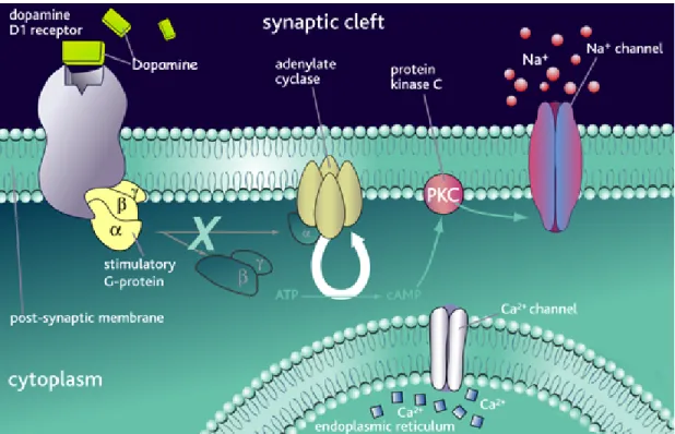
Figure 6. Dopamine D1 receptor. DA binds D1 receptor and leads the activation of the
receptor. The D1 receptor is coupled to stimulatory G-proteins, which dissociate from the receptor on agonist binding and initiate secondary messenger signaling mechanisms. This causes cell depolarisation, which is inhibited by antagonist binding.
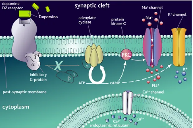
Figure 7. Dopamine D2 receptor. DA binds D2 receptor and leads the activation of the
receptor. The D2 receptor is coupled to inhibitory G-proteins, which dissociate from the receptor on agonist binding and inhibit secondary messenger signaling mechanisms. This causes inhibition of down-stream signaling mechanisms. Antagonist binding inhibits this usual process, resulting in cell depolarisation.
A number of reports have linked the D2R stimulation with a decrease in intracellular Ca2+. The effect seems to be sensible to PTX but independent changes in cAMP levels. The negative regulation of PRL and MSH release, respectively from lactotrops and melanotrps, has been related to the inhibition of of voltage-sensitive calcium channels that leads to a reduction of intracellular Ca2+. (Jackson and Westlind-Danielsson, 1994). In the rat pituitary GH4C1 cell line, D2 receptors inhibit Ca2+ currents through
coupling to Gαo (Liu et al., 1994). In anterior pituitary cells D2 receptors seem to inhibit two voltage-activated Ca2+ currents (Lledo et al., 1992), through coupling to Gαi1 and 3. Interestingly, it has been shown that the ability of D2 receptors to block Ca2+ channels varies between D2L and D2S (Liu et al., 1994).
D2R had also been shown to influence the PKC pathway (Vallar and Meldolesi, 1989; Demaurex and Distelhorst, 2003) although a direct modulation of the phospholipase C (PLC) by D2R is still controversial. Interestingly, the activation of D2R in pituitary lactotrophs inhibits AC, but also Ca2+ release from intracellular stores through the inhibition of IP3 levels (Vallar and Meldolesi, 1989).
Whether this is due to a direct cAMP-independent inhibition of PLC (Rasolonjanahary et al., 2002)by the D2 receptor, or indirectly to the decrease of Ca2+ concentration (Banihashemi and Albert, 2002) via D2 action on Ca2+ channels has to be elucidated.
D2R, acting on intracellular Ca2+ concentration, is also able to influence the DA synthesis by affecting the function of tyrosine hydroxylase, the rate-limiting enzyme in DA production (Meissner et al., 2003). The increase of intracellular Ca2+ activates the calcium/calmodulin dependent protein kinase (CaM kinase or CaM II) by binding to calmodulin (Braun and Schulman, 1995); CaM kinase in turn activates tyrosine hydroxylase.
In addition to the previously described pathways, D2R affects the intracellular concentration of potassium ions. Indeed, D2R mediated activation of K+ channels is mediated by PTX sensitive Gα subunits (Rahman and McBride, 2001). The involvement of D2R in the control of voltage-dependent cAMP-independent K+ channels and of Ca2+-sensitive K+ channels cAMP dependent has also been postulated (Jackson and Westlind-Danielsson, 1994).
The role of D2R in modelling potassium currents has been extensively studied. It has been shown that D2R increases outward potassium currents leading to cell hyperpolarization. Such effects have been observed in rat striatal and mesencephalic neurons as well as in the anterior pituitary (Davison et al., 2004): the activation of potassium currents appears to be modulated by a G-protein mechanism (Congar et al., 2002). The effect of DA on potassium currents in melanotrops is abolished by pertussis toxin (PTX) treatement. In addition, treatment of cells with G-protein antibodies or antisense oligonucleotides blocks the D2R stimulation of potassium currents. In the pituitary, activation of potassium currents appears to be mediated by Gαi3, whereas, in rat mesencephalon cultures by Gαo. K+ influx also decreases Ca2+ concentration through voltage-dependent Ca2+ channels (Koch and Schonbrunn, 1988). The functional significance of cell
hyperpolarization appears to be the inhibition of DA release by autoreceptors in the brain and of prolactin secretion in the pitutary.
D2R can also influence arachidonic acid (AA) synthesis. AA is synthesised from phospholipids by the activity of phospholipase A2 (PLA2): PLA2 needs Ca2+ to work. It has been suggested that D2R acting in synergism with D1R might amplify AA synthesis initiated by the increase of intracellular Ca2+, due to the D1R activated pathway (Piomelli et al., 1991; Piomelli and Di Marzo, 1993). Interestingly, PTX-sensitive G-proteins are involved in these effects, strongly suggesting a D2 receptor mediated effect; PKC, PKA and other kinases can also indirectly activate PLA2 {Balsinde, 2005 #121; Piomelli, 1993 #120; Piomelli, 1991 #119}; consequently, it cannot be excluded that the PLA2 regulation by D2 receptors could also be indirect.
The AA elevation upon D2 receptor activation could also explain the stimulation of K+ channels already described. Released AA can be metabolised by several enzymes, but in the nervous tissue the most important are 12-lipoxygenase (that produces (12s)-hydroperoxyeicosatetraeonic acid or 12-HPETE) and 5-lipoxygenase (that produces 5-HPETE) (Piomelli and Di Marzo, 1993). 12-HPETE could directly activate K+ channels, thus decreasing intracellular [Ca2+]. Moreover, 12-HPETE or its metabolites act directly on CaM kinase II,
playing a role in synaptic vesicle secretion (Piomelli and Di Marzo, 1993). In addition, AA metabolites derived from the epoxygenase pathway are known to inhibit the Na+/K+ ATPase. This ion pump maintains the ion gradient by extruding Na+ and importing K+ in neurones (Tseng and O'Donnell, 2004). D2 receptors acting synergistically with D1 receptors inhibit the activity of this enzyme. This inhibition can lead to a transient membrane depolarization, thus affecting cell excitability and consequently neurotransmitter release (Aizman et al., 2000).
A large array of cellular responses follows the activation of DA D2 receptors. In most of the example given it is clear that the response is dependent on the G-proteins available in the cellular system used. Thus, it can be suggested that the presence of two isoforms of D2R, each coupling to different G-proteins in the same cell, is a mechanism to amplify and diversify the response to DA.
Furthermore, recently it has been indicated that D2R stimulation can lead to both the phosphorylation and activation of CREB and MAPK in vitro as well as to phosphorylation of MAPK in vivo (Lee et al., 2006; Pozzi et al., 2003; Cai et al., 2000; Yan et al., 1999). Interestingly, CREB and MAPK seem activated by two different transduction pathways. Indeed, D2R agonists increase intracellular Ca2+ and PKC activity leading to the activation of the Ras/Raf/MEK/MAPK cascade, while intracellular Ca2+
and CaMK are required for the activation of CREB (Cai et al., 2000). Importantly, it has been shown that activated MAPK in the CNS are mainly cytoplasmic and localised in the cell bodies and dendrites. Conversely, activated CREB is found in the nucleus. The different subcellular localization of these two activated components indicates that DA induced activation of D2R might result in the contemporary stimulation of multiple targets. It has been proposed that MAPK phosphorylation occurring primarily in dendrites would regulate protein synthesis, cytoskeletal dynamics, and ion channel activities at synapses. On the other hand, phosphorylated CREB would regulate gene expression (Yan et al., 1999) by acting at the nuclear level.
In CHO cell line, stably transfected with D2L or D2S , D2R agonists induce a transient ERK phosphorylation (Choi et al., 1999). This stimulation was markedly attenuated by co-expression of the C-terminus of β-adrenergic receptor, which selectively inhibits Gβγ-mediated signal transduction. Further analysis of D2L- and D2S-mediated MAPK activation demonstrated that D2L-mediated MAPK activation was not significantly affected by PKC depletion or partially by genestein. In contrast, ERK activation mediated by D2S was potentially inhibited by PKC depletion and genestein pre-treatment (Choi et al., 1999).
Further levels of regulation of D2R-mediated signal transduction might be dependent upon intracellular interactions with membrane receptors for other
neurotransmitters and modulators. Indeed, a direct intramembrane interaction between D2R and the somatostatin receptor has been recently shown (Rocheville et al., 2000). However, intra-membrane and intracellular modulation of the D2R-mediated signalling was already evoked for the adenosine A2A receptor as well as for other heterologous receptors (Schindler et al., 2004).
In addition, recent studies have shown that D1R and D2R colocalized in striatal neurons (Emamian et al., 2004). These observations are of interest since the described synergistic and antagonistic actions exerted by activation of D1R and D2R subclasses of DA receptors might well be mediated by intracellular rather than exclusively by intercellular mechanisms.
10. D2R isoforms and G protein-coupling
Two isoforms of DA D2R are present in vivo, D2 long isoform (D2L) and D2 short isoform (D2S), generated by alternative splicing of the same RNA transcript. They are co-expressed in all tissue or cell expressing D2Rs, normally in a ratio favouring the D2L with the exception of SN were they are expressed at the same ratio. The long isoform differs from the short isoform in the presence of the sixth exon. This exon codes for 29 amino acids that constitute part of the third intracellular loop (IL3) of the D2R.
D2R belongs to the family of 7TM G protein-coupled receptors. These receptors share similarities in size and structure, with the highest homology in the transmembrane regions (Dohlman et al., 1991): once the binding of a specific ligand activates them, the G-protein/receptor complex dissociates thus leading to the activation of intracellular signaling cascades (Neer, 1995; Hermans, 2003). The third intracellular loop (IL3) is the most variable region among this group of receptors: this region is responsible for the interaction of the receptor with the G-proteins (Fig. 8). In addition to IL3, the second intracellular loop (IL2) be involved in the interaction of the receptors with the G-proteins. Moreover, the generation of several D1R/D2R hybrids has shown that D2R coupling to Gi is due to the IL3 loop in cooperation with regions in the IL2 (Kozell et al., 1997). Mutagenesis studies have shown that the region inside the IL3 loop responsible for the interaction with the G protein is contained in the N-terminal and the C-terminal portions of the loop (Grishina et al., 2000). Interestingly, this is the only variable region between the two isoforms of DA D2 receptors. Indeed, a 29 amino acids insertion is present in a region close to the center of the IL3 of the D2L isoform. All these data suggest that the insertion might play a role in modulating the D2R/G proteins interaction (Montmayeur et al., 1991; Liu et al., 1994; Guiramand et al., 1995).
The expression of D2L and D2S receptor isoforms in JEG3 cells results in the decrease of the intracellular level of cAMP by the inhibition of AC
(Montmayeur et al., 1991) indicating that these receptors are functionally coupled to G-proteins in this cell line. Cotransfection of an antisense Gαi2 vector partially blocked the activity of D2L, but not that of D2S. Taken together, these data clearly indicate that the 29 amino acids insertion present in D2L confers specificity for coupling to Gαi2 and therefore discriminates functionally between the two DA D2R isoforms (Montmayeur et al., 1993). Although the classical signal transduction cascade operates at the plasma membrane, it has been known that heterotrimeric G-proteins are also found on intracellular membranes such as endosomes, secretory granules, the endoplasmic reticulum and the Golgi complex (Helms, 1995). For example, one subclass of Gα can be detected in several membranes depending on the cell type (Wilson et al., 1994). Recent evidences show that G-proteins are involved in diverse functions such as in vesicular transport (Helms, 1995), in the binding of the coatomer to the membranes of the vesicles involved in the endoplasmic reticulum, Golgi and intra-Golgi transport (Serafini et al., 1991; Wilson et al., 1994) as well as in maintaining the Golgi structure (Jamora et al., 1997; Le-Niculescu et al., 2005).
A novel α subunit, sGi2, has been identified in the laboratory of Dr. Borrelli, which is the product of the alternative splicing of the Gαi2 gene (Montmayeur and Borrelli, 1994). A splicing event replaces the last 24 amino acids of Gαi2 with a new segment of 35 amino acids in the C-terminal
the protein in the Golgi apparatus (Downes and Gautam, 1999; Picetti and Borrelli, 2000). A similar mechanism has also been reported for Gαs, in which the N-terminal 46 amino acids are substituted with a new 498 amino-acid sequence (Kehlenbach et al., 1994).
Other G proteins localized intracellularly suggest that they perform different functions from the G proteins at the plasma membrane.
It has been reported that activation of G proteins with AlF4- are involved in the intracellular membrane protein trafficking (Bomsel and Mostov, 1992; Helms, 1995). G proteins have been shown to be involved in secretory (Melancon et al., 1987) as well as in endocytic (Mayogara et al., 1989) mechanisms.
It was previously shown in Cos cells that sGi2 localizes in the Golgi apparatus by a specific 14 amino-acids proline-rich sequence contained in the C-terminal sequence of the protein (Picetti and Borrelli, 2000). In 2002, Wedegaertner reported that the disruption of the normal C-terminus of αi2 causes mislocalization and rapid degradation of sGi2 suggesting the cellular instability of sGi2. Recently, Khan and Gutierrez, in 2004, showed that sGi2 protein is widely expressed in rat and monkey brain regions and that depending on the cell subtype is localized in intracellular compartments such as the endoplasmic reticulum, Golgi, mitochondria and nucleus but also in dendrites, axons and spines suggesting: that this protein is stable as it is transported in the cell after synthesis.
Figure 8: Dopamine D2 Receptor coupled with a G-protein. Dopamine D1-like
receptors are coupled to stimulatory G-proteins and they have a stimulatory effect on neurotransmission when bound by an agonist. The D2-like receptors are coupled to inhibitory G-proteins. Dopamine D2-like receptors have an inhibitory effect on neurotransmission when bound by an agonist. Many neuroleptic drugs are antagonists of the D2 receptors. This class of drug is used to treat psychotic disorders, such as schizophrenia.
11. Gene targeting: D2R-deficient mice
In the beginning of the 80’s, cells from embryonic origin, the embryonic stem (ES) cells, have been isolated for the first time (Evans and Kaufman, 1981);
altering their potential. By a process called homologous recombination, a DNA fragment, which has been incorporated into these ES cells, can integrate into their genome and thus introduce a mutation in the gene of interest. Once implanted into an embryo, these modified ES cells can give rise to mice mutant for the gene of interest.
Recombination between homologous DNA-sequences is a natural mechanism occurring rarely and very little is still know about the molecular mechanisms of this process in vertebrates (Smithies et al., 1985). However, use of appropriate DNA vectors has taken advantage of this event to introduce a fragment of interest into the genome and selection for the cells that have undergone homologous recombination (Thomas and Capecchi, 1987).
The vector for the homologous recombination is a circular DNA plasmid, which contains sequences homologous to the targeted locus. A selection marker (e.g. the gene neo offering resistance to neomycin) interrupts these sequences. Introducing the vector via electroporation into the ES cells can cause chromosomal rearrangements leading to the insertion of the vector into the genome by replacement or integration (Capecchi, 1989). The selection marker has a double function. Its presence in the coding sequence of the gene of interest disturbs the normal transcription of this gene: the gene is thus inactivated. On the other hand, the gene neo enables the modified ES cells to resist to G418, a synthetic analogue of neomycin. Thus, the cells that have integrated by homologous recombination into their genome from the vector
can be isolated (Thomas and Capecchi, 1987). A second selection process may be necessary to eliminate those ES cells that have integrated from the vector randomly into their genome, by non-homologous recombination. The gene coding for the thymidine kinase (TK) of the Herpes simplex virus makes the ES cells vulnerable when applying the modified nucleotide gancyclovir. Introducing the TK gene at one extremity of the vector for homologous recombination allows for a second selection procedure of the electroporated ES cells. Both selection procedures can be combined to augment the frequency of cells that have undergone homologous recombination: integration of the neo gene (resistance to G418) and absence of TK (resistance to Gancyclovir) in the cells that have been harvested after electroporation (Thomas and Capecchi, 1987).
The embryonic stem cells are derived from the inner cell mass of mouse blastocyst (Evans and Kaufman, 1981). The ES cells which have been selected after homologous recombination can be re-injected into mouse host embryos where they can contribute to all different tissues including the germ line (Bradley et al., 1984). The ES cells generally used are derived from the pure genetic mouse strain 129/sv, a mouse strain with agouti coat color. The host embryos are of mixed (heterologous) origin (e.g. C57BL/6J, black mice) and are implanted into a (hormonally-induced) pseudo-pregnant female (Thomas and Capecchi, 1987). The chimera animals that derive from these
derived from wild type blastocysts and agouti for the tissues derived from the injected ES cells. The germline of these chimeras also derives from two genetic backgrounds: C57BL/6J for the wildtype germlayer-cells and 129/sv for the germlayer-cells derived from the modified ES cells. By the end of meiosis, half of the gametes on 129/sv background contain the introduced mutation. The chimeras are then bred into an animal of the host-strain C57BL/6J. The offspring of this crossing with black coat color will then be of wild type phenotype whereas half of the offspring with agouti coat color will be heterozygous for the desired mutation (the agouti color being dominant over the black color). The continuous crossing of the latter ones will allow for the establishment of an animal strain which is homozygous for the modified gene (a “null” mutant) (Thomas and Capecchi, 1987).
Using the gene targeting approach, in the last few years, different DAR mutant mice have been generated (Giros et al., 1989; Drago et al., 1994; Baik et al., 1995; Rubinstein et al., 1997; Xu et al., 1997). These animals have represented a great tool to discriminate the function of each receptor in vivo. In addiction, they allow the overcome the bias created by the use of other model system, in which either chemically or mechanically lesions are induced, which although very useful can be non-selective. Similarly, the use of ligands to test the function of receptors in vivo presents the bias of drugs acting at multiple receptors with the consequent lack of specificity. Targeted mutagenesis of desired genes in vivo, by homologous recombination, is
offering a mean to estimate the physiological effects of genes of interest. Using this technique, null mice for several components of dopaminergic system have been generated, providing animal models to evaluate, in a more selective manner, the role of dopaminergic transmission (Giros et al., 1989; Drago et al., 1994; Baik et al., 1995; Rubinstein et al., 1997; Xu et al., 1997) . Mutant mice have been generated to analyze the physiological involvement of D2R in vivo (Baik et al., 1995). To generate D2R null mice, the first coding exon (exon 2) and flanking introns were deleted and replaced with a neomycin resistance gene under the control of the phosphoglycerate kinase I promoter (Adra et al., 1987). One ES clone was identified that contained the mutated gene inserted by homologous recombination. This clone was injected into the blastocysts of recipient mice and five chimeras’ male mice were obtained. These mice were able to establish a line of D2R heterozygote null mice. Viable homozygous mice were then obtained. The D2R deficient mice presented a reduction of their body weight as compared with normal littermates. The analysis of food and water intake revealed a slight decrease (10-15%) in homozygous mice; the body temperature was reduced of 0.7°C in D2R-KO with respect to the WT littermates. The pharmacological analysis of homozygote mice showed a complete absence of D2R binding site (Baik et al., 1995). D2R mutant mice have helped to establishment the role of D1R and D2R in locomotion. The behavioral phenotype of the D2R-KO is
littermates (Baik et al., 1995). These observations indicate that D2R-KO present a “Parkinson-like phenotype”, intending that lack of D2R signaling affects movements in a similar manner that DA reduction in other models, although to a much lower extent.
D2R-KO mice have been used to elucidate the autoreceptor presynaptic functions of D2R in vivo. Interestingly, D2R-KO mice lead to a total abrogation of the inhibitory effects of DA on the firing of dopaminergic neurons of SN (Mercuri et al., 1997). In addition, also the DA release assessed in the striatal synaptosome in D2R-KO mice suggests a pivotal role of D2R in the control of DA release. More recently, these results have been confirmed by in vivo microdialysis and by voltammetry (Benoit-Marand et al., 2001; Rougé-Ponte et al., 2002). Taken together these results indicate that D2R is the major DA autoreceptor.
12. D2L-deficient mice
Despite all the pharmacological observations, the lack of specific compounds able to discriminate between the two isoforms, D2L and D2S, has not yet allowed to establish their individual contributions in the D2R mediated functions. Two different D2L-KO have been generated (Usiello et al., 2000; Wang et al., 2000) that express only the short isoform, D2S, without changing the total mRNA and protein D2R level (Usiello et al., 2000; Wang et al.,
2000). The analysis of these mice permitted to definitively clarify that the two isoforms have a distinct function, in vivo. The only expression of short isoform in D2L-KO mice at presynaptic level results in an increase of autoreceptor function: the experiments strongly indicate that the D2S can be considered the presynaptic D2R (Usiello et al., 2000; Rougé-Ponte et al., 2002) .
Furthermore at postsynaptic level the over expression of D2S in the striatum of D2L-KO mice completely alters the function of D2R (Centonze et al., 2004). A cooperative/synergism interaction between D1R and D2L can be proposed on the basis of pharmacological and behavioral studies (Centonze et al., 2003; Usiello et al., 2000) while D2S seems to interfere, at postsynaptic sites, with D1R-mediated functions. Moreover, analysis of D2L-KO mice have show that the D2L isoform is the target of the antipsychotic haloperidol, since in D2L-KO mice this compound does not elicit catalepsy any longer as it does in WT mice. Thus, D2L and D2S have a different function in vivo: D2S is principally the D2 presynaptic autoreceptor, which at postsynaptic level negatively modulates D1R-dependent responses. In contrast, the D2R-mediated postsynaptic effects and their cooperative/synergistic activity with D1R seem likely to be mediated by D2L.
These two mouse model, D2R-KO and D2L-KO, have been used during my PhD work.
At the beginning of my PhD studies in the laboratory of Dr. Borrelli, I focused my attention on developing a new D2R monoclonal antibody (4H6-7-3) recognizing both isoforms of the D2R.This antibody is directed against a peptide corresponding to the mouse D2R aminoacid (aa) sequence spanning from aa 309 to 322 (D-P-S-H-H-G-L-H-S-N-P-D-S-P). Since after more than ten years Dr Borrelli’s lab was unable to obtain an antibody which would recognize D2R, we used a different strategy. We reasoned that lack of immunogenicity might depend from an escape mechanism to avoid self destruction of D2R-mediaed functions. We thus immunized D2R-KO mice, indeed these animals were born without D2R and thereby they could have a better response to the immunization protocol.
Monoclonal D2R antibody was produced by fusing single antibody-forming cells to tumor cells grown in culture, the hybridoma. Each hybridoma was able to produce relatively large quantities of identical antibody molecules. By allowing the hybridoma to multiply in culture, it was possible to produce a population of cells, each of which produced identical D2R antibody molecules (Fig. 9). These cells where then injected in nude mice for ascite production.
Figure 9: Monoclonal Antibody Production. Process by which the D2R antibody was
produced.
Once the antibody was made, I tested its specificity by Western blot using striatal membrane protein extracts, rich in dopaminergic receptors from WT, D2L-/- and D2R-/- mice as well as COS cells transfected with the expression vectors of either D2S, D2L or both receptors. I carried outthe solubilization of D2R from striatal membranes, with different detergents and detergent-salt
combination. CHAPS, a zwitterionic detergent
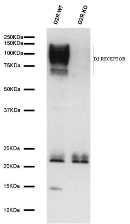
to be the best solubilizing agent, among all detergents used. D2Rs were detected in the striatum as a wide band corresponding to a group of proteins of 70-120 kDaltons (fig. 10). The presence of multiple bands is due to receptor specific posttranslational modifications as well as to the oligomerization of receptors that belong to the seven transmembrane domain G-protein coupled receptor family. As expected, no signal was detected in D2R-/- striatum.
Figure 10: Generation of a specific antibody against D2R. I raised a mouse monoclonal
anti-D2R antibody (4H6-7-3) directed against a peptide corresponding to the mouse D2R aminoacid residues 309-322. Western blot analyses using the 4H6 antibody revealed the presence of positive bands only in WT, but not in D2R-KO membrane preparations. The presence of multiple bands is due to receptor specific posttranslational modifications as well as to the oligomerization of receptors that belong to the seven transmembrane domain
Using this antibody, I was able to perform immunoprecipitation and immunoistochemistry assays, as well as Western blot, that will be presented during my discussion.
The 4H6-7-3 antibody was useful to analyze the subcellular localization of D2R. It has been reported that G-proteins are involved in the intracellular membrane protein trafficking (Bomsel and Mostov, 1992; Helms, 1995). G proteins have been shown to be implicated in secretory (Melancon et al., 1987) as well as in endocytic (Mayogara et al., 1989) mechanisms. A novel α subunit, sGi2, has been identified in the laboratory of Dr. Borrelli, which is the product of the alternative splicing of the Gαi2 gene (Montmayeur and Borrelli, 1994). A splicing event replaces the last 24 amino acids of Gαi2 with a new segment of 35 amino acids in the C-terminal region of the protein. This substitution is responsible for the localization of the protein in the Golgi apparatus (Downes and Gautam, 1999; Picetti and Borrelli, 2000).
In the Dr. Borrelli’s laboratory, I have analyzed the role of sGi2, in vivo. I started by performing in vitro analyses on transtected cells. Cell extracts immunoprecipitated with the D2R-antibody showed that each isoform of D2R could interact with sGi2 in vitro. However, I then analyzed whether these complexes can also form in vivo, by preparing extracts from D2R-KO and D2L-KO mice. Importantly, I was able to show that sGi2 preferentially interacts with D2L in vivo and not with D2S (fig. 11). Indeed, in in D2L-KO mice the anti-D2R is not able to immunoprecipitae sGi2. These findings are