Contents lists available atScienceDirect
Ceramics International
journal homepage:www.elsevier.com/locate/ceramint
X-ray photons attenuation characteristics for two tellurite based glass
systems at dental diagnostic energies
Y. Al-Hadeethi
a, M.I. Sayyed
b,∗, Hiba Mohammed
c,d, Lia Rimondini
c,e aDepartment of Physics, Faculty of Science, King Abdulaziz University, Jeddah, 21589, Saudi ArabiabDepartment of Physics, Faculty of Science, University of Tabuk, Tabuk, Saudi Arabia cDepartment of Health Sciences, Università del Piemonte Orientale UPO, 28100, Novara, Italy dFondazione Novara Sviluppo, 28100, Novara, Italy
eInterdisciplinary Research Center of Autoimmune Diseases (IRCAD), 28100, Novara, Italy
A R T I C L E I N F O Keywords: X-ray attenuation Tellurite glasses WinXcom Geant4 A B S T R A C T
X-ray photons attenuation characteristics for the two tellarite based glasses Bi2O3– B2O3– TeO2– TiO2and PbO–ZnO–TeO2–B2O3have been investigated at dental diagnostic energies (between 30-80 keV) using Geant4 code and WinXcom software. The correlation coefficient (R2
) is utilized to evaluate the extent to which Geant4 results are related to the WinXcom data. For the both series, R2is close to 1 for all samples and this implies a perfect degree of association between the Geant4 and WinXcom data. The linear attenuation coefficient is proportionally increased with addition of TeO2in both series, which implies that there is a decreasing tendency in the X-ray photon transmission corresponding with an increase in the TeO2content in the glasses. The half value layer (HVL) decreases as the density increases and this decreasing is very notable at 70 and 80 keV. The maximum HVL for all samples occurs at 80 keV and this implies that the HVL gradually increases as the energy of the X-ray photons increase. Also, the increment of TO2in the glasses (in both systems) leads to reduce the mean free path and BiTeTi6 and PbTeB6 samples have the lowest MFP. The MFP for both systems was compared with three heavy concretes and the comparison revealed that the selected systems can be utilized to fabricate pro-tection masks used during diagnostic radiation of the head or oral cavity.
1. Introduction
In dental and medicalfields, X-rays are utilized for surgery, radio-therapy and diagnostic treatments. During the diagnostic and ther-apeutic procedures, the organs and tissues near the area of treatment predominately get exposed to the X-ray photons, causing some dama-ging or deleterious effects to the living cells or human organism [1–3]. In order to reduce hazards from this type of radiation, radiographers and patients are required to use special protective clothes such as aprons, lead vest, gloves and masks. In practical applications, the lead is the main constituent of these protective materials since it has high density and can completely absorb or attenuate the X-ray photons [4,5]. Generally, lead has noteworthy drawbacks, mainly toxicity and heavi-ness (the typical lead apron mass is 5–8 kg). Therefore, it is inevitable that medical staffs that use the apron over an extended period of time develop knee and/or back pain. Consequently, a great effort is being made from textile and materials engineers to develop novel non-toxic lead-free protection materials. These alternative materials should have
several characteristics such as malleability and ductility, light weight, and highly efficient in attenuation of X-ray photons [6–8]. In order to improve the attenuation capability of the protection materials, heavy chemical elements such as tungsten, antimony, barium and bismuth must be used during the preparation of these materials. On the other hand, radiation masks are used to protect the face from the potential hazards caused by continuing for a long time exposure to X-ray photons during dental and medical treatment. It is not suitable to use an opaque material to synthesize the protection mask and transparent materials are more convenient to be utilized in this regard. Glasses are amor-phous materials and can transmit the light and therefore they are promising materials to fabricate the radiation protection mask. Several researchers tried to study and report the interaction between the X-ray photons and some glass systems. Kaewjaeng et al. [9] prepared B2O3–CaO– SiO2–La2O3glass system and studied the x rays shielding for the fabricated glasses. They found that the lead equivalent thickness increases with increasing the La2O3and decreased with the increasing of kilovoltage peak (kVp) of the X-ray machine. Waly et al. [10] utilized
https://doi.org/10.1016/j.ceramint.2019.08.258
Received 14 August 2019; Received in revised form 26 August 2019; Accepted 27 August 2019 ∗Corresponding author.
E-mail address:[email protected](M.I. Sayyed).
Available online 28 August 2019
0272-8842/ © 2019 Elsevier Ltd and Techna Group S.r.l. All rights reserved.
MicroShield code and reported the photon attenuation factors for dif-ferent glasses with heavy elements additives between 15 to 300 keV. The authors concluded that the more additives of PbO or Bi2O3to the glass, the superior photon attenuation it provides. The glass with composition 10%SiO2, 55%Bi2O3 and 35% PbO showed the lowest photon exposure rate at low energy (E < 300 keV). MCNPX code helped Almatari et al. [11] to report the attenuation factors for borate based glasses between 20 to 150 keV (medical diagnostic energies) and studied the effect of Li2O, ZnO and BaO on the attenuating behavior of this system. Their results revealed that the sample with high amount of BaO is suitable in medical fields as X-ray protection material. Tekin et al. [12] investigated the boron phosphate glasses as protection ma-terials in the diagnostic radiology fields. They used Monte Carlo si-mulation tofind the photon attenuation factors for the boron phosphate glasses for energy varying from 60 to 120 keV. The authors compared the photon transmission factor for the boron phosphate glasses with other protection materials to evaluate the potentiality of using this glass system in medical radiation facilities. Hongtong et al. [13] prepared Gd2O3–CaF2–P2O5glass system and studied the X-ray shielding prop-erties using transmission method. The tenth value layer for the samples were found to decrease with increasing Gd2O3content from 5 to 15 mol %, and increased with increasing X-ray energy. The authors also found that this glass system has good X-ray attenuation characteristics at low energy.
Tellurite glasses (based on TeO2) have distinctive features like low phonon frequency, large transparency window, low melting tempera-tures, high refractive index and good corrosion resistance [14–18]. Tellurite-based glasses have relatively high density (the density of TeO2 is 5.67 g/cm3) and therefore it is expected that those type of glasses can effectively diminish the effect of the radiation.
In this work, two tellurite based glasses namely Bi2O3– B2O3– TeO2– TiO2(BiTeTi1– BiTeTi6 glasses) and PbO–ZnO–TeO2–B2O3(PbTeB1– PbTeB6 glasses) were selected, and their capability to shield diagnostic X-ray photons (between 30-80 keV) were investigated. We used Geant4 for evaluation the attenuation parameters for the two investigated glasses and then we compared the results with the other commonly used materials for radiation shielding aims.
2. Materials and method
X-ray photons attenuation characteristics for the two tellurite based glasses namely Bi2O3– B2O3– TeO2– TiO2(coded as BiTeTi1– BiTeTi6 glasses) and PbO–ZnO–TeO2–B2O3(coded as PbTeB1– PbTeB6 glasses) have been investigated in this work at dental diagnostic energies (be-tween 30-80 keV). The elemental composition of the BiTeTi1– BiTeTi6 glasses whose densities varying from 4.67 to 5.95 g/cm3and PbTeB1– PbTeB6 glasses whose varying ranging from 4.58 to 6.52 g/cm3are summarized inTable 1andTable 2[19,20]. To achieve the aim of this work, we evaluated the X-ray attenuation parameters for the selected glass systems and then compared the results with the other commonly used materials for radiation sheilding. In this section we will discuss in brief the photon attenuation parameters.
If X-ray photons pass through attenuator (glass specimen in our work), part of the photons is attenuated by the attenuator and the re-sidual photons cross the attenuator. Beer-Lambert law is suitable
relation used to describe the attenuation occurred for the photons [12]:
= −
I I e μx
0 (1)
The previous relation gives an indication of the attenuation capacity of the material. The termμ is called the linear attenuation coefficient that estimates how a photon with specific energy will be attenuated when moving through the glass sample [21].μ for any attenuator de-pends on the density of the attenuator, the energy of the photons and the chemical composition of the attenuator.
The mass attenuation coefficient (μ/ρ) is also a useful quantity to evaluate the capability of a medium to shield the photons. For a sample consists of different elements (such as O, B, Te, Ti and Bi in the first glass system), the mixture rule is convenient way to determine theμ/ρ at any energy [11]:
∑
= μ ρ/ w μ ρ( / ) i i i (2) where wiis the weight fraction for the elements (given inTable 1for BiTeTi1– BiTeTi6 glasses, and inTable 2for PbTeB1– PbTeB6 glasses). Theμ/ρ values for the elements at the investigated energies (dental diagnostic energies 30 < E < 80 keV) were taken from WinXcom software [22]. Also, theμ/ρ for the glasses under study was estimated by Geant4 simulation code [23]. It is worth mentioning that high values of the previous two parameters indicate the good attenuation cap-abilities of the medium.Half-value layer (HVL) practically measures the thickness of the attenuator needed to diminish the photons intensity to 50% of its initial value. This parameter reflects the thickness of the glass sample that can replace the protective garments incorporating lead (Pb)/or lead masks during diagnostic radiation of the head or oral cavity. Mathematically, HVL is expressed as in the next formula [24]:
=
HVL μ
0.693
(3) Generally, to provide an efficient photons shielding, the glass sample must contain heavy elements so the photons can interact easily with the atoms. In our two glass systems, the TeO2, Bi2O3and PbO are helpful in attenuating the incident photons.
The mean free path (MFP) is a density dependent parameter and represents the reciprocal ofμ [25]:
=
MFP μ
1
(4)
3. Results and discussion
Geant4 simulation results ofμ/ρ for the BiTeTi1– BiTeTi6 glasses and PbTeB1– PbTeB6 glasses at dental diagnostic energies (between 30-80 keV) and WinXcom results are summarized inTables 3 and 4 re-spectively. Also, we plotted theμ/ρ (Geant4 and WinXcom data) for BiTeTi1 and PbTeB1 (as an example from thefirst and second series) in Fig. 1andFig. 2. It is evident that the Geant4 results are matching with the calculated values. The correlation coefficient (R2) which estimates the linear association between two variables is used to evaluate the extent to which Geant4 results are related to the WinXcom data. For
Table 1
The chemical composition and the density of the Bi2O3– B2O3– TeO2– TiO2glass series.
Density (g/cm3) Bi Te Ti O B TiO
2 TeO2 B2O3 Bi2O3 Glass code
4.67 0.43903 0.30157 0.02515 0.20587 0.02839 10 45 25 20 BiTeTi1 5.00 0.43538 0.31236 0.01995 0.20416 0.02815 8 47 25 20 BiTeTi2 5.11 0.43179 0.32297 0.01484 0.20248 0.02792 6 49 25 20 BiTeTi3 5.53 0.42827 0.33340 0.00981 0.20083 0.02769 4 51 25 20 BiTeTi4 5.72 0.42479 0.34367 0.00487 0.19919 0.02747 2 53 25 20 BiTeTi5 5.95 0.42138 0.35377 0 0.19759 0.02725 0 55 25 20 BiTeTi6
Table 2
The chemical composition and the density of the PbO–ZnO–TeO2–B2O3glass series.
Density (g/cm3) Pb Te Zn O B B
2O3 TeO2 ZnO PbO Glass code
4.58 0.37103 0.11425 0.11709 0.30083 0.09679 50 10 20 20 PbTeB1 5.22 0.31955 0.29518 0.10085 0.23441 0.05002 30 30 20 20 PbTeB2 5.53 0.29882 0.36804 0.09430 0.20766 0.03118 20 40 20 20 PbTeB3 5.87 0.28062 0.43202 0.08856 0.18418 0.01464 10 50 20 20 PbTeB4 6.22 0.38404 0.39417 0.04039 0.16804 0.01336 10 50 10 30 PbTeB5 6.52 0.36381 0.44809 0.03827 0.14983 0 0 60 10 30 PbTeB6 Table 3
The mass attenuation coefficients (cm2/g) of the Bi
2O3– B2O3– TeO2– TiO2glasses using Geant4 code and WinXcom software.
Mass attenuation coefficients (cm2/g)
Energy (keV)
BiTeTi6 BiTeTi5 BiTeTi4 BiTeTi3 BiTeTi2 BiTeTi1
WinXcom Geant4 WinXcom Geant4 WinXcom Geant4 WinXcom Geant4 WinXcom Geant4 WinXcom Geant4 16.150 15.234 16.200 15.288 16.250 15.343 16.310 15.398 16.360 15.455 16.420 15.512 30 13.660 13.017 13.510 12.871 13.370 12.723 13.210 12.572 13.060 12.419 12.900 12.263 40 7.628 7.154 7.547 7.074 7.465 6.993 7.381 6.910 7.297 6.826 7.210 6.741 50 4.738 4.382 4.689 4.334 4.639 4.285 4.588 4.234 4.536 4.184 4.484 4.132 60 3.173 2.903 3.141 2.871 3.108 2.839 3.075 2.807 3.042 2.774 3.007 2.740 70 2.251 2.040 2.229 2.018 2.207 1.996 2.184 1.974 2.161 1.951 2.138 1.928 80 Table 4
The mass attenuation coefficients (cm2/g) of the PbO–ZnO–TeO
2–B2O3glasses using Geant4 code and WinXcom software.
Mass attenuation coefficients (cm2/g)
Energy (keV)
PbTeB6 PbTeB5 PbTeB4 PbTeB3 PbTeB2 PbTeB1
WinXcom Geant4 WinXcom Geant4 WinXcom Geant4 WinXcom Geant4 WinXcom Geant4 WinXcom Geant4 15.080 14.115 15.300 14.344 13.050 12.211 13.180 12.354 13.330 12.518 13.700 12.924 30 14.720 14.069 13.920 13.268 13.480 12.911 12.460 11.900 11.300 10.750 8.412 7.892 40 8.199 7.728 7.756 7.290 7.501 7.092 6.939 6.539 6.299 5.909 4.711 4.346 50 5.077 4.726 4.808 4.462 4.643 4.338 4.302 4.004 3.912 3.624 2.946 2.680 60 3.389 3.123 3.214 2.952 3.100 2.868 2.877 2.652 2.623 2.405 1.993 1.793 70 2.396 2.188 2.276 2.071 2.192 2.011 2.039 1.863 1.865 1.694 1.432 1.275 80
Fig. 1. Comparison between the mass attenuation coefficients (cm2/g) of the BiTeTi1 glass using Geant4 code and WinXcom.
Fig. 2. Comparison between the mass attenuation coefficients (cm2/g) of the PbTeB1 glass using Geant4 code and WinXcom.
both series, R2is close to 1 for all samples and this implies a perfect degree of association between the Geant4 and WinXcom data.
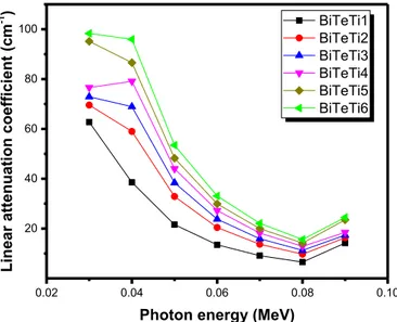
Fig. 3andFig. 4present theμ results of the BiTeTi1– BiTeTi6 glasses and PbTeB1– PbTeB6 glasses respectively. From the curves, the μ is proportionally increased with the addition of TeO2(from 45 to 55 mol% in thefirst series and from 10 to 60 mol% in the second one). This is attributed to the increase of the sample density with increasing TeO2 content and it is known that theμ is depends directly upon the density of the medium. For example, due to increase in the density from 4.67 to 5.95 g/cm3 in the first series, the μ increased from 76.68 to 96.09 cm−1at 30 keV. Similarly, the μ increased from 62.75 to 98.32 cm−1at the same energy (i.e.30 keV) due to the increase in the density for the samples in the second series from 4.58 to 6.52 g/cm3. This finding implies that there is a decreasing tendency in the X-ray photon transmission corresponding with an increase in the TeO2 con-tent in the glasses (or increase the density). Accordingly, the selected tellurite based glasses shielding capability has been increased by the addition of TeO2. Therefore, we can state that the high density glass specimen can effectively absorb the X-ray and used as an alternative protective mask during diagnostic radiation of the head or oral cavity.
It is evident fromFigs. 3andFig.4that the energy is another factor affecting the X-ray attenuation for the glasses. Firstly, the μ is high which means that the magnitude of attenuation is very high and this is attributed to predominant of the photoelectric effect. This process de-pends extremely on the atomic number and the Te, Pb and Bi elements have a significant contribution on this process at low energy, so we noticed the highμ values for low energy.
Moreover, in Fig. 5, the μ of BiTeTi6 and PbTeB6 glasses when compared with four ordinarily utilized protection materials showed some promising outcomes. As BiTeTi6 and PbTeB6 glasses have the highestμ values in series one and two, so we selected these two samples and compared them with barium–Bismuth–Borosilicate glass (G5) [26], erbium zinc tellurite glass (D5) [27], RS-360 glass (contains 45 mol% of PbO) [28] and Ferrite concrete [29]. It is clear that PbTeB6 has higherμ than BiTeTi6 and the both samples have higherμ than the materials used for the sake of comparison. Hence, the selected tellurite based glasses are more effective shielding materials.
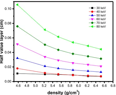
Figs. 6 and 7show the relationship between HVL and sample den-sity of Bi2O3– B2O3– TeO2– TiO2and PbO–ZnO–TeO2–B2O3glass sys-tems between 30 and 80 keV. These twofigures show that the HVL Fig. 3. The linear attenuation coefficient for Bi2O3– B2O3– TeO2– TiO2 glass
system between 30 keV to 90 keV.
Fig. 4. The linear attenuation coefficient for PbO–ZnO–TeO2–B2O3 glass system between 30 keV to 90 keV.
Fig. 5. Chart of linear attenuation coefficients of BiTeTi6 and PbTeB6 samples and other commonly used materials for shielding.
Fig. 6. The variation of half value layer (cm) with the density for Bi2O3– B2O3– TeO2– TiO2 glass system.
decreases as the density increases and this decreasing in HVL is very notable at 70 and 80 keV. The heavier the glass sample, the lower is the value of HVL and can be used as effective x-ray protection materials. As expected, the HVL depends highly on the density of the specimen and hence this parameter can satisfactorily characterize the influence of TeO2 content on the attenuation capability of the tellurite glasses against X-rays. The heavy elements in the glasses (Te, Pb and Bi) pre-sent larger target for the X-ray photons to strike and therefore the likelihoods of interactions are comparatively high and increased with the more amount of TeO2. So, the attenuation should be comparatively high and the X-rays photons have less probability of being transmitted through the high density glass sample. This fact explains the low HVL value for the two samples BiTeTi6 (ρ = 5.95 g/cm3
) and PbTeB6 (ρ = 6.52 g/cm3). Thisfinding is in agreement with that of Agar et al. [30], in which the HVL for P2O5, BaO and MoO3glasses decrease with increasing the density. The same conclusion was reported by Ersundu et al. [31].
Also, fromFigs. 6andFig.7we can see that the maximum HVL for all glasses takes place at 80 keV. This implies that the HVL gradually
increases as the energy of the X-ray photons increases. With the in-crement of the X-rays energy from 30 to 80 keV, the HVL of BiTeTi6 increased from 0.0072 to 0.1023 cm, and for PbTeB6 it increased from 0.0070 to 0.1009 cm. The present dependence of HVL on the energy suggests that the penetrating power of X-ray photons significantly in-creases with increasing the energy of the photons. It can be also noticed fromFig.6andFig. 7that the HVL changes slightly at 30 and 40 keV. This suggests that it is preferable to increase the thickness of the spe-cimen for the applications which require high X-rays photons (E > 40 keV).
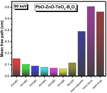
InFig. 8andFig. 9we plot the MFP of the Bi2O3– B2O3– TeO2– TiO2 glass system in comparison with some nuclear engineering materials at 30 keV and 80 keV respectively. The nuclear engineering materials used for the sake of comparison are one commercial glass contains 71 mol% of lead (coded as RS-520, with density = 5.2 g/cm3) [32], three high density concretes namely ferro boron concrete (contains 72% Fe with Fig. 7. The variation of half value layer (cm) with the density for
PbO–ZnO–TeO2–B2O3 glass system.
Fig. 8. Comparison between the mean free path (cm) for Bi2O3– B2O3– TeO2– TiO2 glass system with some nuclear engineering materials at 30 keV.
Fig. 9. Comparison between the mean free path (cm) for Bi2O3– B2O3– TeO2– TiO2 glass system with some nuclear engineering materials at 80 keV.
Fig. 10. Comparison between the mean free path (cm) for
PbO–ZnO–TeO2–B2O3 glass system with some nuclear engineering materials at 30 keV.
density = 3.5 g/cm3) [33], steel magnetite (contains 75.73% Fe, with density = 5.11 g/cm3) and steel-scrap (contains 61.25% Fe, with den-sity = 4 g/cm3) [34]. In Fig. 10andFig. 11we plot the MFP of the PbO–ZnO–TeO2–B2O3glasses in comparison with the aforementioned nuclear engineering materials at 30 keV and 80 keV respectively.
From thesefigures, it is noticed that the increment of TO2in the glasses (in both systems) leads to a decrease in MFP at 30 and 80 keV (this is also true for 40, 50, 60 and 70 keV but we didn't show the re-sults). BiTeTi6 and PbTeB6 have the lowest MFP among the samples under investigation and this is in agreement with the HVL results. This is because TeO2 increases the density which is related to μ of the samples and MFP is the reciprocal ofμ. FromFigs. 8andFig.9we can observe that the three heavy concretes have higher MFP than BiTeTi1– BiTeTi6 glasses indicating that the Bi2O3– B2O3– TeO2– TiO2 glass system can be used as protection glasses at dental diagnostic energies. The results show that the MFP of BiTeTi6 is comparable with RS-520 at 30 keV and slightly lower than RS-520 at 80 keV. InFigs. 10andFig.11 also the results show that the MFP of PbO–ZnO–TeO2–B2O3glasses are lower than steel magnetite, ferro boron and steel-scrap concretes at 30 and 80 keV, while RS-520 has comparable MFP with PbTeB6 at 30 keV.
4. Conclusion
The X-ray attenuation properties of BiTeTi1– BiTeTi6 and PbTeB1– PbTeB6 glasses have been reported. The simulatedμ/ρ was validated by a comparison with the results obtained by WinXcom. The correlation coefficient (R2
) is used to evaluate the extent to which Geant4 results are related to the WinXcom data. Theμ is proportionally increased with the addition of TeO2in both series, which implies that there is a de-creasing tendency in the X-ray photon transmission corresponding with an increase in the density of the samples. The HVL reduces as the density increases especially at 70 and 80 keV. The maximum HVL for all samples occurs at 80 keV and this suggested that the HVL increases as the energy of the X-ray photons increase. Furthermore, the increment of TO2in the both systems leads to a decrease in MFP at all energies and BiTeTi6 and PbTeB6 samples have the lowest MFP. We compared the MFP of both systems with steel magnetite, ferro boron and steel-scrap concretes and the comparison revealed that the selected glasses can be utilized to fabricate protection masks used during diagnostic radiation of the head or oral cavity.
Acknowledgment
This project was funded by the Deanship of Scientific Research, King Abdulaziz University, Jeddah, under grant No. (D-291-130-1440). The authors, therefore, gratefully acknowledge the DSR technical and fi-nancial support.
References
[1] Anil Kumar Singh, Rakesh Kumar Singh, Bhupesh Sharma, Ajay Kumar Tyagi, Characterization and biocompatibility studies of lead free X-ray shielding polymer composite for healthcare application, Radiat. Phys. Chem. 138 (2017) 9–15. [2] Mengge Dong, Xiangxin Xu, Shan Liu, Yang He, Zhefu Li, M.I. Sayyed, O. Agar,
Using iron concentrate in Liaoning Province, China, to prepare material for X-Ray shielding, J. Clean. Prod. 210 (2019) 653e659.
[3] Karoline Günther, Christina Giebing, Antonia Askani, Tilmann Leisegang, Marcus Krieg, Yordan Kyosev, Thomas Weide, Boris Mahltig, Cellulose/inorganic-compositefibers for producing textile fabrics of high X-ray absorption properties, Mater. Chem. Phys. 167 (2015) 125–135.
[4] M.R. Kaçal, F. Akman, M.I. Sayyed, F. Akman, Evaluation of gamma-ray and neu-tron attenuation properties of some polymers, Nucl. Eng. Technol. 51 (2019) 818–824.
[5] Ji Woo Hong, Dae Ho Kim, Seok Won Kim, Seong Hoon Choi, Ga-Eul Lee, Hyun-Kyu Seo, Sang-Hyun Kim, Youngjin Lee, Effectiveness evaluation of self-produced micro- and nanosized tungsten materials for radiation shielding with diagnostic X-ray imaging system, Optik - Int. J. Light Electron Opt. 172 (2018) 760–765. [6] F. Akman, I.H. Geçibesler, A. Kumar, M.I. Sayyed, M.H.M. Zaid, Evaluation of
ra-diation absorption characteristics in different parts of some medicinal aromatic plants in the low energy region, Res. Phys. 12 (2019) 94–100.
[7] Munirah Jamil, Muhammad Hazritz Hazlan, Ramzun Maizan Ramli, Nurul Zahirah Noor Azman, Study of electrospun PVA-based concentrations nanofibre filled with Bi2O3 or WO3 as potential x-ray shielding material, Radiat. Phys. Chem. 156 (2019) 272–282.
[8] N.Z. Noor Azman, N.F.L. Musa, N.N.A. Nik Ab Razak, R.M. Ramli, I.S. Mustafa, A. Abdul Rahman, N.Z. Yahaya, Effect of Bi2O3 particle sizes and addition of starch into Bi2O3–PVA composites for X-ray shielding, Appl. Phys. A 122 (2016). [9] S. Kaewjaeng, S. Kothan, W. Chaiphaksa, N. Chanthima, R. Rajaramakrishna,
H.J. Kim, J. Kaewkhao, High transparency La2O3-CaO-B2O3-SiO2 glass for diag-nosis x-rays shielding material application, Radiat. Phys. Chem. 160 (2019) 41–47. [10] El-Sayed A. Waly, Ghada Shkoukani Al-Qous, Mohamed A. Bourham, Shielding
properties of glasses with different heavy elements additives for radiation shielding in the energy range 15–300 keV, Radiat. Phys. Chem. 150 (2018) 120–124. [11] M. Almatari, O. Agar, E.E. Altunsoy, O. Kilicoglu, M.I. Sayyed, H.O. Tekin, Photon
and neutron shielding characteristics of samarium doped lead alumino borate glasses containing barium, lithium and zinc oxides determined at medical diag-nostic energies, Results Phys. 12 (2019) 2123–2128.
[12] H.O. Tekin, E.E. Altunsoy, E. Kavaz, M.I. Sayyed, O. Agar, M. Kamislioglu, Photon and neutron shielding performance of boron phosphate glasses for diagnostic radiology facilities, Res. Phys. 12 (2019) 1457–1464.
[13] W. Hongtong, S. Kaewjaeng, S. Kothan, P. Meejitpaisan, W. Cheewasukhanont, P. Limkitjaroenporn, J. Kaewkhao, Development of gadolinium doped calcium phosphate oxyfluoride glasses for X-ray shielding materials, Mater. Today: Proc. 5 (2018) 14063–14068.
[14] Helena Ticha, Jiri Schwarz, Ladislav Tichy, Raman spectra and optical band gap in some PbO-ZnO-TeO2 glasses, Mater. Chem. Phys. 237 (2019) 121834.
[15] N. Gedikoğlu, A.E. Ersundu, S. Aydin, M. Çelikbilek Ersundu, Crystallization be-havior of WO3-MoO3-TeO2 glasses, J. Non-Cryst. Solids 501 (2018) 93–100. [16] Kyong-Soo Hong, Miae Kim, Myoung Gyu Ha, Jong Pil Kim, Jang-Hee Yoon, Jong
Hwa Kim, Ho-Soon Yang, Hyun Gyu Kim, Red-emission properties and crystal-lization behavior in Eu2O3-TeO2 glasses, J. Non-Cryst. Solids 505 (2019) 400–405. [17] N. Ghribi, M. Dutreilh-Colas, J.R. Duclère, T. Hayakawa, J. Carreaud, R. Karray,
A. Kabadou, P. Thomas, Thermal, optical and structural properties of glasses within the TeO2-TiO2-ZnO system, J. Alloy. Comp. 622 (2015) 333–340.
[18] M.I. Sayyed, Bismuth modified shielding properties of zinc boro-tellurite glasses, J. Alloy. Comp. 688 (2016) 111–117.
[19] N. Elkhoshkhany, Rafik Abbas, R. El-Mallawany, S.F. Hathot, Optical properties and crystallization of bismuth boro-tellurite glasses, J. Non-Cryst. Solids 476 (2017) 15–24.
[20] Helena Ticha, Jiri Schwarz, Ladislav Tichy, The structural arrangement and the optical band gap in certain Quaternary PbO–ZnO–TeO2–B2O3 glasses, J. Non-Cryst. Solids 489 (2018) 40–44.
[21] M. Mansur Tufekci, Ahmet Gokce, Development of heavyweight high performance fiber reinforced cementitious composites (HPFRCC) – Part II: X-ray and gamma radiation shielding properties, Constr. Build. Mater. 163 (2018) 326–336. [22] L. Gerward, N. Guilbert, K.B. Jensen, H. Levring, WinXCom - a program for
cal-culating X-ray attenuation coefficients, Radiat. Phys. Chem. 71 (3–4) (2004) 653–654.
[23] S. Agostinelli, J. Allison, K.a. Amako, J. Apostolakis, H. Araujo, P. Arce, M. Asai, D. Axen, S. Banerjee, G. Barrand, et al., Geant4a simulation toolkit, Nucl. Instrum. Methods Phys. Res. Sect. A Accel. Spectrom. Detect. Assoc. Equip. 506 (2003) 250–303.
[24] F. Akman, M.R. Kaçal, M.I. Sayyed, H.A. Karatas, Study of gamma radiation at-tenuation properties of some selected ternary alloys, J. Alloy. Comp. 782 (2019)
Fig. 11. Comparison between the mean free path (cm) for
PbO–ZnO–TeO2–B2O3glass system with some nuclear engineering materials at 80 keV.
315–322.
[25] M.I. Sayyed, F. Akman, A. Kumar, M.R. Kacal, Evaluation of radioprotection properties of some selected ceramic samples, Res. Phys. 11 (2018) 1100–1104. [26] R. Bagheri, A. Khorrami Moghaddam, H. Yousefnia, Gamma ray shielding study of
bariumebismutheborosilicate glasses as transparent shielding materials using MCNP-4C code, XCOM program, and available experimental data, Nucl. Eng. Technol. 49 (2017) 216–223.
[27] D5, S.A. Tijani, Salahuddin M. Kamal, Y. Al-Hadeethi, Mehenna Arib, M.A. Hussein, S. Wageh, L.A. Dim, Radiation shielding properties of transparent erbium zinc tellurite glass system determined at medical diagnostic energies, J. Alloy. Comp. 741 (2018) 293–299.
[28] Parminder Kaur, K.J. Singh, Sonika Thakur, Prabhjot Singh, B.S. Bajwa, Investigation of bismuth borate glass system modified with barium for structural and gamma-ray shielding properties, Spectrochim. Acta A Mol. Biomol. Spectrosc. 206 (2019) 367–377.
[29] K.J. Singh, N. Singh, R.S. Kaundal, K. Singh, Gamma-ray shielding and structural properties of PbO–SiO2 glasses, Nucl. Instrum. Methods Phys. Res. B 266 (2008) 944–948.
[30] O. Agar, M.I. Sayyed, H.O. Tekin, Kawa M. Kaky, S.O. Baki, I. Kityk, An in-vestigation on shielding properties of BaO, MoO3 and P2O5 based glasses using MCNPX code, Res. Phys. 12 (2019) 629–634 2019.
[31] A.E. Ersundu, M. Büyükyıldız, M. Çelikbilek Ersundu, E. Şakar, M. Kurudire, The heavy metal oxide glasses within the WO3-MoO3-TeO2 system to investigate the shielding properties of radiation applications, Prog. Nucl. Energy 104 (2018) 280–287.
[32] Parminder Kaur, K.J. Singh, Sonika Thakur, Prabhjot Singh, B.S. Bajwa, Investigation of bismuth borate glass system modified with barium for structural and gamma-ray shielding properties, Spectrochim. Acta A Mol. Biomol. Spectrosc. 206 (2019) 367–377.
[33] Muhammad Khairi Azri Roslan, Mohammad Ismail, Ahmad Beng Hong Kueh, Muhammad Rawi Mohamed Zin, High-density concrete: exploring Ferro boron ef-fects in neutron and gamma radiation shielding, Constr. Build. Mater. 215 (2019) 718–725.
[34] I.I. Bashter, Calculation of radiation attenuation coefficients for shielding concretes, Ann. Nucl. Energy 24 (1997) 1389–1401.