doi:10.1152/ajpheart.00358.2008
296:202-210, 2009. First published Nov 14, 2008; Am J Physiol Heart Circ Physiol
Marco Pallante, Paolo Di Nardo and Alberto Galante
Jacopo M. Legramante, Sergio Sacco, Gianfranco Raimondi, Vito N. Di Lecce,
You might find this additional information useful...
15 articles, 11 of which you can access free at: This article cites
http://ajpheart.physiology.org/cgi/content/full/296/1/H202#BIBL
including high-resolution figures, can be found at: Updated information and services
http://ajpheart.physiology.org/cgi/content/full/296/1/H202
can be found at: AJP - Heart and Circulatory Physiology
about Additional material and information
http://www.the-aps.org/publications/ajpheart
This information is current as of January 3, 2009 .
http://www.the-aps.org/. ISSN: 0363-6135, ESSN: 1522-1539. Visit our website at
Physiological Society, 9650 Rockville Pike, Bethesda MD 20814-3991. Copyright © 2005 by the American Physiological Society. intact animal to the cellular, subcellular, and molecular levels. It is published 12 times a year (monthly) by the American
lymphatics, including experimental and theoretical studies of cardiovascular function at all levels of organization ranging from the publishes original investigations on the physiology of the heart, blood vessels, and
AJP - Heart and Circulatory Physiology
on January 3, 2009
ajpheart.physiology.org
Investigating feedforward neural regulation of circulation from analysis
of spontaneous arterial pressure and heart rate fluctuations in conscious rats
Jacopo M. Legramante,1–4Sergio Sacco,1,3,4Gianfranco Raimondi,5Vito N. Di Lecce,2Marco Pallante,1,4
Paolo Di Nardo,1and Alberto Galante1–3
1Dipartimento di Medicina Interna, Universita` “Tor Vergata,” Roma,2Medicina d’Urgenza Policlinico Tor Vergata, Roma, 3S. Raffaele Hospital, Velletri,4Stazione per la Tecnologia Animale, Universita` “Tor Vergata,” Roma,
and5Dipartimento di Medicina Sperimentale, Universita` La Sapienza, Roma, Italy
Submitted 5 April 2008; accepted in final form 5 November 2008
Legramante JM, Sacco S, Raimondi G, Di Lecce VN, Pallante M, Di Nardo P, Galante A. Investigating feedforward neural
regu-lation of circuregu-lation from analysis of spontaneous arterial pressure and heart rate fluctuations in conscious rats. Am J Physiol Heart Circ Physiol 296: H202–H210, 2009. First published November 14, 2008; doi:10.1152/ajpheart.00358.2008.—It has been suggested in anesthe-tized animals that the occurrence of sequences of consecutive beats characterized by systolic arterial pressure (SAP) and RR or pulse interval (PI) changing in the opposite direction (SAP⫹/RR⫺ and SAP⫺/RR⫹, nonbaroreflex sequences) might represent the expression of neural cardiovascular regulatory mechanisms operating with feed-forward characteristics. The aim of the present study was to study nonbaroreflex sequences in a more physiological experimental model, i.e., in conscious freely moving rats. We studied conscious rats before and after 1) complete autonomic blockade (n⫽ 12), 2) sympathetic blockade (n⫽ 10), 3) ␣ (n ⫽ 7)- and  (n ⫽ 8)-adrenergic blockade, and 4) parasympathetic blockade (n⫽ 10). Nonbaroreflex sequences were defined as three or more beats in which SAP and PI of the following beat changed in the opposite direction. Complete autonomic blockade reduced the number of nonbaroreflex sequences (95.6⫾ 9.0 vs. 45.2⫾ 4.1, P ⬍ 0.001), as did sympathetic blockade (80.9 ⫾ 12.6 vs. 30.9⫾ 6.1, P ⬍ 0.001). The selective ␣-receptor blockade did not induce significant changes (80.9⫾ 12.5 in baseline vs. 79.0 ⫾ 14.7 after prazosin), whereas -receptor blockade significantly reduced nonbaroreflex sequence occurrence (80.9 ⫾ 12.5 in baseline vs. 48.9⫾ 15.3 after propranolol). Parasympathetic blockade produced a significant increase of nonbaroreflex sequences (95.1 ⫾ 6.9 vs. 136.0⫾ 12.4, P ⬍ 0.01). These results demonstrate the physiological role of the nonbaroreflex sequences as an expression of a feedforward type of short-term cardiovascular regulation able to interact dynami-cally with the feedback mechanisms of baroreflex origin in the neural control of the sinus node.
feedforward mechanisms; negative feedback mechanisms; barorecep-tors; nervous system autonomic; nervous system sympathetic
IN THE INTACT CIRCULATION beat-by-beat spontaneous
fluctua-tions in the RR interval have been shown to be linked to beat-by-beat spontaneous changes in arterial pressure (AP) through baroreflex mechanisms. An analysis of the contin-uous relationship between systolic AP (SAP) and RR inter-val revealed that spontaneous increases or decreases in SAP induce directionally similar reflex changes in the RR inter-val (4). On this basis, the “spontaneous baroreflex” or “sequence” technique (1) is able to extract from SAP and RR, or pulse interval (PI) time-series sequences of
sponta-neously occurring consecutive beats in which SAP and PI of the following beat change in the same direction, i.e., hyper-tensive/bradycardic (SAP⫹/RR⫹) and hypotensive/tachy-cardic (SAP⫺/RR⫺) sequences (5, 6). These sequences have been named “baroreflex sequences” and have been consid-ered as an expression of negative feedback mechanisms of baroreflex origin (1, 7).
In contrast, sequences of consecutive beats in which SAP and RR interval (or PI) of the following beat change in the opposite direction [i.e., hypertensive/tachycardic (SAP⫹/ RR⫺), and hypotensive/bradycardic (SAP⫺/RR⫹) sequences] have been reported. These sequences have been defined “non-baroreflex,” and their physiological meaning is not clear.
Based on the consideration that reflex cardiovascular mech-anisms characterized by changes in the same direction of AP and heart rate (HR) (hypertension/tachycardia and hypoten-sion/bradycardia) could contribute to the neural regulation of the cardiovascular system (9, 12, 15), our group performed an experimental study on anesthetized rabbits testing the hypoth-esis that nonbaroreflex sequences represent an expression of the cardiovascular regulatory mechanisms modulated by the autonomic nervous system and operating with feedforward characteristics (7). A complete autonomic blockade drastically and significantly reduced the occurrence of nonbaroreflex se-quences, thus indicating that they were primarily mediated by the autonomic nervous system. In addition, the occurrence of nonbaroreflex sequences was significantly reduced in response to both sympathetic and parasympathetic blockades (7). Even though we concluded that nonbaroreflex sequences might be the expression of neural mechanisms regulating heart beats and operating with positive feedback characteristics, our results did not allow us to completely and definitively clarify the physio-logical modulation exerted by the autonomic nervous system on the occurrence of nonbaroreflex sequences.
The sympathetic contribution to the occurrence of the non-baroreflex sequences was in line with the pivotal study of Pagani et al. (15) who showed the existence of positive feed-back sympathetic cardiovascular reflexes causing hypertension and tachycardia, elicitable by mechanically stretching the tho-racic aorta and reduced or abolished after pharmacological sympathetic blockade (15). Moreover, the existence of cardiac sympathetic reflexes characterized by changes of AP and HR in the same direction has been largely reported in anesthetized animals (9 –11, 16). However, nonbaroreflex sequences
ap-Address for reprint requests and other correspondence: J. M. Legramante, Dip. Medicina Interna, Univ. di Roma “Tor Vergata,” Via Montpellier 1, 00133 Rome, Italy (e-mail: [email protected]).
The costs of publication of this article were defrayed in part by the payment of page charges. The article must therefore be hereby marked “advertisement” in accordance with 18 U.S.C. Section 1734 solely to indicate this fact. First published November 14, 2008; doi:10.1152/ajpheart.00358.2008.
on January 3, 2009
ajpheart.physiology.org
peared to be also under the control of the parasympathetic system, as shown by their significant reduction in response to atropine in anesthetized animals (7).
Even though the complex nature of neural cardiovascular regulation could in part explain these results, we also consid-ered the possibility that the anesthetic agents could have abolished some neural traffic, thus interfering with the physi-ological modulation of the neural mechanisms of cardiovascu-lar regulation. More importantly, the possible selective modu-lation of nonbaroreflex sequences exerted by an␣- and -ad-renergic component of the sympathetic nervous system has not been studied.
Consequently, our aim was to study nonbaroreflex se-quences in a more physiological setting. In the present study, we evaluated the occurrence of nonbaroreflex sequences in conscious, freely moving rats before and after autonomic blockades, and in particular after selective sympathetic block-ades, to test their possible role as an expression of neurogenic cardiovascular regulatory mechanisms controlling the sinus node and operating with feedforward characteristics.
MATERIALS AND METHODS
General procedures. The study was performed on 22 adult Wistar-Kyoto (WKY) rats of both sexes (350 g body wt), and the experi-mental procedures were carried out according to the Association for Assessment and Accreditation of Laboratory Care International and approved by the animal care facility (Stazione per la Tecnologia Animale) of the University “Tor Vergata” and by the Italian Health Minister. Rats were used and housed individually in the animal care facility, allowed normal rat chow and drinking water ad libitum, and kept on a 12-h:12-h light-dark cycle.
Surgical procedures. After having induced the anesthesia by ket-amine (Ketavet 50, 60 mg/kg ip) and medetomidine (Domitor, 0.3 mg/kg ip), a telemetry transmitter (TA11PA-C40, Data Sciences, St. Paul, MN) was implanted for recordings of AP signals according to manufacturer specifications. The tip of the arterial catheter was inserted into the abdominal aorta previously exposed by a midline incision via a hole made by a 21-gauge needle below the bifurcation of the renal arteries just proximal to the iliac bifurcation and secured in place with tissue glue (Vetbond, 3M). The transmitter body was attached to the abdominal wall along the incision line with sutures as the incision was closed. A jugular vein was cannulated by a poly-ethylene cannula that was tunneled subcutaneously and exteriorized at the dorsal cervical region for the infusions of the drugs. The catheter was filled with heparinized saline and capped with an airtight plug. After surgery, the rats were given antibiotics (ceftriaxone) and housed individually in cages for 5–7 days of recovery before any experimen-tal protocol began.
Measurement of AP using radiotelemetry. The system used to record AP consists of three basic elements: 1) a transmitter for monitoring blood pressure (TA11PAC40), 2) a receiver (RPC-1), and 3) an adapter (R11CPA) with an ambient pressure monitor (APR-1) that produces analog output signals of pulsatile AP. The telemetered AP signal was digitized using an analog I/O PC card (National Instrument 6024E, Austin, TX) at a rate of 2,000 Hz, displayed on the computer screen, and processed by an algorithm based on feature extraction to detect and measure the characteristics of AP cycles developed in our laboratory, based on a LabView platform software. PI was measured from the pressure pulses and used to calculate HR. Experimental protocol. We tested three experimental animal groups. Animals were allowed to familiarize themselves with the laboratory environment for 30 min before starting the recording. The three sets consisted of 1) sympathetic blockade, 2) complete auto-nomic blockade, and 3) parasympathetic blockade.
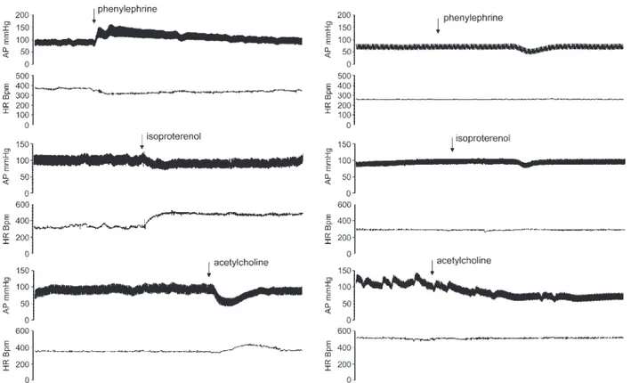
Sympathetic autonomic blockades. Ten rats were studied in base-line conditions and after sympathetic autonomic pharmacological blockade (propranolol, 2 mg/kg iv; plus prazosin, 1 mg/kg iv). In seven rats, we also studied the effect of the selective blockade of ␣-receptors by recording AP in the baseline condition and after the pharmacological blockade (prazosin, 1 mg/kg iv), whereas eight rats were studied in baseline conditions and after selective blockade of -receptors (propranolol, 2 mg/kg iv). The effectiveness of the auto-nomic blockades was tested by measuring the hypertensive responses to phenylephrine (5g/kg iv) and the tachycardic response to isopro-terenol (4g/kg iv), and only the rats in which the responses were abolished by the blocking drugs were accepted for the study (Fig. 1). Complete autonomic blockades. Twelve rats were studied in base-line conditions and after complete autonomic pharmacological block-ade (hexamethonium, 20 mg/kg iv; plus propranolol, 2 mg/kg iv; plus prazosin, 1 mg/kg, and atropine sulfate, 1.5 mg/kg iv). The effective-ness of the autonomic blockades was tested by measuring the cardio-vascular responses to phenylephrine (5 g/kg iv), isoproterenol (4 g/kg iv), and acetylcholine (1 to 2 g/kg iv), and only the rats in which the responses were abolished by the blocking drugs were accepted for the study (Fig. 1).
Parasympathetic blockades. Ten of the twelve rats used for com-plete autonomic blockade experiments (2 rats were discarded for the presence of artifacts in the AP recording) were studied before and after parasympathetic pharmacological blockade (atropine sulfate, 1.5 mg/kg iv). The effectiveness of parasympathetic blockade was tested by measuring the reflex tachycardic responses to acetylcholine (1 to 2 g/kg iv), and only the rats in which the responses were abolished by the blocking drugs were accepted for the study (Fig. 1).
In each experimental condition, we recorded AP for 10 min while the animal was freely moving in the cage. The effectiveness of the autonomic blockade along the whole recording period is demonstrated by the stable cardiovascular values over the entire 10-min recording period (Table 1, and Fig. 2).
Sequence analysis. The sequence analysis was performed as pre-viously reported (7, 8). Briefly, the beat-by-beat time series of SAP and PI were analyzed by a computer to identify spontaneously occurring sequences of three or more consecutive beats in which SAP and PI of the fifth heart beat (i.e., lag 5) (14) changed in the opposite direction, e.g., SAP increasing and PI decreasing (i.e., hypertension and tachycardia) or SAP decreasing and PI increasing (i.e., hypoten-sion and bradycardia). These sequences were identified as “nonbarore-flex” sequences (Fig. 3). A linear regression was applied to each individual sequence, similarly to the Oxford technique employing bolus injections of vasoactive drugs. Only those sequences in which r2
was ⬎0.85 were accepted. The number of nonbaroreflex sequences was calculated. Separate calculations were made for sequences in which SAP increased and PI decreased (SAP⫹/RR⫺, hypertension/ tachycardia) and for those in which SAP decreased and PI increased (SAP⫺/RR⫹, hypotension/bradycardia).
The mean individual slope of the nonbaroreflex sequences, ob-tained by averaging all slopes computed within a given experimental period, was calculated and taken as a measure of the gain of non-baroreflex mechanisms for that period.
The engagement time was also calculated as the fractional occur-rence of the sequences independently on recording time, HR, and the number of heart beats that composed each sequence, as previously reported (8). Briefly, this index has been obtained by dividing the sum of the RR organized in sequences, according to the criteria reported above, by the total recording duration and multiplying it by 100.
The beat-by-beat time series of SAP and PI were also searched for sequences of three or more consecutive beats in which SAP and PI of the same (i.e., lag 0), of the following (i.e., lag 1), and of the second (i.e., lag 2) beat changed in the opposite direction.
Data analysis and statistics. AP data were stored and analyzed by a computerized online system for biological data elaboration devel-oped in our laboratory based on a LabView platform software.
H203
CARDIOVASCULAR FEEDFORWARD NEURAL MECHANISMS
AJP-Heart Circ Physiol•VOL 296 • JANUARY 2009 •www.ajpheart.org
on January 3, 2009
ajpheart.physiology.org
Within-group changes in the reported variables were evaluated by a paired t-test for normally distributed variables and by a Wilcoxon signed rank test for non-normally distributed variables. Between-group changes in the reported variables were evaluated by a one-way ANOVA test. All data are presented as means⫾ SE. A value of P ⬍ 0.05 was considered statistically significant.
RESULTS
No significant differences were found for cardiovascular parameters (Table 2) and for the occurrence of nonbaroreflex sequences (Fig. 4) in the baseline conditions in the three experimental sets.
About 8% of the whole cardiac beats were organized in nonbaroreflex sequences (Figs. 4 and 5), thus showing a greater occurrence of these sequences within the spontaneous AP and HR fluctuations compared with previous studies in anesthe-tized animals (7).
In control conditions, before complete autonomic blockade, the number of spontaneous nonbaroreflex sequences were sig-nificantly lower for lag 5 (95.6 ⫾ 9.0, P ⬍ 0.01) compared with lags 0 and 1 (155.0 ⫾ 16.1 and 134.1 ⫾ 12.3, not significant, respectively). The baseline number of lag 2 non-baroreflex sequences (118.1 ⫾ 13.8) was greater compared with lag 5 without attaining statistical significance. A similar trend was observed before sympathetic and parasympathetic blockade.
Complete autonomic blockade induced a significant de-crease in AP and a significant inde-crease in the RR interval (Table 3).
The occurrence of nonbaroreflex sequences (lag 5) was significantly decreased by complete autonomic blockade (Fig. 5). This reduction was due to a substantial decrease in the number and in the engagement time of both hypertensive/ tachycardic and hypotensive/bradycardic sequences (Fig. 5). The mean slope did not show significant changes for non-baroreflex sequences (5.6 ⫾ 1.0 vs. 4.9 ⫾ 1.0 ms/mmHg).
Similarly, also the occurrence of lags 0, 1, and 2 nonbarore-flex sequences was decreased by complete autonomic blockade (Fig. 6).
Sympathetic blockade induced a significant decrease in AP, and a significant increase in the RR interval (Table 3)
associ-Fig. 1. Original recordings of arterial pressure (AP) and heart rate (HR) responses to phenylephrine, isoproterenol, and acetylcholine before (left) and after (right) selective autonomic pharmacological blockades (seeMATERIALS AND METHODS). AP recordings and HR tachograms clearly show that after prazosin, propranol, and atropine, respectively, the cardiovascular responses were abolished, thus indicating the effectiveness of the pharmacological autonomic blockades. Bpm, beats/min.
Table 1. Comparison of the cardiovascular values
at the beginning and at the end of the recording period during the autonomic blockades
SAP, mmHg DAP, mmHg RR, ms CAB 1 min 88.4⫾3.9 70.4⫾2.6 257.0⫾19.3 Last minute 87.0⫾4.0 68.8⫾2.7 258.4⫾18.0 A 1 min 111.6⫾5.1 91.0⫾3.6 146.7⫾6.7 Last min 111.0⫾6.5 89.2⫾4.7 146.9⫾10.3 SB 1 min 100.3⫾2.5 79.9⫾4.1 226.1⫾5.8 Last min 103.7⫾3.0 82.3⫾4.6 232.3⫾7.1 Values are means ⫾ SE. AP, arterial pressure; SAP, systolic AP; DAP, diastolic AP; RR, RR interval; CAB, complete autonomic blockade; A, parasympathetic blockade (atropine); SB, sympathetic blockade; 1 min, first minute of AP recording; last min, last minute of AP recording.
on January 3, 2009
ajpheart.physiology.org
ated to a significant decrease in the occurrence of lag 5 nonbaroreflex sequences (Fig. 5), which involved both the hypertensive/tachycardic and the hypotensive/bradycardic se-quences (Fig. 5).
Again the mean slope did not show significant changes for nonbaroreflex sequences (4.7⫾ 1.1 vs. 4.6 ⫾ 1.2 ms/mmHg). Similarly, also the occurrence of lags 0, 1, and 2 nonbaroreflex sequences was decreased by selective sympathetic blockade (Fig. 6).
Selective ␣-receptor blockade did not induce significant changes in the occurrence (80.9 ⫾ 12.5 in baseline vs. 79.0⫾ 14.7 after prazosin) and in the mean slope (4.7 ⫾ 1.1 in baseline vs. 5.6 ⫾ 1.0 ms/mmHg after prazosin) of
nonbaroreflex sequences. On the contrary, -receptors blockade induced a significant reduction in the occurrence (80.9 ⫾ 12.5 in baseline vs. 48.9 ⫾ 15.3 after propranolol) but not in the mean slope (4.7⫾ 1.1 in baseline vs. 4.2 ⫾ 1.0 ms/mmHg after propranolol) of nonbaroreflex sequences.
Following the parasympathetic blockade, the RR interval significantly decreased, whereas AP slightly increased (Table 3) and the occurrence of lag 5 nonbaroreflex sequences signif-icantly increased (Fig. 5). The mean slope of the nonbaroreflex sequences significantly decreased (Fig. 7).
Whereas lag 0 nonbaroreflex sequences showed an incon-sistent trend to decrease in response to atropine, the occurrence of lags 1 and 2 nonbaroreflex sequences showed a substantial
Fig. 2. Original recordings of AP and HR recordings during sympathetic (SB), parasympathetic (atropine; A), and complete autonomic blockade (CAB). AP and HR values clearly show stable conditions during the entire recording periods.
H205
CARDIOVASCULAR FEEDFORWARD NEURAL MECHANISMS
AJP-Heart Circ Physiol•VOL 296 • JANUARY 2009 •www.ajpheart.org
on January 3, 2009
ajpheart.physiology.org
and significant increase after parasympathetic blockade similar to the lag 5 nonbaroreflex sequences (Fig. 6).
DISCUSSION
The main goal of the present investigation was to test the hypothesis that nonbaroreflex sequences reflect the expression of feedforward mechanisms modulated by the autonomic ner-vous system and controlling the heart period in physiological conditions.
In the present experiments performed in unanesthetized, conscious, freely moving rats, the nonbaroreflex sequences showed a significant and substantial decrease in response to sympathetic and complete autonomic blockade, whereas atro-pine induced a significant increase in the occurrence of hyper-tensive/tachycardic and hypotensive/bradycardic sequences.
These results must be considered in the context of previous reports to evaluate the possible physiological meaning of nonbaroreflex sequences.
We have hypothesized in a previous study (7) that non-baroreflex sequences could be the expression of feedforward neural mechanisms controlling heart beat because of their similarity with sympathetic reflexes originating from cardiac, pulmonary, and central vessels receptors and characterized by changes of AP and HR going in the same direction. Accord-ingly, our findings of a sympathetic contribution (7 and present data) to the occurrence of the nonbaroreflex sequences are consistent with those reported by Pagani et al. (15) who showed the existence of positive feedback sympathetic reflexes elicitable in conscious dogs by mechanically stretching the thoracic aorta.
The new finding of the present study, compared with previ-ous reports (7), is the significant and substantial increase in the occurrence of nonbaroreflex sequences following parasympa-thetic blockades.
In particular, our data show that when only the sympathetic nervous system is operative, as occurring after an experimental pharmacological blockade of the parasympathetic nervous sys-tem, the occurrence of nonbaroreflex sequences greatly in-creased. This unopposed predominance strongly suggests that nonbaroreflex sequences are mainly under the control of the sympathetic nervous system as previously reported for the sympathetic pressor reflexes mediated by positive feed-back mechanisms (15).
At variance with the present results, we have reported in anesthetized rabbits that in response to parasympathetic block-ade, the occurrence of nonbaroreflex sequences showed a
Fig. 3. Examples of the AP, ECG signals, and corresponding pulse intervals (PIs). The signals refer to sequences during which systolic AP (SAP) and PI of the following beat changed in the same direction, either increasing or decreasing (baroreflex sequences), and to sequences during which SAP and PI of the following beat changed in the opposite direction, e.g., SAP increases and PI decreases or vice versa (nonbaroreflex sequences).
Table 2. Cardiovascular values in baseline conditions
in the three experimental sets
SAP, mmHg DAP, mmHg RR, ms
Baseline 1 107.8⫾5.5 85.6⫾2.9 171.9⫾5.4 Baseline 2 107.6⫾4.7 87.5⫾3.7 176.4⫾5.8 Baseline 3 106.9⫾5.5 86.7⫾4.6 168.4⫾4.0 Values are means⫾ SE. Baseline 1, CAB; baseline 2, SB; baseline 3, A.
on January 3, 2009
ajpheart.physiology.org
similar decrease as in response to sympathetic blockade. Con-sequently, we concluded that both the branches of the auto-nomic nervous system contributed to the modulation of feed-forward neural mechanisms regulating heart beat (7). Species differences might explain the different results. However, both rabbits and rats are rodents, thus sharing, for example, the autonomic pattern regulating the cardiovascular system.
Alternatively, it is possible that anesthetic agents, used in the previous experimental setting (7), abolished, at least in part, the vagal traffic, such that by the time atropine was injected, there was not much efferent vagal activity to oppose. The possible effect of anesthetic agents is also supported by the observation that in conscious rats, a greater number of cardiac beats (about 8%) are organized in non-baroreflex sequences (Figs. 4 and 5) compared with that in previous studies in anesthetized animals (7).
Our experiments following selective sympathetic blockade give further insights into the physiological modulation of the nonbaroreflex sequences. Indeed, the occurrence of nonbarore-flex sequences seems to be modulated mainly by-adrenergic receptors as shown by their significant reduction in response to propranol. These results are in line with those obtained by
Pagani et al. (15) in conscious dogs in which the tachycardic reflex response following the activation of mechanoreceptors induced by aortic distension was abolished by -adrenergic receptor blockade. On the contrary, ␣-adrenergic receptor blockade does not seem to influence the occurrence of non-baroreflex sequences as shown by the fact that prazosin did not significantly affect the number of nonbaroreflex sequences. However, our model defines a priori AP as an input and HR as an output variable in the extrapolation of nonbaroreflex se-quences. Therefore, it is conceivable that in response to spon-taneous AP fluctuations, still existing after having blocked ␣-adrenergic receptors, the nonbaroreflex sequences can occur as a consequence of a HR modulation, possibly due to a -adrenergic stimulation. On the contrary, when the output variable is blocked, as in response to -adrenergic blockade, the spontaneous heart beat fluctuations seem to miss the pos-sibility to be organized in nonbaroreflex sequences.
As previously reported (7), the mean slope of the nonbarore-flex sequences would represent the approximate gain of the feedforward mechanisms responsible for their occurrence, as the baroreflex sensitivity reflects the mean slope of the barore-flex sequences, even though its real physiological significance cannot be elucidated with the sequences technique, and more complex methodologies are needed to clarify this point.
Whereas the mean slope behavior in response to sympathetic and complete autonomic blockade is in line with previous reports (7), showing no significant changes, it is noteworthy that the nonbaroreflex slope, in the present study, was signif-icantly and substantially reduced by atropine.
Our experimental design was not directly focused to study this issue in detail. Nonetheless, we can speculate that in a situation in which feedforward neural mechanisms are mainly operative, as during parasympathetic blockade, the feedfor-ward gain tends to decrease to buffer the possible trend to exaggerated and unopposed AP changes. Even though we have no direct experimental data, we can hypothesize that these modifications of gain are modulated at the central integration level.
We have also studied the possible physiological differences due to different lags used to pair SAP and PI in the study of nonbaroreflex sequences.
Because of the high HR in rats and to exclude the possible interferences linked to acetylcholine kinetics (3), we have used longer lag (lag 5) to pair SAP and PI, according to previous reports (14).
In line with previous reports (7), our data indicate that the lag used significantly affects the physiological meaning of sequences of spontaneously occurring consecutive heart beats. In fact, similarly to lags 5, 0, 1, and 2 nonbaroreflex sequences appear to be neurally modulated because of their significant decrease after complete autonomic and sympathetic blockade (see Fig. 6). However, lag 0 nonbaroreflex sequences seem to be, at least in part, modulated by the arterial baroreflexes as shown by their reduction in response to vagal blockade (see Fig. 6) and as supported by previous studies (2). Therefore, according to the present and previous results (2, 14), we conclude that lag 1 and 2 sequences can be considered as an expression of the feedforward neural mechanisms regulating the cardiovascular function, even though when HR is particu-larly high, as in conscious rats, the use of lag 5 sequences is reliable.
Fig. 4. Number and engagement time (in %) of nonbaroreflex sequences (lag 5) in control conditions in the 3 experimental sets. Data are shown as means⫾ SE. NB, overall nonbaroreflex sequences; SAP⫹/RR⫺, nonbaroreflex se-quences characterized by increase in SAP and decrease in PI; SAP/RR⫹, nonbaroreflex sequences characterized by decrease in SAP and increase in PI. A, atropine. Statistical analysis was performed by one-way ANOVA.
H207
CARDIOVASCULAR FEEDFORWARD NEURAL MECHANISMS
AJP-Heart Circ Physiol•VOL 296 • JANUARY 2009 •www.ajpheart.org
on January 3, 2009
ajpheart.physiology.org
It is noteworthy that both the hypertensive/tachycardic and the hypotensive/bradycardic sequences appear to be modulated by the sympathetic nervous system, inasmuch as both de-creased significantly after sympathetic blockade. Even though the complex nature of the neural cardiovascular regulation limits our attempts to offer a simple schematic view of the physiological mechanisms, we can suggest the following spec-ulation.
As reported in previous studies (11, 16), the activation of sympathetic afferents elicits mainly excitatory but also inhib-itory cardiovascular effects likely depending on the type and location of the receptors. It is possible to speculate that the occurrence of hypertensive/tachycardic or
hypotensive/brady-cardic sequences represents the expression of the dynamic activation of these sympathetic reflexes, respectively excitatory or inhibitory in nature, within the spontaneous fluctuations of AP and HR, as the baroreflex sequences, characterized both by hypotension/tachycardia and by hypertension/bradycardia, rep-resent the expression of the dynamic activation and deactiva-tion of the arterial baroreflexes.
In conclusion, our present results confirm and extend the previous studies showing that the continuous relationship be-tween spontaneous fluctuations in arterial blood pressure and heart period is neurally modulated not only through negative feedback coupling mechanisms, as reflected by the baroreflex sequences, but also through feedforward mechanisms, as it would be reflected by the occurrence of nonbaroreflex se-quences characterized by linearly related changes in SAP and HR both increasing and decreasing. In addition to previous studies, the present results clearly show that nonbaroreflex sequences are mainly under the control of the sympathetic nervous system and strongly suggest that sympathetic reflexes might represent the main physiological source of their occur-rence as the arterial baroreflexes represent the main source for the baroreflex sequences.
Limitation of the study. We have no direct evidence showing
the exact physiological sources modulating the occurrence of nonbaroreflex sequences. Therefore, we cannot exclude a
con-Fig. 5. Number and engagement time (in %) of nonbaroreflex sequences (lag 5) under control conditions, after complete autonomic, sympathetic, and parasympathetic (atropine) blockade. Data are shown as means⫾ SE. *P ⬍ 0.05 vs. control; **P ⬍ 0.01 vs. control by paired t-test or Wilcoxon signed rank test; ***P⬍ 0.001. B, baseline conditions.
Table 3. Cardiovascular responses to CAB, SB, and A
n SAP, mmHg DAP, mmHg RR, ms Control 107.8⫾4.2 85.6⫾2.9 171.9⫾5.4 CAB 12 88.6⫾3.8‡ 71.1⫾2.8‡ 236.2⫾8.7‡ Control 107.6⫾4.7 87.6⫾3.7 176.4⫾5.8 SB 10 89.3⫾5.2‡ 72.3⫾4.8† 233.8⫾8.2‡ Control 106.9⫾5.5 86.7⫾4.6 168.4⫾4.0 A 10 112.6⫾6.6 93.9⫾4.8* 143.0⫾3.9‡
Values are means⫾ SE; n, number of animals. *P ⬍ 0.05, †P ⬎ 0.01, ‡P⬍ 0.001 vs. control.
on January 3, 2009
ajpheart.physiology.org
tribution of the central nervous system (CNS) through the modulation activity of centers directly involved in the control of sympathetic outflow (e.g., the posterior hypothalamus). In particular, due to the continuous and sudden changes in HR within the spontaneous fluctuations (i.e., HR slowing and speeding), it would also be possible to hypothesize a role for some CNS oscillator mechanisms dependent by acetylcholine kinetics. Even though we have no direct evidence on this very
complex issue, we can try some speculations. It is widely accepted that atropine sulfate, used in our experiments (seeMA -TERIALS AND METHODS), crosses the blood-brain barrier, thus being
effective also at the CNS sites. If this is the case, it seems unlikely that acetylcholine effects in the CNS could play some role in the occurrence of nonbaroreflex sequences, since after atropine, the occurrence of these sequences is substantially increased.
Clinical perspectives. The possibility to evaluate the
possi-ble role played by negative feedback and feedforward mecha-nisms in the neural cardiovascular regulation in an unobtru-sive way within the spontaneous fluctuations of AP and HR might allow one to test the fascinating hypothesis that feedforward mechanisms as the expression of sympathetic reflexes might contribute to the cardiovascular instability of some pathophysiological conditions, as suggested by Mal-liani et al. (13).
In this context, further studies are needed to elucidate the possible role played by feedforward mechanisms in some pathological conditions in which exaggerated autonomic re-sponses have been reported, i.e., acute myocardial infarction, heart failure, and arterial hypertension.
ACKNOWLEDGMENTS
We gratefully acknowledge the memory of Prof. Alberto Malliani, recently deceased, whose scientific research has been a guide for the development of our research activity.
Fig. 6. Number of lags 0, 1, and 2 (seeMATERIALS AND METHODSfor details) nonbaroreflex sequences under control conditions, after complete autonomic, sympathetic and parasympathetic (atropine) blockade. Data are shown as means⫾ SE. *P ⬍ 0.05 vs. control; **P ⬍ 0.01 vs. control; ***P ⬍ 0.001 vs. control by paired t-test or Wilcoxon signed rank test.
Fig. 7. Mean slope of nonbaroreflex sequences (lag 5) under control condi-tions and after parasympathetic blockade (atropine). Data are shown as means⫾ SE. *P ⬍ 0.05 vs. control by paired t-test or Wilcoxon signed rank test.
H209
CARDIOVASCULAR FEEDFORWARD NEURAL MECHANISMS
AJP-Heart Circ Physiol•VOL 296 • JANUARY 2009 •www.ajpheart.org
on January 3, 2009
ajpheart.physiology.org
GRANTS
This study was supported in part by the Conto Terzi Grant by Tosinvest Sanita` and in part by the Ricerca Scientifica Ateneo Grant RSA 2005-2006.
REFERENCES
1. Bertineri G, Di Rienzo M, Cavallazzi A, Ferrari AU, Pedotti A,
Mancia G. Evaluation of baroreceptor reflex by blood pressure
monitor-ing in unanesthetized cats. Am J Physiol Heart Circ Physiol 254: H377– H383, 1988.
2. Blaber AP, Yamamoto Y, Hughson RL. Methodology of spontaneous baroreflex relationship assessed by surrogate data analysis. Am J Physiol Heart Circ Physiol 268: H1682–H1687, 1995.
3. Eckberg DL, Eckberg MJ. Human sinus node responses to repetitive, ramped carotid baroreceptor stimuli. Am J Physiol Heart Circ Physiol 242: H638 –H644, 1982.
4. Fritsch JM, Eckberg DL, Graves LD, Wallin G. Arterial pressure ramps provoke linear increases of heart period in humans. Am J Physiol Regul Integr Comp Physiol 251: R1086 –R1090, 1986.
5. Hughson RL, Quintin L, Annat G, Yamamoto Y, Gharib C. Sponta-neous baroreflex by sequence and power spectral methods in humans. Clin Physiol 13: 663– 676, 1993.
6. Iellamo F, Legramante JM, Raimondi G, Castrucci F, Massaro M,
Peruzzi G. Evaluation of reproducibility of spontaneous baroreflex
sen-sitivity at rest and during laboratory tests. J Hypertens 4: 1099 –1104, 1996.
7. Legramante JM, Raimondi G, Massaro M, Cassarino S, Peruzzi G,
Iellamo F. Investigating feedforward neural regulation of circulation from
analysis of spontaneous arterial pressure and heart rate fluctuations. Circulation 99: 1760 –1766, 1999.
8. Legramante JM, Raimondi G, Massaro M, Iellamo F. Positive and negative feedback mechanisms in the neural regulation of the cardiovas-cular function in healthy and spinal cord injured humans. Circulation 103: 1250 –1255, 2001.
9. Lioy F, Malliani A, Pagani M, Recordati G, Schwartz PJ. Reflex hemodynamic responses initiated from the thoracic aorta. Circ Res 34: 78 – 84, 1974.
10. Malliani A, Schwartz PJ, Zanchetti A. A sympathetic reflex elicited by experimental coronary occlusion. Am J Physiol 217: 703–709, 1969. 11. Malliani A, Lombardi F, Pagani M, Recordati G, Schwartz PJ. Spinal
cardiovascular reflexes. Brain Res 87: 239 –246, 1975.
12. Malliani A. Homeostasis and instability: the hypothesis of tonic interac-tion in the cardiovascular regulainterac-tion of negative and positive feedback mechanisms. In: Neural Mechanisms and Cardiovascular Disease. Edited by Lown B, Malliani A, Prosdocimi M. Padova/Berlin: Liviana/Springer Verlag, 1986, p. 1–9.
13. Malliani A, Montano N. Emerging excitatory role of cardiovascular sympathetic afferents in pathophysiological conditions. Hypertension 39: 63– 68, 2002.
14. Oosting J, Struijker-Boudier HA, Janssen BJ. Validation of a contin-uous baroreceptor reflex sensitivity index calculated from spontaneous fluctuations of blood pressure and pulse interval in rats. J Hypertens 15: 391–399, 1997.
15. Pagani M, Pizzinelli P, Bergamaschi M, Malliani A. A positive feed-back sympathetic pressor reflex during stretch of the thoracic aorta in conscious dogs. Circ Res 50: 125–132, 1982.
16. Weaver LC. Cardiopulmonary sympathetic afferent influences in renal nerve activity. Am J Physiol Heart Circ Physiol 233: H592–H599, 1977.
on January 3, 2009
ajpheart.physiology.org