Rhinology
Predictive role of nasal functionality tests
in the evaluation of patients before nocturnal
polysomnographic recording
Ruolo predittivo dei test di funzionalità nasale in pazienti da sottoporre
a polisonnografia
F.M. Passali, l. Bellussi1, s. Mazzone,D. Passali1
Department of surgery, institute of Clinical otorhinolaryngology, “Tor Vergata university of Rome, Rome; 1Department of Human Pathology and oncology, enT Department, university of siena, italy
SummAry
obstructive sleep apnoea syndrome is a disease characterized by a collapse of the pharyngeal airway resulting in repeated episodes of air-flow cessation, oxygen desaturation, and sleep disruption. it is a common disorder affecting at least 2-4% of the adult population. The role of nasal resistance in the pathogenesis of sleep disordered breathing and sleep apnoea has not been completely clarified. Aim of the present study was to establish whether nasal resistance and nasal volumes, measured by means of Active Anterior rhinomanometry and Acou-stic rhinometry together with muco-Ciliary Transport time play a positive predictive role in the evaluation of obstructive sleep apnoea syndrome patients before running a nocturnal polysomnographic recording. A retrospective study was performed analysing 223 patients referred for suspected obstructive sleep apnoea syndrome. All patients were submitted to complete otorhinolaryngological evaluation and underwent nocturnal polysomnography. on the basis of polysomnographic data analysis, the apnoea-hypopnoea index and snoring index, patients were classified into two groups: group 1 (110/223 patients) with a diagnosis of mild-moderate obstructive sleep apnoea syndrome (apnoea-hypopnoea index < 30) and group 2 (113/223 patients) affected by snoring without associated hypoxaemia/hypercapnia. A control group of 76 subjects, not complaining of sleep disorders and free from nasal symptoms was also selected. The results showed, in all the snoring and obstructive sleep apnoea syndrome patients, total nasal resistance and increased muco-Ciliary Transport time compared to standard values. Furthermore, the apnoea-hypopnoea index was significantly higher in patients with higher nasal resistence and signifi-cantly different between the groups. These results allow us to propose the simultaneous evaluation of nasal functions by Active Anterior rhinomanometry, Acoustic rhinometry, and muco-Ciliary Transport time in the selection of patients undergoing polysomnography.
Key wordS: Sleep respiratory disorders • Obstructive sleep apnoea syndrome • Nasal functionality tests • Polysomnography riASSunTo
La sindrome delle apnee ostruttive del sonno, patologia che affligge circa il 2-4% degli adulti, è una malattia caratterizzata dal collasso delle strutture faringee con conseguenti ripetuti episodi di desaturazione, pause respiratorie e risvegli notturni. Ipotizzando che lo studio della funzionalità nasale possa avere un ruolo predittivo in questi pazienti prima di effettuare un esame polisonnografico abbiamo retro-spettivamente analizzato le cartelle di 223 pazienti afferenti all’ambulatorio di Rinologia che riferivano disturbi del sonno. Tutti i pazienti erano stati sottoposti a completa valutazione obbiettiva otorinolaringoiatrica, studio della funzionalità nasale mediante rinomanometria, rinometria acustica, valutazione del tempo di trasporto mucociliare e polisonnografia. I pazienti sono stati suddivisi in due gruppi in base ai valori di polisonnografia (Apnoea-Hypopnoea Index AHI/ Snoring index SI): Gruppo 1 (110/223 pazienti) affetti da OSAS (AHI < 30) lieve e moderata; Gruppo 2 (113/223 pazienti) con diagnosi di russamento semplice senza segni di ipossiemia ed ipercapnia. I risultati hanno evidenziato nei pazienti affetti da russamento semplice e OSAS, valori di resistenze nasali e di tempo di trasporto mucociliare più elevati rispetto al gruppo di controllo. All’analisi statistica lo AHI era significativamente elevato nei pazienti con elevate resistenze nasali totali. Alla luce dei nostri risultati riteniamo utile la simultanea valutazione della funzionalità nasale mediante rinomanometria, rinome-tria acustica e valutazione del tempo di trasporto mucociliare per indirizzare i pazienti verso un esame polisonnografico.
ParOle chiave: Disturbi respiratori del sonno • Sindrome delle apnee ostruttive nel sonno • Prove di funzionalità nasale • Polisonnografia
Introduction
obstructive sleep apnoea syndrome (oSAS) is a patho-logical condition characterized by a collapse of the pha-ryngeal airway resulting in repeated episodes of airflow cessation, oxygen desaturation, and sleep disruption. it is a common disorder affecting at least 2-4% of the adult population and it is increasingly recognized by the pub-lic 1.
Clinically, oSAS is defined by the occurrence of day-time sleepiness, loud snoring, witnessed breathing inter-ruptions, or awakenings due to gasping or choking in the presence of at least 5 obstructive respiratory events (ap-noeas, hypopnoeas or respiratory effort-related arousals) per hour of sleep 1 2. The presence of 15 or more
obstruc-tive respiratory events per hour of sleep, in the absence of sleep-related symptoms, is also sufficient for the diagno-sis of oSAS due to the greater association of this sever-ity of obstruction with important consequences such as increased cardiovascular disease risk 3.
The relationship between obstructed nasal passages and sleep-disordered breathing has been studied for over 30 years 3. it has been speculated that increased nasal
resist-ance (nr) may be associated with increases in snoring activity and apnoic events during sleep 4-6. Some studies
have demonstrated a weak association between nr and the severity of obstructive sleep apnoea 7-10. on the
con-trary, others did not show any correlation between the de-gree of nasal obstruction and the severity of snoring or sleep apnoea 11 12.
in the present study, by focusing on nasal functions, measured by Active Anterior rhinomanometry (AAr), Acoustic rhinometry (Ar) and the evaluation of nasal muco-Ciliary Transport time (mCTt), a group of pa-tients was examined suffering from oSAS and a group of patient suffering from Primary Snoring (PS) i.e., snor-ing without associated hypoxaemia, hypercapnia, sleep disruption or daytime symptoms and these data were then compared with those in a control group without sleep disorders.
The aim was to establish whether the status of the nasal passages has a significant predictive positive role in the evaluation of these patients before running a nocturnal polysomnographic recording.
Materials and methods
Patients
inclusion criteria were age > 18 years, body mass index (Bmi) < 33, tonsil size grades 1 and 2, elongated uvula, all mallampati grades, minimal collapse of the tongue base (< 25%) as seen on the modified müller manoeu-vre 13, Fujita (retro-palatal obstruction) grade i, simple
snorers (apnoea-hypopnoea index Ahi < 5), and patients with mild-moderate oSA (Ahi < 30).
All patients had had a previous complete enT evaluation, including clinical examination, fiberoptic nasopharyngo-scopy with modified müller manoeuvre; then all under-went nasal functionality tests and more precisely: AAr, Ar and nasal mCTt determination. nocturnal polysom-nography was, thereafter, performed on all the patients. Fiberoptic nasopharyngoscopy
The clinical evaluation included a complete traditional enT examination of the upper airways and an endoscopic examination, with a flexible fibroscope (Olympus lF-DP, Tokyo, Japan), of the nasal, nasopharyngeal, and hypo-pharyngeal cavities. during this latter examination, a modified müller manoeuvre was performed in the oropha-ryngeal areas (retropalate region) 1 3 14 15.
otorhinolaryngologic findings of the upper airway were graded as follows:
1) Tonsil size
grade 1: tonsils are in tonsillar fossa, barely seen behind the anterior pillars.
grade 2: tonsils are visible behind the anterior pillars. grade 3: tonsils are extended three quarters of the way to the mid-line.
grade 4: tonsils are completely obstructing the airway. 2) Modified Mallampati grade (MMP)
grade 1: tonsils, pillars and soft palate are clearly vis-ible.
grade 2: uvula, pillars and upper pole are visible.
grade 3: soft palate is partly visible; while tonsils, pillars and the uvula base are all invisible.
grade 4: hard palate only is visible. 3) Fujita scale
Type 1: collapse of palato-pharyngeal arch only.
Type 2: collapse of palato-pharyngeal arch and retroglos-sal space.
Type 3: collapse of retroglossal space only. 4) retroglossal space
grade 1: the retroglossal space is widely patent allowing visualization of the oropharynx.
grade 2: the retroglossal space hardly allows visualiza-tion of the oropharynx but the opposite walls are not in contact.
grade 3: the retroglossal space is very narrow with the barely contacting opposite walls.
grade 4: the retroglossal space is in constant contact with the opposite walls.
Sleep Study
Standard overnight polysomnography (Alice 3, health-dyne, Technologies, ohio, uSA) was performed in a con-ventional manner to record sleep parameters and architec-ture in every patient. The electro-encephalogram (eeg), electrooculogram (eog), electromyogram (emg) and electrocardiogram (eCg) were recorded continuously, and respiration was monitored with oro-nasal thermistors and
thoraco-abdominal piezo sensors. The parameters used in this study were Ahi, snoring index (Si) and minimal oxy-gen saturation (mSAT). Ahi was defined as the total num-ber of apnea and hypopnea episodes per hour of sleep. An apnea episode was defined as cessation of airflow lasting longer than 10 seconds, whereas a hypopnea episode was defined as a reduction of 50% or greater in combined oral and nasal flow lasting longer than 10 seconds. Si was de-fined as the number of spikes in sound intensity exceeding 50 dB per hour of sleep. mSAT was defined as the minimal o2 saturation detected during the polysomnographic test period. The single technician who scored the polysomno-graphic studies was blind to nr measurements 15-17.
Nasal Airflow study
Rhinomanometry
nasal resistance was measured by AAr (ryno Zig rhi-nomanometer by menfis Biomedica srl, Bo, italy) in the daytime. Active rhinomanometry is a quick test that re-quires subjects to generate airflow through the nose by their own effort 18 19. in accordance with the international
Committee on standardization of rhinomanometry, the nasal airflow resistance was measured at standard pres-sure (150 Pa), and the total nasal resistance was calculated
from the unilateral rhinomanometry recordings 20 21. The
measurements were not taken during the symptomatic pe-riod for acute common cold or seasonal allergic rhinitis, and patients in whom nasal resistance was not measurable due to severe nasal obstruction were excluded from this study. in contrast to other studies 15, we measured total
nasal resistance only in the sitting position which allowed us to determine a stable situation of the upper airways (not influenced by wakefulness/sleep or sitting/supine position) and its eventual influence on snoring or sleep apnoea.
Acoustic rhinometry
Acoustic rhinometry (rhinoklack-rK1000, Stimotron Co., wendelstein, germany) was conducted while the patient was breathing quietly after inserting a nocepiece into the nostril. This technique evaluates nasal patency by analysing reflections of a sound pulse introduced by the nostril. By analysing the amplitude of sound reflected from the nasal cavity, an estimate of cavity geometry can be produced by Pc-software as a plot of the cross sec-tional area against the distance from the nostril. in this graph, narrowing is seen as a peak and widening as a dip. minimal nasal cross-sectional areas, cross-sectional ar-eas, at a defined distance from the nostril and volumes of defined regions of nasal cavities, can be derived by further manipulation of the data 21-23. we added these values to
our analysis because, on the basis of our previous studies, acoustic rhinometry is more specific and more sensitive than rhinomanometry in diagnosing rhinopathies in pa-tients with structural anomalies 24.
in this research, it was found that symptom scores, as ra-ted by patients on a visual analogue scale frequently did not correlate with objective measurements, as patients often overestimated the severity of their obstruction. on the contrary, for a few patients, a correlation between symptom scores and mCTt could be observed. This is why we performed also this test in all our patients. Evaluation of Nasal Muco-Ciliary Transport time All subjects underwent nasal mCTt determination using a mixture of charcoal powder and 3% saccharin. nasal mCTt is calculated as being the time elapsing between the moment in which the charcoal powder is placed on the head of the inferior turbinate and the moment in which a blackish colouring appears in the oro-pharynx, as shown by direct pharyngoscopy; saccharin clearance, on the other hand, is calculated at the moment in which the subject being tested notices a sensation of sweet-ness 25.
Statistical analysis
all statistical analyses were made using the SPSS for Win-dows. Continuous variables (age, height, body weight, Bmi) were compared with the independent t test.
The clinical data and PSG variables are expressed as mean values ± Sd. The correlation between nasal resistance and the parameters monitored during PSG were evaluated us-ing the mann-whitney test.
Assuming nasal resistance as a reference value for the severity of oSAS, the specificity, sensitivity, and posi-tive and negaposi-tive predicposi-tive values of nasal function test were calculated using the standard technique to obtain the receiver operator Characteristics (roC) curve. A p value < 0.05 was considered statistically significant.
Results
This retrospective study was performed analyzing 223 patients (77 female, 145 male; age range 25 to 77 years, mean age 53 ± 14 Sd) referred to the e.n.T. Clinic of Siena for suspected oSAS, from January 2002 to de-cember 2005. on the basis of polysomnographic data (Ahi and Si), patients were divided into two groups as follows: group 1 (110/223 patients) with a diagnosis of mild-moderate oSAS (Ahi < 30) and group 2 (113/223 patients) affected by snoring without associated hypoxae-mia, hyper-capnia, sleep disruption or daytime symptoms (Ahi < 5). These two patient groups were compared with a control group (group 3), comprising 76 adults (27 fe-male, 49 fe-male, age range 27 - 75 years, mean age 57 ± 16 Sd) free from nasal symptoms or upper respiratory infec-tions for at least 2 weeks, and without sleep or snoring complaints.
no significant correlation between nasal resistance and other anthropometric parameters (patient age, height, body weight, BMi) was observed (P > 0.05).
The results of AAr, Ar and mCTt in the mild-moderate oSAS group, in snoring group and in a control group are shown in Table i.
Total nasal resistance correlated significantly, and in the expected direction, with Ahi (p = 0.006) (Fig. 1). The values of nasal mCTt were also significantly correlated with Ahi (p = 0.004) (Fig. 2). There was no significant correlation between rhinomanometric parameters and polysomnography. As far as concerns the rhinometric parameters, a decrease of the minimal cross-sectional areas was observed in all the patients, but no statistically significant correlation with Ahi was observed.
Total nr also correlated with Si (p = 0.02): inter-group
comparison revealed a significant difference in Si with the higher nr group showing higher Si.
Table I. Study population data.
Study Groups
Mean ± SD Control GroupMean ± SD
No. 223
(110 Group 1) (113 Group 2)
76
Age (yrs) 53 ± 14 57 ± 16
Body Mass Index 31.3 ± 0.7 (Group 1)
27.4 ± 0.5 (Group 2) 20.4 ± 0.3
AHI 25.3 ± 2.7 (Group 1)
4.78 ± 0.5 (Group 2) < 1
AHI: apnoea-hypopnoea index
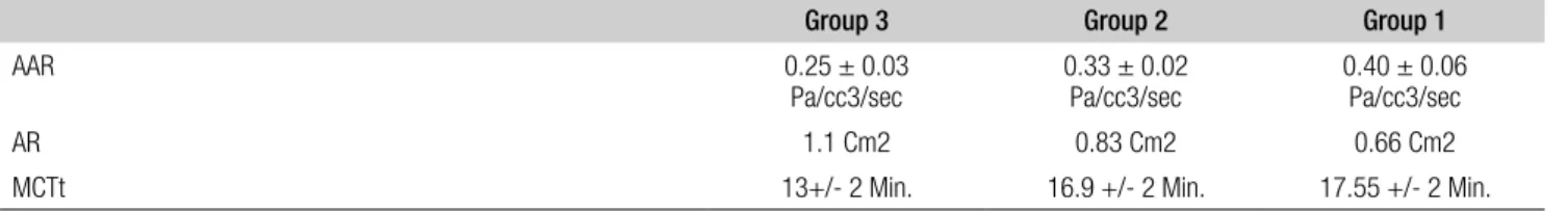
Table II. Results of nasal functionality tests in the three groups of patients.
Group 3 Group 2 Group 1
AAR 0.25 ± 0.03
Pa/cc3/sec 0.33 ± 0.02Pa/cc3/sec 0.40 ± 0.06Pa/cc3/sec
AR 1.1 Cm2 0.83 Cm2 0.66 Cm2
MCTt 13+/- 2 Min. 16.9 +/- 2 Min. 17.55 +/- 2 Min.
Fig. 3. ROC curve of nasal resistance in detecting patients with mild-mod-erate OSAS.
Fig. 1. Nasal resistance value. AAR: Active Anterior Rhinomanometry
Fig. 2. Muco-Ciliary Transport time (MCTt).
MCTt
Stepwise multiple regression models revealed that high nr was predictive of Ahi (p = 0.006).
The roC curve of nasal resistance is shown in Figure 3, without a specific cut-off point; the possibility to identify patients with mild-moderate oSAS is greatly affected by varying the threshold value along the curve.
Discussion
oSAS affects, at least, 2-4% of the adult population and is increasingly recognized by the public 1. The
pathophysiol-ogy of oSAS involves the development of pharyngeal air-way narrowing. The pharyngeal airair-way may behave like a Starling resistor due to decreased upper airway muscle tone and phasic inspiratory activity during sleep.
The collapsibility of the compliant wall is further en-hanced by the increased resistive load caused by obesity which alters the physical characteristics of the wall. The pharynx, connecting the nose to the larynx and act-ing as a Starlact-ing resistor, is more vulnerable to changes in airflow resistance. on the other hand, the nose has a rigid framework to maintain constant resistance both in waking and sleeping states 26-29. Thus, the role of nr in oSAS
remains controversial.
A large number of adult surveys have been conducted in order to establish the presence or absence of oSAS by means of parameters such as age, sex, snoring, witnessed apnoea and daytime sleepiness, and giving a score for each clinical feature suggestive of sleep apnoea29,30.
our results showed that in snoring and mild-moderate oSAS patients the values of nasal resistance and mCTt are increased with respect to standard values whereas minimal cross-sectional areas are decreased.
Specifically, a nasal resistance of 0.40 ± 0.06 Pa/cc3/sec, a minimal cross sectional area of 0.66 cm2 and a mCTt of
17.55 min could be predictors of Ahi and Si (Table ii). in this study, Si was significantly higher in patients with higher nr and significantly different in the three groups (p = 0.02).
Furthermore, there was a significant correlation between total nr and the severity of oSAS.
in this study, mSAT did not correlate significantly with any parameter from rhinomanometry and acoustic rhi-nometry. These results were consistent with previous data, and reinforced the hypothesis that nasal function may not contribute to severe sleep apnoea with hypo-xaemia.
The low correlation level between rhinomanometric and some polysomnographic data, such as mSAT, makes it necessary to perform a careful evaluation of the entire up-per respiratory tract during awakening and with specific manoeuvres or tests (muller manoeuvre, somnendoscopy) feigning sleeping condition 31.
The roC curve (Fig. 3) indicates that nasal resistan-ce value of 0.40 Pa/cm3/s has a sensitivity of 91% and
a specificity of 96%; these values show that AAr offers supplementary diagnostic opportunities and represents a simple and objective system to select patients saving the technician’s time.
Conclusions
This study provides supporting evidence that the status of the nasal fossae has a significant positive predictive role in patients with simple snoring and mild-moderate oSAS. Following the results obtained, we propose the simultane-ous evaluation of nasal function by AAr, Ar and mCTt in patients undergoing PSG recording.
however, as oSAS is a multi-factorial, multi-level disease, where nasal functions play a role, but together with other alterations at different levels of the pharynx (naso, oro, and hypopharynx) our results do not allow us to exclude a “systematic diagnostic and phased protocol” including fiberoptic endoscopy with the evaluation of tonsil size, mallampati grading, Fujita grading with retro-glossal space evaluation, as stated and again stressed recently in the literature 32. These statements are even more important
when considering any surgical approach in patients with severe oSAS 33.
References
1 epstein lJ, Kristo D, Strollo PJ, et al. Clinical guideline for
the evaluation, management and long-term care of obstruc-tive sleep apnea in adults. Adult Obstrucobstruc-tive Sleep Apnea Task Force of the American Academy of Sleep Medicine. J Clin Sleep med 2009;5:263-76.
2 aubert-Tulkens G, hamoir M, van Den eeckault J.
Failu-re of tonsil and nose surgery in adults with long standing severe pulmonary sleep apnea syndrome. Arch intern med 1989;149:2118-21.
3 Olsen KD, Kern eB, Westbrook Pr. Sleep and breathing
di-sturbance secondary to nasal obstruction. otolaryngol head neck Surg 1981;89:804-10.
4 lenders h, Schaefer J, Pirsig W. Turbinate hypertrophy in
habitual snorers and patients with obstructive sleep apnea: findings of acoustic rhinometry. laryngoscope 1991;101(6 Pt 1):614-8.
5 Zeng B, ng AT, Qian J, et al. Influence of nasal resistance on
oral appliance treatment outcome in obstructive sleep apnea. Sleep 2008;31:543-7.
6 Kemppainen T, ruoppi P, Seppä J, et al. Effect of weight
reduction on rhinometric measurements in overweight patients with obstructive sleep apnea. Am J rhinol 2008;22:410-5.
7 Sériès F, St. Pierre S, carrier G. Effects of surgical correction
of nasal obstruction in the treatment of obstructive sleep ap-nea. Am rev respir dis 1992;146:1261-5.
received: december 15, 2010 - Accepted: February 23, 2011
its impact on sleep-related breathing disorders. rhinology 2005;43:242-50.
9 hoffstein v, chaban r, cole P. Snoring and upper airway
properties. Chest 1988;94:87-9.
10 Mates a, Ohki M, cole P. Snoring, apnea and nasal
resis-tance in men and women. J otolaryngol 1991;20:57-61.
11 atkins M, Taskar v, clayton N, et al. Nasal resistance in
ob-structive sleep apnea. Chest 1994;105:1133-5.
12 Miljeteig h, hoffstein v, cole P. The effect of unilateral and
bilateral nasal obstruction on snoring and sleep apnea. lar-yngoscope 1992;102:1150-2.
13 Sher Ae, Thorpy mJ, Spielman AJ, et al. Predictive value
of Mueller’s manoeuver in selection of patients for uvulo-palatopharyngoplasty. laryngoscope 1985;95:1483-7.
14 yagi h, nakata S, Tsuge h, et al. Morphological
examina-tion of upper airway in obstructive sleep apnea. Auris nasus larynx 2009;36:444-9.
15 De vito a, Berrettini S, carabelli a, et al. The importance of
nasal resistance in obstructive sleep apnea syndrome: a study with positional rhinomanometry. Sleep Breath 2001;5:3-11.
16 Mehta a, Qian J, Petocz P, et al. A randomized, controlled
study of a mandibular advancement splint for obstructive sleep apnea. Am J respir Crit Care med 2001;163:1457-61.
17 gotsopoulos h, Chen C, Qian J, et al. Oral appliance therapy
improves symptoms in obstructive sleep apnea: a randomized, controlled trial. Am J respir Crit Care med 2002;166:743-8.
18 Desfonds P, Planès c, Fuhrman c, et al. Nasal resistance in
snorers with or without sleep apnea: effect of posture and nasal ventilation with continuous positive airway pressure. Sleep 1998;21:625-32.
19 li hY, Wang Pc, hsu cY, et al. Nasal resistance in patients
with obstructive sleep apnea. orl J otorhinolaryngol relat Spec 2005;67:70-4.
20 clement Par, Godts F. Consensus report on acoustic
rhi-nometry and rhinomarhi-nometry. rhinology 2005;43:169-79.
21 dunagan d, georgitis J, Kemp S, et al. Intranasal disease
and provocation: diagnostic testing of allergic disease. new york, marcel dekker; 2000, 3539, pp. 151-173.
22 clement Par. Committee report on standardization of
rhi-nomanometry. rhinology 1984;22:151-5.
23 Chung Seop K, Byeong Kweon m, dong hak J, et al.
Cor-relation between nasal obstruction symptoms and objective parameters of acoustic rhinometry and rhinomanometry. Au-ris nasus larynx 1998;25:45-8.
24 Passàli d, mezzedimi C, Passàli GC, et al. The role of
rhi-nomanometry, acoustic rhinometry, and mucociliary trans-port time in the assessment of nasal patency. ear nose Throat J 2000;79:397-400.
25 Passàli D, Bellussi l, De Seta e. Experiences in the
determi-nation of nasal mucociliary transport time. Acta oto-laryn-gologica 1984;97:319-23.
26 Zhong g, Kong w, yue J, et al. The study of the
relation-ship between obstructive sleep apnea syndrome and posi-tional nasal resistance. lin Chuang er Bi yan hou Ke Za Zhi 2003;17:351-3 (Chinese).
27 lee nr. Evaluation of the obstructive sleep apnea patient
and management of snoring. oral maxillofac Surg Clin north Am 2009;21:377-87.
28 madani m, madani F. Epidemiology, pathophysiology, and
clinical features of obstructive sleep apnea. oral maxillofac Surg Clin north Am 2009;21:369-75.
29 Berger g, Berger r, oksenberg A. Progression of snoring
and obstructive sleep apnoea: the role of increasing weight and time. eur respir J 2009;33:338-45.
30 Sugiura T, noda A, nakata S, et al. Influence of nasal
re-sistance on initial acceptance of continuous positive airway pressure in treatment for obstructive sleep apnea syndrome. respiration 2007;74:56-60.
31 campanini a, canzi P, De vito a, et al. Awake versus sleep
endoscopy: personal experience in 250 OSAHS patients. Acta otorhinolaryngol ital 2010;30:73-7.
32 Powell NB. Contemporary surgery for obstructive sleep
ap-nea syndrome. Clin exp otorhinolaryngol 2009;2:107-14.
33 campanini a, De vito S, Frassinetti S, et al. Temporary
tra-cheotomy in the surgical treatment of obstructive sleep ap-nea syndrome: personal experience. Acta otorhinolaryngol ital 2003;23:474-8.
address for correspondence: Prof. D. Passali, via anagnina 718, 00118 morena roma, italy. e-mail: [email protected]