DOTTORATO DI RICERCA IN
SCIENZE CHIRURGICHE: Progetto n.1 METODOLOGIE DI RICERCA NELLE MALATTIE VASCOLARI”
Ciclo XXIV
Settore Concorsuale di afferenza: MED/22
Evaluation of cardiovascular disease markers in patients
submitted to carotid artery stenting or endarterectomy.
Presentata da: Silvia Fittipaldi
Coordinatore Dottorato Relatore
Chiar.mo Prof. Andrea Stella Chiar.mo Prof Gianandrea Pasquinelli
ABSTRACT
Introduction. Microembolization during the carotid artery
revascularization procedure may cause cerebral lesions. Elevated C-Reactive Protein (hsCRP), Vascular endothelial growth factor (VEGF) and serum amyloid A protein (SAA) exert inflammatory activities thus promoting carotid plaque instability. Neuron specific enolase (NSE) is considered a marker of cerebral injury. Neoangiogenesis represents a crucial step in atherosclerosis, since neovessels density correlates with plaque destabilization. However their clinical significance on the outcome of revascularization is unknown. This study aims to establish the correlation between palque vulnerabilty, embolization and histological or serological markers of inflammation and neoangiogenesis.
Methods. Serum hsCRP, SAA, VEGF, NSE mRNA, PAPP-A mRNA
levels were evaluated in patients with symptomatic carotid stenosis who underwent filter-protected CAS or CEA procedure. Cerebral embolization, presence of neurologicals symptoms, plaque neovascularization were evaluated testing imaging, serological and histological methods. Results were compared by Fisher’s, Student T test and Mann-Whitney U test.
Results. Patients with hsCRP<5 mg/l, SAA<10mg/L and VEGF<500pg/ml
had a mean PO of 21.5% versus 35.3% (p<0.05). In either group, embolic material captured by the filter was identified as atherosclerotic plaque fragments. Cerebral lesions increased significantly in all patients with hsCRP>5mg/l and SAA>10mg/l (16.5 vs 2.8 mean number, 3564.6 vs 417.6 mm3 mean volume).
Discussion. High hsCRP, SAA and VEGF levels are associated with
significantly greater embolization during CAS and to the vulnerabiliy of the plaque. This data suggest CAS might not be indicated as a method of revascularization in this specific group of patients.
The data presented in this thesis ar based on the following original articles Faggioli G*, Fittipaldi S*, Pini R., Beltrandi E, Mauro R, Freyrie A, Rapezzi C, Stella A, Pasquinelli G. C-Reactive protein and embolization during carotid artery stenting. A serological and morphological study. Histol Histopathol (2011) 26: 843-853. *first author.
Faggioli G, Pini R, Mauro R, Pasquinelli G, Fittipaldi S, Freyrie A, Serra C, Stella A. Identification of Carotid:‘Vulnerable Plaque’ by Contrast-enhanced Ultrasonography: correlation with Plaque Histology, Symptoms And Cerebral Computed Tomography. Eur J Vasc Endovasc Surg (2011) Feb;41(2):238-48.
Vasuri F, Resta L, Fittipaldi S, Malvi D, Pasquinelli G. RUNX-1 and CD44 as markers of resident stem cell derivation in undifferentiated intimal sarcoma of pulmonary artery. A case report Histopathology. In press.
Vasuri F*, Fittipaldi S*, Buzzi M, Degiovanni A, Stella A, D’Errico-Grigioni A, Pasquinelli G. Nestin and WT1 expression in small-sized vasa vasorum from human normal arteries. Histology and Histopathology. *first author, in press.
Stella A*, Fittipaldi S*, Pini R, Pasquinelli G, Mauro R, Beltrandi E, Freyrie A, Gargiulo M, Faggioli G. High Sensitivity C-Reactive Protein and Vascular Endothelial Growth Factor as Indicators of Carotid Plaque Vulnerability, In revision to Atherosclerosis. *first author.
The main objective of our research project is the cellular and morphological characterization of carotid plaque vulnerability using different approaches; clinical analysis, immunohistochemical and molecular assays. The principal physiopathologic processes studied are inflammation, neoangiogenesis and calcification (osteogenic differentiation).
Clinical study
First we evaluated serological marker of cardiovascular disease in patients submitted to carotid artery stenting (CAS) or endarterectomy (CEA); 84 patients with >70% carotid artery stenosis were preferentially submitted to CEA (50) or CAS (34). On CAS we showed a direct positive correlation between post procedural cerebral lesions, the quantity of thromboembolic material retrieved on the filter and pre-operatory serum levels of inflammation markers. Patients with high hsCRP (>5mg/l), SAA (>10 mg/L) and VEGF (>500pg/ml) are associated with significantly greater embolization during CAS procedure and particularly in dishomogenous plaque. Volume and number of total cerebral lesions post procedural increased significantly in all patients having high hsCRP and SAA values. These data suggest that CAS might not be indicated in this specific group of patients with plaque prone to rupture. Secondly we compared different histological parameters of plaque vulnerability with the Contrast-enhanced Ultrasonography. An increase of dB-E was associated significantly with thinner fibrous cap (TFC <200 um, dB-E: 5.96 vs 3), greater inflammatory infiltrate (7.4 vs 3.2) and greater microvessels density (5.5 vs 2.5). We observed that either in symptomatic patients or in presence of pre-operative cerebral ipsilateral embolic lesions, dB-E values increased significantly (respectively 7.4 vs 3.5 and 5.96 vs 3.0) (3). In addition we observed that patients having neurological symptoms pre-operatively and vulnerable plaque had also high VEGF and hsCRP serum levels compared with
between VEGF serum concentration and fibrous cap thickness. All patients presented an increase of 32% of neuronal specific enolase mRNA level expression after the CAS procedure compared to patients submitted to CEA procedure.
Study on vasculogenic niche
Another aspects of the research project is the vasa vasorum (VV) neovasculogenic potential and involvement in atherosclerosis. We studied Nestin, WT1, dPAPP-A and SAA4 expression in vasa vasorum in human normal arteries (n°20). We saw that Nestin and WT1 in adult VV are expressed in adult VV, especially in VV <50 µm diameter and gathered in “hot spots”. The cytoplasmic colocalization of WT1 and nestin straightens the role of WT1 as a post-transcriptional activator of nestin protein. In our series WT1 was mainly cytoplasmic in the larger vessels, while nuclear expression increased in the smaller vessels (“second-order” VV). The nuclear localization of WT1 could express an increasing transcriptional activity in progenitor-committed Nestin-positive cells. We want to compare WT1/Nestin mRNA and protein hot-spots sub-cellular localization in normal arteries versus pathological arteries to assess a correlation with neovascularization during atherogenesis. The “hot spot” could therefore represent a valid model for the vasculogenic niche and the main source for neovasculogenesis during atherosclerosis. We are now performing the same analysis in atheroma lesions. We saw that dPAPP-A and SAA4 are specific histological and serological markers of atheromasic lesions presence. We also studied (data not presented) a case of pulmonary artery undifferentiated intimal sarcoma that expresses previously unreported markers; RUNX-1, Nestin, WT1 and CD44, commonly seen in different stages of the vascular differentiation hierarchy. These findings raise the question whether this neoplasm derives from a vessel wall-resident stem cell, like the
ACAS: Asymptomatic carotid artherosclerosis study
ACST: Asymptomatic carotid surgery trial AHA: American Heart Association APO: Apolipoprotein
ASA: Acetylsalicylic acid
ATIR: Angiotensin type1 receptor BAV: Bicuspid aortic valves BNP: Brain natriuretic peptide
BMP2: Bone Morphogenetic Protein 2 BSA: Bovin serum albumine
CAD: Coronary artery disease CAS: Carotid artery stenting CCD: Charge-coupled device CCP: Cathepsin cystein protease cDNA: complementary DNA CEA: Carotid endarterectomy
CEUS: Contrast-enhanced Ultrasound CRP: C-reactive protein
CT: Computerized tomography DAPI: Pro long anti-fade reagent dB-E: db-Enhanced
DWI-MR: Diffusion weight resonance magnetic imaging
ECs: Endothelial cells
ELISA: Enzyme-linked immunosorbent assay
FBG: Fibrinogen FFA: Free fatty acid
FLT: FMS-like tyrosine kinase GSM: Gray Scale Measurement HDL: High-density lipoprotein HE: Hematoxylin-eosin HMDS: Hexamethyldisilazene HsCRP: highsensitivity C-reactive Protein
ICAM: intercellular adhesion molecule IGF: Insulin-like growth factor
IHC: immunohistochemical IL: interleukin
IMA: ischemia modified albumi
LDL: Low-density lipoprotein LM: Light microscope
LOX1: Lectin-like oxidized low-density lipoprotein receptor 1
Lp-PLA2: lipoprotein-associated phospholipase A2
MBP: Eosinophil major basic protein MCP-1: Monocytechemoattractant protein-1
miRNA: micro RNA
MMP: Matrix metalloproteinases MPO: Myeloperoxidase
MYG: Myoglobin NO: Nitric oxide
NSE: Neuron specific enolase NT-proBNP: N-terminal prohormone of brain natriuretic peptide
OP: Occluded pore
Ox-LDL: oxidized low-density lipoprotein
PAD: Peripheral artery disease
PAI-1: Plasminogen activator inhibitor PAPP-A: pregnancy-associated plasma protein-A
PCR: Polymerase chain reaction PlGF: Placental growth factor PO: Pore occluded
PTX3: Pentraxin-related protein RBC: Red blood cells
RNA: Ribonucleic acid ROI: Region of interest RT: Room temperature
SAA: Serum amyloid A protein SAA4: Serum amyloid A isoform 4 sCD163: soluble haemoglobin scavenger receptor
sCD40L: soluble CD40 ligand SD: Standard deviation
SEM: Scanning electron microscopy SI: Surface involvement
SMC: Smooth muscle cell
sPLA2: Secretory phopholipase A2 sTREM-1: soluble triggering receptor expressed on myeloid cells
SVS: Society of vascular surgeons TF: Tissue factor
TFC: Thinner fibrous cap TG: Triglyceride
TGF: Tumour growth factor TIA: Transient ischemic attack TIMP: Tissue inhibitors of metallo-proteinases TNF: Tumour necrosis factor TNI: Troponin I
TNT: Troponin T
VCAM: Vascular cell adhesion molecule VEGF: Vascular endothelial growth factor
VV: Vasa Vasorum
VWFC: von Willebrand factor WT1: Wilm’s tumour suppressor
ABSTRACT
Introduction Methods Results DiscussionLIST OF ORIGINAL PUBLICATIONS
ABBREVIATIONS
Overview of the PHD project & data obtained
INTRODUCTION
1
Atherosclerosis 2
Highlights of the introduction 2
Carotid plaque: pathogenesis 4
Effects of hemodynamics on vascular cells Hypercholesterolemia
Oxidized LDL
The fibrous cap 11
Instrumental evaluations
Plaque classification 13
The calcification 15
Media Intima
The lipid core 17
Hypercholesterolemia Instrumental Evaluation
Pharmacological therapies
Biomarkers and vulnerable plaque 20
Vulnerable plaque
Traditional biomarker 24
Emerging biomarker 25
Biomarker: applications always changing 26
Markers of our study: inflammation biomarker 29
C-reactive protein
Serum amyloid A-protein
Pregnancy-Associated Plasma Protein-A Neuron-specific enolase
Markers of our study: neoangiogenesis biomarkers 37
Neoangiogenesis
Vascular Endothelial Growth Factor Nestin
Wilms tumour suppressor
AIM
44MATERIALS AND METHODS
46Carotid artery stenting study 47
Patients
Carotid plaque structure analysis CAS procedure
Filter analysis by light microscopy
Filter analysis by scanning electron microscopy Cerebral lesions assessment: DW-MRI
Carotid endarterectomy specimens 51
Patients CT scan CEUS
Histological analysis of carotid plaque
Immunohistochemical assay on carotid plaque Histological evaluation of vulnerability
Inflammation infiltrate evaluation with ImageJ software, CEUS study Neoangiogenesis evaluation with ImageJ software, CEUS study Sequential double immunofluorescence assay on carotid plaque
Vascular healthy tissue specimens and histopathological analysis 58
Immunohistochemical assay on healthy specimens
Sequential double immunofluorescence assay on carotid plaque Evaluation of vasa vasorum density with an ocular micrometer and morphology at IHC
Atheromasic tissue specimens and histopathological analysis 60
Patients
Evaluation of microvessel density with an ocular micrometer in carotid plaque at IHC
Evaluation of circulating markers’ concentration 62
Measurement of circulating hsCRP and SAA
VEGF and PAPP-A serological evaluation: ELISA assay NSE and PAPP-A mRNA expression level
RNA Extraction from PAX gene tube Extraction with Trizol method
Statistical analysis 66
RESULTS
67CAS, biomarkers and cerebral lesions 68
Patients CAS Drug therapy CAS
Distribution and description of the material covering the filter Filter analysis by light microscopy
Filter analysis by scanning electron microscopy
Evaluation of microvessel density with an ocular micrometer in carotid plaque at IHC
Markers levels pre-procedural, amounts of debris and cerebral lesions post-procedural
Identification of plaque vulnerability in patients submitted to CEA 77
Patients
CEUS evaluation, inflammation and plaque vulnerability
VEGF, hsCRP serum levels and histological plaque vulnerability Thin fibrous cap
Vascular healthy tissue specimens and histopathological analysis 82
CD31 and CD34 Nestin and WT1
Comparison between IHC and vessel morphology Localization of Nestin and WT1 at immunofluorescence
Vascular atheromatous tissue specimens and a new
CD34, Nestin and WT1
Nestin and WT1 “dis-correlation” Dots vessels
Complications in atheromateous plaques Calcified nodules
DISCUSSION
98Cerebral lesions risk and serological markers levels 99
Identification of vulnerable plaque PAPP-A level: a tecnical issue HsCRP and SAA
Circulating molecular markers: peripheral model to study local damage?
Neoangiogenesis and atherosclerosis development 104
Calcified plaques
Main findings of this study 109
Conclusion 110
BIBLIOGRAPHY
111INTRODUCTION
Atherosclerosis
In 1628 William Harvey, St. Bartholomew's Hospital, London, describes the function of the heart, arteries and veins. It is considered to be one of the greatest advances in medicine. Between 1783 and 1793 Edward Jenner, a British country physician who created the smallpox vaccine, while doing an autopsy accidentally discovered coronary arteries disease. He was probably the first to associate angina pectoris with hardening of the arteries (Fig 1).
Figure 1: Edward Jenner and a stomach flattened and injected with wax to
show the veins and arteries.
Highlights of the introduction
Nowadays, in occidental countries cardiovascular events,such as stroke, are the leading causes of death, thus the priority of primary and secondary
prevention (Rosamond et al., 2007). Even if selected patients with acute ischemic stroke are treated with promising experimental therapies, such as tissue plasminogen activator, the best approach to reduce stroke remains still prevention. An early diagnosis allows individuals at higher risk or prone to stroke to be identified and targeted for specific therapies or interventions (Goldstein et al., 2001). The presence of atherosclerotic lesions at the carotid bifurcation and the presence of ischemic cerebral lesions are the main factors leading to cardiovascular events. In fact, the presence of recent focal neurological symptoms ipsilateral to a carotid stenosis is a powerful predictor of ‘high risk’ carotid atherosclerosis. Especially, the risk of major events such as death or stroke, is further increased in patients presenting vulnerable plaque (Wang et al., 2010). The degree of stenosis is still the gold standard to assess stroke risk and subsequent indication to revascularization (R Rosamond et al., 2007). In symptomatic or asymptomatic patients with a stenosis greater than 60%, carotid endoarterectomy procedure (CEA) results respectively in a 56% and 34.5% reduction of the development of major events, thus proving the efficacy of the CEA intervention (ACAS 1991). However waiting time between diagnosis and CEA intervention can result in an interval stroke rate of 9%-15%. The aim of the early intervention is to avoid embolization caused by a vulnerable lesion at the carotid district, nevertheless there is a big issue to recognize “young” vulnerable plaque non-invasively before an acute clinical event occurs. (Setacci, et al., 2008). Different studies estimate the individual risk of stroke such as the Framingham risk score or the ABCD2 score system. The prognostic value of all these score systems improves if completed with clinical information, vascular imaging data and brain imaging data of patients. (A. Bhatt et al., 2011). Cerebro-ischemic symptoms, as transient ischemic attacks or amaurosis fugax, increased the risk of major events such as death or stroke (Liapis et al., 2001). In a magnetic resonance imaging prospective assessment study, it has been
shown, that the occurrence of cerebrovascular events in asymptomatic patients is associated with the characteristics of carotid plaque; thinned or ruptured fibrous caps, intraplaque hemorrhage, larger lipid-rich necrotic cores and larger maximum wall thickness. (Takaya 2006 ).
Stroke risk prediction based only on conventional risk factors is not enough; hence research is oriented on predictive biological biomarkers. Previous studies demonstrated the correlation between serological and structural markers of inflammation and neoangiogenesis related to the risk of cardiovascular events in the coronary district (Ferri et al., 2006). Thus biomarkers knowledge is necessary to allow primary prevention of coronary events (Muller et al., 2006). However there is yet no standard value for biomarkers able to identify a vulnerable plaque in the carotid district. Evaluation of the silent atheroma non-invasively is only possible through recent imaging approaches and computed analysis such as pixel density analysis and elastography at Duplex examination, local temperature probe (Setacci, et al., 2008); these techniques allow to evaluate echostructural alterations, such as ulcers, typical of vulnerable plaque but not molecular composition. Thus, in addition to structural plaque analysis and inflammation protein, particular attention is given to molecular pathway involved in the atherosclerotic lesions development.
Studies related to the discovery of novel biomarkers signature involved in plaque destabilization and stroke are of fundamental importance .
Carotid plaque: pathogenesis
Atherosclerosis is characterized by intimal lesions, the atheromas, which are protrusions within the vascular lumen that can determine alterations in the normal blood flow and direct damage to the vessel structure. At first,
these lesions present a focal distribution in the artery; however, they rapidly begin to increase in number as the disease advances, until affecting the whole circumference of the vessel walls that are more severely damaged
(Fig 2).
The structure of the basic lesion consists in a plaque localised in the intima presenting a central lipid core ( cholesterol and its esters) covered by a fibrous cap.
Figure 2: Structure of a blood vessel (right: healthy type, left: atheromasic
vessels)
Atherosclerotic plaques are mainly composed of cellular elements: smooth-muscle cells, macrophages and leukocytes, connective tissue of extracellular matrix – collagen, elastic fibres and proteoglycans – and intra-and extracellular lipid deposits. (Fig 3). The relative proportion of these components varies depending on the different plaques, which results in a wide spectrum of lesions with different degrees of instability. The overlaying fibrous cap is composed of smooth-muscle cells, few leukocytes and relatively dense connective tissue. The cellular area which is below and
adjacent to the fibrous cap – the “shoulder” of the fibrous cap – is composed of macrophages, smooth-muscle cells and T lymphocytes. The deep necrotic core presents a disorganised mass of lipid material, which is composed of cholesterol crystals, cellular debris, thrombi undergoing organization and other plasma proteins. Lipids are mainly composed of cholesterol and its esters. There are cells that have phagocytized this lipid material: they are called “foam cells” and they are, in particular circulating monocytes activated in macrophages, and activated smooth-muscle cells. Finally, especially at the periphery of the lesions, frequent aspects of neovascularization can be detected and they are represented by the proliferation of blood vessels that can be more or less mature. The variations in the histological characteristics of the plaques depend on the number of smooth-muscle cells and macrophages, the amount of collagen and other extracellular components, besides the lipid contents (Cotran et al., VI Ed)
The pathogenic development of these lesions is still unclear; the endothelium is believed to play a fundamental role in the initial development of the plaque. The most widely accepted hypothesis is that what triggers this process is a reaction to the damage of the intima. Therefore, atherosclerosis would represent an inflammatory response of the vessel wall (Ross et al., 1993). The element that characterizes the primary lesions is the presence of an endothelium that is not morphologically damaged yet. This has led to consider that the most important factor in development of the disease is a cellular dysfunction and activation, together with an increase in the endothelial permeability, rather than a direct damage. This process manifests itself with an increased leukocyte and monocyte adhesion, which is highlighted by alterations in the expression of the endothelial adhesion molecules ICAM-1 and VCAM-1 (InterCellular Adhesion Molecule 1, Vascular Cell Adhesion Molecole 1) (O'Brien et al., 1996; Cybulsky et al., 1991; Gimbrone et al., 1997).
Figure 3: Development and progression of atheromasic lesions (Fortunato
et al, 2007).
The main causes of the endothelial dysfunction and damage are the hemodynamic changes that occur in some specific points of the circulatory tree – major branch points and bifurcations – and the harmful effects of hypercholesterolemia.
Effects of hemodynamics on vascular cells.
In support of the role of the blood flow, and of its changing from laminar to turbulent with a loss of tangential force – and, therefore, a low “shear stress” – it is relevant to consider the location of the plaques, which can be more frequently found at the ostial area or at the bifurcations.
The blood flow undergoing these changes, with variable levels of parietal stress, is believed to cause the endothelial dysfunctions and thus predispose the development of these lesions in these predictable locations (Fig 4). Cyclic strain has been shown to stimulate expression of cellular adhesion molecules such as ICAM-1 and intracellular second messenger systems such as the adenylate cyclase-cAMP, diacylglycerol-IP3, and protein kinase
C pathways (Pradhan et al., 2004).
Figure 4: Hemodynamic forces. A) Severe stenosis proximal left internal
carotid artery as seen in 3D (Rochester Medical Center) B) Endothelial cells are subjected to both a tangential, parallel force (shear stress) as well as a circumferential, perpendicular force (cyclic strain). (Pradhan et al., 2004).
This causes the activation of many pro-inflammatory and pro-atherogenic genes, thereby the production of cytokines, adhesion molecules and coagulation proteins, which produce an increase in the endothelial permeability, the cellular turnover and the cellular endocytosis of the LDL that is present in this area.
Hypercholesterolemia.
Hypercholesterolemia can trigger the development of plaque, due to a higher deposition of cholesterol in the subintimal area. Moreover, hyperlipidemia itself determines an endothelial dysfunction owing a greater production of superoxide dismutase and other free radicals of oxygen that deactivate nitric oxide, which is the main relaxing factor in arteries. The oxidative modifications induced by free radicals, and produced by
macrophages and endothelial cells, lead to the formation of oxidized LDL molecules, which in turn contribute to the formation of lesions in different ways:
Oxidized LDL:
1. can be easily phagocytized by macrophages, resulting in the formation of foam cells,
2. are chemotactic for circulating monocytes,
3. increase the adhesion of the monocytes especially by inducing endothelial adhesion molecules,
4. inhibit the viability of the macrophages that are already in the lesion by facilitating the recruitment and the survival of the macrophages in the plaque,
5. stimulate the release of grow factors and cytokines,
6. are cytotoxic to the endothelial cells and the smooth-muscle cells,
7. are immunogenic, as they induce the production of antibodies against the oxidized lipoproteins.
The idea that hyperlipidemia leads to the formation of lesions by means of the oxidative stress on the endothelium is proven by clinical and experimental studies which show how antioxidant proteins and medications that reduce oxidation have a protective effect on atherosclerosis. Moreover, the decrease in cholesterol and the antioxidant therapy improve the endothelial function (Steinberg, 1997)
During the genesis of the plaque, monocytes subsequently adhere to the activated endothelial surface, thanks to the adhesion molecules, and through these cells they migrate to the subendothelial area. Here they differentiate into macrophages and phagocytize the oxidized LDL molecules, thus
becoming foam cells. Macrophages play a multifactorial role in the progression of atherosclerosis thanks to the secretion of proteins such as the interleukins (IL-1) and the tumour growth factor (TNF), which increase the adhesion of the leukocytes. Furthermore, macrophages produce radicals of oxygen, which cause the oxidation of the LDL molecules in the lesion, and elaborate growth factors that can contribute to the proliferation of the smooth-muscle cells. Prematurely during the development of the lesion, the smooth-muscle cells migrate to the intima, where they proliferate and determine the deposition of extracellular material, thus facilitating the enhancement of the plaque (Ridker et al., 2002).
During the early stages of atherogenesis, the intimal plaque is composed of an aggregation of foam cells, some of which may die and release extracellular lipids and cellular debris, which surround the muscle cells. The adipose cellular atheroma gradually changes, as a consequence of the deposition of collagen and proteoglycans. The connective tissue, particularly abundant in the intimal area, produces the fibrous cap, which then develops in the mature fibroatheroma. Some of these lesions subsequently undergo further modifications:
A. accumulate of larger amount of connective tissue, thus becoming fibrous plaques.
B. calcification deposits.
C. develop a central core that is rich in lipids and foam cells, which increase the potential risk of developing serious complications (Ross et al., 1993).
The fibrous cap
The fibrous cap is the outer fibrous part that covers the atherosclerotic plaque. It is the structure that separates the blood flow from the necrotic core of the lesion, therefore it is very important for the stability of the plaque. If there are lesions on its surface, circulating elements – platelets and coagulation factors – can come into contact with the inner elements of the plaque, thus causing the formation of thrombi (Fig 3).
The fibrous cap originates from the secretory activity of the smooth-muscle cells. These cells migrate from the media to the sub-endothelial area – during the formation of the plaque – where they produce collagen, elastic fibres and proteoglycans, in order to limit the expansion of the inflammatory process of the plaque towards the vascular lumen (Ross et al., 1993; O'Brien et al., 1996; Cybulsky et al., 1991; Gimbrone et al., 1997; Mauriello et al., 2010)
Therefore, the presence of a thin-fibrous cap represents a condition of unstable plaque prone to rupture. The development of a thin-fibrous cap is determined by a series of phenomena:
1. activation of the leukocytes inside the plaque and production of metalloproteinases,
2. degradation of collagen and proteoglycans, which are produced by the smooth-muscle cells,
3. release of cytokines that can reduce the activity of the smooth-muscle cells.34
Histological tests have highlighted that the thin-fibrous cap is associated with higher concentrations of macrophages in the subendothelial area. The
thin-fibrous cap (<65 µm) is a condition of instability of the plaque, because it may cause ruptures on its surface, independently from the size of the necrotic core.
In general, in the areas that present lesions of the fibrous cap that do not determine more severe clinical manifestations, the healing of the ulcerated surface is not complete. These areas present a weaker resistance that can subsequently determine other fractures on the surface, which can be associated with more severe clinical manifestations. This explains the typical manifestations of the carotid plaque. As a premonitory sign we can consider transient ischemic attacks (TIA), which are the epiphenomenon of a microfracture on the surface of the atherosclerotic lesion; in the following days, this may result in a major ischemic event (stroke), due to another rupture of the plaque that had not completely healed (Redgrave et al., 2006; Virmani et al., 2003; Virmani et al., 2006).
Instrumental evaluations. In the ultrasound study, the morphology of the surface of the plaque represents an additional element in the evaluation of the risk associated with a carotid lesion, besides the analysis of the degree of stenosis. Therefore, the identification of alterations on the surface of the plaque, such as the ulcerative lesions (which are identified as introflexions of the surface of the plaque of at least 2 mm per 2 mm of length), is very important in the overall evaluation of the patient, as it allows to identify an additional risk of ischemic events (Fig 5). There are other methods of imaging such as the Computerized Tomography (CT) or the Magnetic Resonance (MR), which allow to identify the structural alterations and the risk of cerebral events due to the presence of a particularly thin-fibrous cap. However, in the clinical practice they are used less than the ultrasound scan (Lovett et al., 2004; Wasserman 2008; Takaya et al., 2006).
Figure 5: Ulcerated plaque on the surface of the fibrous cap (arrow),
identified by means of an ultrasound evaluation. The presence of lesions in the structure of the carotid plaque represents a condition of structural instability and, therefore, of a higher risk of cerebrovascular events
The alterations on the surface of the plaque or the identification of a thin-fibrous cap represent a condition of instability of the plaque and a higher cerebrovascular risk; however, their role is marginal in the decision-making process for the surgical treatment of the carotid lesions. Indeed, the data of most of the clinical trials, from which the current indications originate, refer above all to the evaluation of the degree of stenosis and to the symptomatology that is associated with it.
Plaque classification
The classification of atherosclerotic lesions was initially carried out by macroscopic observation, using descriptive terms such as "Lipid streak" or
atheromatous plaque, fibrous or broken. The terminology varied from study to study so the correlation of data was impossible. In 1994-1995, the American Heart Association (AHA) has established criteria by which plaques are classified according to cellular content and structure, analysing the area of major lesion (Stary et al., 1995) (Table 1a). It is important to differentiate;
– young stable plaque with a low extracellular lipid content, less dangerous (types I–III)
– unstable plaque, more dangerous types including plaque with a high extracellular lipid content (types IV and Va), more prone to rupture and acute thrombosis (Richardson et al 1989; Cheng 1993),
– and older calcified and fibrotic plaque (types Vb and Vc), which are dangerous because of their high degree of stenosis and the risk of rupture (Table 1a, b).
Table 1b: This second table above is a summary of the crucial definitions related
to the vulnerable plaque (Schaar et al., 2004).
The calcification
The calcification at the level of the arteries is a process that is not clear yet and can appear both at the level of the media and at the level of the intima. Media. The involvement of the media, which is defined as Monckeberg’s calcific sclerosis, is a process that begins in areas that are not generally involved in atherosclerotic manifestations, such as the distal vessels of the limbs, the visceral vessels and the thyroid vessels. The calcification is associated with pathologies characterised by ionic alterations: metabolic dysfunctions, electrolyte disorders, diabetes and chronic kidney failure. The calcification can also be determined by alterations of the nerve stimuli and neuropathies. Another possible cause is the lumbar sympathectomy, after which the area of the sympathectomy shows an increased development of calcifications of the media in the vessels of the ipsilateral foot (88% in 2 years) compared to the contralateral foot (18%).
Intima. The most common condition is the intimal calcification that develops at the level of the atherosclerotic lesions. The most widely accepted hypotheses associate the inflammation in the plaque with the deposition of calcium crystals (besides the effect caused by alterations in the metabolism of phosphorus and calcium). The phlogosis at the level of the atherosclerotic lesion is associated with the release of cytokines, which cause the production of molecules that stimulate the formation of calcified nodules. The Bone Morphogenetic Protein 2 (BMP2), which is secreted by the smooth-muscle cells, plays a fundamental role in this process. The stimuli inside the plaque can be very weak – thus determining microcalcifications – or they can be particularly intense, thus causing a progressive differentiation of the smooth-muscle cells into cells with an osteoblastic/ osteoclastic activity and the formation of bone matrix or even bone tissue. In the atherosclerotic plaques, calcium initially develops in nodules that are at a basal level and, subsequently in microcrystals in the whole plaque (Doherty et al., 2004; Abedin et al., 2004: Demer et al., 2003). At the level of the vessels, the identification of calcifications can be an indicator of atherosclerosis. In the coronary arteries, calcium is located only in the atherosclerotic plaques: for this reason, methods such as the TC have been introduced for the non-invasive evaluation of these lesions (Calcium Score). At the level of the carotid stenoses, as the ultrasound scan is the most used method of investigation to easily identify the presence of a plaque, the role of the calcification as an indicator of atherosclerosis is not considered. On the contrary, the presence of calcific deposits in the carotid is an indirect indicator of the instability of the plaque. Indeed, the histological evaluation of the carotid plaques has highlighted in many studies that, in general, the calcified plaques can be found in asymptomatic patients, whereas calcifications are scarce or inexistent in patients who have had a neurological symptomatology and, thus, with unstable lesions. Afterwards, analyses on anatomopathological reports have confirmed that
the calcification inside the carotid plaque is associated with a thicker fibrous cap and a less significant development of the lipid core, which both determine the stability of the plaque (Huang et al., 2001).
In the ultrasound evaluation, the presence of calcifications is identified thanks to the presence of a marked hyperechogenicity, which can also determine the development of an acoustic shadowing due to the total reflection of the ultrasound waves. This makes the evaluation of the atheromatous lesion possible; therefore, in these lesions, the evaluation of the flow modification after the acoustic shadow is fundamental for the identification and evaluation of the degree of stenosis.
Another type of calcification is “a plaque with a calcified nodule” which is a heavily calcified plaque with the loss and/or dysfunction of endothelial cells over a calcified nodule. This is the least common of the three causes of thrombosis described here (Virmani et al., 2000, Table 1b).
Calcification triggers' mechanism in arteries is still unknow. The identification of a calcified carotid lesion is interesting for the indication of a possible surgical treatment.
The lipid core
The lipid core of the carotid plaque is the most interesting element for it represents the main factor determining the stenosis and, consequently, the cerebral ischemic risk. The core is mainly composed of LDL molecules that cross the vascular endothelial barrier from the blood flow and accumulate in the intima. The macrophages are recruited and activated by the subendothelial deposit and differentiate into foam cells. These cells release oxidizing substances, which determine the formation of the oxidized LDL molecules, which have a chemotactic role as they stimulate the evolution of
the atheromatous lesion. The serum concentrations of cholesterol, with high values of LDL and low values of HDL, are fundamental for the development of the carotid plaque and are related to a larger accumulation of lipids inside the plaque. The presence of an abundant lipid component inside the carotid plaque increases plaque instability, for different reasons (Ross et al., 1993)
1. greater fragility with possible events of internal haemorrhage, 2. increased phlogistic component,
3. presence of a thin-fibrous cap,
4. strong stimulus to the aggregation of platelets in case of lesions.
As a consequence, the methods for the identification of these lesions rich in lipids are very important.
Hypercholesterolemia. The evaluation of the serum concentrations of cholesterol is the most helpful element: the study ACST (Asymptomatic Carotid Surgery Trial) highlights how patients with a carotid stenosis over 70% and values of blood cholesterol over 250 mg/dl present a significantly higher risk of ischemic events than the group of patients with lower cholesterol serum levels. The endarterectomy treatment of these lesions determines a total reduction of the risk of events, in a 5-year period, by 11,7% in patients with high levels of cholesterol and by 4,6% in patients with lower levels (MRC (ACST) 2004).
Instrumental evaluation. The evaluation of the carotid plaques by means of imaging methods helps to identify the internal component of the core. Today the B-Mode ultrasound evaluation is used for the internal description of the plaque, which is defined as hypoechogenic if it appears structurally darker than the adventitial component. This is due to the fact that the lipid core does not determine the reflection of the ultrasound waves and,
therefore, appears as dark. However, the direct evaluation does not allow an objectification of the result; for this reason, it is defined as “dependent operator” and considered as liable to individual variations. The analysis of the structure of the plaque by means of the Grey Scale Measurement (GSM) represents a more objective method. This method takes into account the average variation of the grey scale in the area of the plaque, returning a numeric result, which, thus, is not subjective. (Fig. 6)
Figure 6: evaluation of the Grey Scale Measurement (GSM). In the area of
the plaque, the average of the grey scale is calculated and a numeric value indicating the degree of hypoechogenicity is produced.
Evaluations by means of this method have highlighted that symptomatic plaques generally have lower values of GSM (<50), whereas asymptomatic plaques have higher values, which is an indicator of a structure that is more stable and less prone to possible complications. Moreover, long-term evaluations have highlighted how plaques with low GSM levels can determine ischemic events (Kern et al., 2004; Biasi et al., 1999).
to analyse the structure of the core, which confirms that lesions that are structurally richer in lipid component present higher long-term cerebrovascualr risks (Takaya et al., 2006; Cappendijk et al., 2008).
Pharmacological therapies. The use of therapies with pharmaceuticals that can determine a reduction of the blood concentrations of lipids and cholesterol, such as the inhibitors of the HMG-CoA Reductase (statins), has resulted in a reduction of the cerebrovascular risks by both having an effect on the lipid levels and reducing the phlogistic condition that characterises atherosclerosis. It has been demonstrated that high doses of statins are effective for the stabilization of the carotid plaque. Ultrasound evaluations in patients with carotid stenosis between 40% and 60%, and receiving Atorvastatin therapy 80 mg/die (for 12 months), have highlighted a significant ultrasound stabilization of the plaque (with an increase of the GSM from 66 to 100) compared to patients receiving low doses of the same medication (GSM from 64 to 85) (Kadoglou et al., 2010).
Other studies have highlighted how the statin therapy can determine a significant reduction in the concentrations of macrophages and inflammation inside the carotid plaque (Tang et al., 2009).
Statins have resulted as being helpful in the treatment of the carotid stenoses, because they reduce the lipid component of the core and stabilize the plaque. However, in presence of lesions that are hemodynamically significant, the effectiveness of the statins in preventing cerebrovascular events has not been demonstrated yet (Treasure et al.,1995; Anderson et al., 1995).
Biomarkers and vulnerable plaque
Previously, atherosclerosis was thought primarily as a "plumbing" problem. The degree of stenosis on an angiogram, symptoms and signs of ischemia provided the main tools to assess atherosclerosis. In the last decades, studies on the pathogenesis of the disease have grown fast (Libby et al. 2005). The
understanding of the pathophysiology of this disease has now entered a new era based on understanding of the biology and a critical reappraisal of the pathobiology of atherothrombosis (Hansson et al. 2006).
Biomarker are measurable and quantifiable biological parameter such as protein, enzyme or hormone concentration, gene phenotype distribution in a population. Biomarker’s evaluation is a useful indicator of a normal biological process, pathogenic process, or the pharmacologic response to a therapy. The properties of a biomarker depend on their use. Currently the techniques available for biomarker development are genomics, proteomics, metabolomics, pharmacogenetics, integratomics, bioinformatics and molecular imaging technologies. Several categories of biomarker have been described that are pertinent to cancer, namely screening, diagnostic, prognostic, predictive, pharmacological, surrogate response and safety biomarker. Biomarker assays need to be carefully validated and be robust, reliable and reproducible when applied in clinical contexts (Murukesh et al., 2010). Biomarker's application in cardiovascular disease risk assessment is multiple: screening diagnosis, prognostication, prediction of disease recurrence, and therapeutic monitoring. (Vasan, 2006)
For the prediction of atherothrombotic events, markers of inflammation, fibrinolysis, oxidative stress, and altered lipid metabolism are currently in discussion. Also new emerging biomarker are in consideration, for example MPO, PAPP-A, neuron-specific enolase (NSE) represented in the table 4 (Hermus et al. 2010).
During plaque formation different pathological processes involved in the progression of atheromasic lesions are associated with plaque vulnerability,
such as inflammation, neoangiogenesis, lipid accumulation, apoptosis, proteolysis and thrombosis. Also morphological characteristics are related with plaque instability such as ulceration Fig 7 (Hermus L et al. 2010). Biomarker's studies are based on the concept, that a vulnerable plaque contains predictive information for future cardiovascular events, also in other areas of the vascular tree (Lammeren et al., 2011). Results are promising and plaque markers can be used to develop imaging methods to identify patients at risk and to monitor treatment effect. Plaque biomarker studies use the concept of vulnerable plaque to favour the prediction of vascular patients.
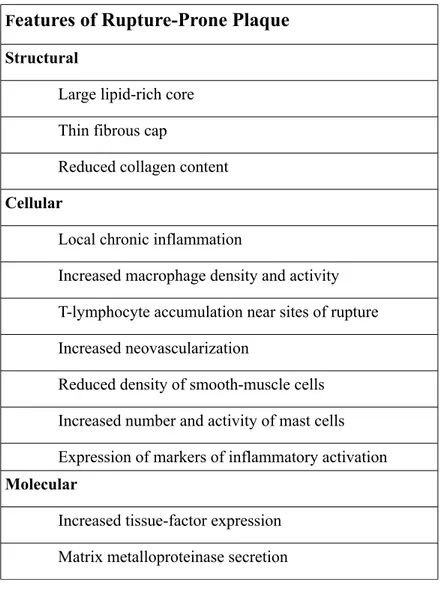
Vulnerable plaque
Vulnerable plaque are characterized by a large lipid core, a thin fibrous cap, a rich infiltrate of macrophagic inflammatory cells and scarce smooth muscle cells. Atherosclerotic plaque prone to rupture have specific structural, cellular, and molecular characteristic listed in Table 2 and illustrated in figure 7. The presence of a thin fibrous cap overlaying a large lipid core increase at high risk for rupture. Plaque rupture usually leads to various degrees of thrombus formation. Thrombosis may result in unstable angina, myocardial infarction, or sudden death, particularly if collateral flow is inadequate.
Early identification of high-risk plaque is critical, but vulnerable plaque is not detectable with the current diagnostic methods. If this plaque could be detected a major advance in healthcare would be achieved. Identification methods available are thermography, spectroscopy, radioisotope scintigraphy, more recently optical coherence tomography and ultrasound imaging. Nowadays the concept of vulnerable plaque is evolving towards patient vulnerability. Vulnerable plaque are not the only cause of symptomatic events, others parameters are involved, i.e. vulnerable blood
(prone to thrombosis) plays an important role in the outcome. Therefore, the term "vulnerable patient" is more appropriate and is now proposed for the identification of subjects with high likelihood of developing cardiac events in the near future (Naghavi et al., 2003).
Figure 7: Overview of a stable atherosclerotic plaque and an unstable
Features of Rupture-Prone Plaque Structural
Large lipid-rich core Thin fibrous cap
Reduced collagen content
Cellular
Local chronic inflammation
Increased macrophage density and activity T-lymphocyte accumulation near sites of rupture Increased neovascularization
Reduced density of smooth-muscle cells Increased number and activity of mast cells Expression of markers of inflammatory activation
Molecular
Increased tissue-factor expression Matrix metalloproteinase secretion
Table 2: Features of plaque prone to rupture
Traditional biomarker
Conventionally, biomarker are identified based on large epidemiologic studies demonstrating the significative statistical association between a phenotype and the studied biomarker. Markers for risk are considered a biochemical signature that correlates with the increased risk of the disease development as determined by clinical and epidemiological studies. C reactive protein (CRP) has emerged as an independent predictor of an incident cardiovascular event in more than 15 large prospective studies,
adding a prognostic value to that conveyed by the Framingham Risk Score. In addition the lack of correlation between LDL-cholesterol and CRP allowed the identification of a subgroup of patients with increased incidence of coronary artery disease but a normal lipid profile (Koenig et al., 2005, Ridker et al., 2002). Some of the most common markers of cardiovascular disease are listed in table 3.
RISK Total Cholesterol mg/dl LDL mg/dl HDL mg/dl TG mg/dl Omocistein umol/l HsCRP mg/l BNP pg/ml LOW <200 <130 >60 >200 2 -15 <1 <100 MODERATE 200-240 130-160 40-60 200 - 400 31-100 1 - 3 HIGH >240 >160 <40 >400 >100 >3 >100 Cardiovascular risk
Hyperlipidemia => increased risk
Cardiovasc ular risk Inflammati on disease Cardiovasc ular risk
Table 3: Markers of dyslipidemia and new marker (omocistein, hsCRP and
BNP).
Emerging biomarker
Serum biomarker representing inflammatory activity in vulnerable carotid plaque may be used to identify high-risk patients for cerebral ischemic events. In a study on 100 patients, the relationship between 4 biomarkers-Neopterin, PTX3, sCD163, sTREM-1- and neurological symptoms, presence of coronary (CAD) and peripheral (PAD) artery disease was analysed. Serum neopterin and sTREM-1 levels may be related to the presence of atherosclerotic disease, but not to carotid plaque vulnerability (Hermus et al.,. 2011).
Recent analysis of inflammatory markers, including hs-CRP, interleukins 6, 10 and 18, soluble CD40 ligand, P- and E-selectin, NT-proBNP, fibrinogen and cystatin C, in patients with acute coronary syndrome showed that all markers by themselves offer only limited incremental information to clinical risk scores. However, a combination of fibrinogen and NT-proBNP contained predictive information in addition to clinical parameters (Lammeren et al .,2011).
Leukocyte telomere length is also associated with the presence of atherosclerotic carotid plaque but is not a proxy for local plaque telomere length. Plaque telomere length is related to plaque characteristics and development of restenosis following endarterectomy (Huzen et al., 2011).
Biomarker; applications always changing.
Biomarker are promising but also have limitations due to their instability or involvement in different pathways; as an example the role of homocysteine is very controversial. In 1969 Kilmer McCully reported an association between increased homocysteine and premature cardiovascular disease, since then many studies and trials demonstrated that homocysteine is an independent risk factor for cardiovascular disease (McCully et al., 1969; Ueland 2008). But, forty years later, this hypothesis has not been conclusively confirmed or refused.
In fact, in the NORVIT trial, enrolling 3,749 subjects who had a recent myocardial infarction there was a trend towards a worse outcome in the group given the combination of folic acid, vitamin B6, and vitamin B12 (Bønaa et al., 2006).
In the HOPE 2 trial (The Heart Outcomes Prevention Evaluation) that also targeted patients at risk of vascular events, 5,522 subjects were randomized to placebo or the combination of folic acid, vitamin B6, and vitamin B12.
Over a 5-year follow-up period, the homocysteine-lowering therapy did not significantly reduce the composite primary end point of death from cardiovascular causes, myocardial infarction, and stroke. A recently published study from Norway in 3,096 subjects undergoing coronary angiography examined the effect of placebo, vitamin B6 alone, folic acid and vitamin B12 administered together, or the combination of folic acid and vitamins B6 and B12. There was no effect of any vitamin therapy on total mortality or cardiovascular events (Ebbing et al., 2008).
So modifying an established risk factor does not always contribute to decrease the risk of cardiovascular disease. Another example is a trial that determines whether obese patients benefitted from treatment with rimonabant in terms of progression of carotid atherosclerosis. There was no difference in atherosclerosis progression between patients receiving rimonabant for 30 months and those receiving placebo. This finding suggests that a 5% loss of body weight over a 30-month period with rimonabant is not sufficient to modify the progression of atherosclerosis in the carotid artery in obese patients with metabolic syndrome (O'Leary et al., 2011).
Still, promising studies of accurate and inexpensive markers for cardiovascular risk assessment are useful in improving the selection of subjects for prevention therapy. Identifying a biomarker can also lead to a preventive or curative therapy aimed to decrease its level of expression. A promising example for the application of a biomarker is a study on the effects of Varespladib methyl on inflammation and cardiovascular risk. As mentioned above, secretory phospholipase A2 (sPLA2) is a family of pro-atherogenic enzymes involved in lipoprotein remodelling and activation of inflammatory pathways. In acute coronary syndrome, high sPLA2-IIA levels predict major cardiovascular events. In this trial on 625 subjects, it
has been shown that Varespladib reduced the levels of LDL-C, hs-CRP, sPLA2-IIA and positive trends were noted for unstable angina/myocardial infarction. Based on these data, a 6,500 subject Phase III trial is planned (Rosenson et al., 2011).
Lectin-like oxidized low-density lipoprotein receptor-1 (LOX1) is a potent regulator of systemic atherosclerosis. In this study the authors developed a LOX1-targeted liposomal rho-kinase inhibitor and examined the therapeutic effect on carotid intimal hypertrophy in rats. Liposomes conjugated with anti-LOX1 antibody inhibited carotid intimal hypertrophy. The new liposomal drug delivery system targeting LOX1 could become a therapeutic strategy for atherosclerotic diseases (Saito et al., 2011). Many targeted therapies have been developed following the discovery of specific markers; i. e. bevacizumab for VEGF, statin for lipid/cholesterol
Biological pathway Example of marker involved Atherosclerotic plaque development processes
Lipid metabolism LDL, HDL, TG LDL lipid accumulation in the core Inflammation IL6, hsCRP, SAA, PAPP-A,
TNF-alfa
SMC and macrophages accumulation in in the lipid core.
Angiogenesis VEGF Formation of new vessels
Proteolysis M M P - 9 , M M P - 1 2 , C C P ’s , TIMP’s
Release of proteolytic enzymes
t h a t d e g r a d e t h e e x t r a c e l l u l a r matrix (cap erosion, rupture, acute neurological events)
Hypoxia Indirect markers: Cytokines, MMP’s
Direct marker unknow
promotes angiogenesis, increases production of cytokines and MMPs (causing plaque destabilization)
Apoptosis Annexin 5,
Others unknow
SMC and Macrophages accumulation in the lipid core causing rupture ?
Thrombosis Instability
Calcification BMP-2, Osteopontin increase the plaque fragility Table 4: Emerging biomarkers
Markers of our study: inflammation biomarker C-reactive protein
The central role of the inflammation during the various stages of the atherogenesis is now clear. The activity of the macrophages determines the
instability of the plaque owing to the erosion of the fibrous cap and the alterations in the internal microvessels. The phlogistic activity manifests itself also with the presence of various molecules – cytokines, chemokines and interleukins – that act as a stimulus for the leucocytes at the level of the plaque (Ross et al., 1993).
Many studies consider these molecular markers as predictive factors of cardiovascular events. In particular, research focuses on cellular adhesion molecules, cytokines, chemokines, acute-phase molecules such as fibrinogen and C-reactive Protein (CRP) (Libby et al., 2002)..
CRP has been widely studied; indeed, the literature includes many publications describing its role as a marker of cardiovascular risk.
CRP is a member of the pentraxin family of proteins; its production in the liver is regulated by serum levels of IL-6, although IL-1 and TNF can also contribute to its release. The half-life of CRP is about 19 hours and it is produced at normal levels in conditions of good health and at higher levels in conditions of disease. The main activity of CRP consists in binding to the macrophage Fc receptors and stimulate the phagocytosis of cells that have died through apoptosis or necrosis. Recent studies have identified areas outside the liver where this protein is produced such as the atherosclerotic lesions (by macrophages and smooth-muscle cells), the kidneys, the neurons and the alveolar macrophages. The lipid peroxidation and the infectious state activate the release of CRP with the release of inflammatory cytokines (Thompson et al., 1999; Yasojima et al., 2001)..
CRP as a marker of risk. Many studies have demonstrated the predictive role of CRP for cardiovascular events such as myocardial infarction, coronary heart diseases, sudden death, peripheral artery disease and stroke in apparently healthy subjects (Ridker et al., 2003; Ridker et al., 2000; Pearson et al., 2003; Jialal et al., 2003).
As regards these data, the American Heart Association and Centers for Disease Control and Prevention have indicated that CRP can be used as a marker of risk for cardiovascular pathologies.
The recommendations indicate that: · CRP< 1mg/l: low risk
· CRP between 1 and 3 mg/l: intermediate risk · CRP> 3 mg/l: high risk.
However, if the levels are higher than 10 mg/l, CRP cannot be used as a marker of cardiovascular risk, because other inflammatory (infectious or traumatic) processes need to be excluded. In the primary prevention of cardiovascular events, it is necessary to use the “high sensitivity” CRP test (hs-CRP) and the patient must not present any acute inflammatory conditions for at least two weeks.
Some of the conditions associated with higher levels of CRP are: obesity, chronic inflammatory diseases, metabolic syndrome and diabetes mellitus type 2.
CRP as an agent of inflammation. The role of CRP as a risk factor has been established; however, its presence at the level of the atherosclerotic lesions has led to look for any possible activity in the development of the plaque. The limitation of these studies lies in the fact that the evaluation of the activity of this protein is carried out in vitro or on animal models but not yet in vivo on human subjects. A list of the cellular activities related to CRP is presented below.
CRP and macrophages: - CRP stimulates the release of reactive oxygen species,a
- it increases the release of MMP-1,c
- it enhances the phagocytosis of cholesterol.d
CRP and endothelial cells: - CRP stimulates the expression of adhesion molecules on monocytes (ICAM-1 and VCAM-1),e
- it correlates with vasoreactivity and endothelial dysfunction by inhibiting the production of nitric oxide (NO),, f,g
- it inhibits the production of prostacyclins, which inhibit the platelet aggregation and the proliferation of smooth-muscle cells,h
-it stimulates the expression of PAI-1 (plasminogen activator inhibitor-1), which presents an atherogenic and procoagulant activity.i
CRP and sm.-musc. Cells: - CRP stimulates the expression of AT1R (angiotensin type1 receptor), which presents an atherogenic activity,j
– it stimulates the cellular proliferation.k
CRP role is well-characterized in the coronary district but its predictive role in vulnerable plaque is still unknown.
*references: a; Tebo et al.,1991; b: Ballouet al., 1992; c: Williams et al., 2004; d:Chang et al., 2002); e: Pasceri et al., 2000; f: Fichtlscherer et al., 2000; g: Venugopal et al., 2002; h: Venugopal et al., 2003; i:. Devaraj et al.,
2003; j: Nickenig et al., 2002;.k: Hattori et al,. 2003
Serum amyloid A-protein
SAA is the most impressive of the acute-phase proteins, increasing within 24 h by up to 1000-fold in response to various injuries including trauma, infection, inflammation, and neoplasia (Urieli-Shoval et al., 1998). SAAs can be divided into two groups; the first group comprises the well-characterized acute phase SAAs that associate with HDL during inflammation, thereby remodelling the HDL particle by displacing apolipoprotein (apo)A-I. The second group consists of the recently discovered constitutive SAAs, mouse SAA5 and human SAA4. SAA4 is found to be associated with a distinct subclass of HDL particles unrelated to those involved in the initial cholesterol transfer from cells (de Beer et al., 1995).
The human SAA gene family is composed of four discrete loci containing two highly homologous genes, SAA1 and SAA2, and two less related genes, SAA3 and SAA4. The gene for SAA4 is constitutively expressed and its protein product is a constituent of normal, non-acute-phase high-density lipoprotein. The liver has been considered the primary site of expression. It was demonstrated that the SAA mRNA and protein are widely expressed in many histologically normal human tissues, including stomach, small and large intestine, tonsil, breast, prostate, thyroid, lung, pancreas, kidney, skin epidermis, and brain neurons. (Urieli–Shoval, 1998).
SAA production is up-regulated by proinflammatory mediators, notably the interleukins IL-1 and IL-6 and the tumour necrosis factor TNF-α [7], and conversely, SAA has been shown to induce the production of cytokines in THP-1 monocytes and in neutrophils.(Niemi et al. 2006) This clustering may be due to the fact that SAA can act as a mast cell chemoattractant
(Niemi et al. 2006). Katri Niemi et al. (2006) showed that SAA can activate human mast cells as indicated by the significant induction of TNF-α and IL-1β production. High levels of SAA activate mast cells to degranulate and to release their stores of cytoplasmic neutral proteases, notably tryptase. SAA4 mRNA (express costituvely) and protein expression in macrophage-derived foam cells of coronary and carotid arteries suggested a specific role of SAA4 during inflammation including atherosclerosis (A Hrzenjak et al., Protein Engineering 14(12):949-952, 2001). Further credence for extrahepatic expression of SAA4 mRNA is derived from studies in human lesion material. This raises the possibility of similar proatherogenic properties of human SAA4 as reported for A-SAA (Andelko Hrzenjak et al 2001).
Ridker et al have shown that serum SAA concentration is a significant predictor of the risk of cardiovascular events more effective than the levels of LDL-cholesterol or total cholesterol, indicating a cardiovascular relative risk factor of 3.
SAA is also expressed in normal cerebral tissue; the pyramidal neurons of the cerebral cortex and Purkinje cells of the cerebellum. (Urieli–Shoval, 1998). A-apoSAA mRNA was detected in Alzheimer's disease and in rheumatoid arthritis synovial tissue and cells (Liang et al., 1997; Kumon et al., 1999). SAA plasma concentration increased after cerebral infarction; the concentration of SAA depending on the clinical severity of the ischemic stroke. SAA was found also to be a sensitive early indicator of possible infectious complications after the cerebral infarction (Hzecka et al., 2000). SAA is expressed in a variety of tissue but at the moment only few studies have analysed its involvement in carotid plaque district in correlation to cerebral injuries.
Pregnancy-Associated Plasma Protein-A
Pregnancy-Associated Plasma Protein-A , a high molecular weight zinc binding metalloproteinase, was first found in 1974 in the third-trimester plasma of pregnant women (Lin et al., 1974). PAPP-A has proteolytic activity when it is not bind to proMBP sub-Unit. The main activity of freePAPP-A is to cleave insulin-like growth factor-1 (IGF-1) from its binding protein-4, thereby increasing the accessibility of free IGF-1 to tissues.
In vitro studies showed that IGF activate macrophages, chemotaxis, LDL cholesterol uptake by macrophages and release of pro-inflammatory cytokines, thus suggesting a pro-atherogenic activity. (Renier et al., 1996; Bayes-Genis et al., 2000).
The study of Sangiorgi et al. suggests that PAPP-A is a specific marker of carotid plaque vulnerability and that an increase of PAPP-A serum level is related to the progress of unstable plaque (Sangiorgi et al., 2006).
However there is contradiction through literature towards the role of PAPP-A; pro-atherogenic or repairing factor? Several lines of evidence indicate that PAPP-A is induced in response to, and within, damaged tissues, as a promoter of repair, in virtue of its IGF-1-dependent actions on vasculogenesis, vasodilation, cell preconditioning, cell survival, and insulin-sensitivity. (Chen et al., 2003). PAPP-A may have important and pivotal roles in local cellular function related to wound healing, bone remodeling, atherosclerotic plaque development, angiogenesis and several aspects of human reproduction (Qin et al., 2002). A study showed that PAPP-A is able to stimulate matrix mineralization; BMP-2 is an inducer of the osteoblast transcription factors RUNX2 and OSX which are crucial for bone formation. OSX sees to operate downstream of RUNX2. The cooperation between PAPP-A and BMP-2 in matrix mineralization could involve Osx, but remains unclear. The data of the study suggests that
PAPP-A, demonstrated to be regulated by BMP-2, is also involved in angiogenesis as new blood vessel formation. (PAPP-A is involved in matrix mineralization of human adult mesenchymal stem cells and angiogenesis in the chick CAM Julie Jadlowiec1, 2, 3, Diana Dongell2, Jason Smith3, Cheryl Conover4 and Phil Campbell)
Some studies reported that circulating PAPP-A is not an early marker of acute myocardial infarction. (Jadlowiec et al., 2005). It is important to underline that the PAPP-A form that becomes elevated in ACS is not complexed with proMBP which is hereby defined as free PAPP-A. In 2010, a study including 267 patients free PAPP-A seems to be superior as a prognostic marker compared to total PAPP-A (Lund et al., 2010). Thus, the concentrations obtained with the assays do not accurately reflect the amount of PAPP-A in acute coronary syndromes. The clinical value is most possibly underestimated when total PAPP-A is measured in ACS patients due to the presence of partial overlap between the distribution of PAPP-A in normal population (complexed form only) and distribution of PAPP-A in ACS patients (complexed and free forms).
The validation results from clinical studies evaluating free PAPP-A serum and tissue level remain to be seen and correlated with angiogenesis and calcification in atheromasic plaque.
Neuron-specific enolase
Several previous studies have indicated that monitoring the levels of neuron- or astroglia-specific proteins in the serum and cerebrospinal fluid could be a useful approach for evaluating the severity of central nervous system injury (e.g. traumatic brain injury, cerebral infraction, intracranial hemorrhage).