http://jtcs.ctsnetjournals.org/cgi/content/full/128/3/402
located on the World Wide Web at:
The online version of this article, along with updated information and services, is
2004 American Association for Thoracic Surgery
Association for Thoracic Surgery and the Western Thoracic Surgical Association. Copyright © is the official publication of the American The Journal of Thoracic and Cardiovascular Surgery
Long-term outcome of Heller myotomy in achalasic sigmoid
esophagus
Tommaso Claudio Mineo, MD Eugenio Pompeo, MD
Objective: We sought to assess the long-term outcome of Heller myotomy and anterior fundoplication in patients with achalasic sigmoid esophagus.
Methods: Fourteen patients with achalasia and sigmoid esophagus (median age, 42.5 years) operated on by the same surgeon through a laparotomy (n⫽ 8) or laparo-scopic approach (n ⫽ 6) between 1985 and 2000 were evaluated. According to a 4-grade classification (1, no symptoms; 4, persistent symptoms), both dysphagia and regurgitation had a median score of 4.0. Five patients complained of respiratory symptoms. Six patients had undergone previous pneumatic dilation. Preoperative and postoperative workup included an esophagogram, esophagoscopy, manometry, and health-related quality-of-life assessment with the Short-Form 36-item question-naire.
Results: Median follow-up was 85 months. At 24 months, esophageal width de-creased by 10 mm (P⫽ .003), and the change correlated inversely with the age of the patients (R⫽ ⫺0.61; P ⫽ .02). Lower esophageal sphincter pressure decreased by 17 mm Hg (P⫽ .001), and both dysphagia and regurgitation scores decreased to 1.0 (P ⬍ .003). Comparison with the results of 37 patients with earlier-stage achalasia showed no difference in changes of esophageal width, lower esophageal sphincter pressure, dysphagia score, and regurgitation score. Quality-of-life Short-Form 36-item questionnaire domains, including general health, social functioning, and vitality, improved significantly. Overall results were classified as excellent or good in 10 patients and as satisfactory and unsatisfactory in 2 patients each. No patient required esophagectomy or had esophageal carcinoma.
Conclusion: In this study Heller myotomy proved effective in improving subjective, objective, and quality-of-life outcome measures in patients with achalasic sigmoid esophagus and should be considered as the first-choice treatment for this severe condition.
A
chalasia is an idiopathic motility disorder of the esophagus char-acterized by a lack of peristalsis in the esophageal body and absent or incomplete relaxation of the lower esophageal sphincter. Because there is no curative therapy for achalasia, both surgical and nonsurgical palliative treatment modalities have been devel-oped with the aim of decreasing the lower esophageal sphincter pressure (LESP), thus facilitating esophageal emptying.1Heller myotomy with anterior fundoplication performed by using either laparot-omy or laparoscopic approaches2,3 represents a reliable and effective surgical treatment for achalasia, with good to excellent palliation of dysphagia in more than 80% of patients4-7 and significant improvements in health-related quality of life (QOL).8-12 Nonetheless, the optimal treatment of patients with achalasic sigmoid esophagus, which is thought to represent the most advanced stage of disease, remains controversial. Some surgeons recommend myotomy as the initial treatment and reserve esophageal resection for patients with persistent symptoms13; others From the Division of Thoracic Surgery,
Policlinico Tor Vergata University, Rome, Italy.
This study was carried out within the Re-search Fellowship Program Tecnologie e Terapie Avanzate in Chirurgia awarded by the Tor Vergata University.
Received for publication Sept 28, 2003; revisions received Feb 3, 2004; accepted for publication Feb 5, 2004.
Address for reprints: Tommaso Claudio Mineo, MD, Cattedra di Chirurgia Toracica, Policlinico Universita` Tor Ver-gata, Via Oxford 81, 00133 Rome, Italy (E-mail: [email protected]). J Thorac Cardiovasc Surg 2004;128:402-7 0022-5223/$30.00
Copyright © 2004 by The American Asso-ciation for Thoracic Surgery
doi:10.1016/j.jtcvs.2004.02.018
recommend esophagectomy as the first-choice treatment, believing that marked esophageal dilation and redundancy predict the impossibility of improving emptying by means of simple myotomy.14-16 For this reason, it is desirable to
perform a comprehensive assessment of the results, includ-ing subjective, objective, and QOL measures, to help iden-tify the most effective therapy for this patient subgroup.
The aim of this study was to analyze the long-term outcome of Heller myotomy and anterior fundoplication performed by the same surgeon as the first-choice surgical treatment in patients with achalasic sigmoid esophagus.
Material and Methods
Between 1985 and 2000, 51 patients with esophageal achalasia underwent Heller myotomy and anterior Dor fundoplication per-formed by the same surgeon (T.C.M.) at our institution. Among these, 14 patients with sigmoid esophagus were evaluated in a retrospective case-control study in which the patient cohort with earlier-stage achalasia (37 patients) was taken as a control group.
In all instances, an identical surgical technique2
was performed through laparotomy (8 patients) or, more recently, through a lapa-roscopic approach (6 patients). The diagnosis was made on the basis of clinical history, esophagograms, endoscopy, and manom-etry. All patients had disabling dysphagia and regurgitation. Me-dian duration of symptoms was 38.5 months. Six patients had undergone a previous pneumatic dilation.
The stage of the disease was established by evaluating the degree of esophageal body dilation expressed in centimeters by measuring the maximum esophageal width from standard pos-teroanterior projection esophagograms. As a result, achalasia was classified in 4 stages: stage I, esophageal width of 4 cm or less; stage II, width of between 4 and 6 cm; stage III, width of greater than 6 cm; stage IV, width of greater than 6 cm and sigmoid-shaped esophagus.
Dysphagia, regurgitation, chest pain, and heartburn were clas-sified preoperatively and postoperatively into 4 grades: grade 1, no symptoms; grade 2, mild symptoms occurring less than once weekly; grade 3, moderate symptoms occurring more than once
weekly but not every day; and grade 4, severe and persistent symptoms. In addition, since 1995, QOL was assessed preopera-tively and postoperapreopera-tively in all patients by means of the Short-Form 36-item (SF-36) questionnaire in the version validated in Italy.17
Finally, the overall result was scored as excellent if the patient had no symptoms, good if the patient had not more than one symptom with a score of 2, satisfactory if the patient’s dysphagia score improved by at least one grade and there was a maximum of one other symptom with a score of greater than 2, and unsatisfac-tory if the postoperative dysphagia score did not improve or the patient had any other symptom with a score of 4.
Descriptive statistics are presented as medians with interquar-tile ranges. Because of the small sample size and the not normal distribution of data, comparison of paired and unpaired data was performed by using the Wilcoxon rank sum test and the Mann-Whitney test, respectively. The relationship between variables was assessed by using the Spearman correlation coefficients. Statistical analysis was carried out with Statistica software (StatSoft, Inc, Tulsa, Okla).
Results
Demographics and baseline data are detailed in Table 1. In the control group achalasia was classified as stage 1, 2, and 3 in 16, 18, and 3 patients, respectively. In the sigmoid group 6 patients had lost some weight before the operation. Eleven patients complained of grade 4 dysphagia, and all had grade 4 regurgitation. Yet 5 patients had respiratory symptoms, including recurrent pneumonia in 4 patients and asthma attacks in 1 patient. Chest pain and heartburn were less frequent preoperative findings, occurring in 2 patients and 1 patient, respectively. Comparison with the control group data demonstrated that apart from the obvious greater esophageal width, patients with sigmoid esophagus had a significantly longer duration of symptoms, a greater inci-dence of respiratory complaints, and a higher regurgitation score.
TABLE 1. Preoperative characteristics of the study group compared with those of a control group with earlier-stage achalasia
Sigmoid esophagus group (nⴝ 14) Control group (nⴝ 37)
Intergroup
P value
Age (y) 42.5 35-52 40 33.5-49 NS
Sex (male/female) 8/6 — 22/15 — NS
Symptom duration (mo) 38 30-96 20 7.5-30 .0001
Esophageal width (cm) 7.0 6.5-8.0 5.0 4.0-5.0 .0001
LESP (mm Hg) 39 29-45 30 25.7-39 NS
Dysphagia score (1-4) 4.0 4.0-4.0 4.0 3.0-4.0 NS
Regurgitation score (1-4) 4.0 4.0-4.0 2.0 1.0-3.0 .0001
Chest pain score (1-4) 1.0 1.0-1.0 1.0 1.0-1.0 NS
Heartburn (1-4) 1.0 1.0-1.0 1.0 1.0-1.0 NS
Respiratory symptoms (N) 5 — 2 — .01
Weight loss (kg) 0 0-3.0 0 0-2.0 NS
Averaged data are expressed as medians with interquartile ranges.
There was no operative mortality, and no major compli-cations occurred after either open or laparoscopic proce-dures. A regular diet was always resumed after an Rx esophagogram performed on the fifth postoperative day confirmed the absence of leaks at the myotomy site.
Subjective Outcome Assessment
Median follow-up was 85 months and ranged from 26 to 139 months. Both dysphagia and regurgitation scores as-sessed at 24 months decreased significantly from 4 to 1, with no intergroup difference in absolute change (Figure 1). In both patients having chest pain preoperatively, the symp-tom resolved soon after the operation. Heartburn disap-peared postoperatively in the patient who had this symptom before the procedure, whereas in 2 patients heartburn ap-peared after the procedure. In both of these patients, esopha-goscopy and 24-hour pH monitoring confirmed the presence of esophagitis, which was satisfactorily treated with proton-pump inhibitors.
Objective Outcome Assessment
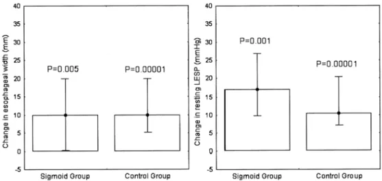
All 6 patients who had lost some weight before the opera-tion returned at least to their ideal body weight 12 months after the operation. Esophageal width decreased by 10 mm or more in 10 patients, by 20 to 30 mm in 6 patients, and by more than 30 mm in one patient. The decrement in esoph-ageal width was statistically significant in both groups, with no intergroup difference (Figure 2, left panel). In patients with sigmoid esophagus, the esophageal width decrease was inversely correlated with age, so that the younger the age, the greater the postoperative decrease in esophageal width (Figure 3). In contrast, no correlation was found between preoperative esophageal width and postoperative resizing of the esophagus (R⫽ ⫺0.08; P ⫽ not significant). Repeated postoperative Rx esophagograms were performed in all patients every 12 months and for at least 2 years, and 9 patients underwent yearly esophagograms for more than 3
years. Comparison of repeated esophagograms showed that maximal esophageal width reduced significantly since the 12th postoperative month and remained stable subse-quently, whereas the esophageal length did not change (Figure 4).
Preoperative and postoperative manometry was per-formed in 13 patients. LESP decreased significantly from 39 to 18 mm Hg (P ⫽ .001). Comparison with the results achieved in 28 patients of the control group who underwent preoperative and postoperative manometry showed no dif-ference in change in LESP (Figure 2, right panel). Postop-eratively, 3 patients had an LESP of greater than the upper normal value of 20 mm Hg, with a median value of 25 mm Hg. All these patients had residual mild dysphagia, and 2 underwent periodic pneumatic dilatation with satisfactory results. Postoperative LESP was inversely correlated with changes in dysphagia score (R ⫽ ⫺0.77; P ⫽ .001) and changes in regurgitation score (R⫽ ⫺0.57; P ⫽ .04), so that the lower the LESP, the greater the change in symptom score.
Esophagoscopy with multiple biopsies of the esophageal mucosa was carried out in 8 patients after a mean of 26 months, showing grade 1 esophagitis in 2 patients and mild squamous dysplasia in 1 patient. Continuous 24-hour pH monitoring confirmed pathologic gastroesophageal acid re-flux in both the patients with clinical and endoscopic evi-dence of esophagitis, with a proportion of time at pH of less than 4 of 19% and 22%, respectively.
QOL Assessment
Baseline SF-36 QOL domains scores in the sigmoid esoph-agus and control group are reported in Table 2. The social function and mental health domain scores were significantly lower in the sigmoid esophagus group than in the control group. At postoperative assessment, general health, social function, and mental health domains improved significantly (Table 3). The postoperative dysphagia score was inversely correlated with preoperative to postoperative changes in role physical (R⫽ ⫺0.61; P ⫽ .04) and mental health (R ⫽ ⫺0.68; P ⫽ .02) domains, whereas postoperative LESP was inversely correlated with changes in role physical (R ⫽ ⫺0.72; P ⫽ .01), role emotional (R ⫽ ⫺0.62; P ⫽ .03), and mental health (R⫽ ⫺0.72; P ⫽ .01). Comparison with the control group results showed that absolute incremental change in social function and mental health were greater in patients with sigmoid esophagus, whereas no difference existed between the groups in the remaining domains.
Up to the most recent follow-up, the procedure result was scored as excellent in 6 (43%) patients, good in 4 (29%) patients, and satisfactory or unsatisfactory in 2 patients each. No patient required esophagectomy or had esophageal carcinoma.
Figure 1. Postoperative change in subjective symptoms score. Data are expressed as medians with interquartile ranges.
Discussion
The main finding of this study was that Heller myotomy provided excellent to satisfactory symptom relief in 12 of 14 patients with achalasic sigmoid esophagus. It is also worth noting that these subjective improvements were accompa-nied by long-lasting improvements in end-expiratory resting LESP, maximal esophageal width, and QOL SF-36 domain scores, which did not differ from those achieved in a control group of patients with earlier-stage achalasia. These results corroborate and extend previous findings suggesting that esophageal myotomy can improve symptoms even in pa-tients with decompensated sigmoid esophagus.11,13,18
Although in no instance have we found restoration of propulsive peristalsis in the sigmoid group, our findings suggest that esophageal tone can be partially restored even in these patients provided that adequate esophageal empty-ing is ensured. This effect can be maximized in young patients, who achieved the greatest reduction in esophageal width and the best clinical results.
In agreement with previous study results,7,19 we have found that postoperative LESP returned to within the nor-mal range in 10 patients and that LESP changes correlated with changes in dysphagia and regurgitation scores.
A further issue relates to whether a sigmoid-shaped esophagus indicates a more advanced stage of disease. We have found that symptom duration was significantly longer in patients with sigmoid esophagus than in the control group. Yet in this patient group we observed a greater regurgitation score and more frequent respiratory symp-toms. In accordance with our results, Shiino and col-leagues20have found that the maximal width of the esoph-ageal body and tortuosity of the esophagus measured on esophagography became greater with longer symptom du-ration. Nonetheless, in our series the postoperative regurgi-tation score decreased to the level of the control group, and
no patient repeated respiratory symptoms postoperatively. These results confirm the efficacy of the Heller myotomy procedure in controlling these symptoms even in patients with sigmoid esophagus. In addition, the operation was not more technically demanding, had no more complications, and resulted in just as good of relief of dysphagia as in achalasic patients with an earlier stage of the disease.
The existence of symptoms leading to modification of eating habits has been shown to impair QOL in achalasia,11 as well as in other benign esophageal diseases.21
The SF-36 questionnaire has proved a reliable instrument to assess QOL after myotomy for achalasia.10Ben Meir and Figure 3. Relationship between esophageal width and age in patients with sigmoid esophagus.
Figure 2. Postoperative change in objective measures score. Data are expressed as medians with interquartile ranges.
associates8have found that physical function, bodily pain, vitality, and social function were significantly improved after surgical intervention. Similarly, Luketich and cowork-ers9have found that after myotomy, all 8 domains of SF-36 data scored at least equal to or better than that of the normal US population used for comparison.
Our findings support the use of the SF-36 as a reliable instrument for preoperative and postoperative QOL assess-ment in patients with achalasia. We have found that patients with sigmoid esophagus had more severely impaired social function and mental health domain scores than the control group. On the other hand, in the sigmoid group greater postoperative improvements occurred in these domains, whereas improvements in role emotional and general health domain scores compared with those of the control group.
We acknowledge some limitations of our study. First, our series is not homogeneous in terms of surgical approach
because it included patients operated on by means of either laparotomy or a laparoscopic approach. However, the tech-nique was identical in all the patients, and all the procedures were performed by the same surgeon. Moreover, there is now sufficient evidence that long-term results of open ver-sus laparoscopic Heller myotomy are comparable.2,3
Second, we had no control group treated with esopha-gectomy, which constitutes the surgical alternative for these patients. However, we believe that any attempt at preserving the native esophagus should be made before deciding to perform an esophagectomy for a benign disease. In fact, despite the fact that esophageal substitution is now a safe procedure that offers optimal clinical and functional out-comes in the majority of the patients, it is more technically demanding and carries a mortality rate of about 2%.14,22
In this study the Heller myotomy proved highly effective in improving objective measures, subjective symptoms, and Figure 4. Preoperative (A) and postoperative (B) esophagograms of a 29-year-old patient showing a significant
reduction in esophageal width 12 months after Heller myotomy.
TABLE 2. Baseline QOL assessment by using the SF-36 questionnaire
SF-36
domain Sigmoid esophagus group (nⴝ 9) Control group (nⴝ 20)
Intergroup P value PF 90 70-100 90 80-97.5 NS RP 55 25-75 75 65-75 NS BP 90 64-90 90 70-95 NS GH 60 55-75 60 55-83.5 NS SF 50 37-55 61 50-72.5 .02 VT 60 45-65 55 45-60 NS RE 66 66-100 66 66-100 NS MH 64 56-68 72 66-76 .03
Data are expressed as medians with interquartile ranges.
QOL, Quality of life; SF-36, Short-Form 36-item; PF, physical functioning; RP, role physical; BP, bodily pain; GH, general health; SF, social functioning; VT,
vitality; RE, role emotional; MH, mental health; NS, not significant.
QOL in patients with achalasic sigmoid esophagus. Yet these improvements were long lasting and did not differ from those achieved in a control group with earlier-stage achalasia. Although confirmation by larger prospective studies is warranted, the results achieved in this series lead us to conclude that independent of the surgical approach, Heller myotomy with anterior fundoplication should be considered as a first-choice treatment option for achalasic sigmoid esophagus.
References
1. Suarez J, Mearin F, Boque R, Zano´n V, Armengol JR, Pradell J, et al. Laparoscopic myotomy vs endoscopic dilation in the treatment of achalasia. Surg Endosc. 2001;16:75-7.
2. Ancona E, Anselmino M, Zaninotto G, Costantini M, Rossi M, Bonavina L, et al. Esophageal achalasia: laparoscopic versus conven-tional open Heller-Dor procedure. Am J Surg. 1995;170:265-70. 3. Dempsey DT, Kalan MMH, Gerson RS, Parkman HP, Maier WP.
Comparison of outcomes following open and laparoscopic esophago-myotomy for achalasia. Surg Endosc. 1999;13:747-50.
4. Graham AJ, Finley RJ, Worsley DF, Dong SR, Clifton JC, Storseth C. Laparoscopic esophageal myotomy and anterior partial fundoplication for the treatment of achalasia. Ann Thorac Surg. 1997;64:785-9. 5. Patti MG, Pellegrini CA, Horgan S, Arcerito M, Omelanczuk P,
Tamburini A, et al. Minimally invasive surgery for achalasia. An 8-year experience with 168 patients. Ann Surg. 1999;230:587-94. 6. Ackroyd R, Watson DI, Devitt PG, Jamieson GG. Laparoscopic
car-diomyotomy ad anterior partial fundoplication for achalasia. Surg Endosc. 2001;15:683-6.
7. Sharp KW, Khaitan L, Scholz S, Holzman MD, Richards WO. 100 consecutive minimally invasive Heller myotomies: lessons learned. Ann Surg. 2002;235:631-8.
8. Ben Meir A, Urbach DR, Yashodhan MS, Khajanchee YS, Hansen PD, Swanstrom LL. Quality of life before and after laparoscopic Heller myotomy for achalasia. Am J Surg. 2001;181:471-4.
9. Luketich JD, Fernando HC, Christie NA, Buenaventura PO, Keenan RJ, Ikramuddin S, et al. Outcomes after minimally invasive esophago-myotomy. Ann Thorac Surg. 2001;72:1909-13.
10. Katilius M, Velanovich V. Heller myotomy for achalasia: quality of life comparison of laparoscopic and open approaches. JSLS. 2001;5: 227-31.
11. Decker G, Borie F, Bouamrirene D, Veyrac M, Guillon F, Fingerhut A, et al. Gastrointestinal quality of life before and after laparoscopic Heller myotomy with partial posterior fundoplication. Ann Surg. 2002; 236:750-8.
12. Ponce M, Ortiz V, Juan M, Garrigues V, Castellanos C, Ponce J. Gastroesophageal reflux, quality of life, and satisfaction in patients with achalasia treated with open cardiomyotomy and partial fundopli-cation. Am J Surg. 2003;185:560-4.
13. Patti MG, Feo CV, Diener U, Tamburini A, Arcerito M, Way LW. Laparoscopic Heller myotomy relieves dysphagia in achalasia when the esophagus is dilated. Surg Endosc. 1999;13:843-7.
14. Devaney EJ, Lannettoni MD, Orringer MB, Marshall B. Esophagec-tomy for achalasia: patient selection and clinical experience. Ann Thorac Surg. 2001;72:854-8.
15. Peters JH, Kauer WKH, Crookes PF, Ireland AP, Brenner CG, De-Meester TR. Esophageal resection with colon interposition for end-stage achalasia. Arch Surg. 1995;130:632-7.
16. Pinotti HW, Cecconello I, Mariano da Rocha J, Zilberstein B. Resec-tion for achalasia of the esophagus. Hepatogastroenterology. 1991;38: 470-3.
17. Apolone G, Mosconi P. The Italian SF-36 Health Survey: translation, validation and norming. J Clin Epidemiol. 1998;51:1025-36. 18. Pechlivanides G, Chrysos E, Athanasakis E, Tsiaoussis J, Vassilakis
JS, Xynos E. Laparoscopic Heller cardiomyotomy and Dor fundopli-cation for esophageal achalasia: possible factors predicting outcome. Arch Surg. 2001;136:1240-3.
19. Yaghoobi M, Mikaeli J, Montazeri G, Nouri N, Sohrabi MR, Male-kzadeh R. Correlation between clinical severity score and the lower esophageal sphincter relaxation pressure in idiopathic achalasia. Am J Gastroenterol. 2003;98:278-83.
20. Shiino Y, Houghton SG, Filipi CJ, Awad ZT, Tomonaga T, Marsh RE. Manometric and radiographic verification of esophageal body decom-pensation for patients with achalasia. J Am Coll Surg. 1999;189(2): 158-63.
21. Revicki DA, Wood M, Maton PN, Sorensen S. The impact of gastro-esophageal reflux disease on health-related quality of life. Am J Med. 1998;104:252-8.
22. Watson TJ, DeMeester TR, Kauer WKH, Peters JH, Hagen JA. Esoph-ageal replacement for end—stage benign esophEsoph-ageal disease. J Thorac Cardiovasc Surg. 1998;115:1241-9.
TABLE 3. Preoperative to postoperative changes in SF-36 domains scores
SF-36
domain Sigmoid esophagus group (nⴝ 9) Control group (nⴝ 20)
Intergroup P value ⌬PF 5.0 0-10 0 0-10 NS ⌬RP 20 0-30 0* 0-25 NS ⌬BP 0 0-20 0 0-20 NS ⌬GH 5* 2.0-15 9.0* 3.5-16 NS ⌬SF 30* 12-38 23.5* 12.5-25 .02 ⌬VT 20* 15-25 17.5* 15-20 NS ⌬RE 0 0-34 0* 0-33 NS ⌬MH 16* 0-20 10* 4.0-16 .03
Data are expressed as median with interquartile ranges.
SF-36, Short-Form 36-item; PF, physical functioning; RP, role physical; BP, bodily pain; GH, general health; SF, social functioning; VT, vitality; RE, role
emotional; MH, mental health; NS, not significant. *Within-group P⬍ .01.
Continuing Medical Education Activities
http://cme.ctsnetjournals.org/cgi/hierarchy/ctsnetcme_node;JTCS
Subscribers to the Journal can earn continuing medical education credits via the Web at
Subscription Information
http://jtcs.ctsnetjournals.org/cgi/content/full/128/3/402#BIBL
This article cites 21 articles, 6 of which you can access for free at:
Citations
http://jtcs.ctsnetjournals.org/cgi/content/full/128/3/402#otherarticles
This article has been cited by 6 HighWire-hosted articles:
Subspecialty Collections
http://jtcs.ctsnetjournals.org/cgi/collection/esophagus_other
Esophagus - other
This article, along with others on similar topics, appears in the following collection(s):
Permissions and Licensing
http://www.elsevier.com/wps/find/obtainpermissionform.cws_home/obtainpermissionform
receipt, is available at:
An on-line permission request form, which should be fulfilled within 10 working days of .
http://www.elsevier.com/wps/find/supportfaq.cws_home/permissionusematerial
can be found online at: