PISA, ANNO 2020
PERFEZIONAMENTO IN NEUROBIOLOGIA TESI:
Upon the way through which astrocytes sense and react to a reduction of extracellular Nerve Growth Factor level
NGF and Astrocytes in neurodegenerative disease models
PERFEZIONANDO:
Dr. Nicola Maria Carucci
RELATORI:
Prof. Antonino Cattaneo, PhD Prof.ssa Simona Capsoni, MV, PhD
ATTIVITÀ CONDOTTE PRESSO IL LABORATORIO DI BIOLOGIA DELLA SCUOLA NORMALE SUPERIORE, PISA
“Siamo amanti e cultori di una sapienza umana che può
considerarsi compiuta quando arriva a scorgere ciò che il senso rivela e ciò che si può desumere dalla similitudine, come i fenomeni percepiti per mezzo del senso. […]
Noi non abbiamo seguito nient’altro che il senso e la natura, la quale, sempre in massimo accordo con se stessa, fa le stesse cose allo stesso modo e sempre produce i medesimi effetti.”
De rerum natura iuxta propria principia (1586),
5
Index
1. NEUROTROPHINS AND ASTROCYTES 11
1.1 Summary 11
1.2 More on NGF and AD 14
1.3 NGF and Alzheimer’s Disease 15
1.4 The AD11 mouse 17
2. GENERAL BACKGROUND 23
2.1 The fall of idols 23
2.3 Astrocytes and Neurons, a well consolidated cooperation 29
3. NATURAL HISTORY OF NEUROGLIA 35
3.1 Neuroglia: from glue to glory 36
3.2 Classification of neuroglia 40
3.3 Phylogenetic importance of neuroglia 43
6
4.1 Unsuspected heterogeneity 49
4.2 Defining an astrocyte: common properties 53 4.3 Morphology of the main type of astrocyte 58
4.4 Human astrocytes 60
5. ASTROCYTES IN ACTION 65
5.1 The tripartite synapse 65
5.2 Ca2+-mediated cellular excitability of astrocytes 69 5.3 Astrocyte Ca2+ is controlled by synaptic activity 70 5.4 Astrocyte Ca2+ signal in vivo 72 5.5 Synaptic information processing by astrocytes 75 5.6 Astrocytes discriminate the activity of synaptic pathways 76 5.7 Astrocyte Ca2+ signals show a nonlinear relationship with
the synaptic activity 79
5.8 Gliotransmission and modulation of synaptic
transmission 80
5.9 Astrocytes and synaptic plasticity 88
5.10 Are all synapses tripartite? 90
6. RESULTS 93
6.1 NGF deprivation induces morphological alterations of
7 6.2 Mechanism(s) at the basis of astrocytes altered
morphology: role of proNGF 112
6.3 In vitro effects of NGF depletion 115 6.4 Astrocytes express NGF receptors in vitro 121 6.5 Atrophic astrocytes induced by anti NGF antibody
treatment display a phenotype of reactive astrocytes 124 6.6 The NGF depletion increases astrocyte calcium
oscillation 127
6.7 The Ca2+ involved in anti-NGF dependent calcium
waves derives from ER-stores 131
6.8 Calcium oscillations induced by anti NGF antibodies are abolished if TrkA expression is chemogenetically inhibited in
astrocytes 135
6.9 Transcriptomic changes at 8, 24 and 48 hours after
anti-NGF antibody administration 138
6.10 In astrocyte-neuron co-cultures, the NGF starvation
leads to neuronal impairment and death 142 6.11 The effects of NGF deprivation onto astrocytes might imply the production of β amyloid oligomers or glutamate
excitotoxicity, impairing neurons 147
6.12 The astrocyte Ca2+ elevations induced by NGF
starvation trigger Ca2+ hyper-activation in neurons 152 6.13 In vivo rescue of astrocytes atrophy by NGF in
different AD models 158
8
6.15 Reducing NGF levels increases astrocyte calcium in the
awake behaving animal 167
6.16 Upregulation of NGF translation after 5 min of NGF
deprivation 172
7. DISCUSSION 177
7.1 Astrocytes and NGF: secretion, signalling and mouse
models 178
7.2 Pathological implications of atrophic astrocytes 180 7.3 Early events after NGF deprivation 182 7.4 Long term events after NGF deprivation 184 7.5 Adaptive homeostatic functions of astrocytes 185 7.6 A new mechanism of neuronal death mediated by
NGF-deprived neurotoxic astrocytes 188
7.7. Widening the potential clinical application of NGF 191
8. METHODS 193 8.1 Animals 193 8.2 Intranasal Delivery 194 8.3 Cell cultures 195 8.4 Immunoblot analysis 197 8.5 Immunocytochemistry 199
9
8.6 Immunofluorescence on slice 200
8.7 Flow cytometry 201
8.8 Microarray transcriptome analysis 202
8.9 Neuron/astrocytes co-cultures 203
8.10 Confocal microscopy and image analysis 204
8.11 Data Analysis and statistics 205
8.12 Transgenic mice for in vivo Ca2+ experiments 206 8.13 In vivo imaging with two-photon microscopy in the
anesthetized mouse 207
8.14 Surgical preparation for imaging awake, head-restrained
mice 208
8.15 Drug through an open window in the skull 211 8.16 Statistical analyses for in vivo Ca2+ experiments 211 8.17 NGF immunoprecipitation and Western Blot 212
10
Astrosphere: astrocytes grown from embryonic induced stem cells. Credit: the laboratory of UW-Madison, Su-Chun Zhang, 2011.
11
1.
Neurotrophins and
Astrocytes
1.1 Summaryy starting observation was that that chronic exposure of hippocampal astrocytes to anti-nerve growth factor (NGF) antibodies is associated with early and progressive changes in astrocytic morphology reminiscent of reactive gliosis. Similar morphology changes are found in the hippocampal astrocytes from proNGF overexpressing
12
transgenic mice and from Alzheimer’s 3XTg and 5xFAD mice at early stages of neurodegeneration. Interestingly, this phenotype can be rescued by intranasal NGF delivery to 3xTG and proNGF mice.
These findings prompted us to investigate the mechanisms whereby interfering with the signalling of NGF causes astrocytes to undergo these changes.
The astrocytic morphological alterations in mouse models of neurodegeneration were reproduced in vitro by culturing naïve hippocampal astrocytes with mAb αD11 anti-NGF antibodies. These dramatic changes of astrocyte morphology are accompanied by robust transcriptomic changes at 8, 24 and 48 hours after anti-NGF administration, which identify a transcriptional fingerprint typical of reactive type A1 neurotoxic astrocytes70. Since cultured astrocytes express both TrkA and p75 NGF receptors and secrete NGF, I postulated that anti NGF antibodies disrupt a homeostatic autocrine or paracrine NGF signalling, inducing their transition to the A1 neurotoxic phenotype.
I went on to investigate intracellular Ca2+ transients in astrocyte cultures. While NGF failed to reveal any effect on Ca2+, anti NGF potently induced within 5 minutes large intracellular Ca2+ transients, from ER intracellular stores.
13 Chemogenetic experiments indicate that the antiNGF-induced Calcium waves are TrkA-mediated.
The functional consequences of anti NGF treatment of astrocytes on neuronal physiology were studied in hippocampal astrocyte-neuronal co-cultures. In these conditions, anti NGF treated astrocytes induced a strong Ca2+ activity in hippocampal neurons, ultimately leading to massive neuronal death.
In conclusion, I have demonstrated that, in vitro and in vivo, astrocytes are sensors of ambient levels of NGF and respond to reduced levels of NGF by the activation of a strong and rapid Ca2+ response that eventually leads them to assume a A1 neurotoxic phenotype and negatively impact neuronal physiology and brain homeostasis.
14
1.2 More on NGF and AD
The role of NGF in neurodegeneration has an old root, linked to its pro-survival and phenotypic maintenance action on cholinergic neurons of the basal forebrain. The discovery that other cells in the brain, besides neurons, like glial cells, could respond to NGF and modulate the neuronal physiology broadened the possible NGF targets to potentially all the brain, boosting the comprehension of some pathologies - like Alzheimer disease - and supporting new therapeutic strategies. Here I propose, based on my experiments, a new mechanism whereby astrocytes can sense locally the NGF level. They respond to a decrease in extracellular NGF, potentially triggering an activation loop sustained by an increase in calcium oscillations that can cause astrocytes to become active and change their morphology.
First experimental evidence reveals that in a short time these paradoxically active astrocytes, when co-cultured with neurons, determine an increase of calcium transients in neurons as well, leading them to death.
More experiments are necessary to unveil these proposed mechanisms and go deeper in the comprehension of astrocyte physiology, but the framework established by the
15 experiments presented in this PhD thesis allow future experiments to be grounded on a solid basis.
1.3 NGF and Alzheimer’s Disease
NGF is not translated as such, but as a pre-pro-protein which is cleaved, typically by furin enzyme, in the trans-Golgi network to yield mature NGF71. ProNGF was initially thought to be simply a chaperone to allow NGF to be folded and mature NGF to be finally secreted. ProNGF can also be released as such and represents the major form of NGF in the brain73 and can be cleaved extracellularly by plasmin and matrix metalloproteases72. ProNGF and NGF have distinct biological activitie43: proNGF has a higher affinity for p75NTR and a lower one for TrkA compared to mature NGF and induces p75NTR-dependent apoptosis.
ProNGF can also induce TrkA-dependent neuronal survival, although less effectively than NGF73.
The pro-domain of NGF interacts with sortilin, a neuronal type-1 VPS10-domain receptor, a co-receptor with p75NTR for proNGF74. Sortilin, together with the VPS10-containing protein sorLA, binds the retromer complex in neurons.
16
The levels of proNGF and of its coreceptor sortilin increase in mild cognitive impairment and early AD brains90, paralleling the progressive decline in TrkA receptors. A diminished conversion of proNGF to mature NGF and an increased NGF degradation in AD brains was definitely reported72.
Thus, the biological effects of proNGF versus NGF influence the balance between cell death and cell survival74, 75 and an imbalance in this complex ligand/receptor system has been correlatively linked to AD neurodegeneration, although no causally direct proof in vivo is available.
17 1.4 The AD11 mouse
NGF (Levi-Montalcini, 1952) is required for the differentiation and the survival of specific neuronal populations during development, including sensory, and sympathetic neurons in the peripheral nervous system and basal forebrain cholinergic neurons in the central nervous system (Levi-Montalcini, 1987). NGF also exerts actions on non-neuronal cell populations (Levi-Montalcini, 1987).
After the early use of anti-NGF antibodies (Levi-Montalcini and Booker 1960), NGF functions in vivo have been investigated with different approaches, including the systemic (Levi-Montalcini and Angeletti, 1966; Gorin and Johnson, 1979, 1980) or local (Li et al., 1995; Van der Zee et al., 1995; Molnar et al., 1998) delivery of anti-NGF antibodies and the disruption of the NGF gene, by homologous recombination, in transgenic mice (Crowley et al., 1994).
The embryonic ablation of NGF function gives rise to a lethal phenotype in the early postnatal period, preventing the analysis of NGF function in adult animals. On the other hand, adult heterozygous NGF knockout mice show only a mild cholinergic phenotype and no other described deficits (Chen et al., 1997).
18
Given the potential clinical relevance of some of the described actions of NGF, the use of specific and not-lethal models represents a desirable tool.
After the demonstration that recombinant antibodies can be efficiently secreted by cells of the nervous system (Cattaneo and Neuberger, 1987), a novel approach for phenotypic knockout, the neuroantibody approach, has been proposed and validated (Piccioli et al., 1991, 1995).
With all these pivotal assumptions, a transgenic mouse that lacks enough mature nerve growth factor was generated, and, among the relevant outcomes, it suffers from a progressive AD-like syndrome. Moreover, the delivery of NGF or of a cholinergic agonist were able to reduce the pathology.
The mouse model, called AD11, was created to explore the possibility that the death of basal forebrain cholinergic neurons in Alzheimer's disease is related to reductions in nerve growth factor levels. AD11 was created by inserting a gene that expresses an anti-NGF antibody (formally, 2 genes for light and heavy chains), named αD11, that efficiently binds only the mature form of the neurotrophin (Ruberti et al. 2000, Paoletti
19 Fig 1.1 Schematic representation of the proNGF/NGF imbalance model
On the left side is the "normal" condition, when nerve growth factor (NGF) signaling is achieved mainly through the NGF/TrkA/p75NTR system. In the middle-right part of the scheme is a
20
right, AD11 model): in both cases, an imbalance in the proNGF/NGF ratio takes place.
From Nerve Growth Factor and Alzheimer’s Disease: New Twists to an Old Story, 2011, MIT Press, 26th chapter by Antonino
21 Capsoni, Cattaneo and their colleagues have described how the depletion of extracellular mature NGF in this model progressively recapitulates features of Alzheimer's disease. The AD11 mouse shows neuronal loss, tau hyperphosphorylation and insolubility, neurofibrillary tangle-like abnormalities, and behavioral deficits linked to cholinergic atrophy (Capsoni et al., 2000).
It also exhibits amyloid plaques (from the endogenous mouse amyloid precursor protein) (Capsoni et al. 2002), and deficits in cortical synaptic plasticity (Pesavento et al. 2002), all of which led Cattaneo and associates to propose this mouse as a comprehensive model for sporadic AD and, among the other numerous outcomes, identifying a novel mechanism of neurodegeneration induced by proNGF/NGF imbalance.
In addition, in 2011 D’Onofrio et al. showed that, in these mice, the onset of neuroinflammation was preceding the appearance of tau hyperphosphorylation and amyloid plaques.
This prompted us to study the effects of NGF deprivation on one cell type involved in neuro-inflammation: the astrocyte.
22
“It’s a crowded party in the body, and biologists want to see what’s going on” From “A Brain in a Plate?”
23
2.
General background
2.1 The fall of idols
he neuron-centred view of the past disregarded or downplayed the role of astroglia as a primary component in the pathogenesis of neurological diseases.
As this concept is changing, so is also the perceived role of astrocytes in the healthy and diseased brain and in the spinal cord. Researchers have started to unravel the different signalling mechanisms that trigger specific molecular, morphological and functional changes in astrocytes and that may be critical for repairing tissue and maintaining function in
24
CNS pathologies, such as neurotrauma, stroke, or neurodegenerative diseases. An increasing body of evidence shows that the effects of astrogliosis on the neural tissue and its functions are not uniform or stereotypic but vary in a context-specific manner. Indeed, astrogliosis can be an adaptive beneficial response under some circumstances but also a maladaptive and deleterious process in another context.
There is a growing support for the concept of
astrocytopathies in which the disruption of normal astrocyte
functions, astrodegeneration or dysfunctional/maladaptive astrogliosis are the primary cause or the main factor in
neurological dysfunction and disease. For this, reason, there is a big interest in the mechanism of dysfunctional/maladaptive astrogliosis and on the factors that execute the toxic actions of astrocytes in neurodegeneration conditions. This thesis proposes a new homeostatic mechanism of astrocytes in the healthy CNS describing how they could react to an unbalance of NGF/proNGF in the microenvironment around them.
This finding increases the diversity of astrocytes responses in neurological disorders and argues that targeting astrocytes may represent an effective therapeutic strategy for Alzheimer Disease (AD) as well as other neurodegenerative diseases.
25 2.2 Astrocytes in the brain: beyond scaffold cells It is nowadays well understood that astrocytes cannot be just be considered as scaffold cells deputed to feed and preserve neuronal activity but, since they are involved in a large number of functions, they are critical to the performance of the Central Nervous System (Verkhratsky et al., 2014; Gallo et al., 2014).
It is more and more evident that the peculiar properties of these cells can actively contribute to the extraordinary complexity of the brain and, on the other hand, to the dysfunctions and diseases classically related to other cell types, especially neurons (Allen et al., 2009; Lin et al., 2012).
New evidence indicates that astrocytes could really make the difference in mammalian brain evolution (Molofsk et al., 2012), corroborated by the impressive discovery that mice enhance some behavioural aspects if expressing human astrocytes(Zang et al., 2013; He et al., 2015).
The essential and various functions astrocytes provide include i) homeostasis of extracellular ions, pH, idratation (Simard et al., 2004); ii) uptake and clearance of neurotransmitters (Schousboeet al., 2004; Yi et al., 2006); iii) energy and metabolism (Allaman et al., 2011); iv) blood flow regulation(Attwell et al., 2010); v) neuronal activity
26
coordination and synaptic functions (Clarke et al., 2013; Halassa et al., 2010; Bergami et al., 2008; Bazargani et al., 2016).
If a large number of evidences are now reported regarding how these functions might involve astrocyte specialization and heterogeneity, the degree to how astrocytes maintain or modulate their participation in these functions, or adopt new functions, is not well understood (Anderson et al., 2014; Colangelo et al., 2014; Trias et al., 2013; Wyss-Coray et al., 2002).
As mentioned before, some aspects traditionally related to neuronal degenerative pathologies, together with some well described neuronal physiological properties, could be reasonably re-interpreted keeping in mind that astrocytes, and their contribution, might have been underestimated
A large number of in vitro studies on bona fide neuronal
preparations include de facto astrocytes, due to the difficulties to
obtain really pure neuronal cultures. Obviously, astrocytes are also present in the majority of the in vivo and ex vivo
27 With the aim of addressing this need, and of turning these limitations into new perspectives,
to explore if and how astrocytes could react to proNGF/NGF imbalance and, eventually, influencing the neuron homeostasis,
I took advantage from our AD11 mouse model, in which the expression of the recombinant αD11 anti Nerve Growth Factor (NGF) antibody causes an imbalance between the mature and immature form of NGF.
This lead, progressively, to a generalized Alzheimer-like phenotype (Ruberti et al. 2000; Capsoni et al., 2000 and 2002) overcoming the concept that the “classical” neuronal target cells for NGF could appear a limiting factor for the model acceptancy (Cattaneo et al., 2008).
The glia cells (microglia and astrocytes) could be the target cells contributing to explain this widespread effect.
The link between Alzheimer's disease and astrocytes is well described and is based on several components: they can take up Aβ, they are involved in plaque progression with death of Aβ loaded astrocytes giving rise to secondary plaques (Nagele et al., 2004); astrocytes have a modulatory effect on neuroinflammation27, on neuron-neuron communication16 and
28
on tripartite (more recently, quadripartite) synapses28,29. In this study, I describe a new way by which astrocytes can respond to a change in brain homeostasis such as NGF local acute deprivation.
In presence of a decrease in the level of NGF astrocytes become active and modify their morphology. I propose also a new way they could influence neuronal activity and cause neuronal death.
29 2.3 Astrocytes and Neurons, a well consolidated cooperation
The multi-partite synapse of the CNS represents a striking example of such a specialization (fig 2.1) with pre- and post-synaptic membranes being packed with exocytotic machinery, neurotransmitter receptors and proteins responsible for plasticity, whereas all “homeostatic” molecules (i.e. transporters and enzymes responsible for ion and transmitter homeostasis in the synaptic cleft, for transmitter catabolism, for metabolic support, etc.) being localized in the perisynaptic astrocytic processes .
The synaptic assembly also includes a microglial cell process that frequently senses the synapse status. Already at this elementary level of CNS organization, the cellular functions are divided. Neuronal compartment assures fast information flow whereas glial elements ascertain functional isolation and support of synapses, maintain synaptic operation through regulation of homeostasis and control synaptic survival or elimination depending on the network demands.
30
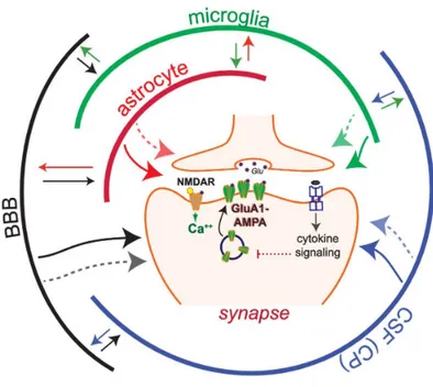
Fig 2.1 Multipartyte synapse involving several CNS players.
This simplified model illustrates LTP, which relies on the NMDAR-dependent insertion of GluA1-containing AMPA receptors at the postsynaptic surface. In this model communication via cytokines is depicted by arrows, which can bi-directionally connect multiple cell populations.
Cytokine networks enable local interactions between neuronal and non-neuronal cells (e.g., astrocytes, microglia, vascular endothelial cells) in the brain, as well as brain-periphery communication via the brain-blood barrier (BBB) and the choroid plexus (CP).
The BBB releases cytokines and regulates the flux of cytokines from the blood; the CP produces cerebrospinal fluid (CSF) and cytokines and regulates the transport of cytokines and immune cells from blood
31
vessels. LTP modulation by cytokines has been widely studied, however, for most cytokines, is unclear if they modulate LTP by directly targeting synapses (one-direction arrows) or by indirect mechanisms relying on cytokine networks maintained by non-neuronal cells interactions.
Cytokines can induce the expression and release of multiple cytokines in their target cells, thus activating cytokine networks, which could modulate synaptic transmission by targeting synapses via both cytokine-dependent and -independent mechanisms (dotted arrows) From Prieto and Cotman 2017.
32
The very same specialization is observed at all levels of CNS organization. Neurons fire and establish multiple contacts whereas neuroglia control local micro-environment and protect neural tissue. In the grey matter, astrocytes divide (through the process known as tiling that starts in late embryogenesis) the parenchyma into relatively independent units traditionally known as neurovascular units, and recently often called astrogliovascular units, that integrate, within an individual astroglial territorial domain, neural and vascular elements. By employing a wide array of molecular mechanisms such as exocytosis, diffusion through plasmalemmal channels or membrane transporters, astrocytes secrete numerous neurotransmitters, neurohormones and trophic factors that regulate synaptic fields, neuronal groups and signal to other cellular elements (e.g. microglia, oligodendroglia, pericytes, endothelial cells).
At the level of the whole brain, astrocytes form glia limitans regulate emergence and function of brain–blood and
brain–cerebrospinal fluid barriers and contribute to overall brain metabolism being the sole producers and repository of glycogen
The homeostatic function of astroglia is linked to their neuroprotective capabilities, as indeed astrocytes are principal
33 elements of CNS defence. Insults to the CNS, regardless of their aetiology, strain the organ homeostasis and in this case astrocytes, through dedicated molecular cascades, protect neurons against glutamate excitotoxicity, extracellular K+ overload, reactive oxygen species, and these are also astrocytes that supply stressed neurons with energy substrates.
The loss of these critical astroglial functions permits and exacerbates progression of various diseases, of which amyotrophic lateral sclerosis, toxic encephalopathies or Alzheimer’s disease (AD) are prominent examples. Defensive function of astrocytes is manifested as reactive astrogliosis, a multi- component and complex remodelling of astroglia triggered by lesions to the CNS. Astrogliosis is an important component of cellular pathophysiology and its suppression often aggravates neuropathology.
34
Mouse Astrocytes, cortex. Credit: Dr. Nybertuc.
35
3.
Natural
history
of
neuroglia
“THE NEUROGLIA is the delicate connective tissue which supports and binds together the nervous elements of the central nervous system. One part of it, which lines the central canal of the cord and ventricles of the brain, is formed from columnar cells, and is called ependyma, while the rest consists of small cells with numerous processes which sometimes branch and sometimes do not.”
36
“As the Greek name implies, glia are commonly known as the glue of the nervous system; however, this is not fully accurate. Neuroscience currently identifies four main functions of glial cells: to surround neurons and hold them in place, to supply nutrients and oxygen to neurons, to insulate one neuron from another, and to destroy pathogens and remove dead neurons. For over a century, it was believed that they did not play any role in neurotransmission. That idea is now discredited; they do modulate neurotransmission, although the mechanisms are not yet well understood.”
Glial Physiology and Pathophysiology, First Edition. 2013
3.1 Neuroglia: from glue to glory
o the continual surprise of everybody working in neuroglial research, the proper characteristics and capabilities of neuroglia are
still a matter of debate. Many existing definitions highlight the supportive role of these cells, and some are based on the process branching and delicate morphology of these cells, but the most common definition assigned to neuroglia is “cells residing in the brain that are not electrically excitable neurons or vascular cells”.
37 As a result, neuroglia has become a generalised term that covers cells with different origins (ectodermal for macroglia and mesodermal for microglia), morphology, physiological properties and functional specialisation. Indeed, in the Central Nervous System (CNS), neuroglia include the cells of the choroid plexus, the oligodendrocytes, the ependymal cells, the radial glia of the retina, the immunocompetent microglia/innate macrophages and the hugely diverse astrocytes; whereas, in the peripheral nervous system (PNS), they include the diverse kinds of Schwann cells, satellite glia, olfactory ensheathing cells and the highly numerous enteric glia.
There is, however, one unifying fundamental property common for all these cell types and this is their ultimate function: homeostasis of the nervous system.
The evolution of the nervous system led to a specialisation of neurons, which become perfect elements for signalling and information processing. This came at the price of losing essential housekeeping functions, as neurons are generally incapable of regulating their own immediate environment and are vulnerable to many kinds of environmental insults. These main housekeeping functions went to the neuroglia, which have specialised themselves into
38
many types of cells to perform specific aspects of nervous system homeostasis.
This homeostatic function of neuroglia is executed at many levels, and includes: whole body and organ homeostasis (e.g. astrocytes control the emergence and maintenance of the CNS, peripheral glia are essential for communication between the CNS and the body, and enteric glia are essential for every aspect of gastrointestinal function);
i) cellular homeostasis (e.g. astroglia and microglia); ii) morphological homeostasis (glia define the migratory pathways for neural cells during development, shape the nervous system cyto-architecture and control synaptogenesis/synaptic pruning, whereas myelinating glia maintain the structural integrity of nerves);
iii) molecular homeostasis (which is represented by neuroglial regulation, of ion, neurotransmitter neurohormone, and neurotrophic factor concentrations in the extracellular spaces around neurons);
iv) metabolic homeostasis (e.g. neuroglial cells store energy substrates in a form of glycogen and supply neurons with lactate);
v) long-range signalling homeostasis (by myelination provided by oligodendroglia and Schwann cells);
39 vi) defensive homeostasis (represented by astrogliosis and activation of microglia in the CNS, immune reactions of enteric glia; all these reactions provide fundamental defence for neural tissue).
Moreover, some neuroglial cells act as chemosensitive elements of the brain that perceive systemic fluctuations in CO2, pH and Na+ and thus regulate behavioural and systemic homeostatic physiological responses.
Therefore, the neuroglia can be broadly defined as homeostatic cells of the nervous system, represented by highly heterogeneous cellular populations of different origin, structure and function.
40
3.2 Classification of neuroglia
Generally, see the scheme below, the neuroglia in the mammalian nervous systems are sub classified into peripheral nervous system (PNS) glia and central nervous system (CNS) glia.
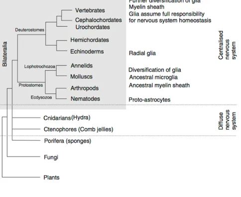
Fig 3.1 The neuroglia in the mammalian nervous system
From Verkhratsky 2013.
The PNS glial cells which surround neurons in peripheral ganglia are known as satellite glial cells, and those in
41 the olfactory system are known as olfactory ensheathing cells. Finally, the PNS includes enteric glia, which reside in the enteric nervous system.
CNS glia are generally subdivided into astrocytes, oligodendrocytes, NG2-glia and microglia. Astrocytes are the main homeostatic cells of the gray matter. Oligodendrocytes are the myelinating cells in the CNS and NG2-glia act as oligodendroglial precursors.
Fig 3.2 The evolutionary origins of glial cells
42
The early evolution of the nervous system can only be speculated upon, because fossils do not provide much material for analysis, and it is likely that many early life forms have not survived to our time. Nonetheless, certain generalisations can be drawn and overall, we are in possession of a rather logical system of views on the milestones of nervous system phylogeny.
There are some indications that glia have appeared in phylogeny on several occasions, and parallel evolution is likely. There is no evidence for the existence of glial cells in diffuse nervous systems and no cells associated with neurons or their processes have been detected in the comb jellies. Similarly, no glial cells were found in Cnidaria polyps, with the exception of scyphomedusae, in which some glia-like cells were apparently reported from Hartline in 2011, but neither their function nor their glial identity, has been analysed in detail.
43 3.3 Phylogenetic importance of neuroglia
44
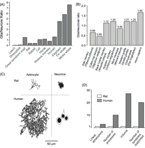
(a) Glia-to-neuron ratio in the nervous system of invertebrates and in the cortex of vertebrates. Glia-to-neuron ratio is generally increased in phylogeny; this ratio more or less linearly follows an increase in the size of the brain.
(b) The glia/neuron ratio in the cortex of higher primates; this ratio is highest in humans (From Sherwood et al., 2006).
(c) Graphic representation of neurons and astroglia in mouse and in human cortex. Evolution has resulted in remarkable changes in astrocytic dimensions and complexity.
(d) Relative increase in glial dimensions and complexity during evolution. Linear dimensions of human astrocytes, when compared with mice, are .ca 2.75 times larger, and their volume is 27 times larger; human astrocytes have .ca 10 times more processes and every astrocyte in human cortex enwraps .ca 20 times more synapses.
45 Most probably, neuroglia appeared with the emergence of a centralised nervous system, when neurons acquired specialisation and subsequently began to amass into sensory organs and ganglia. The very first glial cells are associated with sensory organs and have the same epithelial origin as neurons. More advanced and much more characterised are glial cells in nematodes, as extensively studied in Caenorhabditis elegans.
The brains of primates contain specific astroglial cells which are absent in other vertebrates. Most notable of these are the interlaminar astrocytes, which reside in layer I of the cortex; this layer is densely populated by synapses but almost completely devoid of neuronal cell bodies. These interlaminar astrocytes have a small cell body (10 mm), several short and one or two very long processes. The latter penetrate through the cortex and end in layers III and IV; these processes can be up to 1 mm long. The endings of the long processes create a rather unusual terminal structure, known as the ‘terminal mass’ or ‘end bulb’, which is composed of multilaminar structures containing mitochondria.
Incidentally, the processes of interlaminar astrocytes and size of “terminal masses” were particularly large in the brain of Albert Einstein (Colombo et al., 2006).
The function of these interlaminar astrocytes remains completely unknown, although it has been speculated that they
46
are the astroglial counterpart of neuronal columns, which are the functional units of the cortex, and that they may be responsible for a long-distance signalling and integration within cortical columns. Interestingly, inter- laminar astrocytes are altered in Down syndrome and Alzheimer’s disease.
Human brains also contain polarized astrocytes, which are uni- or bipolar cells that dwell in layers V and VI of the cortex, quite near to the white matter; they have one or two very long (up to 1 mm) processes that terminate in the neuropil. The processes of these cells are thin (2–3 mm in diameter) and straight, and they also have numerous varicosities. Once more, the function of polarized astrocytes remains enigmatic, although they might be involved in para-neuronal long-distance signalling.
The evolution of neurons produced fewer changes in their appearance – that is, the density of synaptic contacts in rodents and primates is very similar (in the rodent brain, the mean density of synaptic contacts is about 1397 millions/mm3, which is not much different from humans, where synaptic density in the cortex is about 1100 millions/mm3). Similarly, the number of synapses per neuron does not differ significantly between primates and rodents.
The shape and dimensions of neurons also have not changed dramatically over the phylogenetic ladder. Human
47 neurons are certainly larger, yet their linear dimensions are only about 1.5 times greater than in rodents. Thus, at least morphologically, evolution resulted in far greater changes in glia than in neurons. Most likely this fact has important, although yet undetermined, significance.
48
Astrocyte cells in the brain of a human fetus. Credit: Dr Amjad.
49
4.
The
Astrocyte
4.1 Unsuspected heterogeneity
enerally, astrocytes arguably are the most diverse glial cells in the CNS. They are variable among different areas and also within the same region. The classic most generally acknowledged definition of astrocyte is based on the morphology and on the expression of specific astroglial markers. Indeed, it is commonly believed that an archetypal
50
feature of astrocytes is their expression of intermediate filaments, which form the cytoskeleton. The main types of astroglial intermediate filament proteins are Glial Fibrillary Acidic Protein (GFAP) and vimentin; expression of GFAP is commonly used as a specific marker for identification of astrocytes.
The astrocyte is therefore generally defined as a cell with star-like appearance expressing GFAP. In reality, most of the astrocytes do not have a star-like morphology and many astrocytes do not express GFAP. Indeed, the normal levels of GFAP expression vary quite considerably between brain regions. For example, GFAP is expressed by virtually every Bergmann glial cell in the cerebellum and by fibrous astrocytes in white matter, whereas only about 15–20 percent of protoplasmic astrocytes express GFAP in the cortex of mature animals. In general, the name ‘astroglia’ is an umbrella term that covers many types of glial cells.
Some astrocytes do, indeed, have a star-like appearance, with several primary (also called stem) processes originating from the soma, although most of them have more complex morphology.
i) Possibly the largest groups of astrocytes are represented by the protoplasmic astrocytes and fibrous astrocytes
51 ii) The second big group of astroglial cells is the
radial glia. These are bipolar cells with an ovoid cell body and
elongated processes. Radial glia usually produce two main processes, one of them forming endfeet on the ventricular wall and the other at the pial surface. They are a common feature of the developing brain, as they are the first cells to develop from neural progenitors. From very early embryonic stages, radial glia also form a scaffold which assists neuronal migration. After maturation, radial glia disappear from many brain regions and transform into stellate astrocytes, although radial glia-like cells remain in the retina (Müller glia) and the cerebellum (Bergmann glia).
iii) In addition to the two major groups of astroglial cells, there are smaller populations of specialised astroglia localised to specific regions of the CNS, namely the velate astrocytes of the cerebellum, the interlaminar and polarised astrocytes
of the primate cortex, tanycytes (found in the periventricular
organs, the hypophysis and the raphe part of the spinal cord),
pituicytes in the neuro-hypophysis, and perivascular and marginal astrocytes.
iv) Astroglia also include several types of cells that line the ventricles or the subretinal space, namely ependymocytes, choroid plexus cells and retinal pigment epithelial cells.
52
All these diverse cell types differ in their morphology and gene expression, in physiological properties, sensitivity to various neurotransmitters and finally in functional features. Studies over recent decades have found that astrocytes from different brain regions differ substantially in expression of genes for the most fundamental proteins responsible for glial function, including genes encoding ion channels and neurotransmitter receptors, glutamate, GABA and glycine transporters, for nitric oxide synthase and for enzymes metabolising dopamine and serotonin (monoamine oxidase) and GABA (GABA transaminase).
Physiological experiments in vitro, in situ and in vivo have
revealed similar regional differences throughout the brain for the functional expression of ion channels and neurotransmitter receptors, the latter most likely being regulated by the local neurotransmitter environment (Verkhratsky et al., 1998, 2011).
Identification of astrocytes, therefore, requires a rather complex set of criteria, which is difficult not only because of the huge astroglial heterogeneity, but also due to developmental changes in astroglial phenotype and because of the wide presence of NG2-glia, which in some past studies were considered a subtype of astrocyte.
53 4.2 Defining an astrocyte: common properties
In some systematic studies, Harald Kimelberg (Kimelberg, 2009, 2010) has elaborated eight criteria for identifying astrocytes, as follows:
i) Absence of electrical excitability (i.e. astrocytes cannot generate action potential).
ii) A very negative membrane potential (-80 to -90 mV) because of a prevalence of K+ permeability of the plasmalemma; the membrane of astrocyte behaves as an almost ideal K+ electrode.
iii) Functional expression of transporters for GABA and glutamate that permits the astroglial role in neurotransmitter homeostasis.
iv) A large number of intermediate filament bundles, which are the sites of the astrocyte specific protein GFAP.
v) Glycogen granules.
vi) Processes from each cell contacting and surrounding blood vessels.
vii) Elaborated perisynaptic processes.
viii) Linkage to other astrocytes by gap junctions formed by connexin 43 and/or 30.
54
This classification, although being straightforward and conceptually simple, omits too many classes of cells that share functional properties considered fundamental and defining for astrocytes. This fundamental property of astrocytes is the maintenance of CNS homeostasis. In this respect, the family of astrocytes can be functionally defined as the homeostatic cells of the CNS that provide for molecular, cellular and organ homeostasis.
55 Fig 4.1 Morphological diversity of astrocytes throughout the brain.
56
Reconstructions from confocal images stacks of astrocytes taken from different regions of the brain and the spinal cord of the adult transgenic mouse expressing enhanced green fluorescence protein under control of glia-specific promoter.
57 Fig 4.2 An astrocyte in culture
An astrocyte cell grown in tissue culture stained with antibodies to GFAP and vimentin. The GFAP is coupled to a red fluorescent dye and the vimentin is coupled to a green fluorescent dye. Both proteins are present in large amounts in the intermediate filaments of this cell, so the cell appears yellow, the result of combining strong red and green signals. The blue signal is DNA revealed with DAPI and shows the nucleus of the astrocyte and of other cells in this image. Image was captured on a confocal microscope in the EnCor Biotechnology laboratory.
58
4.3 Morphology of the main type of astrocyte
Some classical staining techniques, such as Golgi impregnation or gold chloride sublimate staining method of Cajal, as well as immunohistochemical staining with GFAP antibodies (fig 4.1) have provided oversimplified images of protoplasmic astrocytes. These techniques mostly revealed the main astroglial processes, which contributed to our image of astroglia as star-like cells. The introduction of staining techniques that utilised either filling astrocytes with fluorescent dyes, staining with rhodamine 101 (which preferably accumulates in the astrocyte cytosol), or using targeted expression of cytoplasmic fluorescent proteins, have revolutionised morphological studies of astroglia. Indeed, protoplasmic astrocytes have an incredibly complex arborisation of processes that cannot be seen so deeply by GFAP immunolabeling.
The bulk of this arborisation is made of short, feather-like, ultra-fine and extensively ramified processes, 2–10 um long, extending from principal processes to endow protoplasmic astrocytes with a spongiform appearance.
These fine processes also exhibit rapid structural plasticity, especially at the sites contacting to synapses, where
59 astrocytes extend lamellipodia-like membrane protrusions along neuronal surfaces, or filopodia-like extensions, which protrude and retract within tens of seconds (Hirrlinger et al., 2004). The arborisation of protoplasmic astrocytes delineates discrete territorial domains, and there is little overlap (<10 %) between neighbouring cells. On average, protoplasmic astrocytes in rodent hippocampus of rats occupy the volume of ≈43,000–66,000 mm3 (Bushong et al., 2002; Wilhelmsson et al., 2006). The process surface area of protoplasmic astrocytes may reach up to 80,000 mm2 and cover most of neuronal membranes within their domain. Some processes of protoplasmic astrocytes contact blood vessels, forming so-called perivascular endfeet, and some protoplasmic astrocytes also send processes to the pial surface, where they form subpial endfeet, which contribute to glia limitans.
The density of protoplasmic astrocytes is different in various brain regions. A single protoplasmic astrocyte in rodent cortex contacts 4–8 neurons, surrounds ≈300–600 neuronal dendrites and provides cover for up to 20,000– 120,000 synapses residing within its domain (Bushong et al., 2002; Halassa et al., 2007b).
60
4.4 Human astrocytes
Human protoplasmic astrocytes are 2–3 times larger and exceedingly more complex; the processes of a single human protoplasmic astrocyte cover approximately 2 million synapses. The morphology of protoplasmic astrocytes across the brain is highly heterogeneous. Even within the same CA1 hippocampal area, protoplasmic astrocytes have different shapes, with sub-populations of fusiform cells, spherical or markedly elongated astrocytes (Bushong et al., 2002). Protoplasmic astrocytes in the entorhinal cortex are elongated cells with several main processes, whereas astrocytes in other brain regions have distinctly different morphology.
These differences are clearly revealed by both immunocytochemistry with GFAP (which visualises cytoskeleton) and with genetically targeted green fluorescent protein (that is distributed within the cytosol and therefore also shows fine processes).
Polarised astrocytes are a class of cells possibly confined to
the brains of primates, which are positioned in the deep cortical layers very near to the white matter. These cells have one or two long (up to 1mm in length) processes that penetrate into superficial cortical layers.
61 Also the varicose projection astrocytes appear to exist only
in the brains of humans. These cells are characterised by several (up to 5) long (up to 1 mm) unbranched processes that extend in all directions through the deep cortical layers. These processes are endowed with evenly spaced varicosities. Once more, the role of these types of cells remains unknown.
In addition to these classical astrocytes, more types of
astroglia, represented by radial-like glia and glia in specific organs, are distinguished in the brains of humans and, more in general, of mammals.
62
63
A,B. Human astrocytes stained by Golgi method
(A: reproduced from Retzius, 1894; B: drawing of Cajal).
C. Hippocampal mouse astrocyte stained with antibody against GFAP.
D. Similar mouse hippocampal astrocyte stained with antibody against astroglia-specific enzyme, glutamine synthetase.
(C & D both from Olabarria et al., 2011).
E. A single astrocyte labelled with enhanced green fluorescent protein (eGFP). The fine intricate processes of protoplasmic astrocytes are visualised by eGFP fluorescence. Insert shows the eGFP labelled astrocytic processes in higher magnification. The astrocytes were transfected in situ by an intracortical injection of adenoviral eGFP. The brains were processed for histology 2–4 days later.
(From Wilhelmsson et al., 2006).
F. Image of hippocampal astrocyte injected with fluorescent dye Alexa Fluor 568; dye filling reveals a cloud of fine spongiform processes.
64
Differentiation of multipotent human neural progenitor cells into astrocytes. Credit: NIH.
65
5.
Astrocytes
in
Action
5.1 The tripartite synapse
wenty-five years ago, the term tripartite synapse
was proposed to conceptualize the evidence obtained by many laboratories during the 1990s that revealed the existence of bidirectional communication between neurons and astrocytes.
It represents a concept in synaptic physiology wherein, in addition to the information flow between the pre- and postsynaptic neurons, astrocytes exchange information with the synaptic neuronal elements, responding to synaptic activity and regulating synaptic transmission.
66
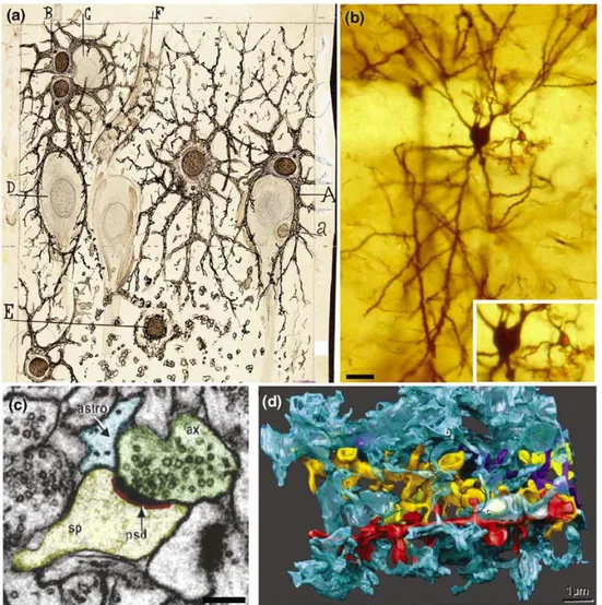
Fig 5.1 Views of the neuron–astrocyte interaction at the tripartite synapse level.
67
(a) Cajal’s drawing showing ‘neuroglia’ of the pyramidal layer and stratum radiatum of the Ammon horn (from an adult man autopsied three hours after death).
Original labels: A, large astrocyte embracing a pyramidal neuron; B, twin astrocytes forming a nest around a cell, C, while one of them sends two branches forming another nest, D; E, cell with signs of ‘autolysis’; F, capillary vessel.
(b) Neuron and astrocyte stained with the Golgi method from a rat hippocampus. Inset: astrocyte and neuronal somas.
(c) Electron microscopy image of astrocyte process at the axon– spine interface:
astrocyte process (astro, blue); postsynaptic density (psd, red); dendritic spine head (sp, yellow); axonal bouton (ax, green).
(d) 3D reconstruction of a single astrocyte process (blue) interdigitating among four dendrites (gold, yellow, red and purple). From Perera et al., 2009.
68
The biology of astrocyte–neuron interaction has emerged as a rapidly expanding field and has become one of the most exciting topics in current neuroscience that is changing our vision of the physiology of the nervous system.
The classically accepted paradigm that brain function results exclusively from neuronal activity is being challenged by accumulating evidence suggesting that brain function might actually arise from the concerted activity of a neuron–glia network.
69 5.2 Ca2+-mediated cellular excitability of astrocytes
The astrocytic revolution in current neuroscience began in the early 1990s when pioneering studies used the fluorescence imaging techniques to monitor intracellular Ca2+ levels in living astrocytes. Those first studies30,31 revealed that cultured astrocytes display a form of excitability based on variations of the intracellular Ca2+ concentration. Until then, astrocytes had been considered as non-excitable cells because, unlike neurons, they do not show electrical excitability. Since these pioneering findings, subsequent studies performed in cultured cells, brain slices and, more recently, in vivo have firmly established the astrocyte excitability, which is manifested as elevations of cytosolic Ca2+ mainly as a result of the mobilization of Ca2+ stored in the endoplasmic reticulum.
The elevated Ca2+ then acts as a cellular signal32. Whereas neurons base their cellular excitability on electrical signals generated across the plasma membrane, astrocytes base their cellular excitability on variations of Ca2+ concentration in the cytoplasm.
70
5.3 Astrocyte Ca2+ is controlled by synaptic activity
Astrocyte Ca2+ elevations can occur spontaneously as intrinsic oscillations in the absence of neuronal activity and they can also be triggered by neurotransmitters released during synaptic activity, which is of crucial importance because it indicates the existence of neuron-to-astrocyte communication.
The synaptic control of the astrocyte Ca2+ signal is based on the fact that astrocytes express a wide variety of functional neurotransmitter receptors. Many of these receptors are of metabotropic type, being associated with G proteins that, upon activation, stimulate phospholipase C and formation of inositol (1,4,5)-triphosphate (Ins(1,4,5)P3), which increases the intracellular Ca2+ concentration through the release of Ca2+ from intracellular Ins(1,4,5)P3-sensitive Ca2+ stores 16-21.
Early studies using cultured cells showed that the astrocyte Ca2+ signal could propagate to neighbouring astrocytes as an intercellular Ca2+ wave involving dozens of cells4,5,22. The synaptically evoked as well as the spontaneous Ca2+ signal originates in spatially restricted areas called
microdomains of the astrocyte processes, 24,25 from where it can eventually propagate intracellularly to other regions of the cell20,25,26. As a single astrocyte might contact up to 105 synapses: the control of the spatial extension of the Ca2+ signal
71 could have relevant functional consequences for the physiology of the nervous system, because not all synapses covered by a single astrocyte are necessarily functionally locked to be similarly and simultaneously modulated.
Therefore, differential neuromodulation of specific synapses would provide an extraordinary increase of the degrees of freedom to the system.
72
5.4 Astrocyte Ca2+ signal in vivo
For many years, technical constraints limited astrocyte Ca2+ signal studies to cultured cells and brain slices. The recent use of novel imaging techniques, that is, two-photon microscopy and specific fluorescent dyes that selectively label astrocytes in vivo, enabled the study of astrocyte Ca2+ signals in the whole animal and has revealed important findings.
First, reports from studies of rat, mouse and ferret have demonstrated that astrocytes in vivo exhibit intracellular Ca2+ variations, indicating that astrocyte Ca2+ excitability is not a peculiarity of slice preparations. Second, like in brain slices, astrocyte Ca2+ variations occur spontaneously 30-33 and are also evoked by neurotransmitters released during synaptic activity 31,33-37, indicating that neuron-to-astrocyte communication is present in vivo.
Finally, and of special relevance, astrocyte Ca2+ elevations might be triggered by physiological sensory stimuli. Indeed, stimulation of whiskers increased the astrocyte Ca2+ in the mouse barrel cortex33 (fig 5.2).
Astrocytes of the sensory cortex also elevate their Ca2+ in response to a robust peripheral stimulation that is known to activate the locus coeruleus or to direct electrical stimulation
73 of this nucleus34, as well as during running behaviour in alert mice 35. Astrocytes from other brain regions also respond to stimuli of corresponding sensory modalities. Astrocytes in the visual cortex not only show Ca2+ elevations in response to visual stimuli but also the properties of these responses indicate the existence of distinct spatial receptive fields and reveal an even sharper tuning than neurons to visual stimuli37.
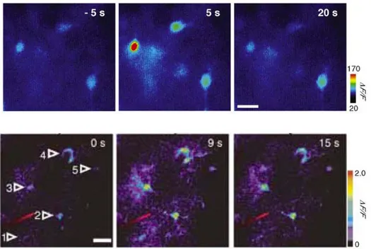
Fig 5.2 Astrocyte Ca2+ signalling in brain slices and in vivo.
Upper line: Pseudocolor images from rat hippocampal slices representing fluorescence intensities indicative of astrocyte Ca2+ levels before
74
(-5 s) and after (5 s, 20 s) electrical stimulation of Schaffer collaterals. Scale bar, 10 um.
Lower line: Two-photon microscopy images of the in vivo astrocyte Ca2+ signal in the barrel cortex. Pseudocolor images represent fluorescence
intensities indicative of astrocyte Ca2+ levels before (0 s) and after (9 s, 15
s) evoked by whisker stimulation. Scale bar, 20 um. From Wang, X. et al., 2006.
In summary, astrocytes in vivo display Ca2+ excitability and respond to neuronal activity. Furthermore, because astrocytes in specific sensory areas respond to a variety of sensory stimuli, it is feasible that astrocytes participate in the brain representation of the external world.
75 5.5 Synaptic information processing by astrocytes
In contrast to the view of astrocytes as passive elements that provide the adequate environmental conditions for appropriate neuronal function and that respond to neurotransmitters, simply performing a linear readout of the synaptic activity, experimental evidence supports the idea that astrocytes integrate and process synaptic information elaborating a complex nonlinear response to the incoming information from adjacent synapses.
As previously affirmed, it is firmly established that astrocytes respond with Ca2+ elevations to synaptic activity. However, to understand the actual role of astrocytes in brain information processing, it is necessary to define whether the astrocyte Ca2+ signal passively results from different neurotransmitter concentrations attained during synaptic activity or, alternatively, whether neuron-to-astrocyte communication presents properties of complex information processing that are classically considered to be exclusive to neuron-to-neuron communication.
76
5.6 Astrocytes discriminate the activity of synaptic pathways
The astrocyte Ca2+ signal does not result from a nonspecific spillover of neurotransmitters. Instead, it is selectively mediated by the activity of specific synaptic terminals (fig 5.3). Astrocytes located in the stratum oriens of
the CA1 area of the hippocampus respond to the stimulation of the alveus (which contains glutamatergic and cholinergic axons) with Ca2+ elevations that are specifically mediated by acetylcholine (ACh) but not by glutamate16.
77
(a) Schematic drawing and pseudocolor images of astrocyte Ca2+
elevations evoked by stimulation of Schaffer collaterals (SC, red) or alveus (green). Astrocytes integrate synaptic information from different synaptic inputs. Scale bar: 15 um.
(b) Hypothesis of astrocyte integration of synaptic information induced by SC and alveus activity (top) and astrocyte Ca2+ signals evoked
by independent and simultaneous stimulation of SC and alveus (bottom). Blue and black traces correspond to the observed and expected responses (i.e. the linear summation of the responses evoked by independent stimulation of both pathways), respectively. Horizontal lines at the bottom of each trace represent the stimuli. Note the lack of correspondence between observed and expected responses, that is, the relative reduction of the observed response versus the linear summation of the responses evoked independently, which is indicative of synaptic integration.
(c) Top, fluorescence images showing astrocytic Ca2+ signals evoked
after electrical stimulation (responding cells are displayed in white) of two contiguous barrel cortex. Bottom, images showing an overlay of the bright-field image with the location of the stimulating pipette and the responding astrocytes (shown in black). Dotted white lines outline the barrels in layer 4. Scale bar: 100 um. Note that astrocytes that respond to the stimulation of the barrel column are located within the stimulated barrel column, and no astrocytes respond to the stimulation of the adjacent barrel column, which indicates the selectivity of astrocyte responses.
(d) Schematic drawing illustrating the discrimination and response selectivity of barrel cortex astrocytes to neuronal activity from layer IV but not from layer II/III of neighbouring barrels. From Schipke, C.G. et al., 2008.
78
By contrast, these astrocytes do respond to glutamate when it is released by different glutamatergic synapses, that is, the Schaffer collateral (SC) synaptic terminals25. Hence, astrocytes selectively respond to different synapses that use different neurotransmitters (i.e. glutamate and ACh), and they discriminate between the activity of different pathways that use the same neurotransmitter (i.e. glutamatergic axons of SC and alveus)25. Likewise, astrocytes in the ventrobasal thalamus respond to the stimulation of either sensory or corticothalamic pathways, but very few respond to the activity of both38. Furthermore, astrocytes in the barrel cortex also respond selectively to the activity of different neuronal inputs, because astrocytes in layer 2/3 respond to glutamatergic inputs from layer 4 in the same column but not to glutamatergic projections from layer 2/3 of adjacent columns39 (fig 5.3). Therefore, astrocytes show selective responses that discriminate the activity of specific synapses.
79 5.7 Astrocyte Ca2+ signals show a nonlinear
relationship with the synaptic activity
The analysis of the astrocyte Ca2+ signal evoked by the activity of different synaptic terminals that release ACh and glutamate indicates that astrocytes integrate synaptic information25. In hippocampal slices, the simultaneous stimulation of alveus and SC (that elicit Ca2+ elevations mediated by ACh and glutamate, respectively) evokes astrocytic responses that are inconsistent with a linear readout of the synaptic activity. The amplitude of the Ca2+ elevations elicited by simultaneous stimulation of both pathways is not equivalent to the linear summation of the Ca2+ signals evoked by independent stimulation25 (fig 5.3). Therefore, the astrocyte Ca2+ signal is nonlinearly modulated by the simultaneous activity of cholinergic and glutamatergic synapses. Moreover, while the Ca2+ signal evoked by simultaneous stimulation at high frequencies (30 and 50 Hz) displays a sublinear summation of the responses evoked independently, it shows a supralinear summation after stimulation at relatively low frequencies (1 and 10 Hz); that is, the Ca2+ signal is relatively depressed or potentiated at relative high and low frequencies of neuronal activity, respectively. Therefore, the astrocyte Ca2+
80
signal is nonlinearly modulated by the simultaneous activity of different synaptic inputs, and the sign of this modulation depends on the synaptic activity level25.
5.8 Gliotransmission and modulation of synaptic transmission
One of the most stimulating topics in current neuroscience is the functional consequences of the astrocyte Ca2+ signal on neuronal physiology. Evidence obtained during the past 20 years has demonstrated that signalling between neurons and astrocytes is a reciprocal communication, where astrocytes not only respond to neuronal activity but also actively regulate neuronal and synaptic activity. Therefore, according to the concept of the tripartite synapse, to fully understand synaptic function, astrocytes must be considered as integral components of synapses where they have crucial roles in synaptic physiology.
Astrocytes release several neuroactive molecules, such as glutamate, D-serine, ATP, adenosine, GABA, tumor necrosis factor a (TNFa), prostaglandins, proteins and peptides, that can influence neuronal and synaptic physiology3. The
81 mechanisms and consequences of this process, called gliotransmission, have attracted considerable interest. Several mechanisms of transmitter release from astrocytes have been proposed.
Compelling evidence demonstrates that some transmitters are released in a Ca2+-dependent manner10,43–48 through vesicle47–51 and lysosome52–54 exocytosis. Furthermore, ultrastructural studies have shown that astrocytic processes contain small synaptic-like vesicles, which are located in close proximity to synapses, apposed either to presynaptic and postsynaptic elements 49,59.
Alternative release mechanisms, including reversal of glutamate transporters, connexin/pannexin hemichannels, pore-forming P2X7 receptors and swelling-induced activation of volume-regulated anion channels, have also been proposed55. Whether Ca2+-dependent and -independent mechanisms coexist and under what physiological or pathological conditions they occur remain unclear.
The original demonstration of astrocyte-induced neuromodulation in cultured cells43,44,56 has been considerably expanded by later studies on acute brain slices.
82
Glutamate was one of the first gliotransmitters released from astrocytes to be identified and has been reported to exert many effects on neuronal excitability. Astrocytic glutamate evokes slow inward currents (SICs) through activation of postsynaptic N-methyl-D-aspartate (NMDA) receptors25,44,60–65 and synchronously excites clusters of hippocampal pyramidal neurons, indicating that gliotransmission increases neuronal excitability and operates as a nonsynaptic mechanism for neuronal synchronization60,62. By contrast, astrocytic glutamate might also activate receptors localized at presynaptic terminals. Through activation of group I metabotropic glutamate receptors (mGluRs)46,26 or NMDA receptors50, astrocytes enhance the frequency of spontaneous and evoked excitatory synaptic currents.
Alternatively, astrocytes induce the potentiation20 or depression of inhibitory synaptic transmission by activation of presynaptic kainate66 or II/III mGlu67 receptors, respectively.
Therefore, a single gliotransmitter can exert multiple effects depending on the sites of action and the activated receptor subtypes, which provides a high degree of complexity to astrocyte–neuron communication. This complexity becomes even higher when considering that other gliotransmitters, such as GABA, ATP, adenosine (a metabolic product of ATP) or D-serine, could act on the same neuron or