Ph.D Course in Infectious Diseases, Microbiology and Public Health
XXXII cycle
Department of Public Health and Infectious Diseases
Innate, non-cytolytic CD8
+T cell-mediated suppression of
HIV replication by MHC-independent inhibition of virus
transcription
Supervisors: Candidate:
Professor Vincenzo Vullo Claudia Pinacchio Professor Gabriella d’Ettorre Student ID:1316049 Co-Supervisor:
Professor Guido Silvestri
Table of Contents Introduction ... 4 HIV-1 Virology ... ………..………5 Virion structure ... 5 Genome structure ... 7 Structural genes ... 8 Regulatory genes ... 9 Accessory genes ... 10 Replication ... 11
Binding, entry and uncoating ... 12
Reverse transcription ... 12
Integration ... 13
Expression of viral proteins ... 13
Assembly, Budding and Maturation ... 14
HIV genetic variation ... 15
Sources of genetic variation ... 15
Genetic variants of HIV ... 15
HIV infection ... 16
HIV-1 Transmission... 16
HIV-1 infection and pathogenesis ………....17
Antiretroviral treatment ... 19
HIV-1 Latency ... 21
Cellular and anatomical reservoirs... 22
HIV-1 molecular mechanisms of latency ... 22
CD8+ T cells ... 25
Role and Importance of CD8+ T Cells During the Antiviral Response ... 25
CD8+ T cells differentiation pathways ... 27
CD8+ T cells function ... 28
The role of CD8+ T cells in HIV infection... 30
Cytolytic activity during HIV pathogenesis ... 32
Non-cytolytic activity during HIV pathogenesis ... 33
CD8+ T cells and ART ... 34
Aim of the study ... 36
Methods ... 37
Study subjects ... 37
Enrichment and Stimulation of Primary Cells ... 37
Virus production ... 37
Dual color co-culture assays ... 38
Flow Cytometry analysis ... 39
Cell Sorting ... 40
Quantification of inducible provirus ... 40
Purification of total viral nucleic acids ... 40
Quantification of integrated HIV-DNA ... 41
RNA-seq library preparation... 42
Cytokine Array... 43
Statistical analysis ... 44
Results ... 45
TCR-stimulated CD8+ T cells effectively mediate suppression of HIV replication in CD4+ T cells ... 45
Virus-independent CD8+ T cell-mediated immunomodulatory mechanisms on the CD4+ T cells activation and proliferation contribute to the suppression of HIV replication………..47
CD8+ T cell control HIV replication through a non-cytolytic, MHC-independent, and partially mediated by soluble factors, activity ... 50
CD8+ T cell mediate viral suppression by increasing the frequency of non-productively infected CD4+ T cells ... 52
Secretome profile analysis reveals the CD8+ T cell-mediated suppressive activity on infected CD4+ T cells ... 54
CD8+ T cell-mediated virus suppression changes the transcriptome profile of infected CD4+ T cells. ... 56
Discussion... 59
Introduction
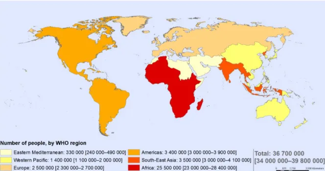
The Human Immunodeficiency Virus (HIV) is the causative agent of the acquired immunodeficiency syndrome (AIDS), characterized by a severe deterioration of the immune cell-mediated response due to the loss of T cells, and infects around 36.7 million people worldwide. UNAIDS estimates 1.8 million new HIV infections annually and 1 million AIDS-related deaths (1). The origin of the HIV has been traced to the simian immunodeficiency virus (SIV), found in African apes and monkeys (2). Epidemiologic and phylogenetic analyses currently available imply that HIV was introduced into the human population through several cross-species transmissions that are estimated to have occurred during the first part of twentieth century in the West Central Africa, but it was only 40 years ago the recognition and identification of the virus began. In 1983, the researchers Dr. Luc Montagnier and Dr. Francoise Barre-Sinoussi and Robert Gallo separately isolated HIV (3,4) and in 2008 they received the Nobel Prize for their findings. HIV is grouped to the genus Lentivirus within the family of Retroviridae, subfamily Orthoretrovirinae. On the basis of genetic characteristic and differences in the viral antigens, HIV is classified into the type 1 and 2 (HIV-1, HIV-2). HIV-1 virus has spread to all continents, but, as of today, Sub-Saharan Africa is the most affected area of the world, where 22.9 million people live with HIV (5) (Figure 1), whereas HIV-2 is still found predominantly in West Africa. The global epidemiology of HIV infection has changed markedly as a result of the expanding access to antiretroviral therapy (ART), with a decrease of global incidence and an increase of global prevalence due to the longer life expectancy of HIV-infected subjects (6). While ART has dramatically reduced the mortality and morbidity of HIV infection and has transformed HIV from a fatal disease to a chronic condition, it fails to provide a cure. The main obstacle to the development of a HIV cure is the presence of HIV-infected cells containing integrated DNA without the expression of the virus, defined as latently infected cells (7). This population is termed “HIV viral reservoir” and occurs primarily within resting memory CD4+ T cells, persisting for the lifetime of the patient.
Figure 1 The global HIV distribution
Data Source: World Health Organization, 2016
HIV-1 Virology
Virion structure
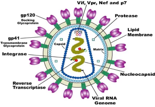
The HIV-1 virion is spherical in shape, with a diameter of about 100 nm (Figure 2). It is delimited by an outer membrane, the envelope, that is composed of a lipid bilayer, derived from the plasma membrane of the producer cell when the virion buds from the cell surface (8). Embedded within the envelope is the viral envelope protein, called Env, composed of two proteins, a surface glycoprotein called gp120 (SU) and a transmembrane protein called gp41 (TM). These two proteins together form a single structural unit. Three of these units comprise the Env trimer. Each Env trimer forms a “spike,” which protrudes away from the surface of the viral envelope. Constituted by the association of the viral protein p17, the matrix of HIV-1 forms an inner coat directly underneath the lipid envelope of the virion and surrounds the capsid, ensuring the integrity of the virion particle. It is known that the myristoylation of the p17, an addition co-translational of myristic acid to the amino terminal glycine residue, plays an important role for the virus assembly
during the late stages of the HIV-1 replication cycle, while, in the early stages, some p17 molecules dissociate from the viral membrane to direct the pre-integration complex to the host-cell nucleus (9). The conical capsid is assembled from the inner capsid protein p24 (CA) and, depending on the section plane, the capsid appears as a cone, a ring or an ellipse in electron micrographs. Its role is to protect the two identical molecules of viral genomic RNA, located inside the so-called nucleocapsid and bound to its associated enzymes reverse transcriptase (RT), protease (PR) and integrase (IN) and to the nucleocapsid proteins p6 and p7. HIV-1 virions adopt two distinct morphological states: the virion buds from a producer cell in its immature form, with a spherical capsid composed of the immature or precursor structural protein called Gag (p55). To become infectious, the immature virion undergoes a morphological change into the mature form at the end of the budding process. This process, termed “Maturation” is triggered by site-specific proteolytic processing or cleavage of the precursor Gag protein by the viral protease (10), that generates new structural proteins including CA, which reassembles into a new, cone-shaped capsid.
Figure 2 The HIV-1 virion structure
Genome structure
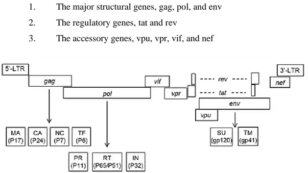
The HIV virus is grouped to the genus Lentivirus within the family of Retroviridae, subfamily Orthoretrovirinae. The HIV-1 genome consists of two identical, unspliced, positive-sense single-stranded RNA molecules of ~9.2 kilobase that are enclosed within the core of the virus particle, non-covalently linked via a palindrome sequence within the dimer initiation site (DIS) stem-loop, 5′-capped and 3′-polyadenylated. Long Terminal Repeat (LTR) sequences, divided in structural (U3, R, U5) and functional (modulatory, enhancer, core) regions (11), flank the genome at both ends; they drive the HIV-1 gene expression, containing crucial regulatory elements (as TATAA sequence), the trans-activation response element (TAR), that interfaces with the viral transcription activator protein Tat, transcription terminator and polyadenylation signals, the primer binding site (PBS), required for initiation of reverse transcription.
HIV-1 genome has several major genes coding for structural proteins that are found in all retroviruses as well as several non-structural genes unique to this virus (Figure 3). In particular, the identified nine open reading frames, encoding fifteen viral proteins, can be divided into three classes:
1. The major structural genes, gag, pol, and env 2. The regulatory genes, tat and rev
3. The accessory genes, vpu, vpr, vif, and nef
Figure 3 Genomic organization of HIV-1
Structural genes
In the direction 5' to 3' the reading frame of the gag (group-specific antigen) gene follows and gives rise to the 55-kilodalton (kda) Gag precursor protein, also called p55, which is expressed from the unspliced viral mRNA and is required for both virion assembly and maturation. After budding, p55 is cleaved by the viral encoded protease (a product of the pol gene) during the process of viral maturation into its final four smaller proteins:
• the outer core membrane protein p17 (MA) participates in the RNA targeting to the plasma membrane prior to viral assembly as well as in the incorporation of the Env glycoprotein into virions.
• the capsid protein p24 (CA) forms the distinctive conical core of viral particles that encapsulates the viral RNA-protein complex, providing structural stability of the virion; the N-terminal domain of the protein is important for infectivity, apparently by participating in viral uncoating through its association with a putative cellular chaperone, cyclophilin A (12).
• the nucleocapsid protein p7 (NC), a nucleic acid chaperon, coats the genomic RNA inside the virion core and is responsible for specifically recognizing the so-called packaging signal of HIV-1. It also has a critical role in mediating highly specific and efficient reverse transcription (13).
• Finally, p6 is a small proline-rich protein important for detachment of maturing virus particles and for packaging of other viral proteins within the virion.
The gag reading frame is followed by the pol (polymerase) reading frame, coding for the enzymes protease (PR, p12), reverse transcriptase (RT/RNAseH, p51/p66) and integrase (IN, p32).
• PR (p12) is an aspartyl protease that acts as a dimer. Protease activity is required for cleavage of the Gag and Gag-Pol polyprotein precursors during virion maturation. Knowledge of this structure has led to a class of drugs directed toward inhibiting the HIV protease function, greatly improving the treatment for HIV-infected individuals.
catalyzes both RNA-dependent and DNA-dependent DNA polymerization reactions and contains an RNAse H domain that cleaves the RNA portion of RNA-DNA hybrids generated during the reverse transcription reaction. RT has also been a major target for drug design.
• The IN (p32) protein mediates the insertion of the HIV proviral DNA into the genomic DNA of an infected cell by an exonuclease activity, a double-stranded endonuclease activity and finally, a ligase activity (14).
Adjacent to the pol gene, the env (envelope) reading frame encodes a glycosylated precursor protein (gp161), which is cleaved to two envelope proteins:
• located on the viral membrane surface, gp120 (SU) is involved in recognition and binding to specific cell surface receptors, causing structural changes in Env that facilitate coreceptor binding and subsequent viral entry.
• The primary function of gp41 (TM), a smaller hydrophobic transmembrane protein, is to mediate fusion between the viral and cellular membranes, following receptors binding.
Regulatory genes
In addition to the structural proteins, the HIV genome codes for several regulatory proteins necessary for the initiation of HIV replication in all host cells:
• tat (Transactivator of Transcription) is essential for HIV-1 replication, enhancing the efficiency of viral transcription. It is an RNA binding protein of two esons that acts by binding to an RNA short-stem loop structure, known as the transactivation response element (TAR), that is located at the 5' terminus of HIV RNAs (15).
• rev (Regulator of Virion Expression) is a 13-kD sequence-specific RNA binding protein, that induces the transition from the early to the late phase of HIV-1 gene expression. The binding of Rev to the RRE (Rev Responsive Element) site facilitates the
export of unspliced and incompletely spliced viral RNAs from the nucleus to the cytoplasm region (16).
Accessory genes
In addition to the gag, pol, and env genes contained in all retroviruses, and the tat and rev regulatory genes, HIV-1 contains four additional genes: nef, vif, vpr and vpu, encoding the so-called accessory proteins. Nef is expressed from a multiply spliced mRNA and is therefore rev independent. In contrast, vpr, vpu, and vif are the product of incompletely spliced mRNA, and thus are expressed only during the late, rev-dependent phase of infection from singly spliced mRNAs.
• Nef (negative expression regulatory factor) is a 27-kD myristylated protein that downregulates the cell surface expression of CD4, the primary receptor for HIV-1, may enhancing the Env incorporation into virions and virion budding. Nef also down-regulates the cell surface expression of Class I MHC, decreasing the efficiency of the cytotoxic T cells killing of HIV infected cells and stimulates the infectivity of HIV virions (17).
• The Vpr protein is incorporated into viral particles and induces the G2 cell cycle arrest. This protein also directs the nuclear localization of the pre-integration complex (PIC) in the nondividing cells, through an NLS (Nuclear localization sequence).
• The 16-kD Vpu polypeptide is an integral membrane phosphoprotein that in HIV-infected cells promotes the degradation of the viral receptor CD4 when it forms complexes with the viral envelope protein in the endoplasmic reticulum, causing the trapping of both proteins to within this compartment, that interferes with virion assembly. It can also increase the release of HIV from the surface of an infected cell.
• Vif (viral infectivity factor) is a 23-kD polypeptide that is essential for the production of infectious virus in a cell type-specific manner. It inhibits an anti-retroviral cellular factor expressed in non-permissive cells named APOBEC3G (18).
Replication
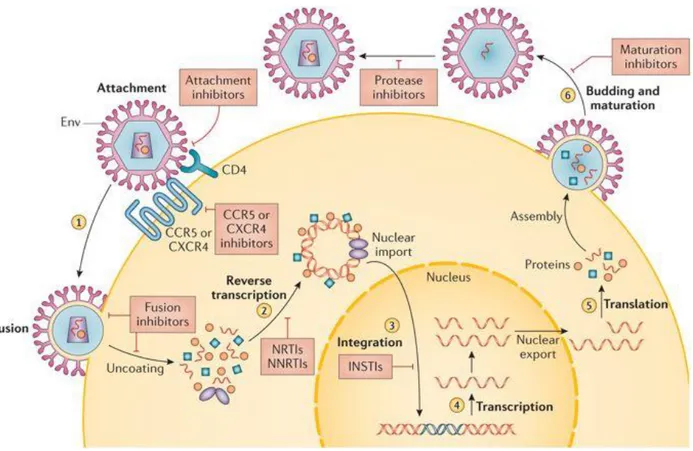
The HIV-1 replication cycle can be divided into an early and a late phase. The early phase encompasses virus binding to cell surface receptors, cell entry, reverse transcription of the viral RNA to DNA, uncoating of the viral capsid, nuclear import of viral DNA and DNA integration. The late phase refers to the transcription of viral RNA, translation and modification of viral proteins and release of progeny virions by budding through the cell plasma membrane (Figure 4).
Figure 4 HIV-1 life cycle
Binding, entry and uncoating
The initial step of entry involves a high-affinity binding of the surface envelope glycoprotein gp120 with the cellular receptor, CD4, which is present on the surface of a subset of helper T lymphocytes, on monocyte-macrophages, dendritic cells and astrocytes (19). This results in a conformational change in gp120 that allows interaction with the coreceptors, chemokine receptor 5 (CCR5) or chemokine receptor 4 (CXCR4 or fusin) on the host cell (20). HIV tropism (the type of cell that the virus is able to infect) is determined by the type of coreceptor recognized by gp120. Macrophage (M-tropic) strains of HIV-1 use the β-chemokine receptor CCR5 for binding and are able to infect macrophages, dendritic cells, and CD4+ T cells. T-lymphotropic HIV-1 attaches first
to the CD4 receptor and then to the α-chemokine receptor CXCR4. Binding of gp120 to CD4 and to the co-receptor triggers an additional conformational change in gp120 and subsequently in gp41 that, presenting its N-terminus on the viral membrane, forms a highly hydrophobic channel inserted into the plasma membrane of the target cell. Fusion of cell membrane and viral envelope is therefore completed, leading to translocation of the viral capsid into the cytoplasm.
Once entered the cytoplasm of the cell, the conical capsid composed of p24 (CA) protein dissociates from the rest of the cytoplasmic viral complex by a process called uncoating (21).
Reverse transcription
Once the viral core enters the cytoplasm of the cell, HIV-1 RT, through the RNA-dependent DNA polymerase activity, converts the single-strand HIV RNA genome into DNA (complementary DNA or cDNA) with a generation of a DNA-RNA duplex, beginning with the annealing of a host transfer RNA (tRNAlys) to a specific primer binding site (PBS) at the 5’ end of the viral genome
(22). In parallel to DNA synthesis, the RNaseH activity, also found in reverse transcriptase, removes the 5’ end of the viral RNA, leaving a purine-rich sequence, called PPT, that serves as a primer for positive-strand DNA synthesis. The nascent minus-strand viral DNA “jumps” and reanneals at the 3’ end of the RNA. After this transfer, DNA synthesis proceeds until the minus-strand completion. Then, there is a second priming event and a second template switch with degradation of the RNA component of RNA-DNA hybrids during the course of the positive-strand
viral DNA synthesis. The complete double-stranded viral DNA copy contains duplicated terminal U3RU5 regions (long terminal repeats), which are necessary for subsequent integration. Finally, the proviral DNA is actively transported into the cell nucleus in the form of a complex (the pre-integration complex or PIC) consisting of the integrase, the Gag matrix protein (p17) and Vpr.
Integration
The first step of the integration process occurs in the cytoplasm of the host cell following the completion of reverse transcription of the HIV RNA into c-DNA and involves binding and recognition of specific sequences within the LTRs of viral cDNA by the integrase protein, that
cleaves the viral DNA at each 3’ end. Subsequently, the pre-integration complex is transported into the nucleus where it binds to the host DNA through the host tethering protein LEDGF/p75 (23). The following step is characterized by the colinear insertion of the HIV DNA into a selected region of the host genome, with the consequent generation of unpaired regions both in the HIV and host DNA junctions, referred to as DNA "gaps". The gap repair process completes the integration of the HIV DNA into the host DNA, with the fully integrated HIV DNA, now being referred to as "proviral DNA". Although viral integration can take place in various locations in the host cell genome as heterochromatin regions, HIV-1 preferentially targets regions that have high gene density and transcriptional activity (24). The viral genome is immediately transcribed in productively infected cells, while it can undergo transcriptional silencing in resting CD4+ T
cells, leading to the generation of a latent viral reservoir.
Expression of viral proteins
The genes in the integrated HIV provirus are only expressed in cells in which cellular transcription factors have been induced. In fact, after activation of infected cells, the 5’ LTR promotor of the proviral genome serves as attachment site for cellular DNA-dependent RNA polymerases and for both viral and cellular transcription factors (as NF-AT, NF-kB, Sp1, AP-1), initiating the synthesis of viral mRNA products. A crucial role in the activation and maintenance of the high levels of
transcription of viral RNAs is played by the Tat protein, that interacts with Tar; it has been also suggested to be involved in remodeling nucleosomes to relieve transcriptional blockage imposed by chromatin (25). The proviral gene expression is a sequential event: in early infection most of the viral mRNAs found in the cytoplasm have undergone multiple splicing events and encode mainly for Tat, Nef, and Rev. Later, the predominant mRNA species are unspliced and singly spliced RNA (Gag, Pol, Env, Vpr, Vif e Vpu). Through interaction with the Rev-response element (RRE) located in the env region of the full-length transcript, the regulatory protein Rev is involved in the export into cytoplasm of unspliced and incompletely spliced mRNAs (26, 27). All mRNAs are translated in the cytoplasm near the endoplasmatic reticulum (ER) by the normal cellular transcription machinery. Finally, viral and cellular proteases cleavage the precursor peptides to generate the mature HIV-1 proteins.
Assembly, Budding and Maturation
HIV-1 virion assembly takes place at the lipid rafts of the plasma membrane, specialized membrane microdomains reached in cholesterol and sphingolipids, that probably serve as platforms for the targeting of the Gag polyprotein precursor. This protein contains all the necessary elements for the assembly and release of virus-like particles from many eukaryotic cell types: the matrix (MA) domain directs Gag to the plasma membrane and promotes the incorporation of the viral envelope glycoproteins into the forming virions; the capsid (CA) domain drives Gag – Gag interactions during assembly; the nucleocapsid (NC) domain packages the viral genomic RNA; and the p6 domain recruits the endosomal sorting complex required for transport (ESCRT) apparatus, which catalyzes the membrane fusion step to complete the budding process of spherical immature particles (28). A final step is required for conversion of the immature virion into its mature infectious form: the processing of the polyproteins by the viral protease with rearrangement of the structural proteins into a cylinder-shaped organization.
HIV genetic variation
Sources of genetic variation
HIV is one of the fastest evolving organisms known. Due to the fast-evolutionary rate, the virus evades the host immune system and has the capacity to develop resistance to antiretroviral drugs during suboptimal treatment. There are several mechanisms that contribute to the high genetic variation of HIV:
1. The error-prone RT enzyme generates on average 0.1-0.3 mutations per genome and replication cycle (29,30) and it is considered to account for most of the point mutations seen in HIV-1. These mutations remain uncorrected since RT lacks proofreading activity. 2. The high rate of virus turnover, approximately 109 virions per day, sustained by HIV-1
infection in vivo, strongly contributes to the continuous generation of new viral variants (31).
3. Retroviral recombination is another important strategy by which HIV generates genetic diversity (32). Each retroviral particle contains two copies of single-stranded RNA, and template switches occur frequently during reverse transcription, thus generating mutations and recombination by intramolecular and intermolecular jumps.
Genetic variants of HIV
Two types of HIV are known to cause AIDS: HIV‐1 and HIV‐2. Geographically, whereas HIV‐1 is responsible to the worldwide AIDS epidemic, HIV‐2 is mainly restricted to West Africa, with small epidemics reported in Portugal, France, Spain and Brazil. Although HIV-2 is morphologically indistinguishable from HIV-1, it is characterized by lower transmissibility and reduced likelihood of progression to AIDS (33). Sequence comparisons suggest that both HIV-1 and HIV-2 are the result of cross-species transmissions of simian immunodeficiency virus from chimpanzees (SIVcpz) and sooty mangabeys (SIVsmm), respectively. The reduced pathogenicity
of HIV-2 in humans is thought to be the result of lower levels of virus replication, perhaps reflecting incomplete adaptation of SIV to the human host.
Due to an extensive genetic heterogeneity, HIV-1 variants are classified into four major phylogenetic groups: group M (main), group O (outlier), group N (non-M/non-O) and group P. Group M is the predominant circulating HIV-1 group, with a global distribution. It splits into 10 phylogenetic subtypes (A to K), or clades, phylogenetically linked strains with a genetic distance from one another of 25 to 35%. Within a subtype, it is possible recognize sub-subtypes, denoted with numbers, with a genetic variation of 15% to 20% (34). HIV-1 group O, relatively rarely found, seems to be endemic to Cameroon and neighboring countries in West Central Africa; likewise, group N is a very distinctive form of the virus and has been identified only in a limited number of individuals from Cameroon. The M and N HIV-1 groups have a common ancestor, closely related to the SIV strain found in chimpanzees, while, recent evidence suggests that the HIV-1 O and P groups may have originated in wild gorillas (35).
All retroviruses have a propensity to recombine with other relatively closely related retroviruses, and HIVs are no exception. Inter-subtype recombinant genomes are common, but many of them, called unique recombinant form (URF) are found only in the one dually-infected (or multiply-infected) individual patient in which they arose. If an inter-subtype recombinant virus is transmitted to many people, it becomes one of the circulating strains in the HIV epidemic, and it can be classified as a "circulating recombinant form (CRF)", an epidemiologically relevant contribution to the HIV-1 M group epidemic.
HIV infection
HIV-1 Transmission
The virus is able to enter the body through three main routes: sexual contact, parenteral inoculation and vertical transmission. The sexual contact is the predominant mode of HIV transmission worldwide, mediated by exposure to HIV-1-infected cells and/or infectious virus in mucosal secretions or semen. The stratified mucosal epithelium and the endocervical epithelium form effective barriers against HIV-1 or HIV-1-infected cells; however, breaches in the integrity of
these barriers are frequent, increasing susceptibility to infection. Once the epithelial barriers have been breached, HIV-1 can target cells in the underlying epithelial layers, including T cells, dendritic cells and macrophages. The probability of HIV-1 sexual transmission has been shown to vary based on different factors; amongst them, the clinical stage of infection is one of the most important because the early and late stages of HIV-1 infection have long been known to be associated with high rates of transmission, related to the high viral loads observed for these periods (36). Also, the sexually transmitted diseases (STD) and the male circumcision are co-factors for the HIV acquisition, increasing the shedding of the virus and the transmission efficiency. Receiving HIV contaminated blood through transfusions and organ/tissue transplants or needles or syringes sharing for drugs injection results in the so-called parenteral inoculation, the most effective mode of HIV-1 transmission. Luckily, the introduction of HIV antibody testing of blood donors significantly reduced the risk of transfusion-associated HIV transmission. HIV-1 can be also spread from mother to child during pregnancy, occurring in utero, intrapartum or postnatally through breastfeeding. Factors associated with higher probabilities of vertical transmission include higher maternal HIV RNA levels, lower CD4+ counts, infant prematurity, chorioamnionitis, and
duration of rupture of membranes. Use of combined antiretroviral therapy, with or without cesarean section to further diminish risk of transmission intra-partum, has resulted in transmission risk of less than 2% among infants born to HIV-1 infected mothers in resource rich areas, while the vertical transmission is nowadays a leading cause of infant mortality in resource-limited countries in sub-Saharan Africa (37).
HIV-1 infection and pathogenesis
There are three stages of HIV infection: the acute infection, the chronic infection and the acquired immunodeficiency syndrome, known as AIDS. As of now there is no cure for HIV-1 infection and, without treatment, chronic HIV infection usually advances to AIDS in 10 years or longer, resulting in the death of the infected subject, with the exception of the elite controllers (EC), HIV-1 infected people who maintain high CD4+ and CD8+ T cell counts and remain therapy naïve.
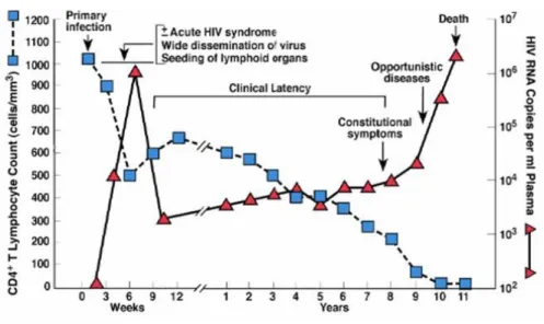
Figure 5 Typical course of HIV infection
Modified from: Fauci, A.S., et al, Ann. Intern. Med., 124:654, 1996
Acute or primary HIV infection is the earliest stage and it generally develops within 2 to 4 weeks following the exposure and transmission. During this time, the virus replicates rapidly in the mucosa and in draining lymphoreticular tissues (especially gut-associated lymphoid tissue), enriching high levels in plasma (106 -107copies per ml). Before the antibodies begin to be produced
by the immune system, the so-called “seroconversion”, there is an “eclipse-phase”, a period generally lasts 7 to 21 days, in which the virus can’t be detected in plasma (38). The early HIV-1 infection is also characterized by a transient drastic decline in the CD4+ lymphocytes count, the
preferred target for infection, since a normalization of the viral values occurring once the antibody response is activated, which is known as the “viral set point”. Importantly, the set point level is correlated with clinical outcome: individuals with high viral load set point typically progress more rapidly to AIDS and death than those with lower viral set point levels (39). Acute infection often occurs asymptomatically or people may experience flu-like illness marked by fever, generalized lymphadenopathy, a nonspecific rash, myalgias and/or malaise. The severity of symptoms is strongly correlated with peak viral load during this phase of the infection.
The second step during the HIV infection is the chronic stage (also called asymptomatic HIV infection or clinical latency). During this stage of the disease, HIV continues to multiply in the body, but at very low levels, with also progressive decrease in the number of circulating CD4+
lymphocytes and the appearance of functional defects affecting both CD4+ and CD8+ lymphocytes
and monocytes and macrophages, associated with a progressive deterioration of the immune system. This phase, clinically latent, can last several years and does not correspond to a biological latency as viral replication continues in the lymphoid organs, but it is rather characterized by lack of symptoms.
AIDS is the final, most severe stage of HIV infection. Because HIV has severely damaged the immune system, the body can’t fight off multiple opportunistic infections and neoplasms, that contribute to a rapid clinical deterioration, such as visceral toxoplasmosis, Pneumocystis carinii pneumonia and Cytomegalovirus. Moreover, the progressive immunodeficiency and chronic immuno-activation are reflected in a decrease in CD4+ count of less than 200 cells/mm3 and in
progressively increase of the viremia (40). People diagnosed with AIDS present severe symptoms as persistent lymphadenopathy (Lymphadenopathy Syndrome, LAS) followed by a phase of weight loss, asthenia, anemia, fever, diarrhea, hypergammaglobulinemia (AIDS-related complex, ARC) and, without treatment, typically survive about 3 years.
Antiretroviral treatment
The development of combination ART (cART) is often considered as one of the greatest achievements of modern medicine, helping people with HIV live longer and healthier and reducing the risk of HIV transmission.
Currently the drugs approved by the FDA (Food and Drugs Administration) for the treatment of HIV infection are divided, according to their action mechanism capable of blocking various stages of viral replication, into four main categories (41):
• Nucleoside and non-nucleoside reverse transcriptase inhibitors
Nucleoside reverse transcriptase inhibitors (NRTIs) act by blocking reverse transcriptase. These drugs are analogues of natural nucleosides and nucleotides, and are preferentially incorporated into HIV DNA, leading to termination of DNA synthesis (Tenofovir, Abacavir, AZT). Non-nucleoside
reverse transcriptase inhibitors (NNRTIs) bind to a pocket near the active site of the reverse transcriptase, which causes a conformational change of the enzyme and inhibition of reverse tran-scription (Nevirapine, Rilpivirine, Etravirine).
• Protease inhibitors
As the virion matures and buds from the cell, long polypeptide chains are enzymatically cleaved into mature functional proteins by the HIV enzyme protease. Protease inhibitors block this late step in the life cycle of the virus (Atazanavir, Darunavir, Ritonavir)
• Entry inhibitors
Entry inhibitors prevent HIV from entering the cell, Maraviroc binding to CCR5, Enfuvirtide binding to HIV directly.
• Integrase strand transfer inhibitors
Integrase strand transfer inhibitors prevent the HIV genome from being integrated into the host genome (Dolutegravir, Raltegravir, Elvitegravir).
The history of HIV infection treatment began in 1987 with the introduction of the first NRTI, zidovudine (42); subsequently, other drugs of the same class were used, first used as monotherapy and then as dual therapy (1994). The real "therapeutic revolution" began in 1996-1997 with the introduction of protease inhibitors and, after, with non-nucleoside reverse transcriptase inhibitors; with the advent of the new drugs the era of cART has started and consists of the use of three or more drugs belonging to two or more different classes of inhibitors (43). Thanks to the combination of antiretroviral therapy, mortality has significantly decreased, turning a deadly disease into a long-term chronic disease. While monotherapy usually produces drug-resistant HIV mutant forms fairly rapidly, the therapeutic combination generally slows the selection of mutants because it acts at various levels of HIV replication. Although the cART can suppress viral replication and reduces the plasma viral load to below limits of detection of the most sensitive clinical assays (<20 RNA copies/mL) for decades, there isn’t currently a cure for HIV infection, due to the persistency of the
HIV-1 Latency
Although antiretroviral therapy suppresses HIV to undetectable levels, interrupting ART causes the virus to rapidly rebound to pretreatment levels, due to the natural phenomenon of HIV latency. Viral latency is a state of reversibly non-productive infection of individual cells, providing an important mechanism for viral persistence and escape from immune recognition, established early during acute infection, likely within days of initial infection. Two forms of viral latency are known: pre- and post-integration latency (44). Pre-integration latency is characterized by an unintegrated virus, due to the inhibition of integration and nuclear import mediated by the low metabolic state of the host cells. The unintegrated forms are located in the cytoplasm for days and constitute the pre-integration complex which eventually decays over time. During post-integration latency, the provirus is able to successfully integrate into the host genome, but exists in a transcriptionally inert state.
For HIV-1, the term latency was initially used in the clinical sense to describe the long asymptomatic period between initial infection and the development of AIDS. However, it became clear that HIV-1 replicates actively throughout the course of the infection, even during the asymptomatic period and that it can establish a state of latent infection at the level of individual T cells.
Naive CD4+ T cells exist in a resting state until they encounter an antigen, after which they undergo
cellular proliferation and differentiation, giving rise to effector cells. Most effector cells die quickly, but a subset survives and reverts to a resting G0 state, persisting as memory cells.
Activated CD4+ T cells are highly susceptible to HIV-1 infection and typically die quickly as a result of the cytopathic effects of the virus or host immune responses. However, some activated infected CD4+ T cells survive long enough to revert back to a resting memory state. Due to the long-lived survival capabilities, they represent an ideal cellular reservoir for the maintenance of a stably integrated, but transcriptionally silent form of the latent virus, even though the frequency of these latently infected cells is very small (in the order of approximately ∼1 × 10⁶ cells per infected
individual). Moreover, the host cell can be activated again by an encounter with antigen or cytokine, leading to the induction of transcription factors like NF-kB and NFAT that, in turn, promote reactivation of the latent HIV proviruses (45).
Cellular and anatomical reservoirs
A reservoir is defined as ‘a cell type or anatomical site in which a replication-competent form of virus accumulates and persists’, in spite of long periods of cART-suppressed viremia (46).
The best-characterized cellular reservoir of HIV comprises resting memory CD4+ T cells. Chomont
et al. identified two viral reservoirs within the memory CD4+ T cells pool, known as Central
memory (TCM)and Transitional memory (TTM) cells, able to survive for decades, whereas Naive
(TN) and effector T cells only marginally contribute to the viral persistence (47). Other immune
cells have been implicated as reservoirs as well, especially myeloid cells. Macrophages are known to be susceptible to infection and more resistant to the cytopathic effects of the virus compared to CD4+ T cells, but their role remains debated due to their highly specialization per organ:
macrophages in the gut-associated lymphoid tissue (GALT) appear to be quite resistant to infection, except in the rectum, whereas alveolar macrophages seem relatively permissive (48). In addition, dendritic cells (DCs), natural killer (NK) cells, and other specialized cell types (e.g., renal, mucosal, and cervical epithelial cells; mastocytes, astrocytes, and microglia in the central nervous system; skin fibroblasts; and bone marrow stem cells) can represent persistent populations with integrated HIV proviruses, but their specific role is not yet clearly understood.
HIV-1 also hides in several immune privileged anatomic sites that are poorly penetrated by antiretroviral drugs: after entering the body, it quickly disseminates to the lymph nodes and later on to the bloodstream, spreading throughout the entire body as individual virions, by the transport of infected immune cells and by cell-to-cell transmission. Indeed, anatomical reservoirs that may harbor such cells include the brain and the central nervous system, the gastrointestinal tract and the gut‐associated lymphoid tissue and other lymphoid organs, and the genital tract (49-52).
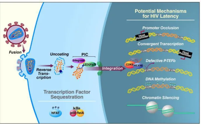
HIV-1 molecular mechanisms of latency
A critical and unanswered question is what distinguishes between productive and latent infection at the molecular level. It is known that the maintaining of the latent state is likely a multifactorial process; it can involve changes in chromatin condensation state in the host cells, subsequently affecting the HIV gene expression: transcribed genes are usually found within “relaxed” chromosomal DNA, termed euchromatin, while analysis of latent integration sites showed that
latent HIV-1 proviruses preferentially integrate within the heterochromatin, a more condensed transcriptionally repressed chromosomal form in which the blocking of the transcriptional machinery is mediated by nucleosomes (53). As fundamental structural unit of chromatin, Nuc-o and Nuc-1 regulate the basal transcriptional activity of the 5′ LTR of HIV genome because they overlap with the binding sites for many key transcription factors that drive HIV-1 gene expression, making the promotor region inaccessible to them; other aspects clearly related to latent conditions are post-translational modifications of the histone tails of the nucleosomes, including acetylation, methylation, ubiquitylation, phosphorylation, sumoylation, and poly ADP-ribosylation. Nuc-1 is in fact rapidly and specifically disrupted following treatment with HDAC (histone deacetylase) inhibitors, thus creating a less-repressive chromatin state and allowing for increased HIV-1 transcription (54).
DNA methylation is another possible mechanism that reinforces HIV-1 latency. Regulation of transcriptional silencing by DNA methylation is catalyzed by DNA methyltransferases and occurs largely at CpG dinucleotides. CpG methylation at the 5’LTR of HIV-1 genome acts by preventing the binding of essential transcription factors such as NF-kB and Sp1. In a study by Kauder et al., it was demonstrated that, during latency, the HIV-1 promoter is hypermethylated at two CpG islands surrounding the transcriptional start site in both J-Lat cell lines and in primary CD4+ T cells (55).
Considering that latent provirus is able to integrate also into actively transcribed regions, it isn’t really surprising that some forms of latency mechanistically involve transcriptional interference. One mechanism depends on the orientation of the HIV-1 provirus relative to the host gene: if both share the same polarity, “promoter occlusion” can occur, in which read-through by the host RNA Pol II displaces key transcription factors on HIV-1 LTR, thereby reinforcing viral latency. Conversely, the “convergent transcription” occurs when the provirus integrates in the opposite orientation or polarity relative to the host gene, leading to collision of the RNA Pol II complexes from the host and viral promoters and early arrest of transcription from both promoters or the weaker of the two (56). This form of transcriptional interference could also generate double-stranded viral RNAs, resulting in silencing of viral transcription or translation through RNA interference, RNA- directed DNA methylation, or the generation of antisense RNA (57).
Initiation of HIV-1 gene expression is critically dependent on a range of host transcription factors, including NF-κB, NFAT, and AP-1, whose activities are induced by extracellular stimuli,
including T-cell receptor ligation or the action of various cytokines. Sp1 also plays a key role by binding at three sites in the core promoter, through its associations with TATA-box-binding protein (TBP), TBP-associated factor (TAF) 250, and TAF55, increasing both basal and Tat-dependent activation of the HIV-1 LTR. Members of the NF-κB/Rel family of transcription factors exert both activating and inhibitory effects on the HIV-1 LTR: in the cytoplasm of resting cells, NF-kB exists in an inactive form, the p50/RelA heterodimer, while in the nucleus of HIV-infected resting cells, NF-kB p50/p50 homodimers are bound to the 5’LTR; the binding of homodimers of p50 recruits HDAC-1 and leads to transcriptional inhibition because of histone deacetylation and chromatin condensation, promoting latency. On the contrary, after their activation and translocation into the nucleus, the p50/RelA heterodimers displace the p50 homodimers and promote a strong transcriptional activation, recruiting histone acetyl- transferases (HATs) like p300/CBP (58).
The HIV transactivator Tat play a major role during the HIV transcription, exclusively stimulating transcription elongation by binding to the transactivation-responsive element TAR, a rather simple RNA stem-loop structure located at the 5′ end of all viral transcripts. Following TAR binding, Tat recruits the P-TEFb complex, that mediates the phosphorylation of the CTD of RNA Pol II, primarily at serine-2. In addition, P-TEFb mediates phosphorylation of two transcription inhibitors, DSIF (DRB sensitivity-inducing factor) and NELF (negative elongation factor). These events result in highly effective RNA Pol II elongation. In the latent proviruses transcription elongation is very inefficient due to the relative absence of Tat, that is able to produce only short abortive transcripts, additionally subjected to elongation restrictions by NELF (59). Transcriptional activation also depends on the acetylation state of Tat, that leads to the dissociation of Tat from TAR, thus promoting the switch between the early and late phases in HIV transcriptional elongation.
Another rather unexpected mechanism has emerged. Specifically, resting CD4+ T cells from
HIV-1-infected patients on antiretroviral therapy retain both Tat and Rev transcripts in their nuclei (60) through the ectopic expression of polypyrimidine tract binding protein (PTB), an RNA export protein. Interestingly, a short form of PTB is present in resting CD4+ T cells, whereas full-length
Figure 6 Potential mechanisms for HIV Latency
Data source: https://www.ncbi.nlm.nih.gov/pmc/articles/PMC4361081
CD8
+T cells
Role and Importance of CD8+ T Cells During the Antiviral Response
CD8+ T cells are a specialized lymphocyte subset in the adaptive immune system that provide defense against intracellular pathogens, mainly against viruses and tumor cells. To produce an effective and robust immune response, Ag-specific CD8 T cells are characterized by the expression of antigen receptors on their surface, known as the T cell receptor (TCR). The TCR receptor is a transmembrane heterodimeric protein, normally consisting of two disulfide-linked polypeptide chains: in humans, the vast majority of T lymphocytes express alpha and beta chains on their surface, but a minority has a TCR made up of a gamma and a delta chains (61); it features a variable region involved in antigen recognition, a constant region, a hinge region, a transmembrane region and a short cytoplasmic tail. The TCR is always expressed with the associated CD3 complex,
comprised of multiple independently expressed units, required for signal transduction once presented antigen is encountered (62). TCRs recognize antigenic peptides that are bound and presented by endogenous major histocompatibility complex (MHC) molecules, which in humans are called HLA. HLA genes are located on chromosome 6 and are known to be the most polymorphic genes in humans (63). Their diversity allows accommodation of a range of different peptides for presentation to T cells. There are two types of HLA molecules, HLA class I and HLA class II. Human HLA class I molecules are glycoproteins generally categorized into two groups: classical (HLA-A, B and C) and non-classical (HLA-E, F and G). HLA genes produce proteins that would form the major histocompatibility complex (MHC) (64). HLA class I molecules present processed peptides to CD8+ T cells, whereas HLA class II molecules present peptides to CD4+ T
cells. MHC I molecules are heterodimeric and composed of two polypeptide chains, α- and β2-microglobulin. The two alpha helices (heavy chains) are highly polymorphic, whereas the light chain β2-microglobulin is less polymorphic. The third α3 domain is quite conserved and interacts with the CTL co-receptor (65). Initial recognition of antigen by naïve Ag-specific CD8 T cells or “priming” usually occurs in a lymph node to which professional antigen-presenting cells (APCs) transport antigen from the site of infection. Through the formation of the so-called “immunological synapse”, α and β domains of the CD8 molecule bind to the α3 domain of the MHC class I molecule (66). This binding is stabilized by the presence of costimulatory molecules, including CD80 and CD86 on the APC (67), and generates a signaling cascade that initiates activation of the CD8+ T
cells. The effective response against virally infected cells requires a “clonal expansion”, an intense proliferation within the lymph nodes that gives rise to many CD8+ T cells of identical TCR
specificity. This expansion is necessary for activated cells to differentiate into effector cells and acquire antimicrobial activity to fight the ensuing infection: the differentiating CD8+ T cells exit
the local lymph node to enter circulation and begins to express chemokine receptors and adhesion molecules appropriate for trafficking to the effector site and gene expression profiles appropriate to carry out effector functions (68). Virus-specific CD8+ T-cell responses can become evident as
early as 4–5 days after infection, with the peak numbers of effector CD8+ T cells usually observed
between 7–14 days after infection (69).At the end of the primary response, in order to preserve the flexibility to respond new foreign Ags and limit immunopathology during a chronic infection, the majority of responding CD8+ T cells dies by apoptosis (contraction phase);
respond with strong proliferation and rapid conversion into effector cells upon re-exposure to the cognate antigen (70, 71), a process defined “the secondary response”.
CD8+ T cells differentiation pathways
T cells develop from lymphoid progenitor cells originating in the bone marrow, but they mature in the thymus. There, they rearrange their TCR genes and undergo the processes of positive and negative selection (72): T cells which weakly recognize self-peptides presented by MHC class I and II molecules on epithelial cells receive survival signals, while T cells that strongly recognize self-peptide-MHC complexes are eliminated, in order to avoid the subsequent effector function mediated against self-antigen. During positive selection, thymocytes bearing “useful” TCRs undergo differentiation to the CD4+ helper or CD8+ cytotoxic lineage, and mature CD4+ or CD8+
single-positive (SP) cells exit the thymus and circulate in the periphery (73), in search of an antigen that matches the specificity of their TCR. To increase their chances of meeting antigen, they use lymph nodes as meeting points with APCs. From its exit from the thymus as a naïve CD8+ T cell,
to its targeted execution of effector function and formation of a memory compartment, there are many complex steps in the development of virus-specific CD8+ T cell responses. Once activated,
they acquire an effector phenotype, characterized by high cytotoxic ability and production of different types of cytokines; therefore, they can be divided in:
(i) CD8+ T cells with a Th1-like cytokine pattern (Tc1), which produce IFN-γ and TNF-α
and exhibit a high cytotoxic function
(ii) CD8+ T cells with a Th2-like cytokine pattern (Tc2), which produce IL-4, IL-5, IL-6,
and IL-10 and have lower cytotoxic ability than Tc1 cells (74).
(iii) CD8+ T cells that produces IL-17, but no granzyme B, with a pattern recently described
as Tc17. These cells exhibit some functional plasticity, since they can produce IFN-γ while losing the expression of IL-17 (75).
Once the pathogen is eradicated, some cells acquire a memory phenotype that persists even in the absence of the antigen (76). They are distinguished from naïve and effector CD8+ T cells that
express the marker CD45RA, by the loss of this marker and the gain of CD45RO after differentiation. These memory CD8+ T cells have been classified into the following subsets,
considering differences in the degrees of effector functions, proliferative capacity, and tissue homing properties (77):
(i) central memory cells, restricted mainly to lymphoid tissues because their expression of the lymphoid homing molecules CD62L and CCR7, which are also considered as the source for self-renewal of the pool of memory cells, generating a second wave of effector T cells (ii) effector memory cells that provide a first line of defense against infections through immediate effector functions and are present in circulation and non-lymphoid tissues due to the low expression of the lymphoid homing molecules CD62L and CCR7 (78)
(iii) a recently described subset of tissue-resident memory cells, that are located at sites of pathogen entry.
The CD8+ T cells can also be classified according to the level of activation; based on the
expression of the activation markers HLA-DR and CD38, four phenotypes of cells have been identified: (i) HLA-DR+CD38+; (ii) HLA-DR+CD38−; (iii) HLA-DR−CD38+; and (iv)
HLA-DR−CD38−. In addition to these described populations, some CD8+ T cells exhibit the ability
to suppress T helper activity and induce anergy, called regulatory CD8+ T cells (79,80). These
cells seem to be related to a memory phenotype since they are CD45RA negative; in addition, they can express the CD25 and the FoxP3 markers, and their regulatory function seems to be mainly achieved by IL-10 production.
CD8+ T cells function
CD8+ T cells display considerable functional heterogeneity. Induction of apoptosis in target cells
is one of their most important effector functions. Direct lysis of infected host cells is caused by the release of pro-apoptotic enzymes and antiviral chemokines. The secretion of perforin from lytic granules in a calcium-dependent process results in the formation of pores (81, 82). With the integrity of the cell membrane destroyed, the cells die rapidly through osmotic lysis. In addition, proteases co-secreted with perforin by lytic granule exocytosis enter the target cell and induce apoptosis. Amongst them, Granzyme B, a serine protease with a caspase cleavage activity, is the major mediator of target cell DNA fragmentation in this perforin-dependent pathway (83). Studies
have shown that the exocytosis of these two secretory proteins is MHC-I restricted and antigen specific (84).
There is a second perforin-independent mechanism of cytotoxicity, Calcium-independent. This process involves specific death-inducing ligands, such as Fas-Ligand (FasL), which trigger apoptosis following the engagement of receptors on the target cell surface. Fas (CD95) is a member of the tumor necrosis factor receptor superfamily; crosslinking of Fas by FasL, which is expressed on activated CTLs, leads to apoptotic target cell death (85,86). Of interest is a study that hinted at the contribution of Fas-mediated cell death to the granule exocytosis-mediated pathway attributed to CD8+ T cells. Kojima et al. reveled by immunoelectron microscopy that FasL was localized to
the outer membrane of the cytoplasmic granules during degranulation (87). These findings indicate that these two lytic pathways may be more intertwined than previously thought.
One non-lytic pathway used by CD8+ T cells to modulate viral infection is through the secretion
of soluble factors that aid in the neutralization of foreign pathogens. This mechanism does not directly eliminate virally infected cells like the perforin/granzyme and FasL-mediated routes, it however does play a large role in the control of viral replication. Almost all CD8+ T cells can
secrete potent amounts of the antiviral cytokine IFN-γ, which can block viral replication and cause up-regulation of MHC molecules on the surface of an infected cell. IFN-γ can also activate macrophages, NK cells and promote isotype switching in B cells (88). Moreover, CD8 T cells are an important source of one of the members of the TNF superfamily, TNF-α. It has been shown to be an important antiviral factor due to its role as a mediator of apoptosis as well as inflammation and immunity (89). TNF-α also promotes an antiviral state by enhancing expression of MHC class I and by inducing expression of IL-12 and IL-18 which are both important for upregulating production of IFN-γ by CD8+ T cells (90). However, similar to IFN-γ, activation of cells induced by TNF-α can also result in increased production of virus.
Although typically considered a CD4+ T cell cytokine, CD8+ T cells are also quite capable of
producing IL-2. It has no direct antiviral effector function, but it does promote expansion of CD4+
and CD8+ T cells, thereby amplifying the effector response to pathogens (91). IL-2 may also be
important for programming CD8+ T cells for better memory generation (92).
Both CD4+ and CD8+ T cells secrete a variety of chemotactic cytokines (chemokines) upon activation. Chief among them are the β-chemokines macrophage inflammatory protein-1α (MIP-1α) and -1β (MIP-1β) and regulated upon activation normal T cell expressed and secreted
(RANTES). MIP-1α and MIP-1β can be found in cytotoxic granules while RANTES is stored in a separate secretory compartment called the RANTES secretory vesicle (RSV) (93). Both types of granules are rapidly released following CD8+ T cells activation. New MIP-1α and MIP-1β
synthesis occurs within a few hours of activation, while RANTES can take several days to be upregulated following its initial release. All three contribute to an inflammatory response primarily by recruiting leukocytes to the site of injury or infection.
The role of CD8+ T cells in HIV infection
Numerous lines of evidence suggest that CD8+ T cells exert potent antiviral effects also during
HIV infection. First, the emergence of HIV-specific CD8+ T cell responses during the primary
infection has been associated with a post-peak decline of HIV viremia, resolution of clinical symptoms and the development of a semi-stable viral setpoint (94). This is consistent with experimental animal model of simian immunodeficiency virus (SIV) of rhesus macaques (RM) showing that the depletion of CD8+ T cells during acute phase of infection results in the abrogation
of the post-peak decline of viremia (95).
Additionally, the immunologic pressure exerted by virus-specific CD8+ T cells is manifested by
the rapid evolution of virus escape variants following acute infection (96, 97). These mutations can result in HIV-infected cells either failing to present the viral epitope or, if presented, evading recognition by CD8+ T cells. This process, termed “escape” is first observed within weeks of
transmission and continues through infection.
Unlike most viral infections, HIV leads to a marked and durable activation not only in HIV-specific (representing only the 10% of total circulating CD8+ T cells), but also in non-specific CD8+ T
cells, referred to as “bystander” activation (98). This mechanism seems to occur through a T-cell-receptor-independent response, conversely initiated mainly by cytokine and chemokine stimulation: in fact, activated CD8+ T cells include subsets specific for persistent pathogens such
as cytomegalovirus (CMV), Epstein-Barr virus (EBV), as well as subsets for non-persistent pathogens, such as influenza virus and adenovirus (99).
While HIV-specific CD8+ T cells cytotoxic activity appear to be essential for the post-peak decline
dysfunction and “exhaustion” of virus-specific CD8+ T cells, due to persistent exposure to HIV
antigen and chronic inflammation with the ongoing viral replication (100). Altered differentiation, impaired function and decreased proliferation characterize the exhausted phenotype of CD8+
population, as well as the expression of a variety of coinhibitory molecules, including programmed death 1 (PD-1), T cell Ig and mucin domain 3 (TIM3), CD160, the NK cell receptor 2B4, lymphocyte activation gene 3 (LAG-3), and cytotoxic T lymphocyte antigen 4 (CTLA-4) (101-103).
CD8+ T cells continue to exert some level of control over HIV and SIV replication after the acute
phase of infection, as shown by studies in which depletion of these cells during chronic SIV infection results in increased viral replication (104). Despite their efforts to maintain the antiviral immune response also during the chronic phase of infection, the combination of the continuous appearance of virus escape mutants (105), the progressive T cell dysfunction and immune exhaustion increasingly limit the HIV specific CD8+ T cells efficacy, contributing to the disease
progression. Additionally, the functional impairment of CD8+ T cells is accompanied by a higher
level of expression of activation markers CD38 and HLA-DR during the chronic phase of infection and most closely associated with shorter patient survival than viral load or CD4+ T cell count (106).
The understanding that CD8+ T cells play a central role in natural control of HIV derives first from
several studies in non-human primates, the best-characterized and most widely studied animal model for the study of AIDS pathogenesis. In particular, SIV infection of sooty mangabeys (SMs), a natural host species, does not cause AIDS despite high-level virus replication. In contrast, SIV infection of non-natural hosts such as RMs induces an AIDS-like disease. In the first study, Friedrich et al. showed that the depletion of CD8+ lymphocytes from controller Rhesus Macaques
(with symptoms resembling human elite controllers) resulted in a transient increase in viremia, suggesting a key role for CD8+ T cells in controlling levels of SIV replication (107). By the
contrast, only minor changes in the levels of plasma viremia were observed in most SIV-infected SMs during the period of CD8+ cell deficiency (108).
Strong evidences suggest the prominent role of HIV-specific CD8+ T cells in keeping the immune
control of the virus also in particular groups of human HIV-infected subjects, named long-term non-progressors (LTNP) and elite controllers (EC), who achieve a spontaneous and sustained control of the infection, maintaining low or undetectable viral loads without cART. Amongst them, very high levels of escape mutations found in the EC population highlight the selective pressure
on the virus exerted by the CD8+ T cells (109). In support, EC cohorts show enrichment with
certain HLA class 1 type alleles, such as HLA-B27 and HLA-B57, that drive potent and effective CD8+ T-cell responses, as the preservation of T cell proliferation coupled to their perforin
expression, which presumably enables them to kill HIV-1-infected cells (110, 111).
Cytolytic activity during HIV pathogenesis
As described before, viruses are typically eliminated by virus-specific CD8+ T cells, which
recognize processed viral proteins that are presented as a complex with an HLA class I molecule at the surface of an infected cell. They are also able to suppress HIV replication in vitro by direct cytotoxicity (112).
However, the specific contribution of CTL responses to the control of HIV infection remains incompletely understood. During acute infection the emergence of HIV-specific CD8+ T cells
correlates with resolution of peak viremia and of clinical symptoms. The critical role of CD8 T cells in controlling viral replication in primary infection is also manifested by CTL escape mutations and the association of certain MHC class I alleles with superior control of viral replication. During the chronic phase of infection, the HIV specific CD8 T cells are functionally impaired, showing a reduced proliferative and differentiative capacity, loss of perforin expression, inability to secrete cytokines in response to antigen re-stimulation, and a reduced capacity to lyse target cells in vitro. This loss of HIV specific CD8 cytolytic function is thought to be a contributing factor to progressive HIV infection (113). Together, these data suggest that CTL activity by CD8+ T cells is present and likely very important during the acute phase of HIV/SIV
infection and in determining the relatively rare EC phenotype, while its role during chronic progressive infection is not clear and possibly much less important.
Non-cytolytic activity during HIV pathogenesis
In addition to cytotoxic activity, CD8+ T cells can suppress HIV-1 replication by releasing soluble
factors. Non-lytic control of HIV-1 infection by CD8+ involves the β chemokines, 1α,
MIP-1β, and RANTES, as major mediators of the suppressive activity against R5 HIV-1 isolates that depend upon CCR5 for entry (114).
HIV/SIV-specific CD8+ T cells also secrete IFN-γ which may play a role in the non-cytolytic
immune response, however there is no demonstrable correlation between IFN- γ expression and viral load, viral set point, viral clearance, or chronicity, with considerable variation between patients (115).
An unidentified soluble factor, called CD8+ cell antiviral factor (CAF), seems to play a critical role
in the non-cytolytic immune response by the CD8+ T cells. Its activity inhibits HIV transcription,
particularly at the step of long terminal repeat (LTR)-driven gene expression through activation of STAT1 (signal transducers and activators of transcription) (116), while having little effect on other stages of the virus life cycle, such as entry into the cell and integration into the host cell genome. The identity of CAF and its precise mechanism for suppressing HIV replication have remained unclear. Interestingly, the relative importance of cytolytic properties of CD8+ T cells has been
questioned by in vivo CD8+ depletion experiments in SIV-infected nonhuman primates, in which
the in vivo lifespan of productively infected cells was measured in CD8+ lymphocyte-depleted
versus non-depleted SIV-infected RM (117, 118). The lack of a prolonged lifetime of infected CD4+ T cells in the absence of CD8+ lymphocytes suggested that non cytolytic responses might
represent another strong element of the protective CD8+ T cell-mediated response to HIV or SIV
infection. In conclusion, a number of independent experimental investigations suggest that conventional CTL activity does not fully explain the antiviral role of CD8+ T cells in HIV/SIV
CD8+ T cells and ART
Although effective ART achieves the viral control and CD4 T-cell recovery in the majority of patients, it fails in restoring the quantitative and functional defects in the CD8 T-cells population also after treatment: elevation and expansion of CD8+ T cells occur from the very early days of
HIV infection, contributing to a partial control of viremia, but, unlike other viral infections, the CD8 counts remain consistently elevated and relatively stable over time, resulting in a persistently low CD4:CD8 ratio even during virologic control (119, 120).
Interestingly, a decrease in CD8 count was reported in HIV-infected patients treated with ART during the early primary infection (121). Moreover, the progressive loss of the proliferative capacity and of the immune function, as the capacity to produce antiviral cytokines and release lytic molecules following antigenic stimulation, that characterize the HIV-specific CD8+ T cell
during the natural course of HIV infection, do not return to normal level in ART-threated HIV-infected subjects (122-124). Factors associated with this phenomenon seem to include the persistent immune activation, exhaustion and immune senescence, as well as increased risks of non-AIDS related events, to which CD8+ T cells are subjects to.
Nevertheless, numerous lines of evidence in animal models suggest that CD8+ lymphocytes exert
a potent antiviral effect also under ART, as recently demonstrated in a group of ART-threated SIV-infected RM, characterized by a rebound of plasma viremia after the CD8+ T cells depletion (125)
In support, in this study the re-emergence of viral control was consistently coupled to the reconstitution of CD8+ T cells. Of note, the longitudinal viral sequencing analysis, performed
during peak viremia, immediately prior the ART initiation and at the time of the virus rebound after CD8+ depletion, suggested that the increased plasma viremia after CD8+ depletion is linked
to the reactivation of virus transcription from a pool of long-lived, latently infected cells, infected prior to ART initiation. In fact, the virus that emerges after CD8+ T cell depletion shows sequences
that overlap with the peak viremia quasi-species and not the pre-ART quasi species. In addition, having observed an increased CD4+ T cell activation and proliferation after the CD8+ T cells
depletion, a CD4+ T cell depletion was also performed in a small group of SIV-infected RM under
play a previously unappreciated, but important direct role in the control of virus production in ART treated SIV infected RM.
Surprisingly, these findings also highlight the possible contribute of CD8+ T cells in maintaining
of the HIV viral reservoirs under ART. In this regard, while clearance of infected cells via CTL activity during the early infection results in a decrease of the reservoir size, non-cytolytic mechanism can act on infected CD4+ T cells to block virus expression and promote latent infection.
This hypothesis is supported by the fact that HIV-1 latent in resting CD4+ T cells under cART
constitutes a barrier to CD8+ T cell mediated eradication (127). The ability of these long-lived cells
to persist against the viral cytopathic effect of the immune response depends on several factors, including presence of viral escape variants. A recent study reported that, while escape mutations of common CD8+ T cells epitopes are relatively rare in individuals treated during acute infection,
more than 98% of proviruses in patients treated during chronic infection harbored escape mutations in dominant epitopes that rendered the proviruses unrecognizable to CD8+ T cells (128).
Moreover, functional defects limit the HIV-specific CD8+ T cells efficacy as the persistent CD8+
T cell exhaustion, due to their chronic immune stimulation. The anatomical immune compartmentalization may also contribute to HIV persistence, considering that HIV-specific CD8+