Research Doctorate School in Biological and molecular Sciences
DOCTORATE
THESIS
Student: Iacopo Petrini
e‐mail: [email protected]Supervisor: Giuseppe Giaccone
e‐mail: [email protected]Doctorate Program: Molecular and Experimental Oncology
Year: 2012
TITLE OF THE PROJECT
Genomic Aberrations of Thymic Epithelial Tumors
Department/Laboratory/Institution: Medical Oncology
Branch/Thoracic Oncology Laboratory/National Cancer
Institute.
Abstract
Thymic epithelial tumors (TETs) are rare neoplasms classified in thymoma and thymic carcinoma. According to the 2004 WHO classification, thymomas are further subdivided into five subcategories (A, AB, B1, B2, B3), depending on cancer cell shape, degree of atypia and number of intratumoral thymocytes. Surgical
resection is recommended for localized tumors, but systemic therapy is the choice for metastatic and not resectable neoplasms. Usually, TETs respond to chemotherapeutic drugs but their effect lasts only for a limited time, thus, new targeted-molecules are currently under evaluation. However, the efficacy of biologic therapies in TET is scant, to date. The main limitation is the lack of a robust rationale to target specific molecules, mainly because the aberrations driving TET growth are obscure.
The aim of this project is to clarify the molecular aberration driving the TET growth.
Firstly, the frequency of aberrations described in TET case-reports has been evaluated. The presence of BRD4-NUT fusion gene was tested in 148 TETs, but resulted in a rare event, observed in only one thymic carcinoma. Similarly, no KIT mutations were detected in the 13 TETs sequenced. Reviewing literature data, KIT was mutated in only 9% of thymic carcinomas.
Therefore, a systematic screening for new genomic aberrations in TET was started using whole genome sequencing to evaluate the presence of mutations, translocation and copy number (CN) aberrations in a B3 thymoma. The translocation t(11;X)(q14.2;q25), the presence of 7 single nucleotide mutations and 1 INDEL were observed together with CN gain of chromosome 1q, 5, 7, X and CN loss of 3p, 6, 13
In order to identify which one of these genomic aberrations was recurrent and therefore candidate to drive TET growth, whole exome sequencing and array
comparative genomic hybridization (CGH) were adopted. 26 tumors were screened by array CGH and 5 selected to be sequenced using tumor and normal DNA. The
identified mutations were confirmed and tested for expression using transcriptome sequencing. The sequencing demonstrated a remarkably heterogeneous pattern of mutations. Recurrent were the amplification of BCL2, observed in 4 thymic
carcinomas, and the inactivation of CDKN2A caused by a frame-shift INDEL and 2 CN losses.
Because BCL2 and CDKN2A are more likely to be mutated by CN
aberrations, 59 formalin fixed paraffin embedded tumors were evaluated using array CGH. Data confirmed the presence of focal CN loss of CDKN2A and amplification of BCL2 as recurrent events in TETs and suggest a possible prognostic role for these CN aberrations. Immunohistochemistry confirmed the lack of P16INK4 expression in tumors with CDKN2A CN loss and showed a poor prognosis for patients with
negative staining. P16INK4 is a well-known tumor suppressor gene involved in the control of cell cycle through the binding of CDK4 and 6.
The role of anti apoptotic BCL2 family proteins was elucidated using 3 TET cell lines because these molecules could represent targets for therapy. A siRNA approach demonstrated that the expression of BCL2 and MCL1, two anti-apoptotic proteins, was necessary for TET cell line growth. GX15-070, a BH3 mimetic inhibitor of anti-apoptotic BCL2 family members, reduced proliferation of TET cell-lines in vitro, through the induction of autophagy and necroptosis. Gx15-070 inhibited tumor growth in the xenograft model established using TY82 thymic carcinoma cell line;
1. Introduction
1.1 Normal thymus
Macroscopic structure Microscopic structure
Physiological function of the thymus
1.2 Definition of thymic epithelial tumors 1.3 Epidemiology
1.4 Etiology 1.5 Staging
1.6 Histological classification of thymic epithelial tumors
Gross pathological and histological features of thymic epithelial tumors
Clinical relevance of WHO classification Reproducibility of WHO classification
1.7 Treatment of thymic epithelial tumors
Surgery
Subtotal resection Salvage surgery
Radiotherapy
Postoperative radiotherapy
Radiotherapy for thymic carcinoma
Systemic therapy
Palliative chemotherapy Neoadjuvant chemotherapy Targeted therapy
1.8 Paraneoplastic syndromes
2. Material and methods 2.1 Patients
2.2 Construction of tissue micro array 2.3 Immunohistochemistry
2.4 Fluorescence in situ hybridization (FISH)
2.5 DNA extraction from formalin fixed paraffin embedded material 2.6 Conventional Sanger sequencing for c-KIT
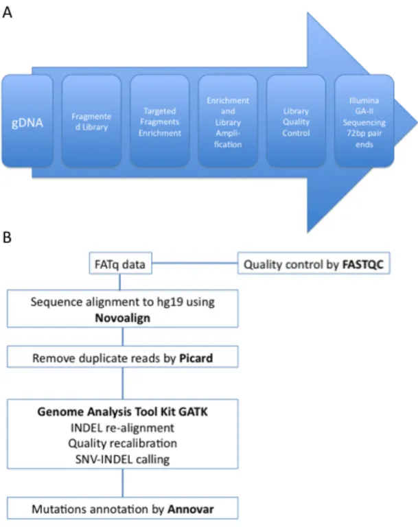
2.7 Whole genome sequencing
2.8 Confirmation of candidate mutations
2.9 DNA and RNA extraction from frozen material 2.10 Array comparative genomic hybridization (CGH) 2.11 Exome sequencing
2.12 Exome sequencing data analysis 2.13 RNA quality evaluation
2.14 Transcriptome sequencing
2.15 Transcriptome sequencing data analysis 2.16 Cell lines and cell line work
2.17 ShRNA, siRNA and transfection experiments 2.18 Statistical analysis
3. Results
3.1 Overview and strategy of the performed experiments 3.2 NUT-BRD4 fusion gene in thymic epithelial tumors
Clinical Features of studied thymic epithelial tumors NUT rearrangement in thymic carcinomas is uncommon
3.2 Expression and mutational status of c-Kit in thymic epithelial tumors
c-KIT immunohistochemistry Sequencing analysis of KIT
Patient’s clinical history
Preparation and validation of tumor material Whole genome sequencing results
Junction sequences
Copy number (CN) aberrations
Expression of mutated genes in patient’s tumor
Function of mutated genes and their relevance in cancer
3.4 Exome sequencing and transcriptome sequencing
CGH results
Exome sequencing results
Transcriptome sequencing results
3.5 CGH evaluation of a series of formalin fixed paraffin embedded thymic epithelial tumors.
Correlation between chromosome arm-level aberrations and thymic epithelial tumors histotypes
Chromosome arm-level CN loss of 13q is a candidate marker of poor prognosis
Identification of significant CN aberrations in thymic epithelial tumors by GISTIC algorithm
CDKN2A CN loss correlates with low p16INK4 expression and poor prognosis
Deregulation of BCL2 family genes in thymic epithelial tumors Thymic epithelial tumor cell lines are resistant to ABT263 treatment but sensitive to the combination with sorafenib
Gx15-070 inhibits thymic epithelial tumor cell growth through autophagy and necroptosis
4. Discussion and conclusions 5.References
6. Acknowledgments 7. Abbreviations
1.1 Normal thymus
Macroscopic structure
The thymus is a median organ located in the anterior mediastinum. Starting from fetal life, it increases its dimension reaching maximum size during adolescence and decreases thereafter. In the adolescent the thymus extends from the fourth costal cartilage upward to the lower border of the thyroid gland. The sternum and the sternohyoidei and sternothyreoidei muscles cover the thymus. Back, the thymus confines with the pericardium and a layer of fascia, which covers the aortic arch and the great vessels. In the neck, it partially wraps the frontal part of the trachea. The thymus consists of two lateral lobes that are occasionally united and frequently differ in size. This organ presents a soft lobulated surface when cut and a pinkish-gray color.
Figure1.1: Normal thymus. (A) Schematic representation of the thymus. (B)
Microscopic structure
A fibrous capsule wraps each lateral lobe of the thymus and protrudes septa of a delicate areolar tissue within the organ; therefore it presents a lobular aspect. The lobules vary in size and have an irregular shape being, in the interior portion fused together. Each follicle is divided into two main regions on histological grounds, the cortex and the medulla; and each of these regions contains several ultrastructurally and phenotypically distinct types of thymic epithelial cells and immature lymphocytes (thymocytes).
The thymic cortex is composed by network of epithelial cells mixed with abundant thymocytes; these stroma cells form an adventitia to the blood vessels in the periphery of lobules.
In the medulla the epithelial cells are prevalentand are organized in a coarse reticulum whereas the thymocytes are relatively fewer in number. Hassall corpuscles are nest-like bodies observed in thymic medulla. These corpuscles are composed of central granular cells wrapped by several layers of epithelioid cells. The cortico-medullary junction separates the two distinct portions. Just outside the cortex, the sub-capsular zone defines a functionally differentiated region of the thymus.
Stromal cells, mainly thymus epithelium, interdigitating cells, and macrophages constitute the thymic microenvironment, but other components, such as nerves, endothelium, fibroblasts, and connective tissue are represented as well.
Distinct regions of the thymus contain different populations of epithelial cells. Electron microscopy analyses have identified six types of thymic epithelial cells: type 1 cells are present in the capsule and septa and surround the perivascular spaces of the
cortical capillaries; types 2, 3 and 4 form a graded series in the cortex, ranging from electron-lucent metabolically active to electron-dense dying forms; type 5 cells are rare, medullary unspecialized epithelial cells, while type 6 cells form the major medullary population and contribute to Hassall’s corpuscles.
Expression of cytokeratin 5 (K5) and K8 distinguishes epithelial cell subpopulations of the thymus. The more prominent cortical and medullary subsets are K8+K5– and K8–K5+, respectively. Cells of the cortico–medullary junction are K5+K8+. The presence of a common stem cell of endodermic origin, able to generate such epithelial subpopulations, has been demonstrated and is characterized by MTS20+MTS24+K8+K5+ phenotype1. Therefore, the epithelial component of the thymus entirely originates from the endoderm of the third pharyngeal pouch, whereas the fibroblast and the capsule originate from mesoderm of the neural crest. The cells originating from the neural crest wrap the thymic primordium and this interaction is necessary to the normal development of thymus2. Moreover, the precursors of thymic epithelial cells are able to start their maturation process but necessitate the interaction with thymocytes to differentiate into the mature phenotype3.
Physiological function of the thymus
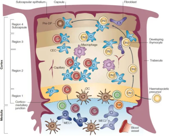
The thymus has a central role in the immune system because it is required for T-cell differentiation and repertoire selection. The stromal cells of the thymus sustain and drive the maturation of the thymocytes that migrate in this organ from the bone marrow. T-cell development is characterized by the progression through several phenotypically distinct stages, defined as double negative (DN), double positive (DP) and single positive (SP) based on expression of the co-receptors CD4 and CD8. The
differential expression of CD44 and CD25. Thymocytes occupy different regions of the thymus according to their maturative stages suggesting an highly ordered migration (Figure 1.2).
T-cell precursors access the thymus at the cortico–medullary junction and migrate progressively to the subcapsular zone of the outer cortex; thereafter thymocytes move back through the cortex and into the medulla, from where they egress to the periphery in the form of mature T lymphocytes. Migrating through the cortex, thymocytes undergo the positive selection for those cells presenting a T cell receptor able to recognize the major histocompatibility complex. Passing through the medulla, thymocytes are exposed to the negative selection that removes those T-cell precursors able to recognize self-antigens. This process is also known as central tolerance and it is promoted by the ectopic expression of tissue-restricted self-antigens in the medullary cells of the thymus. The expression of tissue-restricted self-antigens mirrors virtually all tissues of the body, irrespective of developmental or spatio-temporal expression patterns. The autoimmune regulator gene (AIRE) promotes the expression of peripheral antigens in medullary cells. The subversion of thymic architecture and the abnormal properties of neoplastic thymic epithelial cells in thymomas can brake the central tolerance and are considered responsible for the increased number of autoimmune diseases observed in these tumors.
Figure 1.2: Modified from Blackburn CC, et al.3; the thymic lobule is divided into cortex and the medulla. Different types of epithelial cells constitute the stroma of thymic medulla (MEC) and cortex (CEC). The thymic cortex has been separated into 4 regions. In region 1, the cortico–medullary junction, is the site of entry into the thymus and contains DN1 cells. In region 2, cells differentiate to the DN2 stage, undergo a proliferative clonal expansion, and lose B- and natural killer (NK)-cell potential. In region 3, DN3 cells rearrange their T-cell receptor (TCR) β-chain. In region 4 DN cells become CD4+CD8+ DP. DP cells migrate back through the cortex to differentiate into the medulla either in CD4+ or CD8+ SP cells. B: B lymphocytes and DC: dendritic cells.
1.2 Definition of thymic epithelial tumors
Thymoma and thymic carcinoma are neoplasms originating from the epithelial cells of the thymus; therefore, they are defined thymic epithelial tumors to distinguish them from lymphomas that arise from thymic lymphocytes and thymocytes (immature lymphocytes of the thymus). Levine and Rosai have introduced such distinction in 19764. Previous publications indiscriminately named thymomas also those tumors originating from thymic lymphocytic component.
1.3 Epidemiology
Thymic epithelial tumors (TETs) are the most frequent primary tumors of the mediastinum, however they are rare. In the mediastinum, metastases are the most commonly diagnosed masses, while primary tumors are among the most rare human neoplasms comprising less than 1% of all adult cancers. TETs comprise about 25% of primary mediastinal cancers (Figure 1.3 A).
Three population-based studies have estimated the incidence of TETs in the Netherlands, Sweden and USA. According to the Dutch cancer register, the incidence of TETs is 0.32/100 000 person-year5, while, according to the Swedish register, 0.23 in female and 0.27 in male6. In the Nederlands, the incidence progressively increased by 6% between 1995 and 2003. Data on US population are more difficult to interpret, because the Surveillance, Epidemiology and End Results (SEER) database collects only data on malignant thymomas. The definition of malignant thymomas was based on an obsolete classification and limited to those cases with capsular invasion. Due to the stage at the diagnosis and the evidence of capsular invasion, not all thymomas have been captured by SEER registries, because they could be interpreted as benign tumors by pathologist or clinicians. Because such cases have not been recorded in the US cancer registries, the reported estimated incidence of 0.13/100 000 person-year underestimates the global incidence of TETs in US, including only a subgroup of cases considered “malignant thymomas”7. Such results come back in line with European estimations, considering that 35.5% of TETs were classified malignant and 64.5% benign in the Swedish population6.
presentation is 60 years, with most patients older than 40 years6. Surprisingly, the increase in thymoma incidence with age appears in striking contrast to the progressive age-related involution of the thymus. Similarly, the incidence decline at the oldest ages is unexplained.
TET incidence is higher in Asians/Pacific Islanders and African Americans than Caucasian and Hispanics, among the ethnic groups studied in SEER database7.
Patients affected by TET present a reduced overall survival6, despite the possible indolent behavior of a subgroup of them (Figure 1.3B). Patients with either malignant or benign tumors have a shorter survival compared to the control group which was constituted by subjects randomly selected from the Swedish population database, without any history of cancer and matched by gender and age6.
The evaluation of secondary malignancy after the diagnosis of TET is relevant to understanding the etiology and the biology of these tumors. An increased risk of specific neoplasms could indicate that those cancers share genetic or environmental risk factors. Moreover, an elevated risk for specific cancers succeeding thymoma may suggest that the immune dysregulation caused by thymoma predisposes to those cancers. Retrospective reports suggest a broadly increased risk for cancer in patients treated for thymoma, with possible relation to genetic predisposition or to immune disturbance. In those studies the frequency of secondary malignancy ranged between 28 and 8%8-10. In the SEER database, 66 out of 733 (9%) patients with a diagnosis of malignant thymoma successively developed a secondary tumor. However, the corresponding standardized incidence ratio (SIR)@, that approximates the relative risk for cancer associated with thymoma, was only 1.5; which was a more modest elevation than that depicted in the hospital-based series7. Among the specific
malignancy, only an increased risk for non-Hodgkin’s lymphoma (7 cases) was supported by SEER data for a SIR of 4.77. The increased risk for secondary non-Hodgkin’s lymphoma was confirmed in the Swedish register (SIR 6.3) and reported together with an increased risk of non-melanoma skin cancer (SIR 10.6) and cervix cancer (SIR 6.9) that were not evaluated in the US study6. These observations have been related to the immune system alteration associated with thymic impairment, since a significantly risk increase of non-melanoma skin cancer, non-Hodgkin’s lymphoma and cancer of the cervix, are strongly associated with severe immune disorders such as acquired immunodeficiency syndrome (AIDS). A complementary approach can be adopted to examine cancer registry data searching for thymoma risk following other primary malignancy. A report, based on SEER data, showed only a modestly elevated risk of developing thymoma after different cancers (SIR 1.33), without any relation to a particular tumor type11. The lack of an increased risk for
developing thymoma in non-Hodgkin’s lymphoma patients supports the hypothesis that the elevated risk of secondary non-Hodgkin’s lymphoma after thymoma arises from immune disturbance consequent to thymoma. Moreover, the absence of an increased thymoma risk in lung or breast cancer and in Hodgkin’s lymphoma patients, all of which are frequently treated by radiation therapy to the chest, argues against a role played by ionizing radiation as a risk factor for thymoma. Such data regarding risk of secondary cancers do not suggest tobacco or alcohol as candidate risk factors for thymoma. There are not data concerning the role of occupation, environmental exposures, or diet and nutrition.
@ Note: the standardized incidence ratio (SIR) is the ratio of the observed-to-expected number of cases: if SIR =1 the incidence of secondary malignancy is the same of the reference population whereas it is increased when SIR >1.
Figure 1.3: Epidemiology data of thymic epithelial tumors. (A) Depicts the frequency
of primary tumor of the mediastinum in adults. (B) Modified from Gadalla SM, et
al.6; Survival curves for benign and malignant thymoma patients and their matched
1.4 Etiology
The etiology of TETs is largely unknown. Based on case-reports, viral etiology has been evaluated as a possible cause of TETs. Two cases of human foamy virus in patients with thymoma or myasthenia gravis have been reported12,13, however subsequent larger studies did not confirm these findings14,15. Similarly, the infection of human T-cell lymphotropic virus type 1 has been described in myasthenia gravis patients, some of whom had thymoma16, but, again, these findings were not confirmed14.
More convincing evidences indicate Epstein Barr virus (EBV) as a possible cause of lymphoepithelioma-like thymic carcinoma, since viral infection has been confirmed in these tumors17-20 and its histological features resemble those of
nasopharyngeal carcinoma: tumors caused by EBV in a subset of patients.
An higher frequency of neuroendocrine thymic carcinomas has been reported in patients affected by Multiple endocrine neoplasm 1 (MEN1) syndrome21,22, however, the incidence (8%) of thymic tumors remains an infrequent finding in MEN1 patients. About 25% of patients with thymic carcinoids have a positive history of MEN123.
1.5 Staging
Thymomas are an heterogeneous group of tumors according to histological features and malignant behavior. There are tumors that present a favorable prognosis growing into the thymic capsule; such tumors can even reach large dimensions, up to bulky masses, but they remain limited to the thymus borders. Other tumors present a more aggressive behavior invading the mediastinal structures. Usually, these more aggressive tumors start invading the thymic capsule, thereafter the mediastinal fat and finally, infiltrate other mediastinal organs. Diffusion in the pericardial and pleural cavities are the most common ways of dissemination. Less frequently, advanced thymomas present a metastatic spread colonizing distant organs such as liver, lung or bone than regional lymph nodes. On the other hand thymic carcinomas usually are aggressive diseases with early invasion of regional structure as well as metastatic spread24.
To evaluate TETs it is necessary to organize them according to their extent of spread, therefore they are classified according a specific staging system. The stage at the diagnosis is one of the most important prognostic factors for TETs and contributes to define the framework of treatment of these neoplasms. In 1981, Masaoka has proposed a staging system that has been widely accepted during the last decades and is currently adopted in most centers (Table 1.1). Beside Masaoka staging system, recently, several authors have proposed a TNM based classification: Yamakawa and Masaoka et al.25, Tsuchiya et al.26 at the National Cancer Center Hospital of Japan,
the WHO committee24 and Bedini et al.27 at the National Cancer Institute of Italy. The TNM schema proposed by Yamakawa and Masaoka is reported in table 1.2. The Masaoka staging system empathizes the importance of local invasion and is mainly
Conversely, a larger number of thymic carcinomas display nodal spread (32.6%), thus, the presence of N1, N2 or N3 diffusion could segregate patients upon survival.
Kondo et al. have evaluated the prognostic implication of nodal spread among the stage IVB in a large series of TETs. However, the authors did not show any significant difference. Consequently, they concluded that Yamakawa and Masaoka TNM system represents an excellent predictor for the prognosis even in thymic carcinomas where the N and/or M influence the prognosis more than the T factor29. A proper sub classification of the N and/or M factors necessitates large-scale studies including resectable and unresectable tumors29. Therefore, in the flowing chapters stages are referred to the system reported in table 11.
Table 1.1: Masaoka staging system
I Macroscopically encapsulated tumor, with no microscopic capsular invasion IIa Microscopic invasion into the capsule
IIb Macroscopic invasion into surrounding fatty tissue or mediastinal pleura
IIIa Macroscopic invasion into neighboring organs without invasion of great vessels IIIb Macroscopic invasion into neighboring organs with invasions of great vessels IVa Pleural or pericardial metastases
Table 1.2: TNM Classification of Thymic Epithelial Tumors (Yamakawa and Masaoka 25)
T factor
T1: Macroscopically completely encapsulated and microscopically no capsular invasion T2: Macroscopically showing adhesion or invasion into surrounding fatty tissue or mediastinal
pleura, or microscopic invasion into capsule
T3: Invasion into neighboring organs, such as pericardium, great vessels, and lung
T4: Pleural or pericardial dissemination
N factor
N0: No lymph node metastasis
N1: Metastasis to anterior mediastinal lymph nodes N2: Metastasis to intrathoracic lymph nodes except anterior mediastinal lymph nodes
N3: Metastasis to extrathoracic lymph nodes
M factor M0: No hematogenous metastasis M1: Hematogenous metastasis Stage I T1 N0 M0 Stage II T2 N0 M0 Stage III T3 N0 M0 Stage IVA T4 N0 M0
Stage IVB any T N1-3 M0
1.6 Histological Classification of thymic epithelial tumors
The classification of TETs based upon their histological features has been one of the most debated topics between pathologists. In 1961, Barnatz et al. from the Mayo clinic presented the first widely accepted classification of TETs by which thymomas were divided in four categories: predominantly lymphocytic,
predominantly epithelial, mixed and predominantly spindle cells. During the following years, several subsequent classifications (Table 1.3) have been proposed and criticized, until 1999, when the World Health Organization (WHO) committee chaired by Dr. Rosai presented a proposal of classification that encountered large agreement between pathologists. That proposal has been integrated and accepted as a formal classification in 2004 by a WHO committed chaired by Dr.
According to this classification, a clear cut distinction has been defined between thymomas, organotypic tumors that mimics the structure of normal thymus, and thymic carcinomas, more aggressive neoplasms that do not resemble the structure of normal thymus but those of carcinomas originating in organs other than thymus.
Gross pathological and histological features of thymic epithelial tumors
Thymomas are classified in A, B histotypes, depending on the shape of epithelial cells and their nuclei.
A type thymomas present bland spindle/oval epithelial tumor cells with few or no lymphocytes. Grossly, they are usually encapsulated and easily separable from the surrounding organs even in case of tumors with conspicuous dimension. When cut, the surface appears white with vague lobulation. Cystic structures and calcification of the capsules can be observed. Microscopically, the tumor cells present bland nuclei with disperse chromatin and inconspicuous nuclei. Tumor cells are arranged in solid sheets without any particular pattern, neither distinct lobules nor dissecting fibrous bands typical of other type of thymomas. Type A cells can form cystic o glandular structure, glomeruloid bodies, rosettes or meningioma-like whorls. Perivascular spaces are less commonly seen than in other types of thymomas and even if type A is a thymocyte poor thymoma, spindle cells micronodules in a lymphoid stroma can be observed at places. Mitotic figures are infrequent. Necrosis can be appreciated in case of lobular infarct. Rarely, thymic carcinoma can arise in type A thymoma, usually accompanied by necrotic areas, which examination reveals hyperchromatic anaplastic nuclei and or mitotic figures indicating the presence of carcinoma24.
Type B thymomas show epithelial cells with a predominantly round or polygonal appearance and are further classified in B1, B2 and B3, according to an increasing degree of atypia of the tumor cells and to a decreasing extent of thymocytes24.
thymus cortex. Usually, B1 thymomas are encapsulated grayish masses. Thick fibrous septa protruding from the capsule can be observed, as well as cystic spaces or small hemorrhagic and necrotic areas. Microscopically, the neoplastic epithelial cells are scant, small, with very little atypia and submerged by non-neoplastic thymocytes. The tumor cells are oval with pale round nuclei and small nucleoli, although some cells may be large and occasionally have conspicuous nucleoli. B1 thymomas are usually organized in a lobular structure even if sheets architectures can be sometime appreciated. Either thick or thin fibrous bands separate lobules of various sizes. Perivascular spaces are not as frequent as in other B thymomas. Pale areas of medullary differentiation are always present, composed of more loosely packed thymocytes; Hassall’s corpuscles are less frequent than in medulla of normal thymus24.
Type B2 thymomas are characterized by large polygonal epithelial tumor cells arranged in a loose network containing numerous immature T lymphocytes. Grossly, B2 thymomas can be encapsulated, circumscribed or invasive of the surrounding tissues and organs. When cut, the surface presents tan-colored nodules separated by white fibrous septa and can be soft or firm. Cysts, hemorrhages and fibrosis can be appreciated. Microscopically, the large polygonal tumor cells present large nuclei with an open chromatin pattern with prominent central nucleoli. Commonly, B2 thymomas are organized in large lobules with delicate septa, resembling the normal architecture of thymic cortex. The tumor epithelial cells form palisades around perivascular spaces and along septa, large sheets of tumor cells occasionally can be observed. Medullary islands are missing or infrequent and Hassall’s corpuscles are an exceptional finding. Tumor cells are usually outnumbered by non-neoplastic immature lymphocytes24.
B3 thymomas are composed from medium size round or polygonal epithelial tumor cells with slight atypia; these cells are mixed with a minor component of intraepithelial thymocytes. Commonly, B3 thymomas are not encapsulated and present an infiltrative border with extension to the mediastinal fat or the surrounding organs. The cut surface appears firm and lobulated in grey and white nodules by fibrous septa. Soft yellow or red foci, cyst formation or hard calcification are common both in large and small tumors. Usually, neoplastic cells are polygonal with round or elongated nuclei often folded or grooved. Commonly, B3 cell nuclei are smaller and with less prominent nucleoli than in B2 thymomas. In a minority of cases, more atypical, enlarged and hypercromatic nuclei are observed. Other rare variants present either polygonal cells with nuclei similar to B2 thymomas or partial clear cell changes. Focal or extensive structure of spindle cells can be observed. B3 thymomas with anaplasia are a small group of tumor showing an high degree of atypia but conserving the organoid structure of thymomas. Tumor cells form lobules that are separated by thick fibrous and hyalinized septa. Tumor cells grow in palisades around perivascular spaces and along septa resulting in the formation of tumor cell sheets with a vaguely solid or epidermoid appearance24.
AB thymomas are composed of a lymphocytes-poor type A and a more lymphocyte-rich type B component. Usually, AB thymomas are encapsulated. The cut surface shows a lobular structure divided, by fibrous septa, in tan colored nodules of variable sizes. All the histological features of type A thymomas are represented in the A component. Conversely, the B component is peculiar and differs from B1, B2 or B3 thymomas; it is characterized by small polygonal tumor cells with small round, oval or spindle nuclei showing disperse chromatin and small nucleoli. The A and B
components either form discrete separate nodules or intermix together. The growth pattern consists of nodular and diffuse areas24.
The 2004 WHO classification depicts separate additional categories: the micronodular thymoma with lymphoid stroma, the metaplastic thymoma and a group of rare thymomas. Micronodular thymoma is characterized by cystic tumor areas of variable size and by multiple discrete or confluent nodules of epithelial cells, separated by lymphocytic stroma that might contain follicles with germinal center. Tumor epithelial cells of microcystic thymoma are slender or pulp spindle with bland looking oval nuclei and small nucleoli. Metaplastic thymomas do not present a lobulated growth pattern and are characterized by a biphasic architecture comprising epithelial islands intertwining with boundless of spindle cells. The tumor cells are polygonal, ovaloid or plump spindle, with oval vesicular nuclei, small distinct nucleoli and an amount of lightly eosinophilic cytoplasm. Rare thymomas are defined: microscopic thymoma, sclerosing thymoma and lipofibroadenoma of the thymus, a neoplasm that occasionally occurs adjacent to a conventional type B1 thymomas. Microscopic thymoma depicts the multifocal proliferation (<1mm in diameter) that can occur in the thymus of myasthenia gravis patients without a macroscopically evident tumor. The sclerosing thymoma exhibits the features of a conventional thymoma in terms of epithelial cell morphology and thymocytes content, but with an exuberant collagen rich stroma24.
Table 1.4: Thymoma histotypes Type A Type AB Type B1 Type B2 Type B3 Micronodular thymoma Metaplastic thymoma Rare thymomas Microscopic thymoma Sclerosing thymoma
Lipofibroadenoma of the thymus
Thymic carcinomas regroup all non-organotypic malignant epithelial neoplasms other than germ cell tumors. Carcinomas are named according to their differentiation and are listed in table 1.5.
Table 1.5: Thymic carcinoma histotypes Squamous cell carcinoma
Basaloid carcinoma
Mucoepidermoid carcinoma Lymphoepithelioma-like carcinoma Sarcomatoid carcinoma
Clear cell carcinoma Papillary adenocarcinoma Non-papillary adenocarcinoma Carcinoma with t(15;19) translocation Undifferentiated carcinoma of the thymus Thymic neuroendocrine tumors
Well differentiated neuroendocrine carcinomas Typical carcinoid Atypical carcinoid Poorly differentiated neuroendocrine carcinomas
Non-small cell neuroendocrine carcinoma Small cell neuroendocrine carcinoma
Figure 1.4: microscopy appearance of thymic epithelial tumors according to their
histotype. Images were captured at 20x magnifications.
Squamous cell carcinoma of the thymus is a tumor of the epithelial cells of the thymus resembling the features of those squamous cell carcinomas originating in organs other than thymus. Squamous cell carcinomas at the diagnosis usually infiltrate the surrounding organs and tissues. At the cut surface, lobulation and fibrous septa are not evident, whereas foci of necrosis and hemorrhage are common features. Tumor cells are large polygonal with or without evidence of keratinization and presenting vesicular or hyperchromatic nuclei. Cells are arranged in nests and cords divided by fibrohyaline-stroma24.
Basaloid carcinoma is composed by compact lobules of tumor cells with peripheral palisading and a basophilic staining pattern due to a high
nuclear-cytoplasmatic ratio. Basaloid carcinoma frequently originates in micronodular thymic cysts. Generally, basaloid carcinomas are well-circumscribed, grey masses surrounded by a thin fibrous capsule with focal hemorrhage and cystic formations. Microscopically, the cells are small, columnar, round or spindle with hyperchromatic round nuclei, scant cytoplasm and indistinct borders24.
Mucoepidermoid carcinoma is a rare variety of thymic carcinomas characterized by the presences of squamous cells and cells producing mucous. The cut surface of these tumors looks nodular with fibrous bands and mucinous appearance24. Lymphoepithelioma-like carcinoma is characterized by a sincitial growth of undifferentiated carcinoma cells accompanied by lymphoplasmacytic infiltration. It may or may not be associated with EBV infection. Macroscopically, the tumor is solid with yellow/white areas of necrosis. The tumor cells present large vesicular nuclei with open chromatin and show indistinct plasma membrane. Lymphocytes are admixed with the carcinoma cells, that growth in nests or anastomosing cords24.
Sarcomatoid carcinoma resembles soft tissue sarcoma morphology and, usually, is constituted by a sarcomatoid and a carcinoma component. The carcinomatous component comprises clusters and sheets of poorly differentiated epithelial cells with nuclear prominent atypia and occasional squamous differentiation. Spindle tumor cells with pleomorphic nuclei characterize the sarcomatoid component, these cells show distinct nucleoli and frequent mitotic figures organized in fascicules and storiform arrays24.
Clear cell carcinoma is a rare variety composed from polyhedral tumor cells with optically clear cytoplasm with slight cellular pleomorphism and nuclear atypia.
or necrosis. Tumor cells grow in nests, lobules or sheets surrounded by a dense fibrous stroma that confers a lobular architecture24.
Papillary adenocarcinoma is rare and characterized by a prominent papillary pattern of growth. Tumor cells have eosinophilic clear cytoplasm and round ovaloid nuclei with condensed chromatin and few small prominent nucleoli. Few cells are positive for mucine stating. Tumor cells grow in tubulopapillary structure of uniform cuboid to columnar cells, mainly lying in a monolayer but occasionally showing a glomeruloid arrangement. Non-papillary adenocarcinoma differentiation of thymic carcinoma is more extremely rare with only three cases described in literature24.
Carcinoma with t(15;19) translocation is a rare carcinoma of unknown histogenesis arising in the mediastinum and other midline organs of young people. Undifferentiated cells, vigorously mitotic, grow in sheets that form syncytia with inter epithelial lymphocytes24.
Undifferentiated thymic carcinomas are characterized by a solid growth of undifferentiated cells. The diagnosis is one of exclusion with other primary sites24.
Thymic neuroendocrine carcinomas are predominantly or exclusively composed of neuroendocrine cells and have to be distinguished by those thymic carcinomas that may contain scattered groups of neuroendocrine cells. The neuroendocrine differentiation of epithelial tumor cells can be demonstrated by immunohistochemistry when positive for chromogranin, synaptophysin NSE and CD56. These tumors are sub-classified in well-differentiated neuroendocrine carcinomas: typical and atypical carcinoid and poorly differentiated neuroendocrine carcinomas: small cell and large cells neuroendocrine carcinomas. Grossly, they are frequently invasive at the diagnosis and on the cut section are firm, grey-white or tan
with a gritty consistency. Typical carcinoids are devoid of necrotic area and exhibit a low mitotic rate (<2 mitosis in 2mm2); conversely to the more common atypical carcinoids that display either necrosis or 2-10 mitosis in 2mm2. Large cells
neuroendocrine carcinomas present more than 10 mitoses in 2mm2; whereas, small cell neuroendocrine carcinomas are characterized by small size round, oval or spindle cells with very scant cytoplasm and high mitotic activity24.
Finally, the 2004 WHO classification introduces the combined TETs, with at least two distinct areas each corresponding to one of the histological thymoma and thymic carcinoma types. The description and the frequency of these combined tumors underlines the morphological heterogeneity that TETs can display. Moreover, this can partially explain the limited reproducibility of histological classification and the difficulty to predict prognosis upon morphological appearance24.
Clinical relevance of WHO classification
Difference between thymomas and thymic carcinomas is relevant for the clinical practice mainly because thymic carcinoma exhibits a more aggressive behavior and is infrequently paraneoplastic syndromes.
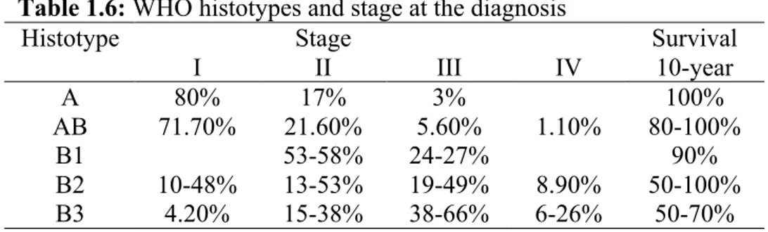
B2 and AB are the most common thymomas, whereas, A and B1 are less represented (Table 4). Myasthenia gravis (MG) and red pure cell aplasia have been reported in all kind of thymomas, even if, with a different frequency of association. Paraneoplastic hypogammaglobulinemia and Good syndrome were described in B tumors. For all histotypes, symptoms at the diagnosis can be either related to paraneoplastic syndromes or consequent to the presence of a mediastinal mass24. In type A, AB and B1 the diagnosis is frequently incidental, being the patients asymptomatic. Usually, B2 and B3 tumors manifest themselves with local symptoms such as chest pain, cough or dyspnea up to vena cava syndrome as they tend to be invasive due to their more malignant behavior. At the diagnosis, A and AB thymomas are usually circumscribed at the stage I or II (Table 1.6) and show a favorable outcome with survival reported up to 100% at 10-year24 (Table 1.7). B1 thymomas
present limited malignant behavior; more than 90% of tumors are resectable and local recurrences or late metastases occur in less than 10%24. Consequently, B1 thymoma 10-year survival is about 90%24. Conversely, B2 and B3 thymomas are more malignant tumors with reported 10-year survival between 100-50% and 70-50%, respectively24. 3% of B2 and 7% of B3 thymomas are metastatic at the diagnosis.
5-15% of B2 and 17-47% of B3 are not resectable at presentation. After surgery, B2 and B3 thymomas present local recurrence in 5-9% and 15-17% and metastasize in 11% and 20% of cases, respectively. Such data support a prognostic segregation of low
malignant grade tumors (A, AB and B1) and intermediate grade malignant neoplasms24 (B2 and B3; Figure 1.5).
Micronodular thymomas are 1-5% of TETs that occur at a mean age of 58 years and are rarely associated with paraneoplastic syndromes (<5%). Micronodular thymoma outcome is usually favorable due to stage I diagnosed in more than 90% of patients. Only few cases of metaplastic thymomas have been described, none of them associated with paraneoplastic syndromes. Metaplastic thymoma diagnoses were incidental with 75% of the tumor stage I24.
Thymic carcinomas are 10-20% of TETs; they are frankly aggressive neoplasms with a 10-year survival <50%. At the diagnosis, most tumors are stage III and IV24. Rare paraneoplastic polymiosites but not MG have been described in
thymic carcinomas. The most frequent thymic carcinomas are those with squamous cell differentiation; 90% in Asian and 30% in western series. Undifferentiated carcinoma is the second most common and it is characterized by a more severe prognosis24. Basaloid carcinomas are rare with few cases described; they show a remarkable tendency to originate in multilocular thymic cysts and are characterized by a less malignant behavior24. The remaining subtypes of thymic carcinoma are considerably rare and their description is mainly based on case reports. Out of these tumors lymphoepithelioma-like and clear cell carcinomas present a remarkable aggressive behavior. Only 6 cases of carcinomas with the translocation t(15;19) have been described for which a thymic origin has been postulated24. These belong to a
group of neoplasom named midline tumor, characterized by a remarkably aggressive behavior, a young age of insurgence and the fusion of NUT gene with BRD4 in 2/3 of
Neuroendocrine thymic carcinomas are 2-5% of TETs; most of them are atypical carcinoid. Rare small cell neuroendocrine carcinoma and exceptionally rare typical carcinoid and large cell neuroendocrine carcinoma have been described24.
Outcome is more favorable for atypic carcinoid 84% and 75% at 5 and 10-year survival, respectively24. 30-50% of small cell neuroendocrine carcinomas are diagnosed in stage IV. Small cell neuroendocrine carcinomas present a severe prognosis with 5 and 10-year survival of 50 and 30%, respectivelly24. Symptoms at the presentation can be related to the mediastinal mass or to the correlated endocrine syndromes. The most frequently observed endocrine syndromes are ACTH induced Cushing, present in 17-30% of adults and >50% of children, acromegaly, hypercalcemia/hypophosphatemia whereas carcinoid syndrome is rare (<1%). 25% of thymic neuroendocrine carcinomas present a MEN1 syndrome and 8% of patients affected by MEN1 syndrome present a neuroendocrine thymic carcinoma24.
Table 1.6: WHO histotypes and stage at the diagnosis
Histotype Stage Survival
I II III IV 10-year A 80% 17% 3% 100% AB 71.70% 21.60% 5.60% 1.10% 80-100% B1 53-58% 24-27% 90% B2 10-48% 13-53% 19-49% 8.90% 50-100% B3 4.20% 15-38% 38-66% 6-26% 50-70%
Table 1.7 Histotypes’ characteristics
Histotype Prevalence Mean age Myasthenia gravis
A 4-19% 61 24%
AB 15-43% 55 14%
B1 6-17% 41-47 18-56%
B2 18-42% 47-50 30-82%
B3 7-25% 45-50 30-77%
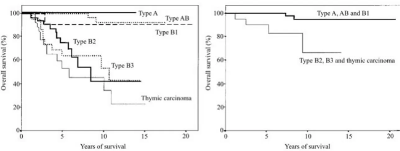
Figure 1.5: modified from Chen et al.31 (A) Overall survival reported for each histological subtype. Type B2-3 and C thymomas have a significantly (P=0.001) increased risk of death. (B) Overall survival of stage I and II tumors (n 102). There were two deaths among patients with Type A, AB, and B1 thymomas (n 78), but four deaths in patients with Type B2, B3, and C thymomas (n 24). The difference is statistically significant (P =0.003)
Reproducibility of WHO classification
The 2004 WHO, as well as previous classifications, presents disputed issues regarding reproducibility and its prognostic value. The inter observer agreement has been evaluated using the material described in the chapter 3.5 and is currently under consideration for publication but do not represent a result of this dissertation. Briefly, a series of 132 TETs has been reviewed by 3 independent pathologists at 3 different institutions Humanitas Cancer Center (Rozzano-Milan, Italy), Seoul National University Bundang Hospital, Seongnam-si (Gyeonggi, Republic of Korea) and National Institute of Health (Bethesda, MD) and compared to the original diagnosis (CLAS1). The aims of this comparison were to evaluate the reproducibility of the WHO schema and determine if reproducibility can affect its prognostic value. The original histological diagnosis reported after surgery, paraffin blocks and slides, as well as clinical history were available in 129 cases. For this analysis combined thymomas were re-classified according to the more aggressive histotype (for example B2-B3, were considered B3 thymomas).
Table 1.8 summarizes histotype frequencies according to the 4 observers. The global agreement was moderated; taking into consideration all the WHO histotypes and all the pathologists at the same time: Fleiss’ kappa coefficient 0.53. To compare agreement between different pathologists, Cohen’s Kappa coefficient was calculated for each comparison pair (Table 1.9). The best agreement was observed between CLAS2 and CLAS1 (K coefficient: 0.84). A substantial agreement was also observed between CLAS3 and CLAS2 (K coefficient: 0.70) and CLAS3 and CLAS1 (K coefficient: 0.64). A moderate strength of agreement was observed between CLAS4 and each of the other three observers, with a Kappa correlation coefficient ranging
different observers. In 43/129 (33.3%) cases three out of four pathological diagnoses were identical; in 15/129 (11.6%) cases the diagnoses were identical by pair; in 8/129 (6.2%) cases three different pathological diagnoses were reported; complete disagreement (four different diagnoses) was never observed. More frequently (9/23, 39%) the discordant diagnosis interested B thymomas, especially the discrimination of B1 and B2 (6/9). In 6/23 (26%) cases, the diagnostic discrepancy involved type AB thymomas versus B thymomas. In 7/23 (30%) cases, inconsistency regards fairly different histological subtypes: 4 cases of thymic carcinoma vs. type B3; 2 cases type B3 vs. type A; and in one case thymic carcinoma vs. type A.
The interpretations of 2 different pathologists (CLASS1 and CLASS4) were able to predict prognosis in univariate analysis of time to progression (TTP; CLAS1 p=0.001; CLAS4 p<0.001) and disease relate survival (DRS; CLAS1 p=0.039; CLAS4 p=0.027).
Previous reports have evaluated the reproducibility of WHO classification and have described an high agreement strength between pathologists interpretation (Kappa coefficient 0.9-0.8731,32, respectively). Chen et al.31 have reported inter-observer
agreement between pathologists from different institutions after a training introduction from Dr. Muller-Hermelink; whereas in Riekel et al. evaluation included pathologists from the same background. Our results are in line with these reports when referred to pathologists of similar backgrounds: CLAS1 and CLAS2 were evaluated within the Humanitas Cancer Center and showed a high agreement K=0.84). Conversely, among pathologists of different backgrounds, the reproducibility was only substantial or moderate. Similarly, the evaluation of 95 TETs
These data denote the importance of a proper training for an acceptable reproducibility of the WHO classification. The most challenging diagnoses appear to be those discriminating between B1, B2 and AB thymomas in most of the reports. Consequently, more clear and defined criteria needs to be established for these histotypes. The discrimination of B2 from B1 and AB tumors is relevant for clinical practice because of their more severe prognoses; consequently, the prognostic value of WHO were, in our series, subject to variation according to pathologist interpretations.
Suster and Moran30 proposed an exemplified classification in order to increase reproducibility and better predict prognosis. Table 1.3 summarized this classification. However, the WHO classification better highlights the morphological differences between TETs and, even if it seems to be of limited immediate clinical interest, it is remarkably important for research purpose. Thus, we are going to demonstrate that different WHO histotypes present different genomic background and may necessitate different targeted therapeutic approaches in the results section.
Table 1.8: WHO histotypes according to classification of 4 different pathologists
A AB B1 B2 B3 TC
CLAS1 16.4% 21.1% 17.2% 15.6% 21.1% 8.6%
CLAS2 11.6% 20.2% 19.4% 13.9% 25.6% 9.3%
CLAS3 12.7% 19.8% 17.5% 15.9% 19.0% 15.1%
CLAS4 17.6% 19.2% 3.2% 33.6% 18.4% 8.0%
Table 1.9: Kappa correlation coefficient between each pathologist pair
CLAS1 CLAS2 CLAS3 CLAS4
CLAS1 0.84 (0.77-0.91) 0.64 (0.55-0.74) 0.52 (0.41-0.62) CLAS2 0.70 (0.61-0.79) 0.52 (0.41-0.62) CLAS3 0.53 (0.43-0.63)
CLAS = pathologist’s classification; 95%CI was reported with each coefficient in parenthesis. The agreement’s strength of reputed: low: <0.41; moderate: ≥0.41<0.61;
1.7 Treatment of thymic epithelial tumors
Surgery is the mainstream of treatment for TETs and completeness of resection is the goal of the therapy, being the most relevant prognostic factor24. Neoadjuvant chemotherapy can improve the rate of radical resection in stage III patients who are not eligible for a complete resection34. If a radical resection is not achieved by surgical treatment, postoperative radiotherapy can reduce tumor recurrences. Although, the efficacy of adjuvant radiotherapy in radically resected patients is still controversial, many clinicians adopt such treatment. In patients experiencing tumor relapse, salvage surgery is often practicable but depends on the site and the number of lesions. Metastatic or not operable patients are treated by systemic chemotherapy with a palliative intent.
Surgery
A complete resection is usually practicable in stage I (100%) TETs and it is sufficient to be curative in the vast majority of patients. Conversely, the resectability rate conspicuously varies among reports in infiltrating thymomas: 43-100% for stage II, 0-89% for stage III and 0-78% for stage IV35. However, the reported survival is more favorable than that observed for other thoracic tumors (Table 1.10). The 15-year overall survival is 78% for stage I, 73% for stage II, 30% for stage III and 8% for stage IV36. The completeness of resection can not be achieved in all stage III and IV patients despite a carefully selection and a proper diagnostic workup. Relapse rate and disease free survival measure the effectiveness of surgical treatments and are summarized in Table 1.10 and Figure 1.6. Five years is the average time to recurrence, although relapses have been described up to 32 years later36. Stage I
to relapse 3 year)36. Among all recurrences the large majority are local (average 81%), whereas distant metastases occur in 9%. 10% of patients presented concomitantly with both local and distant relapses36.
Table 1.10: Overall Survival of Patients With Thymic Tumors
% 5-year Survival % 10-year Survival
n % R0 I II III IV I II III IV
Kondo and Monden28 924 92 100 98 89 71 100 98 78 47
Regnard et al.36 307 85 89 87 68 66 80 78 47 30
Maggi et al.37 241 88 89 71 72 59 87 60 64 40
Verley and Hollmann38 200 85 60 80 42
Nakahara et al.39 141 80 100 92 88 47 100 84 77 47 Wilkins et al.40 136 68 84 66 63 40 75 50 44 40 Blumberg et al.41 118 73 95 70 50 100 86 54 26 Quintanilla-Martinez et al.42 116 94 100 100 70 70 100 100 60 Pan et al.43 112 80 94 85 63 41 87 69 58 22 Elert et al.44 102 83 90 46 Average 83 92 82 68 62 88 71 57 38
Figure 1.6: Modified from Sakamoto M, et al.45. Disease free survival reported according to Masaoka’s stage after extended thymectomy. Survival for Stage III disease was significantly worse than that for Stage I (P = 0.0002) and Stage II (P = 0.002).
Subtotal resection
Despite neoadjuvant treatments, a complete resection can not always be planned. The advantages offered by a subtotal resection are still debated because of the inconsistence of reports and trial methodology limitations. Authors have reported either no advantage for debulking surgery46,47 or an increase of 5-year overall survival about 30%28,37,39,48. However, a subtotal surgical resection is indicated for the relief of
symptoms. Adjuvant treatments should be considered after an incomplete resection.
Salvage surgery
In those cases experiencing a relapse, the re-intervention is a valuable option because many recurrences are local and some of them can be radically removed by a salvage surgery. Increase of 5-year survival has been reported up to 40-50% beside some negative studies41,49-53. Although, in those cases presenting unique or few local metastases re-intervention is a valid option but it is not the standard treatment for those cases experiencing extensive pleural dissemination. The chance of resection of pleural implants is sometime feasible but has still to be considered an experimental procedure54.
Radiotherapy
Radiotherapy has been proposed with different intent in TETs either in neoadjuvant, adjuvant, in incompletely resected tumors or as exclusive treatment. Currently, the preferred modality of administration is conformal radiotherapy with respiratory gating. The use of intensity modulated radiotherapy (IMRT) is encouraged and consists of a real-time modeling of the contours and the amount of photons delivered within the radiation beam, using a programmed movement of the blades of the collimator55. The dose to be delivered varies according to the purpose of the treatment. It has been suggested to deliver to the thymic area a total dose of 45 Gy in a neoadjuvant setting, 45 to 55 Gy in an adjuvant setting, and 60 to 66 Gy as an exclusive treatment using a standard fractionation scheme (one 1.8- to 2-Gy fraction per day)55. Patients with TETs localized in the mediastinum, but not eligible for surgery, can be considered for radiotherapy that is rarely exclusive being more frequently associated with chemotherapy56. Neoadjuvant radiotherapy aim is to increase the rate of complete resections. Neoadjuvant radiotherapy can induce response in up to 80% of treated patients, thus, a radical resection can be achieved in 50-75% of cases56. Despite its promising results, the role of neoadjuvant radiation therapy remains controversial being currently preferred neoadjuvant chemotherapy followed by adjuvant radiotherapy55. Because thymomas are considered radiosensitive tumors and they tend to relapse locally, postoperative radiotherapy has been evaluated in several studies.
Postoperative radiotherapy.
Postoperative radiotherapy has been offered for adjuvant treatment of completely resected tumor or to consolidate results of subtotal resections. Because stage I thymomas relapse in about 2% of cases, adjuvant radiotherapy is not recommended for these patients. The role of adjuvant radiotherapy in R0 resected stage II and III thymomas remains controversial because the rarity of the disease, reports are heterogeneous, include a limited number of cases and are not prospectively designed. In 2009, a meta-analysis evaluated the role of adjuvant radiation therapy in stage II and III radically resected tumors that were included in retrospective studies reporting a control arm of exclusively surgically treated patients. Among stage II, 273 patients received a radical surgical resection and 197 a radical resection plus adjuvant radiotherapy; relapse were observed in 11 and 8% of cases, respectively (Figure 1.7A). Out of the radically resected stage III patients, 53 received adjuvant radiotherapy and 69 did not; the relapse rate was 32% and 26%, respectively (Figure 1.7B). Significant overall survival improvement was not achieved for the patients who received adjuvant radiotherapy neither a different pattern of relapse, in favor of distant metastases, was described for treated patients57. The analysis of SEER database has revealed a survival advantage for those stage II and III patients who underwent adjuvant radiation therapy. However, in the same evaluation, patients completely resected did not benefit by the adjuvant radiotherapy57. Moreover, mediastinal radiation frequently can present both short and long term adverse events including, but are not limited to, secondary malignancies, pulmonary fibrosis, esophageal strictures, coronary disease, cardiac valvular fibrosis, and pericarditis57. Although, the role of adjuvant radiotherapy in stage II and III is discouraged by data,
it is currently recommended by many clinicians for stage III patients with more aggressive histotypes (B2, B3 and TC)56.
Postoperative radiotherapy is systematically recommended after a subtotal resection58, including the boost in areas of macroscopic invasion. Radiotherapy has consistently decreased recurrence rates from 60-80% to 21-45% after a R1 or R2 resection28,53,59-64. Frequently, after local recurrence salvage surgery is practicable but for those cases not operable or resectable radiation therapy could be a valuable option. Small retrospective series depicted promising response rates and 5-year survival rate of 70-80%53.
Radiotherapy for thymic carcinoma
Peculiar considerations are appropriate for thymic carcinomas because theirs more pronunced aggressive behavior. Thymic carcinomas frequently are diagnosed when invasive and neoadjuvant chemo-radiotherapy may represent a potent approach to downstage unresectable tumors55. Although, the role of radiotherapy is expected to be more relevant in thymic carcinoma than in thymoma because the minor importance of the surgery, Kondo K., et al.28 did not show any significant advantage for the adjuvant treatment. In the 186 thymic carcinomas underwent radical resection, 5-year survival rate was 74% after adjuvant radiotherapy and 72% after surgery alone.
Figure 1.7: Modified from Korst RJ et al.57 Forest plot generated using extracted recurrence data for patients with stage II (A) and stage III (B) TET. The squares represent the odds ratios of the individual studies, and the size of the squares reflects the calculated weight of the study in the meta-analysis. The horizontal bars running through each square represent the 95% confidence interval (CI). Studies had been weighted and the Diamond at the bottom of the plot illustrates the combined odds ratio using a fixed effects model.
Systemic therapy
Thymomas are reputed chemo-sensitive tumors. Thus, chemotherapy is indicated either with neoadjuvant or palliative intent.
Palliative chemotherapy
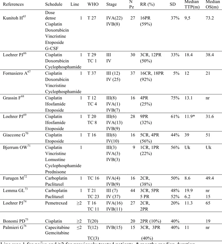
Chemotherapy is indeed the treatment of choice for patients with metastatic disease or with unresectable diffuse disease and recurrences. The intent of systemic treatment is to delay as much as possible tumor progression and eventually to reduce disease related symptoms. Conventional chemotherapeutic such as platinum based drugs (Cisplatin and Carboplatin), anthracyclines (Doxorubicin and Epirubicin), alkylating agents (Cyclophosphamide and Ifosfamide), vinca alkaloids, Etoposide and Pemetrexed have shown activity in TETs. Combination schedules have shown more encouraging results when adopted for the first line treatment (Table 1.11). Thus, schedules containing platinum and anthracyclines induce response rate from 50 to 92%. Since TETs, and especially thymomas, frequently present a slow rate of growth, multiple subsequent lines of chemotherapy are commonly offered to patients after disease progression. Responses to second or further chemotherapy lines are reported in up to 40% (Table 1.11). Therefore, overall survival improvement conferred by a single schedule is often difficult to understand. Although, thymomas frequently respond to chemotherapy, thymic carcinomas are much more resistant tumors. Interesting results in thymic carcinoma (response rate 30-24%) have been obtained by the combination of carboplatin and paclitaxel (Table 1.11).
Table 1.11: Chemotherapy regimes for palliative therapy of thymic epithelial tumors
References Schedule Line WHO Stage N
Pz RR (%) SD
Median TTP(m)
Median OS(m) Kunitoh H65 Dose dense 1 T 27 IVA(22) 27 16PR 37% 9,5 73.2
Cisplatin IVB(8) (59%)
Doxorubicin Vincristine Etoposide G-CSF
Loehrer PJ66 Cisplatin 1 T 29 III 30 3CR, 12PR 33% 18.4 38.4
Doxorubicin TC 1 IV (50%) Cyclophosphamide
Fornasiero A67 Cisplatin 1 T 37 III (12) 37 16CR, 18PR 5% 12 21
Doxorubicin IV (25) (92%) Vincristine
Cyclophosphamide
Grassin F68 Cisplatin 1 T 12 III(8) 16 4PR 75% 13.1 nr Ifosfamide TC 4 IVA(1) (25%)
Etoposide IVB(7)
Loehrer PJ69 Cisplatin 1 T 20 III(6) 28 9PR 61% 11.9* 31.6
Ifosfamide TC 8 IVA(13) (32%)
Etoposide IVB(9)
Giaccone G70 Cisplatin 1 T 16 III(6) 16 5CR, 4PR 44% 39 51
Etoposide IV(10) (56%)
Bjerrum OW71 Cisplatin 1 III(3) 9 1CR, 1PR 56% Uk Uk
Vincristine IVA(3) (22%)
Lomustine IVB(3)
Cyclophosphamide Prednisone
Furugen M72 Carboplatin 1 TC 16 IVA(4) 16 2CR, 50% 8.6 49.4
Paclitaxel IVB(9) (38%)
Lemma GL73 Carboplatin 1 T 21 III (7) 44 3CR, 5PR 48% 19.9 nr
Paclitaxel TC 23 IV (37) 5 PR 52% 6.2 15 Loehrer PJ74 Pemetrexed ≥2 T 16 IVA(16) 27 2CR, 20% 11.3 65
TC 11 IVB(11) 2PR
Bonomi PD75 Cisplatin ≥2 T(20) 20 2PR (10%) 40% 19
Palmieri G76 Capecitabine ≤2 T(12) IVB(15) 15 3CR, 3PR 40% 11 nr
Gemcitabine
TC(3) (40%)
Line was 1 for naïve and ≥2 for previously treated patients. * months median duration response, T=Thymoma, TC=Thymic carcinoma; N Pz=number of patients; RR response rate, CR complete response; PR partial response, SD stable disease; TTP=time to progression in months; OS overall survival in months; Uk unknown, nr not reached.
Neoadjuvant chemotherapy
Mostly cisplatin-based regimens have been adopted for neoadjuvant down-staging of stage III and IVa TETs. The aim of neoadjuvant chemotherapy is to induce shrinkage of locally advanced tumors in order to increase the rate of complete resection. Authors have reported promising results (Table E 1.12), despite the relevant inter-trial variability. The inconsistence of the results was probably due to the different proportion of stage III and IV patients, to the presence or not of thymic carcinomas, to the aggressiveness of the surgery and to the different efficacy of chemotherapy schedules. Among the 246 patients enrolled in table E 1.12 trials 18 complete responses (CR) and 101 partial responses (PR) were observed for a response rate (PR+CR) of 48% (range 40-100%). After induction chemotherapy, 57% of tumors received a complete resection. These results appear remarkably improved compared to those reported in historical cohorts for stage III and IV patients exclusively treated by surgery77. Moreover, a conspicuous number of patients received a surgical resection with curative intent after neoadjuvant chemotherapy even if they initially had been considered not resectable.