POLYTECHNIC UNIVERSITY OF MARCHE
School of Medicine
_______________________________________
Department of Experimental and Clinical Medicine
CEREBRAL HEMODYNAMICS
AND COGNITIVE PERFORMANCE
IN CAROTID ARTERY DISEASE
Academic Supervisor:
Prof. Dr. Mauro SILVESTRINI
Ph.D. Dissertation of:
Dr. Simona LATTANZI
TABLE OF CONTENTS
TABLE OF CONTENTS ... 1
LIST OF ORIGINAL PUBLICATIONS ... 2
ABBREVIATIONS ... 3
SUMMARY... 4
CHAPTER I ... 6
INTRODUCTION ... 6
1.1 Atherosclerotic disease of internal carotid artery ... 6
1.2 Aims of the study ... 7
CHAPTER II ... 8
MATERIALS AND METHODS ... 8
2.1 Study participants ... 8
2.2 Cerebral hemodynamics assessment ... 9
2.3 Neuropsychological assessment ... 10 2.4 Statistical analysis ... 10 CHAPTER III ... 12 RESULTS ... 12 CHAPTER IV ... 25 DISCUSSION ... 25 4.1 Conclusion ... 28 REFERENCES ... 29
LIST OF ORIGINAL PUBLICATIONS
This thesis is based on the following original articles referred to in the text by their Roman numerals (I-II).
I. Lattanzi S, Carbonari L, Pagliariccio G, Bartolini M, Cagnetti C, Viticchi G, Buratti L, Provinciali L, Silvestrini M. Neurocognitive functioning and cerebrovascular reactivity after carotid endarterectomy. Neurology. 2018;90:e307-e315.
II. Lattanzi S, Carbonari L, Pagliariccio G, Cagnetti C, Luzzi S, Bartolini M, Buratti L, Provinciali L, Silvestrini M. Predictors of cognitive functioning after carotid revascularization. J Neurol Sci. 2019 Aug 28;405:116435. doi: 10.1016/j.jns.2019.116435. [Epub ahead of print]
ABBREVIATIONS
BH Breath-holding BHI Breath-holding index (ca)VF Categorical verbal fluency CEA Carotid endarterectomy CFCT Complex Figure Copy Test CPM Coloured Progressive Matrices CVR Cerebral vasomotor reactivity ICA Internal carotid artery
MFV Mean flow velocity (ph)VF Phonemic verbal fluency TCD Transcranial Doppler TIA Transient ischemic attack
SUMMARY
Background and aim: The inter-relationships between atherosclerotic disease
of internal carotid artery (ICA), surgical revascularization and cognitive outcome are not fully understood. The aim of this study was to explore the relationship between cerebral hemodynamics and neurocognitive functioning in patients with high-grade ICA stenosis undergoing carotid endarterectomy (CEA).
Materials and methods: Patients with history of transient ischemic attack
within the past 6 months and ipsilateral high-grade stenosis of ICA undergoing CEA were prospectively enrolled. Cerebral vasomotor reactivity (CVR) to hypercapnia was measured through transcranial Doppler ultrasonography. Coloured Progressive Matrices plus Complex Figure Copy Test, and phonemic plus categorical (ca) Verbal Fluency tests were performed to test cognitive functions of right and left hemispheres, respectively. Cerebral hemodynamics and cognitive functions were assessed before and 6 months after CEA.
Results: One hundred and eighty-three patients were included. The mean age
was 73.1 (6.9) years and 122 (66.7%) were males. Before CEA, patients had decreased CVR on the side of ICA stenosis and reduced performance in the cognitive tests exploring the ipsilateral cerebral hemisphere. At 6 months from CEA, cerebral hemodynamics and neurocognitive functioning were significantly improved. The performance change in cognitive tests exploring the
revascularized hemisphere was inversely associated with pre-operative ipsilateral CVR and positively associated with the improvement in cerebral hemodynamics.
Conclusions: In patients with symptomatic high-grade ICA stenosis, cognitive
performance was enhanced at 6 months since CEA. The cognitive improvement was related to the increase in CVR on the side of stenosis correction and
predicted by baseline cerebral hemodynamic status. Cerebral hemodynamics may be an independent and potentially reversible determinant of cognitive dysfunction in severe carotid artery disease.
CHAPTER I
INTRODUCTION
1.1 Atherosclerotic disease of internal carotid artery
Extracranial carotid artery disease affects about 10% of subjects over 70 years, and its burden is expected to raise with the progressive aging of population. Being a recognized risk factor for stroke, transient ischemic attack (TIA) and cognitive impairment, it represents a growing public health issue.1
Carotid endarterectomy (CEA) is currently recommended as secondary prevention of cerebral ischemia in patients presenting with stroke or TIA and ipsilateral moderate to severe internal carotid artery (ICA) stenosis.2 Conversely, the influence of carotid revascularization on cognition still remains unclear.3 Cerebrovascular hemodynamic failure is one main mechanism of cognitive dysfunction in patients with atherosclerotic carotid disease, and the recovery of cerebral perfusion following ICA stenosis correction may have favorable effects. To date, however, the relationship between hemodynamic and cognitive changes following carotid revascularization have been not fully addressed and many questions remains unanswered.
1.2 Aims of the study
This study has been designd to explore the relationship between cerebral hemodynamics and neurocognitive functioning in patients with atherosclerotic ICA disease and evaluate the effects induced by surgical stenosis correction. To this end, cognitive performance and cerebrovascular reactivity have been assessed before and after surgery in patients with symptomatic high-grade ICA stenosis who underwent CEA.
CHAPTER II
MATERIALS AND METHODS
2.1 Study participants
Participants were patients who underwent CEA at the Vascular Surgery Unit of the Ospedali Riuniti of Ancona (Ancona, Italy) from January 2014 to December 2016, presented TIA within the past 6 months and had ipsilateral severe (70%-99%) ICA stenosis according to the North American Symptomatic Carotid Endarterectomy Trial criteria,4 as documented by noninvasive imaging. All patients underwent complete bloodwork, clinical history, neurologic and cardiologic examination, brain MRI, ECG, and transthoracic echocardiography before CEA.
Exclusion criteria were carotid occlusion, vertebrobasilar or intracranial steno-occlusive disease, contralateral ICA stenosis ≥50%, cerebral infarct in the territory of stenotic ICA on neuroimaging, cardiac failure (left ventricular ejection fraction <50%), cardiac arrhythmia or other sources of cardiogenic embolism, history of stroke, any severe psychiatric disease, absence of temporal acoustic window/noncompliance with the cerebrovascular reactivity assessment with transcranial Doppler ultrasonography, and lefthandedness as by the Edinburgh Handedness Inventory.5
Age- and sex-matched controls were recruited in a 1:1 ratio from patients who performed evaluation of neck vessels for the presence of vascular risk factors at the Ultrasound Laboratory of the Neurologic Clinic of the Marche Polytechnic University Hospital after the exclusion of extra- and intra-cranial arteries plaque or stenosis through ultrasonographic examination.
Cerebral hemodynamics and cognitive functions were assessed at 6-month interval in both patients and controls. In patients undergoing CEA, the first assessment was performed before surgery.
Neurosonologic and neuropsychological evaluations were performed by trained operators blinded to the cognitive performance and hemodynamic status of the study’s participants.
2.2 Cerebral hemodynamics assessment
Intracranial vessels were examined by means of a MultiDop X/TCD instrument (DWL Elektronische Systeme GmbH, Singen, Germany). Cerebral hemodynamics was assessed through the cerebral vasomotor reactivity (CVR) to hypercapnia as measured by the breath-holding index (BHI).6 Participants were requested to hold their breath for a period of 30 seconds, which was monitored by a capnometer. The BHI was obtained by dividing the percent increase in mean flow velocity (MFV) of middle cerebral artery occurring during breath-holding (BH) by the length of time (in seconds) that patients held their breath
after a normal inspiration ([MFV at the end of BH − rest MFV]/rest MFV × 100/seconds of BH).6 Three recordings were obtained at each side for any participant and their mean was considered.
2.3 Neuropsychological assessment
Coloured Progressive Matrices (CPM)7 plus Complex Figure Copy Test (CFCT),8 and the phonemic (ph) plus categorical (ca) Verbal Fluency (VF) tests9 were performed to assess right and left hemisphere functions, respectively. The selection of neuropsychological tests was based on prior studies demonstrating an asymmetry in cerebral function with increased activity of right-hemisphere during nonverbal, visual-spatial cognitive performances,10,11 and selective prevalence of left hemisphere during verbal tasks.12,13 All test scores were corrected for age and education according to normative data. Detailed description of all tests has been previously provided [I].
2.4 Statistical analysis
Values are presented as mean (SD) or median (interquartile range) for continuous variables and as the number (%) of patients for categorical variables. Univariate comparisons were made through the Student's t-test, one-way Analysis of Variance, or Chi-squared test, as appropriate. Tukey's honestly significant difference post hoc test was used. Changes in cognitive performance
were calculated by subtracting the baseline from 6-month follow-up values obtained in neuropsychological tests. In order to account for practice effect, Z scores for CEA patients were derived from the reference control group’s performance, as follows: Z score = ([change score CEA - mean change score control]/SD of change score control). Change in CVR was the delta (δ) BHI obtained
as the difference between postoperative and preoperative BHI values. A composite cognitive score was also estimated for any patient as the sum of the Z-scores from the two neuropsychological tests exploring the hemisphere ipsilateral to the ICA stenosis.
First, linear regression model was used by considering the Z score from any neuropsychological test as the dependent variable with δ BHI as predictor, and adjusting for age, sex, years of education, hypertension, and diabetes mellitus.14 Second, uni- and multivariate linear regression analyses were performed by considering the composite cognitive score as the dependent variable and the baseline characteristics of patients as the independent factors.
Results were considered significant for p values <0.05 (2 sided). Data analysis was performed using STATA/IC 13.1 statistical package (Stata-Corp LP, College Station, TX).
CHAPTER III
RESULTS
Hemodynamic and neuropsychological assessments before CEA were performed in 210 patients. Twenty-seven patients did not contribute any follow-up data and were excluded. Accordingly, 183 patients were included in the current analysis, whose 94 (51.4%) and 89 (48.6%) had right and left high-grade ICA stenosis, respectively. Some of the included patients contributed to previously published reports [I-II].
The mean age of the patients was 73.1 (6.9) years and 122 (66.7%) were males. Baseline characteristics of the patients are summarized in Table 1.
Before CEA, patients with right ICA performed worse on the CPM and CFCT and patients with left ICA stenosis performed worse on the (ph) and (ca) VF tests than either patients with contralateral ICA stenosis or controls. The BHI values on the side of ICA stenosis were lower than those observed on the contralateral side and in controls (Table 2).
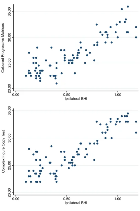
The relationships between cerebral hemodynamics and cognitive performance at baseline are displayed in Figure 1.
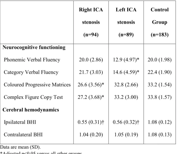
Table 1. Baseline demographic and clinical characteristics Right ICA stenosis
(n=94)
Left ICA stenosis (n=89) Demographics Age (years) Male sex Education (years) 73.2 (7.3) 64 (68.1) 8.2 (3.8) 73.0 (6.4) 58 (65.2) 8.3 (4.1) Clinical history Hypertension Diabetes mellitus Dyslipidaemia
Coronary artery disease Current smoking
Medications
Antihypertensives Antidiabetics
Lipid lowering drugs Antiplatelets 57 (60.6) 18 (19.2) 36 (38.3) 15 (16.0) 15 (16.0) 64 (68.1) 18 (19.2) 71 (75.5) 94 (100.0) 54 (60.7) 22 (24.7) 31 (34.8) 13 (14.6) 17 (19.1) 59 (66.3) 20 (22.5) 61 (68.5) 87 (97.8)
Data are mean (SD) for continuous variables, and n (%) for categorical variables. *Adjusted p<0.05 versus all other groups.
Table 2. Baseline neurocognitive functioning and cerebral hemodynamics Right ICA stenosis (n=94) Left ICA stenosis (n=89) Control Group (n=183) Neurocognitive functioning
Phonemic Verbal Fluency Category Verbal Fluency Coloured Progressive Matrices Complex Figure Copy Test
Cerebral hemodynamics Ipsilateral BHI Contralateral BHI 20.0 (2.86) 21.7 (3.03) 26.6 (3.56)* 27.2 (3.68)* 0.55 (0.31)† 1.04 (0.20) 12.9 (4.97)* 14.6 (4.59)* 32.8 (2.66) 33.2 (3.00) 0.56 (0.32)† 1.05 (0.19) 20.0 (1.98) 22.4 (1.90) 33.2 (1.54) 33.8 (1.57) 1.08 (0.12) 1.08 (0.13)
Data are mean (SD).
*Adjusted p<0.05 versus all other groups. †Adjusted p<0.05 versus control group.
Figure 1. Baseline cerebral hemodynamic and cognitive performance A. Right internal carotid artery stenosis
B. Left internal carotid artery stenosis
Point to point graphs showing the relationship between BHI values on the side of carotid stenosis and scores in tests exploring cognitive functions of the ipsilateral hemisphere. Abbreviation: BHI=breath-holding index.
At the 6-month assessment, the CVR on the side of surgery and the scores obtained in neuropsychological tests evaluating the functions of the revascularized hemisphere were significantly improved in both patient groups (Table 3). No meaningful changes were observed in cerebral hemodynamics or cognitive performance in controls.
In patients who underwent right CEA, the mean Z-score was 1.54 (1.37) for the CPM and 1.73 (1.64) for the CFCT. In patients who performed left CEA, the mean Z-score resulted 1.61 (1.43) and 1.46 (1.35) for the tests evaluating (ph) and (ca)VF.
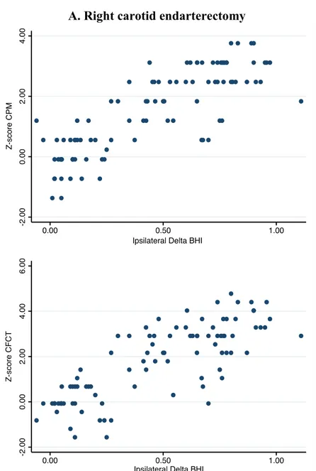
The relationships between the changes observed in CVR and neurocognitive functioning after CEA according to the side of carotid stenosis are shown in Figure 2.
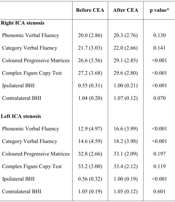
Table 3. Neuropsychological performance and cerebral hemodynamics before and after carotid endarterectomy
Before CEA After CEA p value* Right ICA stenosis
Phonemic Verbal Fluency Category Verbal Fluency
Coloured Progressive Matrices
Complex Figure Copy Test Ipsilateral BHI
Contralateral BHI Left ICA stenosis
Phonemic Verbal Fluency Category Verbal Fluency Coloured Progressive Matrices Complex Figure Copy Test Ipsilateral BHI Contralateral BHI 20.0 (2.86) 21.7 (3.03) 26.6 (3.56) 27.2 (3.68) 0.55 (0.31) 1.04 (0.20) 12.9 (4.97) 14.6 (4.59) 32.8 (2.66) 33.2 (3.00) 0.56 (0.32) 1.05 (0.19) 20.3 (2.76) 22.0 (2.66) 29.1 (2.85) 29.6 (2.80) 1.00 (0.21) 1.07 (0.12) 16.6 (3.99) 18.2 (3.98) 33.1 (2.09) 33.4 (2.12) 1.00 (0.19) 1.05 (0.12) 0.130 0.141 <0.001 <0.001 <0.001 0.070 <0.001 <0.001 0.197 0.119 <0.001 0.601
Data are mean (SD). *Paired t test.
Abbreviations: BHI=breath-holding index, CEA=carotid endarterectomy, ICA=internal carotid artery.
Figure 2. Cerebral hemodynamic and cognitive changes after surgical stenosis correction
B. Left carotid endarterectomy
Change in CVR was the difference between post- and pre-surgery BHI values. Change in each neuropsychological test was expressed by means of Z-score derived from control group’s performance change. Abbreviations: BHI=breath-holding index, (ca)VF=categorical verbal fluency, CEA=carotid endarterectomy, CFCT=Complex Figure Copy Test, CPM=Coloured Progressive Matrices, CVR=cerebral vasomotor reactivity, (ph)VF=phonemic verbal fluency.
According to multiple regression analysis, the change observed after stenosis correction in tests exploring the cognitive functions of the revascularized hemisphere was positively associated with ipsilateral δ BHI, being the increase in CVR related to improved neuropsychological performance (Table 4).
Table 4. Associations between cognitive and hemodynamic changes after carotid endarterectomy Dependent Variable Ipsilateral δ BHI Unadjusted Adjusted* β 95% CI p β 95% CI p Right ICA stenosis
CPM CFCT
Left ICA stenosis
(ph)VF (ca)VF 3.60 4.16 3.60 3.33 3.06-4.09 3.51-4.81 3.02-4.18 2.77-3.89 <0.001 <0.001 <0.001 <0.001 3.52 4.10 3.62 3.39 3.02-4.03 3.43-4.76 3.06-4.17 2.85-3.93 <0.001 <0.001 <0.001 <0.001
Values are from linear regression model with Z-score of change in neuropsychological test performance as dependent variable.
*Adjustment for age, sex, education years, hypertension and diabetes mellitus.
Abbreviations: BHI=breath-holding index, CI=confidence interval, (ca)VF=categorical verbal fluency, CFCT=Complex Figure Copy Test, CPM=Coloured Progressive Matrices, ICA=internal carotid artery, (ph)VF=phonemic verbal fluency.
The mean composite cognitive change scores were 3.26 (2.94) and 3.07 (2.73) (p=0.645) in patients who underwent right and left ICA stenosis correction, respectively. Linear regression analyses were modelled to identify the baseline predictors of the post-operative cognitive outcome (Table 5).
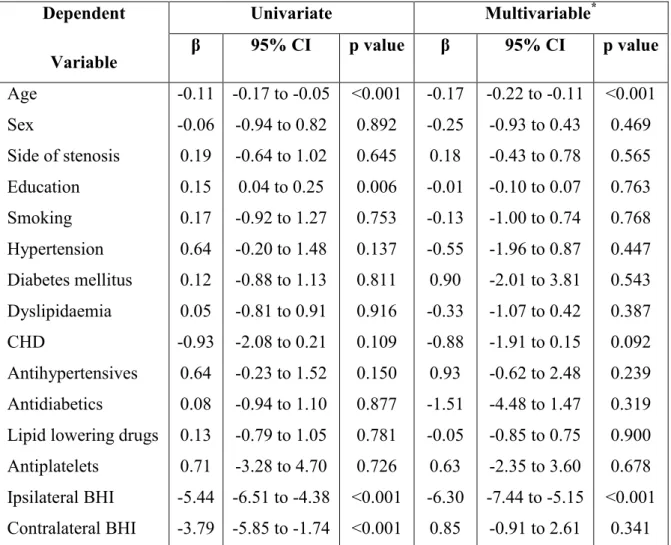
According to the multivariable model, the 6-month performance change in the neuropsychological tests exploring the re-vascularized hemisphere was inversely associated with age [ß=−0.17, 95% confidence interval (CI) -0.22 to -0.11; p<0.001] and pre-operative ipsilateral BHI (ß=−6.30, 95% CI -7.44 to -5.15; p<0.001).
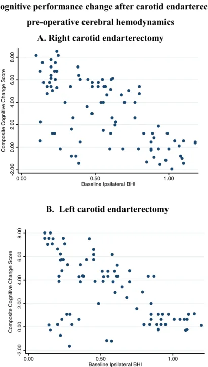
Scatterplots of the relationships between cerebral hemodynamics at baseline and cognitive performance change after CEA according to the side of carotid stenosis are shown in Figure 3.
Table 5. Associations of baseline characteristics with neuropsychological performance change at 6 months from carotid endarterectomy Dependent Variable Univariate Multivariable* β 95% CI p value β 95% CI p value Age Sex Side of stenosis Education Smoking Hypertension Diabetes mellitus Dyslipidaemia CHD Antihypertensives Antidiabetics
Lipid lowering drugs Antiplatelets Ipsilateral BHI Contralateral BHI -0.11 -0.06 0.19 0.15 0.17 0.64 0.12 0.05 -0.93 0.64 0.08 0.13 0.71 -5.44 -3.79 -0.17 to -0.05 -0.94 to 0.82 -0.64 to 1.02 0.04 to 0.25 -0.92 to 1.27 -0.20 to 1.48 -0.88 to 1.13 -0.81 to 0.91 -2.08 to 0.21 -0.23 to 1.52 -0.94 to 1.10 -0.79 to 1.05 -3.28 to 4.70 -6.51 to -4.38 -5.85 to -1.74 <0.001 0.892 0.645 0.006 0.753 0.137 0.811 0.916 0.109 0.150 0.877 0.781 0.726 <0.001 <0.001 -0.17 -0.25 0.18 -0.01 -0.13 -0.55 0.90 -0.33 -0.88 0.93 -1.51 -0.05 0.63 -6.30 0.85 -0.22 to -0.11 -0.93 to 0.43 -0.43 to 0.78 -0.10 to 0.07 -1.00 to 0.74 -1.96 to 0.87 -2.01 to 3.81 -1.07 to 0.42 -1.91 to 0.15 -0.62 to 2.48 -4.48 to 1.47 -0.85 to 0.75 -2.35 to 3.60 -7.44 to -5.15 -0.91 to 2.61 <0.001 0.469 0.565 0.763 0.768 0.447 0.543 0.387 0.092 0.239 0.319 0.900 0.678 <0.001 0.341
Values are from linear regression model with composite cognitive change scores as dependent variable.
*Fully adjusted model including all the variables considered in the univariate analysis.
Abbreviations: BHI=breath-holding index, CHD=coronary artery disease,
Figure 3. Cognitive performance change after carotid endarterectomy and pre-operative cerebral hemodynamics
A. Right carotid endarterectomy
B. Left carotid endarterectomy
Point to point graph shows the relationship between composite cognitive performance change at 6 months after surgical stenosis correction and pre-operative cerebal hemodynamics on the side of revascularization. Composite cognitive change score was obtained for each patient as the sum of the Z-scores from the neuropsychological tests
CHAPTER IV
DISCUSSION
The main findings of the current study were the relationship between the cerebral hemodynamic status and cognitive performance in patients with symptomatic unilateral high-grade ICA stenosis and the association between the increase in cerebral vasomotor repsonse and improvement in neurocognitive functioning following surgical correction of carotid stenosis.
Patients with severe ICA stenosis and history of TIA showed decreased CVR on the side of the carotid narrowing and reduced scores on the tests exploring the cognitive domains of the ipsilateral hemisphere.
The cognitive performance improved after surgical stenosis correction and the improvement was independently associated wiht the increase in cerebral vasomotor response on the side of revascularization. In addition, the patients whose cerebral hemodinamics was more compromised at baseline were those who presented the greater enhancement in neurocognitive functioning following CEA.
These findings build up the notion that cerebrovascular hemodynamic insufficiency can act as one pathogenic mechanism underpinning the cognitive impairment associated with the atherosclerotic disease of the carotid artery.14-16 In this regard, cerebrovascular resactivity can synthetize the degree of the
compensatory dilation of cerebral arterioles that occurs to preserve blood flow distally to a carotid stenosis, and represent a reliable index of cerebral hemodynamic status.17,18 Remarkably, the redcution of BHI values on the side of
ICA stenosis can reflect the degree of impairment of cerebral vasomotor reserve and identify a condition of cerebral hypo-perfusion that may be responsible for the reduction in cognitive abilities involving the function of ipsilateral brain areas.19
Surgical revascularization can enhance cerebral perfusion and restore brain autoregulation. The increase in CVR following stenosis correction can, hence, translate into the improvement of cognitive abilities that were detrimentally affected on a hemodynamic basis for the effects of the carotid stenosis. The hypothesis of reversible cognitive dysfunction is further supported by the evidence that the benefit in neurocognitive functioning was obtained at a greater extent by those patients who had a greater impairment of CVR at baseline and achieved a more pronounced enhancement in cerebral vasomotor response after CEA.
The current results can also contribute to explain the conflicting results coming from the studies that, so far, have explored the changes in cerebral hemodynamics and cognitive outcome in patients with ICA stenosis undergoing surgical stenosis correction.3 Indeed, patients with different hemodynamic status
have been enrolled and variations in cognitive tests have not been assessed according to changes occuring in brain perfusion and vasomotor reactivity after revascularization.
Some shortcomings need to be taken into account when interpreting the current findings, like the observational, single-center, non-randomized and placebo-uncontrolled nature of the study. In addition, data about brain atrophy and burden of white matter lesions have not been sistematically collected and their contribute to the cognitive status and hemodynamic changes have not been addressed.
The key strenghts of the study included the prospective design and the sound methodology, including the estimate of Z scores and the domain-specific assessement of cognitive performance.
The reference to a control group and the 6-month time frame between the evaluations allowed to minimize the learning and practical effects and provide a reliable mesure of the hemodynamic and cognitive changes observed in patients due to carotid revascularization.20,21 The selective inclusion of patients with history of TIA avoided the potential confounding effects of acute cerebral damage and post-stroke plasticity.
4.1 Conclusion
Cerebral hemodynamics can contribute to cognitive impairment in patients with symptomatic high-grade ICA stenosis and CEA can be associated with
significant improvement in neurocognitive functioning by the enhancement of cerebral blood flow and hemodynamic reserve.
Carotid stenosis correction may offer more than the prophylaxis of acute cerebal ischemic events and result into cognitive improvement in patients whose
neuropsychological performance is negatively affected by the redcution in cerebral vasoreactivity.
Further investigations are warranted to investigate whether these findings can be extended to patients with ICA stenosis and no history of cerebral and/or ocular ischemic events referable to the carotid stenosis itself. This would allow to refine the definition of “asymptomatic” carotid artery disease and criteria for surgical revascularization, and pre-operatively identify the patients with the greater likelihood to gain a benefit in their cognitive performance.
REFERENCES
1. Abbott AL, Paraskevas KI, Kakkos SK, Golledge J, Eckstein HH, Diaz-Sandoval LJ, Cao L, Fu Q, Wijeratne T, Leung TW, Montero-Baker M, Lee BC, Pircher S, Bosch M, Dennekamp M, Ringleb P. Systematic Review of Guidelines for the Management of Asymptomatic and Symptomatic Carotid Stenosis. Stroke. 2015;46:3288-3301.
2. Kernan WN, Ovbiagele B, Black HR, Bravata DM, Chimowitz MI, Ezekowitz MD, Fang MC, Fisher M, Furie KL, Heck DV, Johnston SC, Kasner SE, Kittner SJ, Mitchell PH, Rich MW, Richardson D, Schwamm LH, Wilson JA; American Heart Association Stroke Council, Council on Cardiovascular and Stroke Nursing, Council on Clinical Cardiology, and Council on Peripheral Vascular Disease. Guidelines for the prevention of stroke in patients with stroke and transient ischemic attack: a guideline for healthcare professionals from the American Heart Association/American Stroke Association. Stroke. 2014;45:2160-236.
3. Plessers M, Van Herzeele I, Vermassen F, Vingerhoets G. Neurocognitive Functioning after Carotid Revascularization: A Systematic Review. Cerebrovasc Dis Extra. 2014;4:132-148.
4. North American Symptomatic Carotid Endarterectomy Trial Collaborators. Beneficial effect of carotid endarterectomy in symptomatic patients with high-grade carotid stenosis. N Engl J Med. 1991;325:445-453.
5. Oldfield RC. The assessment and analysis of handedness: the Edinburgh inventory. Neuropsychologia. 1971;9:97-113.
6. Markus HS, Harrison MJ. Estimation of cerebrovascular reactivity using transcranial Doppler, including the use of breath-holding as the vasodilatory stimulus. Stroke. 1992;23:668-673.
7. Raven JC. Mental Tests Used in Genetic Studies: The Performance of Related Individuals on Tests Mainly Educative and Mainly Reproductive [MSc Thesis]. University of London, London, 1936.
8. Osterrieth PA. Le test de copie d'une figure complexe: contribution a l'e'tude de la perception et de la memoire. Arch. Psychol. 1944;30:286-350.
9. Lezak MD, Howieson DB, Bigler ED, Lezak DT. Neuropsychological Assessment. Oxford University Press, New York, 1995.
10. Kastner S, Pinsk MA, De Weerd P, Desimone R, Ungerleider LG. Increased activity in human visual cortex during directed attention in the absence of visual stimulation. Neuron. 1999;22:751-761.
11. Corbetta M, Kincade MJ, Lewis C, Snyder AZ, Sapir A. Neural basis and recovery of spatial attention deficits in spatial neglect. Nat Neurosci. 2005;8:1603-1610.
12. Binder JR, Rao SM, Hammeke TA, Frost JA, Bandettini PA, Jesmanowicz A, Hyde JS. Lateralized human brain language systems demonstrated by task subtraction functional magnetic resonance imaging. Arch Neurol. 1995;52:593-601.
13. Stroobant N, Vingerhoets G. Transcranial Doppler ultrasonography monitoring of cerebral hemodynamics during performance of cognitive tasks: a review. Neuropsychol Rev. 2000;10:213-231.
14. Marshall RS, Festa JR, Cheung YK, Chen R, Pavol MA, Derdeyn CP, Clarke WR, Videen TO, Grubb RL, Adams HP, Powers WJ, Lazar RM. Cerebral hemodynamics and cognitive impairment: baseline data from the RECON trial. Neurology. 2012;78:250-255.
15. Silvestrini M, Paolino I, Vernieri F, Pedone C, Baruffaldi R, Gobbi B, Cagnetti C, Provinciali L, Bartolini M. Cerebral hemodynamics and
cognitive performance in patients with asymptomatic carotid stenosis. Neurology. 2009;72:1062-1068.
16. Buratti L, Balucani C, Viticchi G, Falsetti L, Altamura C, Avitabile E, Provinciali L, Vernieri F, Silvestrini M. Cognitive deterioration in bilateral asymptomatic severe carotid stenosis. Stroke. 2014;45:2072-2077.
17. Alexandrov AV, Sloan MA, Tegeler CH, Newell DN, Lumsden A, Garami Z, Levy CR, Wong LK, Douville C, Kaps M, Tsivgoulis G; American Society of Neuroimaging Practice Guidelines Committee, Practice standards for Transcranial Doppler (TCD) ultrasound: part II: clinical indications and expected outcomes. J Neuroimaging. 2012;22:215-224.
18. Kargiotis O, Safouris A, Magoufis G, Georgala M, Roussopoulou A, Stamboulis E, Moulakakis KG, Lazaris A, Geroulakos G, Vasdekis S, Tsivgoulis G. The role of neurosonology in the diagnosis and management of patients with carotid artery disease: a review. J Neuroimaging. 2018;28:239-251.
19. Balucani C, Silvestrini M. Carotid atherosclerotic disease and cognitive function: mechanisms identifying new therapeutic targets. Int J Stroke. 2011;6:368-369.
20. Siddiqui AH, Hopkins LN. Asymptomatic carotid stenosis: the not-so-silent disease changing perspectives from thromboembolism to cognition. J Am Coll Cardiol. 2013;61:2510-2513.
21. Huang CC, Chen YH, Lin MS, Lin CH, Li HY, Chiu MJ, Chao CC, Wu YW, Chen YF, Lee JK, Wang MJ, Chen MF, Kao HL. Association of the recovery of objective abnormal cerebral perfusion with neurocognitive improvement after carotid revascularization. J Am Coll Cardiol. 2013;61:2503-2509.