Amber imitation? Two unusual cases of Pinus
resin-coated beads in Iberian Late Prehistory
(3
rd
and 2
nd
millennia BC)
Carlos P. OdriozolaID1*, Jose´ A´ ngel Garrido Cordero1, Joan Daura2, Montserrat Sanz2, Jose´ Marı´a Martı´nez-Blanes3, Miguel A´ ngel Avile´s3
1 Departamento de Prehistoria y Arqueologı´a, Universidad de Sevilla, C/ Marı´a de Padilla S/N, Sevilla, Spain, 2 Grup de Recerca del Quaternari (GRQ-SERP), Departament d’Història i Arqueologia, Universitat de Barcelona, Carrer Montalegre, 6, Barcelona, Spain, 3 Instituto de Ciencia de Materiales de Sevilla, Centro mixto CSIC-Universidad de Sevilla, Sevilla, Spain
Abstract
A group of beads from the artificial cave of La Molina (Lora de Estepa, Sevilla) and Cova del Gegant (Sitges, Barcelona) were made from a biogenic raw material and intentionally cov-ered by a layer of resin. This is the first time this type of treatment has been documented on elements of adornment in the Late Prehistory of the Iberian Peninsula. The composition and nature of the coatings are analysed and the symbolic role of such alterations and imitations of prehistoric adornments is discussed.
Introduction
Since the Upper Palaeolithic, the unique translucent reddish-yellow colour of amber has made this resinite sought after for personal ornamentation, e.g. beads, pendants, charms. . . [1], con-ferring on an important symbolic value in European Prehistory. The allure and rarity of amber triggered the exchange and use of this resinite, but also the development of imitations by the use of other local translucent minerals [2] or the application of coatings, as described by this paper, to reproduce the colour of amber.
The utmost importance to prehistoric societies of colour categories and their impact to show, emphasize and materialize codes, metaphors and narratives has been highlighted by sev-eral authors [3–10]. Although altering the genuine colour of raw materials or products by means of technically controlled processes to obtain a desired effect seems to have been a usual practice in past societies, this technical behaviour has been occasionally recorded and studied. Colour modification practices in personal ornaments goes back to the Upper Palaeolithic, when in Franchthi Cave (Greece) perforated shell beads were thermally altered to obtain black hues [11]. A similar intentional effect has been also recorded in discoidal beads made from shells at the Chalcolithic site of Perdigões (Portugal) [12]. Colour modification by heat has also been recorded in carnelian quartz beads in the Varna necropolis (Bulgaria), creating darker hues that were complemented with a complex faceting technique to enhance surface gloss [13]. However, during the Neolithic much more complex processes of thermal colour a1111111111 a1111111111 a1111111111 a1111111111 a1111111111 OPEN ACCESS
Citation: Odriozola CP, Garrido Cordero JA´, Daura J, Sanz M, Martı´nez-Blanes JM, Avile´s MA´ (2019) Amber imitation? Two unusual cases of Pinus resin-coated beads in Iberian Late Prehistory (3rd and 2ndmillennia BC). PLoS ONE 14(5): e0215469. https://doi.org/10.1371/journal.pone.0215469
Editor: Peter F. Biehl, University at Buffalo - The State University of New York, UNITED STATES Received: October 31, 2018
Accepted: April 2, 2019 Published: May 3, 2019
Copyright:© 2019 Odriozola et al. This is an open access article distributed under the terms of the
Creative Commons Attribution License, which permits unrestricted use, distribution, and reproduction in any medium, provided the original author and source are credited.
Data Availability Statement: All relevant data are within the paper.
Funding: This research was funded by the MINECO/AEI/FEDER -EU under contract HAR2012-34620 and HAR2017-83474-P. Jose´ A´ngel Garrido Cordero acknowledges the University of Seville for a PhD grant under the V Plan Propio de Investigacio´n de la Universidad de Sevilla. Montserrat Sanz acknowledges the program Juan de la Cierva for a postdoctoral grant (FJCI-2014-21386). Daura holds a postdoctoral grant (SFRH/
Estepa, Sevilla, 3 millennium BC) and Cova del Gegant (Sitges, Barcelona, 2 millennium BC) (Fig 1). In all the case studies, biogenic beads have been coated with a resinite that resem-bles the appearance of amber beads. These amber resembling beads and their coating tech-nique are currently the only cases known for European Late Prehistory.
First, the nature and technology of the bead cores and coatings are characterised by means of electron microprobe (SEM-EDX), infrared spectroscopy (FTIR), x-ray diffraction (XRD) and Raman spectroscopy. Then the role of raw materials as exotica is discussed, considering the importance of imitations in cases where procurement of highly appreciated raw material is disrupted by political or economic factors.
Case studies
La Molina (Lora de Estepa, Sevilla)
The artificial cave CE17 (Fig 2) at La Molina was discovered in 2008, next to another hypogean burial that was partially destroyed (CE16) and a series of negative structures in the form of silos [17]. The structure consists of a small descending corridor 1.5m long with two steps, lead-ing to the entry of an oval-shaped chamber with a maximum diameter of 3.5m [18]. The mor-tuary space, which was divided into two areas that were differentiated both spatially and in the funerary treatment, contained a minimum number of ten individuals [18]. The grave goods consisted of several pottery vessels, bone awls and singular objects in ivory, as well as abundant chipped and polished lithic artefacts, particularly two arrowheads in rock crystal [19]. The excavators dated this mortuary site in the early third millennium BC by material and contex-tual comparisons after absolute dating failure [18].
Two beads with a more or less discoidal shape (Fig 5.23 in [19] were found in association with the mortuary layer of stones called Structure III (SU 106a) [18]. This part of the chamber (Zone 1) contained practically all the cinnabar in the tomb, where it was found on all the indi-viduals (MNI = 3), compared with Zone 2, where it was only occasionally found. It also con-tained most of the ivory in the tomb [19], attesting the differential and special treatment of the individuals buried in this part of the chamber in comparison with those in Zone 2. The indi-viduals (N1, an adult male in anatomical connection; E1, a young adult female; and E4, an adult male) were deposited in the Zone 1 near the chamber wall on top of some prominent stones [18]. Only E1 possessed grave goods unmistakably placed near her head, including a carved elephant’s tusk, a flint knife with ivory handle, and other ivory and lithic artefacts, which indicate the prestige of the inhumation.
Cova del Gegant (Sitges, Barcelona)
Cova del Gegant (Fig 3) is a cave located in the NE part of the Iberian Peninsula, ~40 km south of Barcelona (1˚ 46’ 27.33”E, 41˚ 13’ 24.75”N, 0m abmsl). It consists of a main chamber (GP), now eroded by wave action, and its inner part (GP2), where a small passage (GLT) leads to the adjacent Cova Llarga. Two galleries branch off on the right side of GP, one towards the interior
(GL2) and the other nearer to the sea (GL1). The original entrance is partially flooded and faces the sea.
At least eight site formation episodes from the Upper Pleistocene to the Holocene have been recorded in the Cova del Gegant stratigraphic sequence [21]. As regards the Holocene layer, Episode 4 is the oldest (Bronze Age), represented by Levels VI and Ic2 (Sectors GP and GL2) and Layer XXV (Sector GP2) with which the beads are associated (Fig 3,Table 1). Two additional pit-storages (Silo-1 and Silo 2) have been documented in the same formation epi-sode [21].
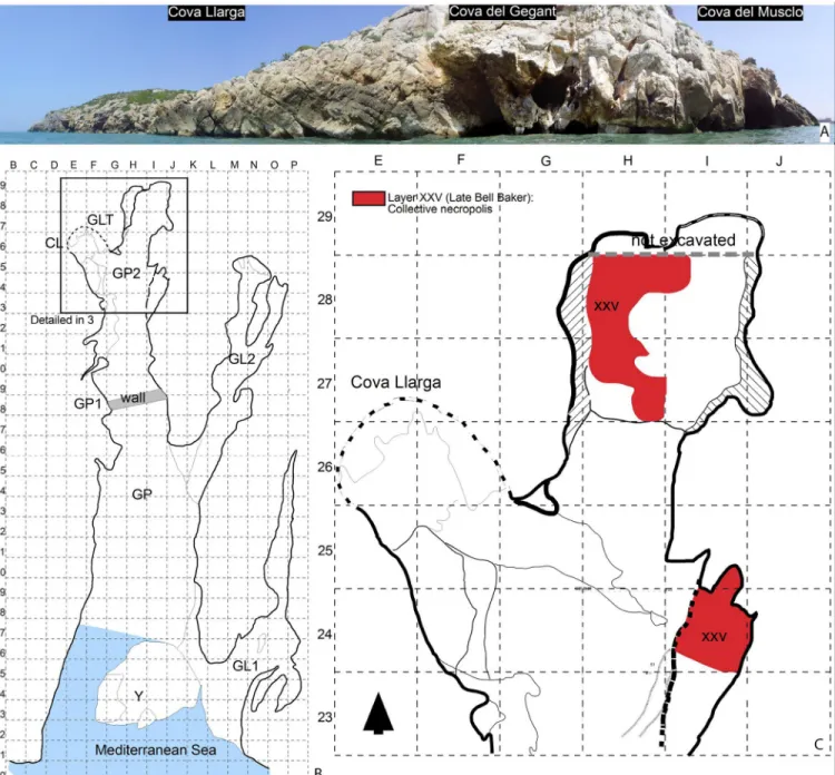
The archaeological layer ascribed to the Bronze Age (XXV), mainly preserved at the back of the main gallery (GP2), corresponds to the grid delimited area between squares H27 and H28 (~1.8 m2) [20]. To the south, archaeological layer XXV is not preserved due to the ongoing marine erosion. This layer corresponds to a collective burial accumulation radiocarbon dated to the Middle Bronze Age, 1600–1400 BC. Among the archaeological remains, the bulk of the assemblage is formed by 1,728 human bones ascribed to a Minimum Number of Individuals (here after MNI) of 19 disarticulated individuals. The artefacts accompanying the recovered human remains comprise Late Bell Beaker pottery (NR = 71) generally ascribed to the Early Bronze Age. Body ornaments are scarce and consist of beads manufactured from a variety of raw materials. For example, three of them are made of lignite, two of amber [2], one of coral and one of a shell fragment (Cypraea). Four beads were manufactured from shell and covered by a resinite; these are the pieces analysed in the present study (Table 1). The Sicilian origin of Fig 1. Location of the studied sites.
the amber materials evidences that the area participated in long-distance exchange networks [2]. Additionally, the two gold artefacts (tutuli) recovered from the same layer are very rare ornaments in the SW of Europe and reinforce the idea that the site formed part of an exchange network along the Mediterranean shore of the Iberian Peninsula [20].
Materials & methods
Administrative permits to study La Molina beads were granted to Carlos P. Odriozola and Jose´ A´ ngel Garrido Cordero by the Junta de Andalucı´a Delegacio´n territorial de Educacio´n Cultura y Deporte, Museo Arqueolo´gico de Sevilla (DCI/ES/2014/19). The ID numbers inTable 1are those used by the Museo Arqueolo´gico de Sevilla (Sevilla, Spain) to label the material. Archae-ological excavations and beads sampling from Cova del Gegant are approved by the project “Els canvis climàtics durant el plistocè superior a la costa central catalana i l’impacte en les poblacions neandertals i humans anatòmicament moderns” (CLT009/18/00022, Generalitat de Catalunya). The ID numbers for Cova del Gegant materials inTable 1are those given by the site inventory number. These materials are housed at the University of Barcelona.
Infrared spectroscopy (FTIR), Scanning Electron Microscope Microprobe (SEM-EMP), x-ray diffraction (XRD) and confocal dispersiveμ-Raman spectroscopy (DcμRS) were used to investigate the nature and technology of the coated beads from both sites (Table 1,Fig 4). These instrumental techniques are able to obtain full details of the chemical composition and structure of both the bead core and the coating, with the double aim of understanding their nature and technology.
Infrared spectroscopy is capable of satisfactorily identifying resinites, their composition and botanical origin, to classify them and can be performed either non-destructively, by means infrared microscopy (μ-FTIR), or destructively on a very small sample of no more than 1 mg. It has become the standard technique applied in archaeological research to determine Fig 2. A. general plan of the excavated area at La Molina (After Fig 4.2 in [17]). B. Human remains of La Molina´s artificial cave CE17. Indicated by circle/ ellipsis and labels are bone assemblages and individuals (After Figs 4.3 to 4.6 in [17]).
Fig 3. A. Site plan of the Cova del Gegant indicating the position of the different galleries mentioned in the text. B. Location of the excavated area. C. Stratigraphy (section CC’ as detailed in B) (After Fig 1 in [20].
https://doi.org/10.1371/journal.pone.0215469.g003
Table 1. Brief description of the beads studied in this paper (H: height, W: width, P: perforation diameter, Wg.: weight).
# ID Site Core H W P Wg. Color Chronology Arch. context
4316 Cova del Gegant Shell 2.07 8.04 2.62 0.22 Reddish yellow Early 2ndmillennium BC Natural cave, col. burial
4472 Cova del Gegant Shell 2.11 8.12 2.61 0.21 Reddish yellow Early 2ndmillennium BC Natural cave, col. burial
4473 Cova del Gegant Shell 1.93 9.52 2.63 0.12 Reddish yellow Early 2ndmillennium BC Natural cave, col. burial 4476 Cova del Gegant Shell 2.16 6.20 2.46 0.11 Reddish yellow Early 2ndmillennium BC Natural cave, col. burial
M-89 La Molina ¿? 5.55 7.82 2.41 0.16 Red Early 3rdmillennium BC Artificial cave, col. burial
M-90 La Molina ¿? 3.30 13.63 2.43 0.22 Red Early 3rdmillennium BC Artificial cave, col. burial https://doi.org/10.1371/journal.pone.0215469.t001
the nature and origin of resinites. We have therefore followed this well-known methodology [22] to study the six amber-like (resinite) coated beads.
Approximately 1mg of sample was ground by hand using an agate mortar and mixed with a small amount of KBr, before pressing (8 T) the mixture to produce discs 1mm thick. The spec-imens were analysed using a JASCO FT/IR-6200 spectrometer. The data were collected as infrared transmission spectra after scanning each specimen 32 times in the range 4000–400 cm−1, with a resolution of 4 cm−1.
X-ray diffraction is an inexpensive high-resolution technique which provides information about the mineralogical composition. It has been used to identify the pigments the coatings are composed of. After the baseline calculation with the X’Pert Highscore Plus 3.0 software, and when all the peaks in the diagram have been identified, the numerical values obtained were compared with the 2004 ICDD (International Centre for Diffraction Data) PDF (Powder Diffraction File) database, with the aim of identifying the minerals that the sample is composed of.
A Panalytical X’Pert Proθ/θ X-ray diffraction equipment with Cu Kα (1.5406 Å) radiation operated at 45 kV and 40 mA, equipped with a PixCel detector and parabolic incident beam mirrors was used. The diagrams were acquired with a step of 0.026˚ 2θ between 10˚ and 90˚ 2θ with an acquisition time of 247 s per step at room temperature (25˚C).
Confocal dispersiveμ-Raman spectroscopy, also a non-destructive technique, is used to identify solids through vibrations in the crystalline lattice, as it can detect the sample composi-tion, bonds, coordination environment and crystalline structure [23–24].
The data was obtained with a HORIBA Jobin Yvon LabRAM HR dispersive confocal μ-Raman spectrometer system. The laser diode, when operated at a wavelength of 532.06 nm, produces up to 15 mW of power in the source. Filters to reduce the laser’s power were not used. The acquisition time was 32 s per acquisition, up to a maximum of 20. The spectral mea-surement range chosen was between 100 and 1800 cm-1using a 100x lens with a CCD multi-Fig 4. Samples studied in this paper.
channel detector. The selected measurement is accurate to 1 cm-1. The measurement area selected was 1000μm in diameter.
A Hitachi S4800 high resolution (1–3 nm) field emission scanning electron microscope, equipped with a 1.33 eV resolution Bruker X Flash 4010 EDX detector equipment was used for the composition analysis.
Additionally, routine close-up inspection of the bead coatings with a Nikon Shuttlepix P-4000R digital microscope (up to x80 magnification) was performed to gain full image details of the bead coatings.
This analytical study was conducted jointly at the Institute for Material Science in Seville (CSIC–University of Seville) and the Prehistory and Archaeology Department (University of Seville) laboratories.
Results
Cova del Gegant
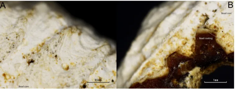
Taking advantage of the cracked coating that leaves the beads’ white core exposed, SEM-EMP analysis was performed on both the surface of the white core and the surface of the reddish-yellow coating. The elemental analysis revealed that the core of the beads is mainly composed of calcium (Fig 5A). While when performed on the bead coating surface, it revealed a mixture of calcium and phosphorus (Fig 5B).
Both, the radial ribs of a mollusc valve (4474) shown inFig 6, and the chemical composition of the bead core, points to the use of a valve for the bead making. Most mollusc shells are made of aragonite, a type of calcium carbonate [CaCO3].
FTIR microscopy of the surface of the white core material showed, for specimens 4472 and 4316, the presence of a band centred atc. 1087 cm-1and a band atc. 1488 cm-1assigned to the υ1andυ4symmetric stretching modes of the aragonite [25] (Fig 7A). The band atc. 1087 cm-1
is active only in aragonite and is not active in calcite; it has therefore been used to identify the nature of the white calcium carbonate core. The presence of these aragonite assigned bands suggests that a mollusc was used to produce the core of the coated beads from Cova del Gegant.
FTIR microscopy of the surface of the reddish-yellow coating (Fig 7B) yielded similar spec-tra to those for the white core of the beads.μ-FTIR identified υ3andυ1hydroxyapatite
vibra-tion modes [26], together withυ1symmetric stretching mode of the aragonite [25], and
calcite’sυ3,υ2andυ1vibration modes [25] overlapped by other signals that remain
unassigned.
FTIR band assignment suggests that the white outer layer is most likely the result of tapho-nomic processes. The basic pH of the cave sediment,c. 8.4 [2], has dissolved the hard bone tis-sue of the buried humans which have subsequently precipitated as some sort of apatitic calcium phosphate [27] together with calcite on the beads’ surface. This taphonomic process is clearly observed onFig 8where a white layer (calcium and phosphate layer) overlays a reddish one. Indeed, it can be seen inFig 8Dthat calcite coming from the cave has precipitated on the top of every layer.
This means that the outermost layer of the coating is the result of taphonomic process occurred since burial in the sediment and this is not an intentional coating layer. FTIR micros-copy of the surface of this white core material showed additional bands to theυ1symmetric
stretching mode of the aragonite (c. 1087 cm-1) atc. 881, 1432 and 712 cm-1that can be assigned to the vibrational active modes of calcite [25] (Fig 7B). This fully supports the above stated precipitation of calcite on the surface of the beads from Cova del Gegant.
However, the band assignment ofμ-FTIR spectra collected on the bead surface coating and white core (Fig 7) proved challenging because spectra were poorly defined and too noisy. Although the appearance of the reddish yellow coating under microscope magnification resembles that of an amber,μ-FTIR spectra of the coating surface do not show the chara-cteristic Baltic shoulder (succinite) nor the charachara-cteristic bands of Sicilian amber (simetite), Cretaceous Iberian amber, or any fossil resins archaeologically recorded. Thus, the signal com-parison of the recorded spectra to natural resin reference spectra failed to give a positive
Fig 6. Detailed microphotographs of bead 4474 in a region where the coating is lost. A) Detail of the radial ribs of a mollusc valve. B) Detail of the conserved coating.
https://doi.org/10.1371/journal.pone.0215469.g006
Fig 5. Cova del Gegant beads 4472 and 4476 SEM-EMP analysis of the bead core (A) and superficial coating (B).
match. However, it was quite clear under microscope magnification that the reddish-yellow coating was made of some sort of surface oxidized resin.
Natural resins and their type can be identified by their infrared spectral features. The diag-nostic IR absorption bands for natural resins, as described by Table 1 in [28], are bands atc. Fig 7. Micro FTIR spectra of (A) white core surface. (B) coating surface.
https://doi.org/10.1371/journal.pone.0215469.g007
Fig 8. (A-C) detailed imaged of the 4472 bead coating. A white layer can be observed over a reddish-yellow one. (A) Zenithal view of the bead surface. (B) Detailed view of the surface coating. (C) Section view of the coating. (D-F) Detailed imaged of the 4474 bead coating. A white layer can be observed over a reddish-yellow one. (D) Zenithal view of the bead surface. (E) Detailed view of the reddish amber-like layer. (F) Section view of the coating.
1695, 1448–1467, 1382–1387, 1178–1184, 1078–1092, 1028–1038 and 887–897 cm-1. These may be used to distinguish tree resins from shellac. All of these bands although present are hardly appreciated inFig 7.
Natural terpenous resins. Apart from shellac, which is of insect origin, natural resins or
terpenoids [according to [29] the term terpenoid is now preferred as the generic name for nat-ural resins] exuded natnat-urally from certain trees as viscous liquids which subsequently harden by evaporation of volatiles or the partial oxidative polymerization of some components [30] (Mills and White 1977, p. 13). Diterpenoids comprise the bulk composition of resins from the families Coniferae (Pinaceae, Cupressaceae, and Araucariaceae) and Leguminosae. The most abundant source of terpenous resins are trees of the genusPinus.
Diterpenoid resins possess mainly abietane, pimarane, and labdane skeletons. In non-poly-merized structures abietane and pimarane compounds are predominant. The Pinaceae, and especially the genusPinus, have resins with a high content of abietic acid (Fig 9), and a small number of abietane isomers [30], but resins fromAbies and Picea species also contain large amounts of labdanes, as do Cupressaceae resins [29].
Terpenous resins are a complex mixture of molecules that changes considerably with time due to their high susceptibility to oxidation or polymerization. Their identification has the rep-utation of being a complex analytical task due to the broad range of oxidation products result-ing from the oxidative ageresult-ing process observed [29–33]. In contrast, the first molecular changes to natural resins can be observed a few weeks after collection [32].
Terpenoids are susceptible to a number of alterations mediated by oxidation and reduction reactions as well as changes brought about by microbial activity [29]. For example, the ageing of diterpenoid resins at molecular level is predominantly the result of the oxidation / dehydro-genation of abietanes. Non-oxidized abietanes quickly convert to abietic acid (Fig 9A) and that to dehydroabietic acid (Fig 9B). Further oxidation leads to the formation of 7-oxodehydroabie-tic acid (Fig 9C) and 15-hydroxy-7-oxodehydroabietic acid (Fig 9D). Intermolecular reactions occurring between two carboxylic acid groups from abietanes or pimaranes give rise to the for-mation of acid anhydrides [34]. Although pimaranes are more stable, some studies also revealed their diminution during ageing [30,32–36].
Besides, many studies of Pinaceae resin [this genus includes many species (Pinus, Larix and Abies), mainly trees, found mostly in the northern hemisphere. One of the features of Pinus species is that they exudate a terpenous resinous secretion [32]] made by FTIR Spectroscopy and Raman Spectroscopy indicate that resins of different species show spectra directly related to the precise amount of abietanes and a smaller portion of pimaranes [28,32]. The dehydro-genation process in terpenous natural resins as well as the effects of soil pH after deposition Fig 9. (A) Abietic acid structure. (B) Dehydroabietic acid structure. (C) 7-Oxodehydroabietic acid structure. (D) 15Hydroxy-7oxo-Dehydroabietic acid structure.
must be considered when trying to identify the nature of the resin used for coating the beads. This requires quality spectra where the overlaps due to compound mixture other than those naturally occurring in resins are absent. Because of this, we decided to take a micro sample of the reddish-yellow coating to record spectra in FTIR transmission mode (KBr pellet) in the hope of acquiring spectra of the coating material not mixed with carbonates and phosphates recorded on bead surface.
Spectra discussion. The transmission spectra for specimen 4476 seem to be polluted by
the post-depositional phosphate precipitation. Fortunately, that helped to clearly identifyυ1
hydroxyapatite vibrational modes atc. 564 and 602 cm-1, in part due to the 200 cm-1broader spectral range scanned by the DTGS detector of the FTIR transmission mode compared to the nitrogen-cooled MCT detector of the FTIR microscope. This supports the above-described post-depositional precipitation of apatitic calcium phosphate. In addition, the terpenous resin diagnostic region, between 1500 and 700 cm-1, is dominated by the presence of an intense and broad band where C-C skeletal vibrations of the abietanes (abietic/dehydroabietic acid) are overlapped by theυ3PO4vibrations of the post-depositional phosphates (Fig 10). However,
the transmission mode recorded spectra showed better defined and less noisy bands, which clearly improved band assignment in the diagnostic region where most of the molecular bond vibrations occur. The presence of C-C skeletal vibrations of abietic/dehydroabietic acid, the most abundant molecules in aged resins [30,35], points to the use of a natural terpenous resin to coat the bead. That is, the coating is made out of a tree resin that has undergone molecular transformation over time.
Fig 10shows the FTIR spectra in the diagnostic region (1500–700 cm-1). In this figure the second derivative spectrum is plotted against the recorded spectra and reference spectra from [32] of differentPinus species resins and a 14th
centuryPinus resin used as a painting varnish. The second derivative spectrum identified bands and molecular structure diagnostic bands are indicated over the samples’ recorded spectra. Literature identified bands forPinus halepensis are indicated on the y upper axis. Band assignment to specific molecular structures was based primarily on the available literature about aged resins [32–33,36,39]. It is possible to observe a fairly good match between thePinus halepensis fingerprint, the 14thcenturyPinus resin and the recorded spectra, especially that of Sample 4474. However, some differences between the references and the recorded spectra can be appreciated (Fig 10).
According to [28,32,34] it is possible to first identify the resin class (e.g., tree, insect, wax) by their characteristic functional group vibration and second to identify the type of resin (Pinacae, Cupressaceae, Araucariaceae, Anacardiaceae. . .) by the amount of abietanes and of pimaranes in the spectra.
Here, the assignment of bands to specific molecular structures for resin type identification was based primarily on available literature on fresh and aged natural terpenous resins (see
Table 2). However, although the match of at least half of the bands with the reference material is a strong indication of the type of resin, visual comparison of the sample spectrum with that of the reference material is mandatory to determine resin type [28].
In the untransformed recorded transmittance spectra, band positions are difficult to iden-tify due to occurring overlaps. Therefore, spectra second gap derivatives were used for band recognition using the gap derivative tool of R‘s prospect package [37] and a custom built mini-mum/maxima identification function.
The main compounds observed for Samples 4474 and 4476, in addition to PO4for
speci-men 4476, are oxidized abietanes. Whereas the main spectral features identified below 1500 cm-1are due to C-H, C-O and C-C wags and bends of the abietanes aging products, for exam-ple, hydroabietic acid and 15-hydroxy-7-oxo-dehydroabietic acid. However, other compounds in minor amounts are also present in the recorded spectra. Bands atc. 1182 and 1020 cm−1are
Fig 10. FTIR spectra in the diagnostic region (1500–700 cm-1). A) Second gap derivative of the spectra 4474 and 4476. B) 4474 and 4476 spectra, bands identified with the custom-built minimum/maxima identification function applied to the second gap derivative are labelled. C) Reference spectra discussed in the text are plotted, labels correspond to band position identified for 4474 and 4476 spectra.
Table 2. FTIR band assignments for Cova del Gegant samples. Band
(cm-1)
Band assignments Reference
3400 O–H stretching, increases with aging [32]
2936 C-H stretching [28]
2929 C-H stretchingPinus pinea, Pinus halepensis ν(CH2),ν(CH3) in abietic acid
2869 C-H stretchingPinus pinea, Pinus halepensis ν(CH2),ν(CH3) in abietic acid [28,32,38]
2652 C-H stretching oxidized [32]
2534 C-H stretching oxidized [28,32]
1823 ν(C = O) of abietic acid. This band is assigned to unsaturated ketones, which form as primary oxidation products by the decay of hydrogen peroxides.
[33]
1800sh Acid anhydride groups formed by the reaction of two carboxylic acid groups from different molecules (abietanes or pimaranes)
[32]
1739 Acid anhydride groups formed by the reaction of two carboxylic acid groups from different molecules (abietanes or pimaranes)
[32]
1725sh C = O stretching of the ketone group of the 7-oxodehydroabietic and 15-hydroxy-7oxo-dehydroabietic acids
[32]
1713 Pinus halepensis ν(C = O) of abietic acid [39]
1695sh C = O stretching (COOH) dehydroabietic acid [28]
1690 C = O stretching group isopimaric acid [34]
1685 ν(C = O) of abietic acid. This band is assigned to unsaturated ketones, which form as primary oxidation products by the decay of hydrogen peroxides.
[33]
1664 Pinus halepensis ν(C = O) of abietic acid [32,38]
1645 C = C stretching of isopimaric acid (Beltran et al. 2017),Pinus halepensis abietic acid ν
(C = C) trans conjugated (Brody et al. 2002)
[32,38]
1612 Pinus halepensis abietic acid ν(C = C) aromatic [32,38]
1607w C = C stretching [32]
1575 C = C stretching (ring) 15-hydroxy-7-oxodehydroabietic acid [34]
1514 C = C stretching [32]
1500 Abietic acidν(C = C) aromatic [32]
1469 C-H deformation bending dehydroabietic acid [34],Pinus halepensis abietic acid δ(CH2),δ
(CH3) [38]
[34,38]
1449 Pinus halepensis C-H deformation bending (ring) COOH [34]
1415– 1411
Pinus halepensis abietic acid δ(CH2),δ(CH3)
1383 C-H bending [28]
1337 Pinus halepensis abietic acid δ(CH2),δ(CH3) [38]
1303 Pinus halepensis abietic acid δ(CH2),δ(CH3) twisting [38]
1281 Pinus halepensis abietic acid δ(CH2),δ(CH3) twisting [38], Coupled C-O/O-H deformation
[34]
[34,38]
1260 C-C-O stretching (-OH) [34]
1241 C-C-O stretching (-OH)
O–H deformation in–COOH associated with 15-hydroxy-7-oxo-dehydroabietic acid (increase intensity with aging)
[34]
1191– 1196
15-hydroxy-7-oxodehydroabietic acid skeletal vibrations [34]
1187– 1192
15-hydroxy-7-oxodehydroabietic acid skeletal vibrations [34],Pinus halepensis abietic acid
ν(CC) ring breathing [38]
[32,34,38]
1152 Acid anhydride groups formed by the reaction of two carboxylic acid groups from different molecules (abietanes or pimaranes)
[32,34]
1138 Pinus halepensis abietic acid ν(CC) ring breathing [38]
1130 Pinus halepensis abietic acid ν(CC) ring breathing [28]
1020 Related to common compounds [32,34]
probably related to common compounds, and the 1020 cm−1band is proposed as a pimarane marker (CH2= CH wagging) [32].
The strongest absorptions in specimen 4474 between 1065 and 963 cm-1are related to the skeletal vibrations of oxidized abietanes: abietic, dehydroabietic acid, 15-hydroxy-7-oxodehy-droabietic acid. . . However, strong absorbance bands assigned to C-H bending modes occur atc. 1469, 1649, 1383 and 1028 cm-1[28,32,34], and moderate to weak absorbance at 1281 and 1245 cm-1, are assigned to the coupled C-O/O-H deformation associated with the com-mon oxidized abietane structure [34]. The weak bands atc. 1191–1196, 887 and 718 cm-1are assigned to 15-hydroxy-7- oxodehydroabietic acid skeletal vibrations. Furthermore, according to [32,34], the band at 1260, 1241 and 1152 cm-1is specific to acid anhydride groups formed with aging by the reaction of two carboxylic acid groups from different molecules (abietanes or pimaranes).
The C = C and C = O region (1900–1500 cm-1) is where terpenous resins show the strongest hydrocarbon and carbonyl-stretching band (Fig 11). In this region multiple overlaps from the various resin aging products occurs [33]. For example, a broad band formed by the overlap of theν(C = O) associated with the ketone group of the 7-oxodehydroabietic and 15-hydroxy-7oxo-dehydroabietic acids atc. 1725 cm-1[32], theν(C = O) at c. 1713 cm-1[39], theν(C = O) in dehydroabietic acid atc. 1695 cm-1[28] and theν(C = O) of abietic acid at c. 1685 cm-1[33]. The latter band is assigned, according to [33], to unsaturated ketones, which form as primary oxidation products by the decay of hydrogen peroxides. Additionally, the presence of bands peaking atc. 1690 and 1645 cm-1associated with theν(C = O) and ν(C = H) in isopimaric acid [34], the bands atc. 1575 cm-1assigned to the C = C stretching in 15-hydroxy-7- oxodehydroa-bietic acid, the band centred atc. 1823 cm-1related toν(C = O) of abietic acid, and the bands atc. 1612 and 1500 cm−1related to the aromatic groups, agrees with the identification of those acids.
Finally, inFig 12is shown the C-H and O-H stretching region (4000–2500 cm-1), where it is possible to observe the bands associated with the hydroxyl groups atc. 3400, 2652 and 2530 cm−1(O–H stretching) and the C-H stretching at c. 2936, 2929, 2869, 2652 and 2534 cm−1of aged resins.
The identification of the main characteristic bands of abietane and primarane aging prod-ucts, for example, abietic, dehydroabietic, 7-oxodehydroabietic, 15-hydroxy-7oxo-dehydroa-bietic, and isopimaric acid, points to the use of a tree resin, most probably of thePinus genus. Additionally, the partial match of the spectral features of the recorded samples in the diagnos-tic region with those of 14thcentury agedPinus resin reference material and the identification ofPinus halepensis in Level XXV charcoals (anthracological analysis by E. Allue´ -personal communication) might point to the use of an exudate ofPinus halepensis.
718 Skeletal vibration abietic/dehydroabietic [32,34],ν(CC) isolated [38] [32,34,
38]
Fig 11. FTIR spectra in the carbonyl region.
La Molina (C17)
Observation under microscope shows that a thin white and red layer is on the top of the bead coating (Fig 13). Unlike the Cova del Gegant reddish-yellow beads, the La Molina beads’ pink-ish-red in appearance is most likely due to a post-depositional cinnabar (HgS) layer. It seems that the red pigment is somehow dispersed over a white matrix that is all over the bead surface, and that this white material is clogging the bead perforation. Therefore, the bead coating stra-tigraphy consists of the bead core, an amber-like resin, a white matrix and a red pigmentation layer. The white layer is interpreted as a post-depositional layer formed over time. On top of this post-depositional white layer, a red pigment sprinkled all over the bead surface can be observed. Although the beads were not found within a stratigraphy, this fact seems to indicate that the beads belong to the first inhumation phase and therefore they are not associated with the female individual (E1), the last individual to be buried in the tomb [18]. Opening a possible association of the beads with one of the two male individuals buried in the earlier time of use of this artificial funerary cave.
Chemical analysis performed with an EDX handheld device on the bead surface shows that it is mainly composed by calcium,c. 70 atomic %, and in a minor amount of sulphur and Fig 13. Detailed microphotographs of La Molina’s beads.
precisely characterize the red pigment.
XRD analysis (Fig 14) shows that the bead red coating is formed by a mixture of calcite, cin-nabar and an oxidized and/or polymerized abietane. The broad band peaking between 14.3 to 15.55˚ 2θ in the diffractogram has been associated with oxidized and/or polymerized abietanes [42]. Therefore, XRD analysis shows that the beads’ coating is formed by calcite, cinnabar and an oxidized abietane, most likely a natural terpenous resin. Indeed, DcμRS yielded the typical cinnabar spectrum (Fig 15).
Fig 14. X ray powder diffraction pattern of La Molina samples coatings compared to cinnabar, calcite and amber patterns.
Unlike Cova del Gegant, La Molina samples do not display bands that can be associated with isopimaric acid or with the formation of anhydride groups by the reaction of carboxylic acids groups. This is most likely due to the different pH conditions of the burial sediment. Nevertheless, the FTIR spectra in the carbonyl and diagnostic regions (Table 4) show bands compatible with those of abietane aging products, for example abietic, dehydroabietic, 7-oxo-dehydroabietic and 15-hydroxy-7-oxo-7-oxo-dehydroabietic acids, that is, to a tree resin.
Discussion
The use of beads covered by tree resin has been documented for the first time at the artificial cave of La Molina (Lora de Estepa, Sevilla) and Cova del Gegant (Sitges, Barcelona), dated in the third and second millennia BC respectively.
Organic materials easily acquired and available in the environment were used both for the cores and the coating. This is especially clear in the wayCardium shells were used at the coastal site of Cova del Gegant and most likely seeds at La Molina. Similarly, pines are abundant in the whole Mediterranean basin and in the Iberian Peninsula in general.
In our opinion, the ultimate goal of using this technology was to imitate the organoleptic properties of amber. With this type of surface coating it was possible to emulate effectively the Fig 15. Raman spectra of La Molina samples coatings compared to the cinnabar reference spectrum (www.rruff.info, R070532).
translucence, shine and colour that made amber such an appreciated material. The use of translucent reddish-yellow minerals, aesthetically similar to amber and possible substitutes for amber, has been documented from the late fourth millennium BC [2]. Many reasons may have led to this use of such amber equivalents as citrine quartz and carnelian. One of them may lie in the insufficient supply of amber to meet an increasing demand. The dynamic of change towards increasingly complex and unequal societies accelerated greatly in the early third mil-lennium BC and as a result the number of exotic items accumulated around important indi-viduals with a different or special treatment in both life and death [6,43]. However, few raw materials look like amber and they are equally unusual in the archaeological record. Therefore, in a situation of increasing demand it is logical to imagine that alternatives were found in the coatings, particularly in cases of scarcity of those raw materials.
The increase in demand and the need for exotic elements to be exhibited by certain individ-uals is clear in burials like those at Anta Grande do Zambujeiro (E´vora, Portugal) and the tho-los at Montelirio (Castilleja de Guzma´n, Seville) which concentrate nearly 90% (in weight and number of items) of amber in the Iberian Peninsula in the third millennium BC [2].
1607w C = C stretching [32]
1563 Pinus halepensis abietic acid ν(C = C) [34]
1469 C-H deformation bending dehydroabietic acid [34],Pinus halepensis abietic acid δ(CH2),δ
(CH3) [38]
[34,38]
1443 Pinus halepensis C-H deformation bending (ring) COOH [34]
1415 Pinus halepensis abietic acid δ(CH2),δ(CH3)
1383 C-H bending [28]
1332 Pinus halepensis abietic acid δ(CH2),δ(CH3) [38]
1303 Pinus halepensis abietic acid δ(CH2),δ(CH3) twisting [38]
1281 Pinus halepensis abietic acid δ(CH2),δ(CH3) twisting [38], Coupled C-O/O-H deformation
[34]
[34,38]
1188 15-hydroxy-7-oxodehydroabietic acid skeletal vibrations (Beltran et al. 2017),Pinus halepensis
abietic acidν(CC) ring breathing [38]
[23,34,
38]
1182 Pinus halepensis abietic acid ν(C = C) ring breathing [38]
1152 Acid anhydride groups formed by the reaction of two carboxylic acid groups from different molecules (abietanes or pimaranes)
[32,34]
1139 Pinus halepensis abietic acid ν(CC) ring breathing [38]
1107 Pinus halepensis abietic acid ν(CC) ring breathing [38]
1028 C-H deformation (aromatic ring) [34]
981 Pinus halepensis abietic acid ρ(CH2),ρ(CH3) [38]
887 Skeletal C-C stretching dehydroabietic (isopropil group) (Beltran et al. 2017),Pinus halepensis
abietic acid
[32,34,
38] 718 Skeletal vibration abietic/dehydroabietic (Beltran et al. 2016, 2017),ν(CC) isolated [38] [32,34,
38]
In this respect, the large amount of ivory and cinnabar in the hypogean tomb of La Molina seems to reinforce the idea that the individuals in the most exclusive sector of the collective tomb were able to acquire exotic raw materials. It is however strange that individuals who pos-sessed ivory and cinnabar were unable to obtain amber, at a time when the Sicilian sources had not yet become exhausted and Sicilian amber was reaching the southern Iberian Penin-sula. Perhaps the individuals at La Molina were less able to acquire amber than those buried in thetholos of Montelirio (Castilleja de Guzma´n, Sevilla) due to the high demand or cost, or sim-ply the exchange networks of which they formed part did not have access to amber. However, ivory and Sicilian amber may have been arriving together through North Africa, so the scarcity of amber compared with ivory and its association with wealthy burials may indicate that amber enjoyed greater social value as it was a scarce raw material (always based on the propor-tion of the number of items in each category).
In this context, the amber equivalents may have been: i) a substitute to meet the high demand for that raw material which was impossible to satisfy by the suppliers; ii) a low-cost product with the same social function as amber used by segments of society that were not wealthy enough to acquire the real product; iii) products used by middlemen to cheat the pur-chasers. The last possibility might be the case in Cova del Gegant where the four resin-covered beads were found together with two beads of Sicilian amber [2] of very similar size and shape. This supports the idea that the resin-covered beads can be regarded as amber equivalents as there are no apparent differences between them and they form a homogeneous and coherent group of adornmentsde visu.
The identification of those two beads as Sicilian amber [2] poses a problem regarding the interaction networks of the Bronze Age groups. Unlike the situation in the third millennium BC when most of the amber in the Iberian Peninsula came from Sicily, most of the second mil-lennium BC beads came from the Baltic region [2]. At that time, the sources of Sicilian amber were exhausted, and the central and eastern Mediterranean were the poles of attraction for most of the exotic products that were circulating in Europe and the Mediterranean. Thus, the epicentre of exchange networks moved towards the central and eastern Mediterranean while the source of the materials changed. This would have led to a lack of supply in the Iberian Pen-insula, which would be reached by very little amber, mostly from the Baltic. It is therefore sig-nificant that Sicilian amber reached Cova del Gegant, possibly some of the last Sicilian batches, together with other elements clearly of a southern origin, such as the goldtutuli, probably through exchange networks that had been operative in the previous millennium. Again, the presence of the two goldtutuli in the mortuary level shows that the inhumed individuals had been able to acquire exotic elements. Therefore, as no doubts can be harboured about the wealth of the individuals, it is possible that the middlemen who supplied the elite with their prestige items, in a situation of shortages in supplies, high demand, exhaustion of traditional sources and the inability to compete with the central and eastern Mediterranean decided to create the amber-equivalent beads. However, it is also possible that, owing to the difficult access to this prestigious raw material, the two Sicilian amber beads were re-used and come from earlier burials, and the resin-covered beads were produced locally to increase the number of items for display.
In summary, the use of resin-covered beads, that has never been documented before, was an alternative developed in areas with a high capacity of acquiring exotic raw materials. How-ever, in the case of La Molina the buried individuals did not possess amber or any other ele-ments of personal adornment, and at Cova del Gegant the buried individuals formed part of eccentric dynamics in the acquisition of amber elements. These amber-equivalent beads attest the technical and symbolic complexity in the choice of raw materials in Late Prehistory and the search for alternatives in times when it was impossible to acquire a particular raw material.
Author Contributions
Conceptualization: Carlos P. Odriozola, Jose´ A´ ngel Garrido Cordero.
Data curation: Carlos P. Odriozola, Jose´ Marı´a Martı´nez-Blanes, Miguel A´ ngel Avile´s. Formal analysis: Carlos P. Odriozola, Jose´ Marı´a Martı´nez-Blanes, Miguel A´ ngel Avile´s. Funding acquisition: Carlos P. Odriozola.
Investigation: Carlos P. Odriozola, Jose´ A´ ngel Garrido Cordero, Joan Daura, Montserrat
Sanz.
Methodology: Carlos P. Odriozola, Jose´ Marı´a Martı´nez-Blanes, Miguel A´ ngel Avile´s. Project administration: Carlos P. Odriozola.
Resources: Carlos P. Odriozola, Joan Daura, Montserrat Sanz. Software: Carlos P. Odriozola.
Supervision: Carlos P. Odriozola. Validation: Carlos P. Odriozola. Visualization: Carlos P. Odriozola.
Writing – original draft: Carlos P. Odriozola, Jose´ A´ ngel Garrido Cordero.
Writing – review & editing: Carlos P. Odriozola, Jose´ A´ ngel Garrido Cordero, Joan Daura,
Montserrat Sanz, Miguel A´ ngel Avile´s.
References
1. Clark G. Symbols of Excellence. Cambridge University Press. 1986.
2. Odriozola CP, Sousa AC, Mataloto R, et al (2017) Amber, beads and social interaction in the Late Pre-history of the Iberian Peninsula: an update. Archaeological and Anthropological Sciences, 1–29.https:// doi.org/10.1007/s12520-017-0549-7
3. Sahlins M. Colors and cultures. Semiotica. 1976; 16 (1): 1–22.
4. Hodder I. Symbols in Action. Cambridge University Press. Cambridge; 1982.
5. DeMarrais E, Castillo LJ, Earle T. Ideology, Materialization, and Power Strategies. Current Anthropol-ogy. 1996; 37:15–31.
6. Chapman R. Producing Inequalities: Regional Sequences in Later Prehistoric Southern Spain. Journal of World Prehistory. 2008; 21: 195–260.
7. Tilley C. Metaphor and material culture. Blackwell Publishing; 1999.
8. Jones A, MacGregor G. Introduction: Wonderfullthings—colour studies in Archaeology from Munsell to materiallity. In: Jones A, MacGregor G, editors. Colouring the past. The significance of colour in archae-ological research. BERG; 2002. pp. 1–22
9. Gaydarska B, Chapman J. The aesthetics of colour and brilliance–or why were prehistoric persons interested in rocks, minerals, clays and pigments. In: Geoarchaeology and Archaeomineralogy. Pro-ceedings of the International Conference. Citeseer; 2008. pp 63–66.
10. Bar-Yosef Mayer DE, Porat N. Green stone beads at the dawn of agriculture. Proceedings of the National Academy of Sciences. 2008; 105: 8548–8551.https://doi.org/10.1073/pnas.0709931105 PMID:18559861
11. Perlès C, Vanhaeren M. Black Cyclope neritea Marine Shell Ornaments in the Upper Palaeolithic and Mesolithic of Franchthi Cave, Greece: Arguments for Intentional Heat Treatment. Journal of Field Archaeology. 2010; 35: 298–309.https://doi.org/10.1179/009346910X12707321358874 12. Dias MI, Kasztovszky Z, Prudêncio MI, Harsa´nyi I, Kova´cs I, Szőkefalvi-Nagy Z, et al. Investigating
beads from Chalcolithic funerary cremation contexts of Perdigões, Portugal. Journal of Archaeological
Science Reports. 2018; 20: 434–442.https://doi.org/10.1016/j.jasrep.2018.05.030.
13. Kostov RI, Pelevina O. Complex faceted and other carnelian beads from the Varna chalcolithic necropo-lis: archaeogemmological analysis. In: Geoarchaeology and Archaeomineralogy. Proceedings of the International Conference, 29–30 October 2008 Sofia. St. Ivan Rilski, 2008. pp 62–72
14. Taniguchi Y, Hirao Y, Shimadzu Y, Tsuneki A. The First Fake? Imitation Turquoise Beads Recovered from a Syrian Neolithic Site, Tell El-Kerkh. Studies in Conservation. 2002; 47:175–183.https://doi.org/ 10.1179/sic.2002.47.3.175
15. Reiche I, Vignaud C, Champagnon B, Panczer G, Brouder C, Morin G. From mastodon ivory to gem-stone: The origin of turquoise color in odontolite. American Mineralogist. 2001; 86:1519–1524.https:// doi.org/10.2138/am-2001-11-1221
16. Baysal EL. Personal Ornaments in Neolithic Turkey, the Current State of Research and Interpretation. Arkeoloji ve Sanat. 2017; 155: 1–22.
17. Jua´rez Martı´n JM, Moreno E, Ca´ceres P, Lacalle R, Guijo J, Nieto J. El enterramiento en cueva artificial de La Molina (Lora de Estepa, Sevilla). Arqueologı´a monografı´as, Consejerı´a de Cultura, Junta de Andalucı´a. Sevilla; 2010.
18. Jua´rez Martı´n J M, Moreno Alonso E, Ca´ceres Misa P. Sector C. La cueva artificial de La Molina (Estructura CE17). In: Jua´rez Martı´n JM, coordinator. El enterramiento en cueva artificial de La Molina (Lora de Estepa, Sevilla). Arqueologı´a monografı´as, Consejerı´a de Cultura, Junta de Andalucı´a. Sevilla; 2010. pp. 52–87.
19. Jua´rez Martı´n JM, Moreno Alonso E, Ca´ceres Misa P, Rico Ramı´rez E. El registro material. In: Jua´rez Martı´n JM, coordinator. El enterramiento en cueva artificial de La Molina (Lora de Estepa, Sevilla). Arqueologı´a monografı´as, Consejerı´a de Cultura, Junta de Andalucı´a. Sevilla; 2010. pp. 88–125. 20. Daura J, Sanz M, Soriano I, Pedro M, Rubio A´ , Oliva M, et al. Objetos de oro y epicampaniforme en la
Cova del Gegant. Relaciones en la costa mediterra´nea de la Penı´nsula Ibe´rica durante la Edad del Bronce. Trabajos de Prehistoria. 2017; 74:149.https://doi.org/10.3989/tp.2017.12188
21. Daura J, Sanz M, Pike AWG, SubiràME, Forno´s J J, Fullola J M, et al. Stratigraphic context and direct dating of the Neandertal mandible from Cova del Gegant (Sitges, Barcelona). Journal of Human Evolu-tion.2010; 59:109–122.https://doi.org/10.1016/j.jhevol.2010.04.009PMID:20570316
22. Beck CW. The provienence analysis of amber. American Journal of Archaeology. 1995; 99: 125–127. 23. Edwards Howell GM, Chalmers JM. Raman Spectroscopy in Archaeology and Art History. Vol. 9.
Royal Society of Chemistry. 2005.
24. Smith GD, Clark RJH. Raman Microscopy in Archaeological Science. Journal of Archaeological Sci-ence. 2004; 31(8): 1137–60.https://doi.org/10.1016/j.jas.2004.02.008.
25. Cifrulak S. High pressure mid-infrared studies of calcium carbonate. American Mineralogist. 1970; 55 (5–6): 815–824.
26. Fowler BO. Infrared Studies of Apatites. I. Vibrational Assignments for Calcium, Strontium, and Barium Hydroxyapatites Utilizing Isotopic Substitution. Inorganic Chemistry. 1974; 13(1): 194–207.
27. Posner A, Blumenthal NC, Betts F. Chemistry and Structure of Precipitated Hydroxyapatites. In: Nriagu JO, Moore PB, editors. Photosphate Minerals. Springer Berlin Heidelberg, Berlin, Heidelberg; 1984. pp 330–350
28. Derrick M. Fourier Transform Infrared Spectral Analysis of Natural Resins Used in Furniture Finishes. Journal of the American Institute for Conservation.1989; 28(1), 43–56.https://doi.org/10.2307/ 3179466
29. Pollard AM, Heron C. Archaeological chemistry, 2nd ed. Royal Society of Chemistry, Cambridge, UK; 2008.
30. Mills JS, White R. Natural Resins of Art and Archaeology Their Sources, Chemistry, and Identification. Studies in Conservation. 1977; 22:12.https://doi.org/10.2307/1505670
35. Mills JS, White R. The organic chemistry of museum objects. Butterworth’s, London; Boston. 1987. 36. Berg KJ van den, Boon JJ, Pastorova I, Spetter LF. Mass spectrometric methodology for the analysis of
highly oxidized diterpenoid acids in Old Master paintings. Journal of mass spectrometry. 2000; 35:512– 533https://doi.org/10.1002/(SICI)1096-9888(200004)35:4<512::AID-JMS963>3.0.CO;2-3PMID: 10797648
37. Stevens A, Ramirez–Lopez L. An Introduction to the Prospectr Package. R Package Vignette, Report No.: R Package Version 0.1 3. 2014.
38. Brody RH, Edwards HGM, Pollard AM. Fourier transform-Raman spectroscopic study of natural resins of archaeological interest. Biopolymers. 2002; 67:129–141.https://doi.org/10.1002/bip.10059PMID: 12073935
39. Russo MV, Avino P (2012) Characterization and Identification of Natural Terpenic Resins employed in “Madonna con Bambino e Angeli” by Antonello da Messina using Gas Chromatography–Mass Spec-trometry. Chemistry Central Journal 6:59–59.https://doi.org/10.1186/1752-153X-6-59PMID: 22721351
40. Rogerio-Candelera MA´ , Herrera LK, Miller AZ, Sanjua´n LG, Molina C M, Wheatley DW, et al. Allochtho-nous red pigments used in burial practices at the Copper Age site of Valencina de la Concepcio´ n (Sevilla, Spain): characterisation and social dimension. Journal of Archaeological Science. 203; 40:279–290.https://doi.org/10.1016/j.jas.2012.08.004
41. Odriozola CP, Villalobos Garcı´a R, Bueno Ramı´rez P, Bermejo RB, Ferna´ndez RF, del Rı´o Español PD. Late Prehistory body ornaments, Exchange and social dynamics in the middle Tagus basin. In: Key resources in Archaeology. University of Tubiguen. 2016
42. Frondel JW. X-Ray Diffraction Study of Some Fossil and Modern Resins. Science. 1967; 155:1411. https://doi.org/10.1126/science.155.3768.1411PMID:17839614