A broad spectrum of genomic changes in
latinamerican patients with EXT1/
EXT2-CDG
M. A. Delgado1, G. Martinez-Domenech1, P. Sarrio´n2, R. Urreizti2, L. Zecchini3, H. H. Robledo4, F. Segura5, R. Dodelson de Kremer1, S. Balcells2, D. Grinberg2& C. G. Asteggiano1,6,7
1Centro de Estudio de las Metabolopatı´as Conge´nitas (CEMECO), Facultad de Ciencias Me´dicas, Universidad Nacional de Co´rdoba, Hospital de Nin˜os de la Santı´sima Trinidad, Co´rdoba, Argentina,2Universitat de Barcelona, IBUB, Centro de Investigacio´n Biome´dica en Red de Enfermedades Raras (CIBERER), Departament de Gene´tica, Facultat de Biologı´a, Barcelona, Espan˜a,3Servicio de Traumatologı´a, Hospital de Nin˜os de la Santı´sima Trinidad, Co´rdoba, Argentina,4Servicio de Bioima´genes, Hospital de Nin˜os de la Santı´sima Trinidad, Co´rdoba, Argentina,5IIdaCa´tedra de Ortopedia y Traumatologı´a, Facultad de Ciencias
Me´dicas, Universidad Nacional de Co´rdoba, Argentina,6Ca´tedra de Farmacologı´a, Facultad de Medicina, Universidad Cato´lica de Co´rdoba, Argentina,7Consejo Nacional de Investigaciones Cientı´ficas y Te´cnicas (CONICET), Argentina.
Multiple osteochondromatosis (MO), or EXT1/EXT2-CDG, is an autosomal dominantO-linked glycosylation disorder characterized by the formation of multiple cartilage-capped tumors
(osteochondromas). In contrast, solitary osteochondroma (SO) is a non-hereditary condition.EXT1 and EXT2, are tumor suppressor genes that encode glycosyltransferases involved in heparan sulfate elongation. We present the clinical and molecular analysis of 33 unrelated Latin American patients (27 MO and 6 SO). Sixty-three percent of all MO cases presented severe phenotype and two malignant transformations to chondrosarcoma (7%). We found the mutant allele in 78% of MO patients. Ten mutations were novel. The disease-causing mutations remained unknown in 22% of the MO patients and in all SO patients. No second mutational hit was detected in the DNA of the secondary chondrosarcoma from a patient who carried a nonsenseEXT1 mutation. Neither EXT1 nor EXT2 protein could be detected in this sample. This is the first Latin American research program onEXT1/EXT2-CDG.
M
ultiple osteochondromatosis (MO; MIM# 133700, 133701), also known as EXT1/EXT2- CDG in the Congenital Disorder of Glycosylation (CDG) nomenclature1,2is an autosomal dominant disease. MO isgenetically heterogeneous and 70–90% of patients present mutations in one of two genes: EXT1 (MIM 608177) (8q24.11-q24.13)3or EXT2 (MIM 608210) (11p12-p11)4,5. Both are ubiquitously expressed
tumor-suppressor genes of the EXT gene family. All members of this gene family have been cloned encode glycosyl-transferases involved in the adhesion and/or polymerization of heparan sulfate chains (HS)6–10.
Heparan sulfate proteoglycans (HSPG) are ubiquitously expressed at cell surfaces and in extracellular matrices. They are composed of a core protein and one or more heparan sulfate glycosaminoglycan chains (linear poly-saccharides formed by alternating N- acetylated or N-sulfated glucosamine units and uronic acid) that interact with numerous proteins, including growth factors, morphogens and extracellular matrix proteins11. Each HS
binds to a serine unit of a proteoglycan core protein via O-linked-xylosylation binding11,12. The truncated HSPG
disturb specific growth-factor–binding in chondrocytes, resulting in abnormal signaling and altered endochon-dral ossification, thus leading to MO13.
MO is characterized by the formation of multiple cartilaginous tumors (osteochondromas), that mainly affect the metaphyses of long bones or the surface of flat bones14–18. Complications may involve bone and surrounding
tissue deformities, fractures or mechanical joint problems, vascular compression, arterial thrombosis, aneurysm, pseudoaneurysm formation, and venous thrombosis. Pain, acute ischemia, signs of phlebitis or nerve compres-sion occur alongside the most severe complication, the malignant transformation of osteochondroma to second-ary peripheral chondrosarcoma (0.5–5% of patients)16–23. EXT1 and EXT2 have been analysed using different
techniques to search for point mutations and structural alterations. Intragenic deletions involving single or multiple exons of EXT1 or EXT2 genes have been found in about 10% of cases24–29. Additionally, the promoter
of EXT1 was analysed in some cases. The EXT1 core promoter region was reported to map to approximately 2917 bp upstream of the EXT1 start codon, within a 123-bp region30. One SNP within this region, rs34016643,
SUBJECT AREAS: DISEASE GENETICS CANCER GENOMICS Received 7 January 2014 Accepted 22 July 2014 Published 18 September 2014 Correspondence and requests for materials should be addressed to C.G.A. (asteggianocarla@ hotmail.com)
was shown to have a significant effect on EXT1 promoter activity (the C-allele resulting in a 56% rise in promoter activity) compared to the G-wild-type allele30. The presence of an additional MO-causing gene
has been proposed to explain the absence of an EXT1 or EXT2 muta-tion in a small percentage of MO patients (15–30%)17,31,32. To date,
more than 600 different EXT1 and 345 EXT2 mutations have been found worldwide and an update on all reported mutations is depos-ited at http://medgen.ua.ac.be/LOVD)20.
This study represents the first Latin American research program in MO, with a broad spectrum of genomic changes detected, including 10 novel pathogenic mutations identified in EXT1/EXT2-CDG patients. Twenty-one different mutant alleles in the EXT1 or EXT2 genes were found in a cohort of 27 MO patients, most of them with a severe phenotype, including two patients with malignant trans-formation to chondrosarcoma. No mutation was found in six MO patients after performing sequencing and MLPA analyses.
Results
Phenotypic characterization. We observed multiple osteochon-dromas in 27 out of 33 patients, who ranged from 3 to 55 years at diagnosis. Orthopedic deformities of the forearm, shortening of limbs, ankle, varus or valgus of the knee, short metacarpal bones, scoliosis, synostosis, arthritis, and vessel or nerve compression were some common manifestations. The lesions were located in the femur (71%), tibia (67%), humerus (67%), fibula (62%), radius (52%) and pelvis (29%), a frequent site of malignant transformation to chon-drosarcoma. Phenotypic data were available for 78% of the MO patients (n 5 21), of whom four presented with a moderate pheno-type (15% of all MO patients) and 17 with a severe presentation of the disease (63% of all MO patients) (Table 1, Figure 1A). A severe phenotype ranging from grade IS to IVS was observed in most of the MO patients (Figure 1B). Seventy six percent of them presented an age of onset below 5 years and 59% manifested familial inheri-tance (Table 1). Two patients developed malignant transformation as a large chondrosarcoma on the pelvis that led to severe vascular and organ compression: P06, a 32 year-old female with a type IV severe phenotype, reported by Delgado et al. and P38, a 42 year-old male with a type IIS severe phenotype (Table 1)22.
Gene sequence and dose analyses of EXT1 and EXT2 exons.Exons and flanking regions of the EXT1 and EXT2 genes were sequenced from the genomic DNA of the 33 patients and MLPA analysis was performed in DNA samples of those with negative results for sequencing analysis. The mutant allele was found in 78% of the MO patients including one patient with solitary presentation (P36) (Table 1). We identified 21 pathogenic mutations, 15 in EXT1 and 6 in EXT2 (five nonsense, six frame-shift, four missense, three splice-site mutations, and three large deletions identified by MLPA) listed in Table 2. Six of the EXT1 mutations were novel (p.Val78Glyfs*111, p.Leu264Pro, p.Lys306*, p.Arg346Thr, c.1164 1 1C . A, and p.Gln583Arg) as were four of the EXT2 mutant alleles (p.Asp307-Valfs*45, p.Trp394*, p.Asp539Glnfs*5 and a deletion of exon 4 to 14).
Bioinformatic predictions for the EXT1 missense mutations sug-gested a pathogenic role for these genomic changes. In particular, the p.Leu264Pro change was considered ‘‘disease causing’’ by Mutation Taster (score: 0.999, amino acid sequence changed, protein features might be affected with potential luminal loss and splice site changes) and ‘‘probably_damaging’’ by PolyPhen2 (score: 0.997 sensitivity: 0.27; specificity: 0.98), while ESE Finder predicted an increased level of the enhancer splicing proteins SF2/ASF (score changed from 20.21685 to 1.24048), and SF2/ASF (score changed from 0.4979 to 1.75265). The novel missense mutation, p.Arg346Thr, change from a basic amino acid (Arg) to a non-polar one (Thr) and it was predicted to be ‘‘disease causing’’ by Mutation Taster (score 0.999, amino acid sequence changed, protein features might be affected with potential
luminal loss and splice site changes) and ‘‘probably_damaging’’ by PolyPhen2 (score: 0.993, sensitivity: 0.47; specificity: 0.96). ESE Finder predicted diminished levels of enhancer splicing protein SRSF2 (SC35). The other novel EXT1 missense mutation, p.Gln-583Arg, is a change from an uncharged polar amionacid (Gln) to a basic one (Arg) and it was predicted to be ‘‘disease causing’’ by Mutation Taster (score: 0.999, amino acid sequence changed, protein features might be affected with potential luminal loss and splice site changes) although PolyPhen2 predicted it to be ‘‘benign’’ (score: 0.002 sensitivity: 0.99; specificity: 0.30). The Protein Homology Fold Recognition Engine Phyre2, (http://www.sbg.bio.ic.ac.uk/ phyre2/html, last accessed March 2014) was used to predict the effect of missense mutations on 3D structure and the missense mutation p.Arg346Thr removes two fragments of alpha helix between aa 345 and 347, and from aa 635 to 639, and a beta sheet from aa 361 to 368 in EXT1 protein. The p.Leu264Pro mutation adds an alpha helix structure from aa 161 to 166 and removes an alpha helix from aa 344 to 346, while removing a beta sheet structure from aa 360 to 365 and introducing a segment of beta sheet from aa 724 to 726. The other novel missense mutation, p.Gln583Arg removes two fragments of alpha helix between aa 39 and 41, and from aa 635 to 639 in the EXT1 protein.
In silico analyses for one novel intronic mutation (c.1164 1 1 G . A) predicted the use of cryptic donor splice sites: Human Splice Finder (http://www.umd.be/HSF/, last accessed March 2014), con-sidered the use of a cryptic donor splice site (score: 91.85%) located 74 nucleotides downstream from the wild-type sequence, while NetGene2 (http://www.cbs.dtu.dk/cgi-bin/webface?jobid5netgene2/ last accessed March 2014) predicted the use of a cryptic donor splic-ing site (score: 0.76) 201 nucleotides downstream from the wild-type (score:0.83).
In three patients large deletions were detected by MLPA (Tables 1 and 2). In EXT1 (exon 1, P36) and a deletion of 11 exons (6–16) in (P12), and the third one was a deletion in EXT2 (exon 6, P04). Normal MLPA profiles were obtained for 19 patients.
No mutation was found in 12 cases (6 MO and 6 SO) after per-forming sequencing and MLPA analyses. Most of these patients did not have a positive family history of osteochondromatosis (Table 1). Analysis of the EXT1 promoter.We sequenced 435 bp upstream of the EXT1 gene including the 123-bp region described to contain the basic promoter elements30in samples from patients and 9 controls,
but no mutation was detected. We found that four patients (P18, P21, P34 and P41), and one control individual, were heterozygous carriers of the C allele of SNP rs34016643, which has been previously shown to have a significant effect on EXT1 promoter activity, with the C-allele resulting in a 56% rise in promoter activity compared to the G (wild–type) allele30. No pathogenic mutation was identified in EXT1
or EXT2 in three of these four patients, while patient P41 bore a nonsense mutation (c.1219C . T, p.Gln407*) in exon 4 of the EXT1 gene (Table 1).
Loss of hetereozygosity analysis in a chondrosarcoma. We had access to a chondrosarcoma sample from P06. We have detected the heterozygous p.L283* mutation in EXT1 in the tumor sample. We further analysed both genes by MLPA and we did not detect any dose alteration in the chondrosarcoma from this patient. The patient was heterozygous for the single nucleotide polymorphism rs11546829 in exon 3 of the EXT1 gene. Loss of heterozygosity for this marker was not observed in the analysis of DNA in the tumor tissue.
Discussion
This work represents the first clinical, biochemical and molecular research on multiple hereditary osteochondromatosis (EXT1/EXT2-CDG) in Latin American patients. Thirty-three unrelated patients
Table 1 |Overview of EXT1 and EXT2 mutations and the phenotype found in this cohort Patient Sex Gene DN A Ded uced protein chang e EXT 1 promot er SNP rs34 016643 MLPA Fam ily Hist ory Pheno type A ge of onset Other clinic al fea tures P01 male EXT2 c.1182G . A p.Trp394 * Wt NA No MO /IIIS 1,5 y-o Vertebral loc ation P02 fem EXT1 c.14 69delT p. Leu49 0Argfs *9 W t N A N o M O /IIS 5 y-o Surgery /Sinostoses P03 fem ND …… …….. … … … … .. W t Norm al No SO 5 y-o Exosto ses in hume rous P04 male EXT2 ex 6 del Unkno wn Wt Abno rmal No MO /IIS 5 m Shortenini ng of limbs P05 male EXT1 c.75 2delT p.Leu 251 * Wt NA Y es M O /IVS 4 y-o Surgery /Axis deviations (cu bito and radious) P06 1 fem EXT1 c.84 8T . A p.Leu 283 * Wt Norm al Y es M O /IVS 12 y-o Chon drosar coma /Surger y P07 fem ND …… …….. … … … … .. W t Norm al No SO 6 y-o Surgery P08 fem EXT1 c.1037G . C p. Arg346T hr Wt NA No MO /IIS 3 y-o Scholiosis P12 male EXT2 ex 4-14del Unkno wn Wt Abno rmal Y es M O /IVS 2 m Surgery /Scapu lar and rib s location. Abnorm al kar yotype (18 q deletion) P13 male EXT2 c.92 0_ _929del10 insTG p. Asp307Val fs *45 Wt NA Y es M O /IIS 2 m Scapular osteo chondroma s P14 fem EXT1 c.36 9_370d elAG p.Lys126Asnfs *62 Wt Norm al Y es M O /IS 1 y-o Deformity of the heel P15 male EXT1 c. 232insG p. Val78Gl yfs *111 Wt NA No MO /M 8 y-o Decreased bone dens ity P16 male EXT1 c.916A . T p. Lys306 * Wt NA No MO /M 4 y-o Restricted join tmot ion P17 male EXT1 c.791T . C p.Leu 264Pro Wt NA Y es M O /IIS 1 m Ribs location P18 male ND …… ……… …… …… ... G / C Norm al No SO 14 y-o NA P19 male EXT2 c.626 1 1G . A --W t Norm al Y es M O /NA 8 y-o Ribs location P21 male ND …… ……… …… …… ... G / C Norm al No MO /NA 9 y-o Surgery P24 male ND …… …… ... …… …… .. Wt Norm al No MO /IIS 2 y-o Surgery /Short ening and deformi ties of limbs P25 fem EXT1 c.1748A . G p. Gln583Arg Wt Norm al Y es M O /IIIS 5 y-o Vertebral loc ation P26 fem ND …… …….. … … … … .. W t Norm al No MO /M 11 y-o Surgery /Scolio sis P27 fem EXT2 c.1616 __16 23del8ins 10 p.Asp539Glnfs *5 W t Norm al No MO /NA 2 m Deformity of the hip P28 male EXT1 c.1164 1 1G . A --Wt Norm al Y es M O /NA 10 m Surgery /Short ening and deformi ties of limbs P29 fem EXT1 c. 172 2 1 1G . A --W t Norm al No MO /IVS 1 m Scoliosis P30 fem ND …… …….. … … … … .. W t Norm al No SO 4 y-o Restricted Joint mot ion P31 male ND …… …….. … … … … .. W t Norm al No SO 8 y-o Surgery /Bilater al valg us P32 fem ND …… …….. … … … … .. W t Norm al No SO 9 y-o NA P34 fem ND …… ……… …… …… ... G / C Norm al No MO /IS 3 y-o Bilateral valgus, ve rtebral P36 fem EXT1 ex 1del Unkno wn Wt Abno rmal No MO /M 10 y-o NA P37 male EXT1 c.24 8_2 49insA p. Gln84Alafs *105 Wt NA No MO /NA 5 m Distrophy in rib s P38 1 male ND …… …….. … … … … .. W t Norm al Y es M O /IIS 10 y-o Chon drosar coma /Seve re vascu lar compres sion, phlebitis P39 fem ND …… …….. … … … … .. W t Norm al NA MO /NA 12 y-o Deformity of ankl es P40 male EXT1 c.10 18C . T p . Arg34 0Cys Wt NA Y es M O /IIS 2 m Scapular osteo chondroma s P41 male EXT1 c.12 19C . T p.Gln 407 * G / C N A Y es MO /IIS 1 y-o Pelvic and Sca pular O steocho ndrom as Novel mutations are indica ted in bold . 1Patients w ith mali gnant transformations to chondr osarcoma. (Wt) wild type 5 G/G. (ND) No mutati o n detected by sequencing and MLPA analysis; (NA) Not Available; (S) Severe phenotype; (M) M ild phenotype; (MO) Multiple osteocho nd roma; (SO) Solit ary o steochond roma.
were studied, 27 of which presented with MO. The mutant allele was identified in 21 of these patients (78%). EXT1 mutations (71%) were more common than EXT2 mutations (29%) and most of the EXT1 mutations were located in the first six exons. These results are con-sistent with recent studies reporting that EXT1 is responsible for ,65–75% of MO cases20,27.
The 67% of EXT1 mutations (10/15), were located in exon 1 or 2, which encode the exostosin domain of the EXT1 protein (from amino acid 111 to 396). Most of these patients presented a severe
phenotype (67%). Twenty per cent of EXT1 mutations (n 5 3) were located in the glycosyltransferase domain (from amino acid 480 to 729) (Table 2). In contrast, EXT2 mutations (n 5 6) were more frequent in the last exons. Thirty three per cent of the EXT2 muta-tions (n 5 2) were found in the exons that encode the glycosyltrans-ferase domain (from exon 10 to 14) (Table 2). The structure of the different EXT1 and EXT2 protein domains was analyzed using Phyre2 to predict a decrease or loss of protein function according to the detected mutations and the altered structure of protein
Figure 1|Genotype–phenotype association in MO patients (n 5 27). (A) Graph showing the proportion of severe phenotype (blue), mild phenotype
(red) and patients with phenotype not available (green) and the distribution of EXT1 and EXT2 mutations or no mutations identified (NM) within each category. (B) Grade of phenotype severity among severely affected patients and distribution of EXT1 and EXT2 mutations or no mutations identified (NM) within each category.
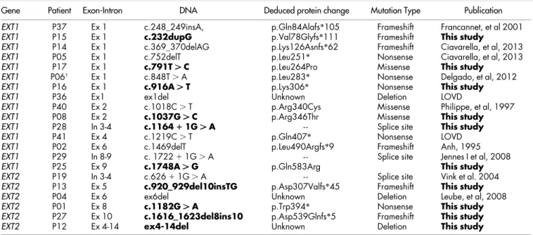
Table 2 | List of mutations in EXT1 or EXT2 gene in MO patients
Gene Patient Exon-Intron DNA Deduced protein change Mutation Type Publication
EXT1 P37 Ex 1 c.248_249insA, p.Gln84Alafs*105 Frameshift Francannet, et al 2001
EXT1 P15 Ex 1 c.232dupG p.Val78Glyfs*111 Frameshift This study
EXT1 P14 Ex 1 c.369_370delAG p.Lys126Asnfs*62 Frameshift Ciavarella, et al, 2013
EXT1 P05 Ex 1 c.752delT p.Leu251* Nonsense Ciavarella, et al, 2013
EXT1 P17 Ex 1 c.791T . C p.Leu264Pro Missense This study
EXT1 P061 Ex 1 c.848T . A p.Leu283* Nonsense Delgado, et al, 2012
EXT1 P16 Ex 1 c.916A . T p.Lys306* Nonsense This study
EXT1 P36 Ex1 ex1del Unknown Deletion LOVD
EXT1 P40 Ex 2 c.1018C . T p.Arg340Cys Missense Philippe, et al, 1997
EXT1 P08 Ex 2 c.1037G . C p.Arg346Thr Missense This study
EXT1 P28 In 3-4 c.1164 1 1G . A - - Splice site This study
EXT1 P41 Ex 4 c.1219C . T p.Gln407* Nonsense LOVD
EXT1 P02 Ex 6 c.1469delT p.Leu490Argfs*9 Frameshift Anh, 1995
EXT1 P29 In 8-9 c. 1722 1 1G . A - - Splice site Jennes I et al, 2008
EXT1 P25 Ex 9 c.1748A . G p.Gln583Arg This study
EXT2 P19 In 3-4 c.626 1 1G . A - - Splice site Vink et al. 2004
EXT2 P13 Ex 5 c.920__929del10insTG p.Asp307Valfs*45 Frameshift This study
EXT2 P04 Ex 6 ex6del Unknown Deletion Leube, et al, 2008
EXT2 P01 Ex 8 c.1182G . A p.Trp394* Nonsense This study
EXT2 P27 Ex 10 c.1616__1623del8ins10 p.Asp539Glnfs*5 Frameshift This study
EXT2 P12 Ex 4-14 ex4-14del Unknown Deletion This study
Novel mutations are indicated in bold. We considered as new mutations those not published and/or not mentioned in the LOVD databases. LOVD:http://medgen.ua.ac.be/LOVDv.2.0/
domains32 (Protein Homology Fold Recognition Engine, http:// www.sbg.bio.ic.ac.uk/phyre2/html/page.cgi?id5index). Regarding the 3-D prediction for two novel EXT1 missense mutations, p.Leu-264Pro and p.Arg346Thr, the Phyre2 bioinformatic tool showed that both mutations produce impairment in protein folding or alterations in the exostosin or glycosyltransferase domains. For the third one, p.Gln583Arg, Phyre2 showed the removal of alpha helix structures with possible alterations in EXT1 glycosyltransferase domain. A type II severe phenotype (IIS) was found in patients carrying the two first missense mutations (P17 and P08), and a severe phenotype (IIIS) was observed in P25 carrying the p.Gln583Arg mutation.
Six out of the 14 mutations in the EXT1 gene (p.Val78Glyfs*111, p.Leu264Pro, p.Lys306*, p.Arg346Thr, c.1164 1 1C . A and p.Gln583Arg) and four of the six EXT2 mutant alleles (p.Asp-307Valfs*45, p.Trp394*, p.Asp539Glnfs*5, and exon4-14del) were novel. Although some mutation hotspots have been reported20 (http://medgen.ua.ac.be/LOVDv.2.0/), we did not observe recurrent EXT1 or EXT2 mutations in patients in this study. The missense mutation c.1018C . T (p.Arg340Cys) observed in P40 and the exon 6 deletion c.1469delT (p.Leu490Argfs*9) found in P02 were previously described to cause the impairment of heparan sulfate synthesis20,33.
The MLPA analysis in gDNA of patient P36, showed the complete deletion of EXT1 exon 1. This patient had previously been reported as an SO case because he only had one lesion, but this detection in germline DNA allow us to change the diagnosis into MO patient with very mild symptoms (only a single osteochondroma lesion)33
(Table 1 and 2). Two molecular defects in EXT2 were detected by MLPA: the deletion of exon 6 (P04) and of 10 exons (from exon 4 to exon 14) in patient P12. This patient also carried an abnormal karyo-type (an 18 q deletion).
Splice-site mutations were detected in EXT1 in two patients. One of them, c.1164 1 1G . A in intron 3, was a novel mutation observed in patient P28. This novel mutation was analyzed in silico and the use of alternative cryptic donor sites was predicted. The phenotype in these patients was severe, presenting deformity of the limbs, valgus, restricted joint movement and scoliosis. Furthermore, one splice site mutation previously described was detected in P19 in EXT2. Clinical data were not available for this patient25, There are several possible
explanations for the lack of identification of mutations in some of the MO patients (22%). The mutation may have been in the EXT1 or EXT2 genes but in regions that were not analyzed. We did not look for mutations in deep intronic regions or in the 59 and 39 UTR sequences. Instead, the promoter region was genotyped and no mutation was detected. A recent study described a regulatory role for a G/C SNP (rs3401643) located at position 21158 bp, within a USF1 transcription factor binding site30. These authors observed that
the presence of the C-allele resulted in a ,56% increase in EXT1 promoter activity. The effect of this allele in the four patients of the present study who are heterozygous for it will require further studies. It is well established that methylation of cytosine residues in the promoter region leads to transcription repression in tumor sup-pressor genes; nevertheless this does not seem to be the case for EXT1 and EXT2 promoters in osteochondromas or in chondrosar-comas34,35. Finally, the possible existence of other genes responsible
for MO should also be considered. A putative EXT3 gene, located on the short arm of chromosome 19, has been proposed to explain the absence of an EXT1 or EXT2 mutation in a small percentage of MO patients (15–30%). Nevertheless, the existence of this third locus is generally accepted to be a false linkage result.
Inactivating mutations in the EXT1 and EXT2 genes were prev-iously reported as the most common event in MO patients resulting in the formation of non-functional EXT1 or EXT2 proteins with a variable degree of expression in tissues27,36. We observed 11
truncat-ing mutations that create premature stop codons presenttruncat-ing a high grade of severity in patient’s phenotype (Table 1 and 2). One of these
patients (P06) presented malignant transformation to chondrosar-coma and we detected the p.Leu283* mutation in the EXT1 gene. Very low or null levels of EXT1 and EXT2 proteins were detected by Western blot in this patient. However, in these experiments, the bands corresponding to GAPDH (control protein) were very weak and the lack of additional sample precluded repetition. We think that in spite of the technical problems, this observation should be reported to allow comparisons with other studies. Obviously, further cases should be analyzed to confirm these findings. The loss of EXT2 protein suggests that EXT1 mutations probably interfere with the function of exostosin’s complexes in the Golgi, inactivating the holoenzyme, degrading the whole protein, or interfering in some other function in the Golgi37.
Several studies have suggested that MO patients present a more severe phenotype due to EXT1 mutations than EXT2 mutations16,18,21
while other studies could not confirm this observation23,36. Pedrini
et al 2011 recently performed a genotype–phenotype association study in a large cohort of MO patients and identified some specific correlation according to a new clinical classification system31. Our
patients presented some of the most common manifestations, including orthopedic deformities of the forearm, ankle, varus or valgus of the knee, arthritis, vessels and nerve compression and very short stature (below the third percentile). The bones most often affected were tibia, femur, radius, humerus and fibula. Never-theless, we observed a severe phenotype (12% type IS, 53% type IIS, 12% type IIIS and 23% type IVS) in 63% of MO patients (Figure 1). The remaining 15% presented with a moderate phenotype without a family history of the disease. We observed that the grade of severity differed between the proband and other affected members in the family, according to previously reported intra-familial
variabil-ity18,38. Nevertheless, no family history for MO was reported in 56%
of MO patients. Patients with a mutation in the EXT2 gene showed a smaller number of affected bones (data not shown) consistent with a recent study39. The most frequently observed skeletal deformations
in our patients were shortening of limbs, varus or valgus knee, short metacarpal bones, scoliosis, shortened stature and synostosis, with no evidence of differences between the grade of severity in the phenotype observed in patients with EXT1 or EXT2 (Figure 1). Genotype–phenotype correlations are difficult to establish in MO patients because most of the EXT1 and EXT2 variants are private mutations20.
Malignant transformation to a chondrosarcoma is the most important complication in MO, and has been estimated to occur in 0.5–5% of patients17. Patients P06 and P38 developed malignant
tumors, which gives a frequency of malignant transformation of 7% in our cohort of patients. Patient P06 bore the pathogenic muta-tion c.848T . A (p.Leu283*) in the first exon of the EXT1 gene22,
while no mutation was detected in P38, neither in EXT1 nor EXT2 (Table 1). It has been shown that hereditary osteochondromas and secondary chondrosarcomas are associated with a second mutational hit in the EXT genes40,41. We thus investigated this possibility in DNA
extracted from the osteochondroma tissue resected from P06 by Sanger sequence and MLPA but we found no evidence of a somatic mutation as a second hit in any of these genes. The presence of genetic rearrangements at the EXT1 and EXT2 loci (as the second mutational hit) in P06 osteochondromas and secondary chondrosar-comas was ruled out.
In conclusion, we have identified the disease-causing mutation in 21 out of 27 MO patients, including 10 mutations described for the first time. No mutation was identified in SO cases. Structural analyses predicted a disruption of important domains of EXT1 proteins bear-ing missense mutations. A potentially functional promoter poly-morphism was found in three patients with no other mutation, in one patient with a disease-causing mutation and in one control. No second hit was identified in a sample from a chondrosarcoma. Further studies are needed to identify the molecular bases of the
disease in 22% of the patients of this cohort and to understand the mechanisms underlying the malignant transformation process.
Methods
Patients and control individuals.We investigated 33 patients (18 males and 15 females), from unrelated pedigrees with osteochondromatosis from Chile and Argentina (27 MO and 6 SO). Nine control samples from healthy subjects were included in the promoter studies. Diagnosis was made on the basis of clinical manifestations and confirmed by physical and/or radiographic examinations at the Orthopedic and Imaging Departments, Children’s Hospital of Co´rdoba, National University of Co´rdoba, Argentina. DNA and tissues samples from patients and their relatives were obtained together with their informed consent in accordance with the Helsinki Declaration as revised in 2000. The study was approved by the Ethics Committee (CIEIS) Act Nu 95/2007. Genomic DNA was obtained from peripheral blood leukocytes and tissue samples from discarded tissues obtained by surgery, using the Wizard Genomic DNA purification Kit (Promega, Madison, WI). DNA was extracted according to the manufacturer’s instructions.
Clinical studies and phenotypic data.Clinical variables were analyzed according to a scale established by the Musculoskeletal Tumoral Society with some modifications18.
This scale includes the evaluation of all palpable lesions, patient’s height, deformities, and functional limitations. Lesion quality and the severity of the disease were assessed according to age of onset (before/after 3 years), number of exostoses (more/less than 10 osteochondromas), vertebral location of the exostoses (absence or presence), stature (above/below 10th percentile), and functional rating (good or fair). The degree of severity was classified as mild (M) or severe (S). Four subcategories were defined in patients with a severe phenotype (from types IS to IVS)16,18.
Genotyping and mutation analysis.The 11 EXT1 and 13 EXT2 coding exons and their intronic flanking regions were amplified by PCR from genomic DNA. Primer sequences and PCR conditions were as described by Sarrio´n et al 201342. All
fragments, except those corresponding to exon 1 of EXT1, could be amplified by PCR simultaneously. Exon 1 of EXT1 and exon 2 of EXT2 were split into several overlapping fragments, to obtain amplification products that did not exceed 650 bp. PCR was performed in a 50-ml reaction volume, containing ,100 ng of genomic DNA, 1–2 mM MgCl2, 0.2 mM of each dNTP, 0.4 mM of each forward and reverse primer and 0.7 U of GoTaqR Flexi polymerase (Promega, Madison, WI). All PCR programs included an initial denaturation of 4 min at 95uC, followed by 35 cycles of 30 sec at 95uC, 30 sec at annealing temperature (Ta) and 1 min at 72uC. An extension at 72uC was then performed for 5 min. The annealing temperature was 60uC for all primer combinations, except during the amplification of overlapping regions of exon 1 of EXT1. For these primer combinations, Ta was set at 55uC for ex1.1 and 57uC for ex1.2 and ex1.3. The EXT1 promoter region, between positions 21285 and 2851, was also analyzed by sequencing. The PCR reaction was performed as described above with a Ta of 55uC. All PCR products were purified using a PCR purification kit (GE Healthcare) and sequenced with BigDye 3.1 (Applied Biosystems Life Technologies). The sequences were analyzed in an ABI PRISM 3730 DNA Analyzer (Applied Biosystems Life Technologies).
The presence of all detected mutations was confirmed by digestion with the appropriate restriction enzyme. Novel mutations were confirmed by analyzing 100 control alleles. The mutations were given the official HGVS nomenclature (www. hgvs.org). The reference sequences were NM_000127.2 for EXT1 and NM_000401.3 for EXT2.
MLPA.The number of copies of the EXT1 and EXT2 exons present in the patient’s genomic DNA was analyzed using the multiplex ligation-dependent probe amplification (MLPA) technique designed by MRC-Holland (code #P215-B1 EXT, MRC-Holland, Amsterdam, The Netherlands) following the manufacturer’s instructions. PCR products were run on an ABI 3730 DNA Analyzer capillary sequencer (Applied Biosystems, Forster City, CA, USA). Peaks were analyzed using Coffalyser v9.4 software (MRC-Holland Vs 05; 30-08-2007). The proportion of each peak relative to the height of all peaks was calculated for each sample and then compared to proportions for the corresponding peak averaged for a set of at least ten normal DNA samples. Samples with ratios between 0.7 and 1.3 were considered as bearing a normal copy number. Ratios below 0.7 were considered to correspond to deletions, and above 1.3 to duplications. Each positive result was confirmed in a second independent MLPA reaction.
Assessment of functionality of missense mutations.In order to assess the possible pathogenic effect of the new missense mutations, the changes were analyzed using three in-silico online tools: PolyPhen-2 (Polymorphism Phenotyping v2; http:// genetics.bwh.harvard.edu/pph2/, last accessed March 2014), Mutation Taster (http:// www.mutationtaster.org/, last accessed March 2014, and ESE Finder 3.0 (ESE: Exonic Splicing Enhancer; http://rulai.cshl.edu/cgi-bin/tools/ESE3/esefinder.
cgi?process5home, last accessed March 2014). Protein Homology Fold Recognition Engine (http://www.sbg.bio.ic.ac.uk/phyre2/html, last accessed March 2014) was used to predict the implications of missense mutations for EXT1 3D structure Human Splice Finder (http://www.umd.be/HSF/, last accessed March 2014) and NetGene2 (http://www.cbs.dtu.dk/cgi-bin/webface?jobid5netgene2, last accessed March 2014) online tools were used to assess the possible effect of novel intronic mutations on splicing.
Ethics statement.The methods were carried out in accordance with the approved guidelines and in accordance with the Helsinki Declaration as revised in 2000. The study was approved by the Ethics Committee (CIEIS) Act Nu 95/2007.
1. Martinez-Duncker, I., Asteggiano, C. & Freeze, H. H. [Congenital Disorders of Glycosylation] [59–83] [Biochemistry Research Trends, Nova Science Publishers Inc Nueva York, 2012].
2. Jaeken, J. Congenital disorders of glycosylation. Ann. N. Y. Acad. Sci. 1214, 190–198 (2010).
3. Ahn, J. et al. Cloning of the putative tumour suppressor gene for hereditary multiple exostoses (EXT1). Nat. Genet. 11, 137–143 (1995).
4. Stickens, D. et al. The EXT2 multiple exostoses gene defines a family of putative tumour suppressor genes. Nat Genet. 14, 25–32 (1996).
5. Wuyts, W. et al. Positional cloning of a gene involved in hereditary multiple exostoses. Hum. Mol. Genet. 5, 1547–1557 (1996).
6. Busse, M. et al. Contribution of EXT1, EXT2, and EXTL3 to heparan sulfate chain elongation. J. Biol. Chem. 282, 32802–32810 (2007).
7. Kim, B. T. et al. Human tumor suppressor EXT gene family members EXTL1 and EXTL3 encode alpha 1,4- Nacetylglucosaminyltransferases that likely are involved in heparan sulfate/heparin biosynthesis. Proc. Natl. Acad. Sci. 98, 7176–7181 (2001).
8. Van Hul, W. et al. Identification of a third EXT-like gene (EXTL3) belonging to the EXT gene family. Genomics. 15; 47, 230–7 (1998).
9. Wuyts, W. et al. Identification and characterization of a novel member of the EXT gene family, EXTL2. Eur J Hum Genet. 5, 382–9(1997).
10. Wise, C. A., Clines, G. A., Massa, H., Trask, B. J. & Lovett, M. Identification and localization of the gene for EXTL, a third member of the multiple exostoses gene family. Genome Res. 7, 10–16 (1997).
11. Gallagher, J. & Lyon, M. Structure and function of heparan sulphate proteoglycans. Biochem. J. 236, 313–325 (1986).
12. Carlsson, P., Presto, J., Spillmann, D., Lindahl, U. & Kjelle´n, L. Heparin/heparan sulfate biosynthesis: processive formation of N-sulfated domains. J. Biol. Chem. 283, 20008–20014 (2008).
13. Shi, X. & Zaia, J. Organ-specific heparan sulfate structural phenotypes. J. Biol. Chem. 284, 11806–11814 (2009).
14. Zak, B. M., Crawford, B. E., & Esko, J. D. Hereditary multiple exostoses and heparan sulfate polymerization. Biochim. Biophys. Acta 1573, 346–355 (2002). 15. De Andrea, C. E. & Hogendoorn, P. C. Epiphyseal growth plate and secondary peripheral chondrosarcoma: the neighbours matter. J. Pathol. 226, 219–228 (2012).
16. Alvarez, C., Tredwell, S., De Vera, M. & Hayden, M. The genotype-phenotype correlation of hereditary multiple exostoses. Clin. Genet. 70, 122–130 (2006). 17. Bove´e, J. V. Multiple osteochondromas. Orphanet J. Rare Dis. 13, 3 (2008). 18. Francannet, C. et al. Genotype-phenotype correlation in hereditary multiple
exostoses. J. Med. Genet. 38, 430–434 (2001).
19. Hameetman, L., Bove´e, J. V., Taminiau, A. H., Kroon, H. M. & Hogendoorn, P. C. Multiple osteochondromas: clinicopathological and genetic spectrum and suggestions for clinical management. Hered. Cancer Clin. Pract. 2, 161–173 (2004).
20. Jennes, I. et al. Multiple osteochondromas: mutation update and description of the multiple osteochondromas mutation database (MOdb). Hum. Mutat. 30, 1620–1627 (2009).
21. Alvarez, C. M., De Vera, M. A., Heslip, T. R. & Casey, B. Evaluation of the anatomic burden of patients with hereditary multiple exostoses. Clin. Orthop. Relat. Res. 462, 73–79 (2007).
22. Delgado, M. A. et al. A novel nonsense mutation of the EXT1 gene in an Argentinean patient with multiple hereditary exostoses: a case report. J. Bone Joint Surg. Am. 94, e76 (2012).
23. Kitsoulis, P. et al. Osteochondromas: review of the clinical, radiological and pathological features. In Vivo 22, 633–646 (2008).
24. Jennes, I. et al. Mutation screening of EXT1 and EXT2 by denaturing high-performance liquid chromatography, direct sequencing analysis, fluorescence in situ hybridization, and a new multiplex ligationdependent probe amplification probe set in patients with multiple osteochondromas. J. Mol. Diagn. 10, 85–92 (2008).
25. Vink, G. R. et al. Mutation screening of EXT1 and EXT2 by direct sequence analysis and MLPA in patients with multiple osteochondromas: splice site mutations and exonic deletions account for more than half of the mutations. Eur. J. Hum. Genet. 13, 470–474 (2005).
26. Lonie, L. et al. Determination of the mutation spectrum of the EXT1/EXT2 genes in British Caucasian patients with multiple osteochondromas, and exclusion of six candidate genes in EXT negative cases. Hum. Mutat. 27, 1160 (2006). 27. Pedrini, E. et al. Novel EXT1 and EXT2 mutations identified by DHPLC in Italian
patients with multiple osteochondromas. Hum. Mutat. 26, 280 (2005). 28. White, S. J., Sterrenburg, E., van Ommen, G. J., den Dunnen, J. T. & Breuning,
M. H. An alternative to FISH: detecting deletion and duplication carriers within 24 hours. J. Med. Genet. 40, e113 (2003).
29. Wuyts, W. et al. Mutations in the EXT1 and EXT2 genes in hereditary multiple exostoses. Am. J. Hum. Genet. 62, 346–354 (1998).
30. Jennes, I. et al. Identification and functional characterization of the human EXT1 promoter region. Gene 492, 148–159 (2012).
31. Pedrini, E. et al. Genotype phenotype correlation study in 529 patients with multiple hereditary exostoses: identification of ‘‘protective’’ and ‘‘risk’’ factors. J. Bone Joint Surg. Am. 93, 2294–302 (2011).
32. Ciavarella, M. et al. 20 novel point mutations and one large deletion in EXT1 and EXT2 genes: report of diagnostic screening in a large Italian cohort of patients affected by hereditary multiple exostosis. Gene 515, 339–348 (2013). 33. McCormick, C., Duncan, G., Goutsos, K. T. & Tufaro, F. The putative tumor
suppressors EXT1 and EXT2 form a stable complex that accumulates in the Golgi apparatus and catalyzes the synthesis of heparan sulfate. Proc. Natl. Acad. Sci. USA 97, 668–673(2000).
34. Ropero, S. et al. Epigenetic loss of the familial tumor-suppressor gene exostosin-1 (EXT1) disrupts heparan sulfate synthesis in cancer cells. Hum. Mol. Genet. 13, 2753–2765 (2004).
35. Hameetman, L. et al. Decreased EXT expression and intracellular accumulation of heparan sulphate proteoglycan in osteochondromas and peripheral
chondrosarcomas. J. Pathol. 211, 399–409 (2007).
36. Signori, E. et al. A combined analytical approach reveals novel EXT1/2 gene mutations in a large cohort of Italian multiple osteochondromas patients. Genes Chrom. Cancer 46, 470–477 (2007).
37. Bernard, M. A. et al. Diminished levels of the putative tumor suppressor proteins EXT1 and EXT2 in exostosis chondrocytes. Cell Motil. Cytoskeleton 48, 149–162 (2001).
38. Hennekam, R. C. Hereditary multiple exostoses. J. Med. Genet. 28, 262–266 (1991).
39. Stancheva-Ivanova, M. K. et al. Clinical and molecular studies of EXT1/EXT2 in Bulgaria. J. Inherit. Metab. Dis. 34, 917–921 (2011).
40. Bove´e, J. V. EXTra hit for mouse osteochondroma. Proc. Natl. Acad. Sci. USA. 107, 1813–1814 (2010).
41. Hecht, J. T. et al. Hereditary multiple exostoses (EXT): mutational studies of familial EXT1 cases and EXT-associated malignancies. Am. J. Hum. Genet. 60, 80–86 (1997).
42. Sarrio´n, P. et al. Mutations in the EXT1 and EXT2 genes in Spanish patients with multiple osteochondromas. Sci. Rep. 3, 1346 (2013).
Acknowledgments
The authors thank the patients and their families for their cooperation. They wish to acknowledge Joanne Ferrier for revising the English. The authors are also grateful for the support from the Centro de Investigacio´n Biome´dica en Red de Enfermedades Raras (CIBERER), which is an initiative of the ISCIII. Grant support: Scientific and Technical National Research Council (CONICET); FONCyT PICT 2006–2350 and BID Nu 2437/ OC-AR PICT2010-2824, Catholic University of Cordoba Grant 2010/2011/2012/2013, Spanish Ministry of Education and Science (SAF2011-25431, PIB2010AR00473) and the Catalan Government (2009SGR971).
Author contributions
Study design: S.B., D.G., C.G.A. Collection and data samples: M.A.D., G.M.-D., P.S., L.Z., H.H.R., F.S. Performance of experiments: M.A.D., G.M.-D., C.G.A. Data interpretation and analysis: M.A.D., G.M.-D., P.S., R.U., R.D.-K., S.B., D.G., C.G.A. Draft composition: M.A.D., G.M.-D., S.B., D.G. C.G.A. conceived of the study, and participated in its design, coordination and helped to draft the manuscript. All authors reviewed the manuscript.
Additional information
Competing financial interests: The authors declare no competing financial interests. How to cite this article:Delgado, M.A. et al. A broad spectrum of genomic changes in latinamerican patients with EXT1/EXT2-CDG. Sci. Rep. 4, 6407; DOI:10.1038/srep06407 (2014).
This work is licensed under a Creative Commons Attribution-NonCommercial-NoDerivs 4.0 International License. The images or other third party material in this article are included in the article’s Creative Commons license, unless indicated otherwise in the credit line; if the material is not included under the Creative Commons license, users will need to obtain permission from the license holder in order to reproduce the material. To view a copy of this license, visit http:// creativecommons.org/licenses/by-nc-nd/4.0/