NEUROBEHAVIORAL FUNCTIONS, SERUM PROLACTIN AND PLASMA
RENIN ACTIVITY OF MANGANESE-EXPOSED WORKERS
Q. NIU
I,
H. SHUCHANG
2,W. SHENG
2,M. DI GIOACCHIN03, N. VERNA3,
A.
R.
VOLPE4,
L.
DI GIAMPAOLO" M. CARMIGNANI
4 andP. BOSCOL03*
'Department of Occupational Health, Shanxi Medical University, Taiyuan, 030001 (P. R. China),
2Department of Occupational and Environmental Health, Health Science Center,
Peking University, 100083 (P. R. China); 'Section of Occupational Medicine,
Allergology and Clinical Immunology, Department of Medicine and Science of Ageing,
"G. D'Annunrio" University, Via dei Vestini, 66100 Chieti (Italy); "Section of Pharmacology and
Toxicology, Department of Basic and Applied Biology,
Univ.ersity of
L'Aquila, Via Vetoio, 67010 Coppito, Italy
Objective of this study was to assess effects of manganese (Mn) exposure on 56 workers employed in a Mn welding workshop of a machine building factory in Taiyuan (Shanxi Province, P.R. China) for a mean period of 16.1 years. The mean air Mn level in the workshop was 138.4 ug/m". Neurobehavioral Core Test Battery (NCTB), including the Profile of Mood States (POMS), was performed. Blood pressure (BP) increase following immediate stand-up (BP-IS), serum prolactin (PRL) and plasma renin activity (PRA) in supine position were also determined. Most of the NCTB scores of the Mn-exposed workers were lower than those of the controls, while the POMS scores were higher, indicating a Mn-induced impairment of neurophysiological functions and a deflection of mood towards negative emotion states. PRL values of the
Mn-exposed workers were higher than those of the controls. BP-IS of Mn-exposed workers was
significantly lower than that of the controls. PRA of the same workers was augmented more than 200 %. In the Mn-exposed workers, the higher PRL values are possibly due to a reduced inhibitory effect on pituitary lactotrope cells by the tubero-infundibular dopamine system; the decreased BP-IS was referred to imbalance between the sympathetic and parasympathetic activities, whereas the higher basal PRA was thought to depend on neuroendocrine changes (including increased central sympathetic tone) and/or on a direct effect of Mn on renal juxta-glomerular cells. On the whole, this study demonstrates that occupational Mn exposure is responsible for neurobehavioral changes coexisting with alterations of neuroendocrine and humoral systems.
Manganese (Mn) is a ubiquitous constituent of the environment comprising about 0.1 %of the earth crust. Today, occupational exposure to Mn occurs during several activities including arc welding. High levels of Mn in air, water and soil, which are able to affect human health, may depend on pollution produced by working activities and/ or derive from geological sources(1). Moreover, methylcyclopentadienyl Mn was used in Canada since 1976 as an additive anti-knock agent in unleaded gasoline (2).
Mn is considered an essential element required for the human diet (3).Italso acts as a component of several metallo-enzymes including superoxide dismutase, a scavenger of reactive oxygen species (ROS) (4). Mn is known to catalyze autoxidation of dopamine (DA), in the presence of L-cysteine, in buffered solution (5). Within its selective neurotoxicity for DA neurons of the central nervous system (6), Mn has been implicated in degeneration of the nigrostriatal dopaminergic neurons in idiopathic and chemically-induced Parkinson's
Key words: manganese, neurobehavioral function, prolactin, plasma renin activity, sympathetic system Mailing address: Prof. Paolo Boscolo
Medicina del Lavoro, "G.D'Annunzio" University Via dei Vestini 66100 Chieti (Italy)
Tel/fax: +39 8713556704
e-mail address:[email protected]
17
0394-6320 (2004)
Copyright©hy BIOLlFE, s.a.s.. This publicationand/or article is for individual usc only and may not be further
disease. Such selectivity was found to depend upon interactions of Mrr" with the DA transport system in these neurons (7). Mn (along with iron) acts as a pro-oxidative agent at synaptic level. Mn'" also appears to influence Ca'" transport kinetics in the mitochondria, thus leading to defective mitochondrial function, decreased oxidative phosphorilation and A TP production, and accumulation of ROS (8). Moreover, intrathecal administration of MnCI2 to young male rats caused DA depletion in the caudate-putamen correlating with a decrease in spontaneous motor activity (9). In aged rats, Mn exposure was able to increase formation of ROS more than in young rats and an age-related impairment of the neuronal antioxidant systems was suggested to play an enabling role in Mn neurotoxicity (10).
Manganism is a central nervous system disease first described in the 1800s following exposure to high concentrations of Mn oxides (11). On the other hand, Mn exposure can cause a Parkinsonian syndrome within one or two years. This syndrome includes gait disorders and postural instability, psychiatric disorders with compulsive behavior, emotional liability and autonomic dysfunction (12-14). In this regard, Mn-exposed workers may present not only reduction of motor functions but also increased olfactory perceptions (15). Since increased serum prolactin (PRL) is a common finding among Mn-exposed subjects, the assay of PRL has been proposed, as a neuroendocrine biomarker, to assess the effect of this metal on the tubero-infundibular DA system, which exerts a tonic inhibitory effect on the pituitary lactotrope cells (16,17).
Although Mn was found
invitro
to playa protective role against free radicals in ventricular myocytes (18), otherin vitro
andex vivo
assays as well asin vivo
acute and subacute toxicity tests evidenced cardiotoxic effects of the metal (19). In this respect, higher incidence of electrocardiographic abnormalities in Mn-exposed workers was reported (20).There are only few and controversial studies in humans on neurogenic, neurohumoral and autacoidal mechanisms regulating cardiovascular homeostasis and BP levels. This study, therefore, aimed to investigate the relationship between the effects of Mn exposure on neurobehavioral parameters and those on neuroendocrine and humoral (autacoidal) pathways.
MA TERIALS AND METHODS
A Mn welding workshop of a machine building factory was investigated. The ventilation of the premises was efficient. Air Mn samples were collected by air sampler in the welding operation sites at the highness of workers' breathing zone (about 160 em) with flow speed 15 Umin for 10minutes. 20 samples were collected. The air Mn, Cd, Ni and Fe concentration were measured by graphite furnace atomic absorption spectrophotometry according to Wang (21).56 welders (Mn-exposed) men and 34 control men were recruited for the study. Welding workers had been exposed to Mn for (mean
±
SEM) 16.17±
0.73 years (range 3 -32 years) working for 4 hours per day and 5 days per week. Subjects of the two groups had similar age, family income, smoking habit (22 and 24 %, respectively) and education degree. Subjects suffering from diseases, in treatment with drugs, taking a large amount of alcohol (>500 mL ethanol/week) and smoking more than 40 cigarettes/ day were excluded from the study. The age of the Mn-exposed subjects was (mean±
SEM) 33.7±
0.6 years and that of controls 33.2±
0.9 years. All the examined subjects were living in the same area of the South District ofTaiyuan City (Shanxi Province, P.R. China) with similar exposure to urban environment [recently, we found mean blood lead (Pb) levels of 12 mg/dL in the women resident in this area (22)].The Mn-exposed workers and the control subjects attended the hospital in the early morning, in fasting condition, after a sleeping night. The neurobehavioral tests were performed in a clinic room with the same examiner, from 8.30 to 10.30 a.m., lasting about 30 minutes for each subject. The clinic room was quiet, illuminated with a daylight lamp, and the temperature in the room was 20-22° C.
The WHO recommended Neurobehavioral Core Test Battery (NCTB) was used for interviewing all the examined subjects (23). NCTB is composed of two parts. One part is the inventory of Profile of Mood States (POMS) questionnaire including Anger-Hostility (POMSA), Confusion-Bewilderment (POMSC), Depression-Dejection (POMSD), Fatigue-Inertia (POMSF), Tension-Anxiety (POMST), and Vigor-Activity (POMSV), which reflect neuropsychological variations caused by neurotoxicants; the other part is a test battery including six tests: Simple Reaction Time (SRT), which reflects response speed and attention; Digit Span (DSP), which reflects attention and auditory memory; Santa Ana Dexterity (SAN), which presents
manual coordination and speed; Digit Symbol (DSY), which shows cognition and motor speed; Benton Visual Retention (BVR), which reflects visual cognition and memory, coordination and speed; and Pursuit Aiming II (PA), which presents manual coordination and speed. SRT is divided into SRTF (fastest simple reaction time) and SRTS (slowest simple reaction time); DSP is divided into DSPF (forward) and DSPB (backward); SAN includes SANP (preferred hand) and SANN (non-preferred hand); PA includes PAC (correct) and PAE (erroneous).
Serum PRL and plasma renin activity (PRA) and BP increase following immediate standing-up (BP-IS) were determined in 21 Mn exposed workers and 16 control men. These 21 Mn-exposed workers and 16 controls were included in the previous groups of 56 Mn-exposed workers and 34 controls. The age and the smoking habit of 21 Mn-workers and 16 controls were similar to those of the groups composed of 56 workers and 34 controls. After a sleeping night, in fasting condition, the recruited subjects were implanted with a needle (connected to a heparinized cannula), which was inserted into a forearm vein. Afterwards, blood samples were collected after 30 minutes of standing in supine position. Serum prolactin (PRL) and plasma
renin activity (PRA) were determined by
radioimmunoassay according to Garden and Weintraub (24).
BP-IS was determined from 8.30 to 9.30 a.m.; the recruited men remained in supine position for 5 minutes
before testing BP and then stood up quickly. BP-IS was the difference in systolic BP during lying down and after standing up; the value of BP-IS (mmHg) was the mean of 3 determinations with 5 minutes of interval; general procedures were in accordance with Ewing (25).
Kolmogorov-Sminorv tests showed that the groups of 56 workers and 34 controls conformed more the normal distribution than the smaller groups of21 workers and 16 controls. Therefore, parametric test were utilized in the larger groups and non-parametric (Mann-Whitney U-test) in the smaller ones.
Data were compared by analysis of covariance in order to exclude the influences of age, family income, smoking habit and education All sets of data were expressed as means
±
SEM, P values less than 0.05 were considered as significant.RESULTS
Air Mn level in the premises of the factory was (mean
+
SEM) 138.4±
11.6Il-g/m3 (range 38-198; determinations n 20); air Cd was 12.6±
2.8 ng/m' (range 3.0-48.3); air Fe was 581±
45 u.g/rn' (range 354-950); air Ni was 3.8±
0.53u.g/rrr'
(range 1.2-8.7).The accumulative exposure of Mn, Cd, Fe and Ni were 2.23
±
0.19 Il-g/m3.year, 0.204±
0.046 Il-g/m'. year, 9.40
±
0.73 mg/m'iyear and 0.062±
0.08mg/m3.year, respectively.Items Mn-eIposed (n=56) Controls (n=34) F P Tab. I. Neurobehavioral tests of Mn-exposed workers and controls
SRT 242.58 ± 6.21 325.85 ± 26.48 3.578 0.001*"
Values (mean
±
SEM) represent the scores obtainedSRTF 172.57 ± 7.78 227.79 ± 21.89 2.054 0.050· for each item of the Neurobehavioral Core Test Battery
SRTS 466.16 ± 30.69 563.50 ± 56.98 2.205 0.054 (NCTB). Scores were processed by covariance analysis
DSP 14.55 ± 0.39 18.50 ± 0.68 2.758 0.010· in order to exclude the influence ofage, family income,
DSPF 9.64 ± 0.26 12.67 ± 0.44 2.559 0.015· smoking habit and education degree. ns: not significant.
DSPB 4.91 ± 0.21 5.82 ± 0.37 2.038 0.052 Difference statistically significant: *p<0.05, **p<O.OJ
SRT: Simple Reaction Time; SRTF: Fastest SRT; SRTS:
SANP 42.92 ± 0.85 41.79 ± 1.13 0.051 0.846 Slowest SRT; DSP: Digit Span; DSPF: DSP (forward);
SANN 39.92 ± 0.67 40.00 ± 1.46 0.555 0.811 DSPB: DSP (backward); SANP: Santa Ana Dexterity
DSY 41.55 ± 1.66 50.38 ± 2.03 4.172 0.001·· (preferred hand); SANN: Santa Ana Dexterity
(non-BVR 7.12 ± 0.16 6.85 ± 0.41 0.426 0.902 preferred hand); DSY: Digit Symbol; BVR: Benton
Visual Retention; PA: Pursuit Aiming II; PAC: PA
PA 203.88 ± 4.68 220.08 ± 5.20 3.882 0.001··
(correct); PAE: PA (erroneous)
PAC 203.46 ± 4.22 226.23 ± 5.85 3.798 0.001··
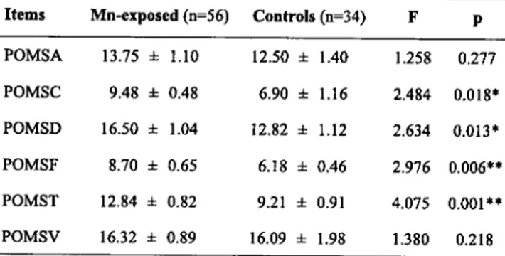
Tab. II.Profile ofmood states (POMS) of Mn-exposed workers and controls
Items Mn-exposed (n=56) Controls (n=34) F p Values (mean
±
SEM) represent the scores obtainedfor each item of the Profile of mood states (POMS).
POMSA 13.75 ± 1.10 12.50 ± 1.40 1.258 0.277 Scores were processed by covariance analysis in POMSC 9.48 ± 0.48 6.90 ± 1.16 2.484 0.018* order to exclude the influence ofage, family income,
smoking habit and education degree. ns: not significant.
POMSD 16.50 ± 1.04 12.82 ± 1.12 2.634 0.013* Difference statistically significant: *p<0.05, **p<O.O1
POMSF 8.70 ± 0.65 6.18 ± 0.46 2.976 0.006** POMSA: Profile of Mood State
(POMS)/Anger-Hostility; POMSC: POMSIConfusion-Bewilderment;
POMST 12.84 ± 0.82 9.21 ± 0.91 4.075 0.001** POMSD: POMSIDepression-Dejection; POMSF: POMSIFatigue-lnertia, POMST:
POMSITension-POMSV 16.32 ± 0.89 16.09 ± 1.98 1.380 0.218
Anxiety; POMSV: POMSIVigor-Activity.
Tab. III. Serum prolactin (PRL) and plasma renin activity (PRA) of Mn-exposed workers and controls
Mn-exposed (n=21) Controls (n=16) p
PRL (rrg/L) 21.75 ± 0.55 17.01 ± 0.47 <0.05 Values are expressed as mean
±
SEM. Data are processed byPRA (ng/mL/h) 3.17 ± 0.22 1.49 ± 0.19 <0.01 Mann- Whitney U test
Results of the neurobehavioral tests are listed in Tab. 1. The scores of SRT, SRTF, DSP, DSPF, DSY, PA and PAC of the Mn-exposed workers were significantly lower than those of the control subjects. Moreover, in the Mn-exposed workers, also the scores of SRTS and DSPB were lower than those of the controls, but only with a borderline statistical significant difference, while SANP, SANN, BVR and PAE were similar to those of the controls.
The results of POMS are reported in Tab.II. The scores of POMSC, POMSD, and, mostly, POMSF, POMST, of the Mn-exposed workers were significantly higher than those of the controls. However, POMSA and POMSV of the Mn-exposed men did not show significant changes.
BP-IS of the Mn-exposed workers was 5.6 ± 0.5 mmHg, while that of the controls was 8.0± 0.9
mmHg (p<0.05 processed by Mann-Whitney U
test).
Serum PRL and PRA values of the Mn-exposed workers were significantly augmented in relation to those of the control men (Tab. III).
DISCUSSION
The welding workers had been exposed to Mn for long time (mean 16.1 years). When the workers were investigated, mean air Mn concentration in the factory was 138.4u.g/m:',However, a previous determination in some premises of the factory showed mean values exceeding 200 p.g/m". It may thus not be excluded that Mn exposure of the workers had been recently reduced by environmental improvements.
Welding workers were exposed to very low Cd levels (mean 12.6 u.g/rn") able to affect neither renal mechanisms of cardiovascular regulation or nervous functions (26). Moreover, the levels of occupational Ni and Fe exposure were not harmful for human health. Therefore, only Mn exposure was able to affect the nervous and cardiovascular parameters of the investigated workers.
Neurobehavioral tests measure integrated activities of the nervous system. Such tests are considered very sensitive functional indicators of neurotoxicity, being the behavioral performances
the expression of sensory, motor, neuroendocrine,
neurovegetative and integrative processes involving
both central and peripheral nervous system (27,28).
The sensitivity, reliability, validity and
cross-cultural consistency of NCTB as an integrated
battery have been confirmed, with some disparity
of different tests dependent on cultural groups,
and with some influence of age and education
degree (Anger et aI., 1997). In this regard, many
reports on neurobehavioral assessment for
neurotoxicity of chemicals have been published,
mostly dealing with the exposure to Pb, Mn,
aluminum, organic solvents and pesticides
(21,23,29-31).
In this study, the majority of the scores of
NCTB (neurobehavioral tests and POMS) was
significantly altered by Mn exposure. On the basis
of these results, Mn appears to represent, at levels
of occupational
exposure,
a
factor
of
neurobehavioral impairment. This impairment
involves
reduced
performances
of
neurophysiological functions (including decreased
attention and auditory memory, cognition, manual
coordination and performance speed) and deflection
of the mood state toward depressive, anxious and
asthenic components [as indicated by the elevated
scores of confusion-bewilderment (POMSC),
depression-dejection (POMSD), fatigue-inertia
(POMSF) and tension-anxiety (POMST) in the
Mn-exposed men)]. However, Mn did not affect
active emotion/reaction scores [anger-hostility
(POMSA), vigor-activity (POMSV)], thus appearing
that Mn-exposure affects quite specifically the
neuropsychological determinants of mood state.
In this respect, animal experiments showed the
emotionally negative states to be induced by
hyperactivity of serotoninergic pathways and/or
hypoactivity of noradrenergic, dopaminergic and
opioid ones, while the emotionally positive states
result from opposite activities of these central
neurogenic systems; moreover, the serotoninergic,
noradrenergic and dopaminergic systems playa
central role in the processes of reinforcement,
acquisition of the evaluative capacities and
realization of all types of behaviour (32). On the
other hand, PRL, an index of functionality of the
hypothalamo-pituitary axis, is regarded as a
modulator of immunity (33,34)
and
behavior
showing anxiolytic and anti-stress effects (35).
Therefore, a relationship between higher values of
serum PRL and impairment of neurophysiological
function (with shift towards emotionally negative
states) of the Mn-exposed workers appears to be
suggestive. On the other hand, the observed higher
levels of serum PRL are in agreement with the
results of previous studies, in which increased
serum PRL was a common finding among subjects
exposed to Mn. The involved mechanism in the
serum PRL increase was supposed to consist in a
Mn inhibiting effect on the tubero-infundibular
DA system, in turn inhibiting production and/or
release of PRL from the pituitary lactotrope cells
(16,17,36).
Autonomic nervous system function test battery
recommended by Ewing (1993), including BP-IS,
was used not only on patients (in clinical studies)
but also on non-symptomatic workers exposed to
neurotoxic agents (30,37). This study showed that
BP-IS of the Mn-exposed workers was significantly
reduced, thus evidencing that sympathetic and/or
parasympathetic activities may be affected by Mn
exposure. Our results confirm those of Barrington
et aI. (20) who found decreased chronotropic
reactivity, depending on sympathetic-vagal
imbalance, in eight Mn-exposed workers by using
the 24 hr-electrocardiographic monitoring (Holter).
There are no data on the effects of Mn on the
renal juxta-glomerular (JG) apparatus and
renin-angiotensin-aldosterone (RAA) system for
explaining the increased PRA of the Mn-exposed
workers. This apparatus, with a rich sympathetic
nerve supply, is composed of macula densa and
modified granulated smooth muscle cells of the
afferent arteriole of the glomerulus, with a minor
contribution by those of the afferent arteriole and
by interstitial cells (38). The JG apparatus has
baroreceptors in the arterioles, responding to change
of transmural pressure gradient, and chemoreceptors
in the macula densa, detecting changes in the
composition of fluids of the distal tubule. The
secretion of renin is stimulated by changes in
sympathetic activity, modification of body position,
reduction of renal perfusion pressure, extracellular
volume depletion, changes in plasma levels of
electrolytes (such as sodium, potassium and calcium)
and circulating hormones. In particular, the release
of renin from JG cells is stimulated by substances
(such as b
t-and
b
2-
adrenoreceptor agonists)
leading to an increase of the intracellular
concentration of cyclic adenosinemonophosphate
(cAMP). The levels of intracellular free calcium in the JG cells and the transmembrane sodium gradient seem to playa major role in the release of renin (39). Moreover, it is known that DA-l and DA-2 receptor subtypes are localized in various regions within the kidney (lacking DA neurons), including vasculature (DA-l) and sympathetic nerve endings in blood vessels (DA-2). Activation of the DA-l and DA-2 receptors (by circulating DA and by DA released from renal sympathetic endings) involves stimulation of adeny late cyclase activity and inhibition of the inositol 1,4,5-triphosphate-dependent Ca'" release, respecti vely, with inhibition of sodium reabsorption from tubules and consequent increased natriuresis (40,41). DA causes vasodilation in the renal circulation, suppresses aldosterone secretion, directly stimulates renal sodium excretion, suppresses noradrenaline release at the sympathetic nerve terminals by a pre-synaptic inhibitory mechanism, and influences the secretion of renin. Therefore, considering the selectivity ofMn in affecting catecholamine (DA) neurogenic systems and metabolic pathways (42), the marked PRA increase of the Mn-exposed workers is likely to depend on Mn-induced alterations .of catecholamine (DA) metabolism and/or adrenergic neurotransmission in the kidney. However, effects of Mn on other (renal) systems and pathways, involved in renin production and release (e.g., the kallikrein-kinin and nitric oxide ones) and interacting with the catecholamine ones, cannot be excluded (41,43).
Neuroendocrine, immune, humoral and renal mechanisms (responsible for Pb-induced arterial hypertension) have been described in previous studies on Pb-exposed workers (21,44). In this respect, workers with a short period ofPb exposure showed high increase of plasma renin activity (PRA) in response to the change of position, while there was a low PRA response and high PRA values in supine position in workers suffering from blood hypertension and/or nephrosclerosis with prolonged Pb exposure (45,46). The results of these studies on Pb-exposed workers suggest that the reduced BP increment with the change of position, along with the increased PRA in supine position of the Mn-exposed workers, may in part be explained by an increased central sympathetic tone and/or by an increased response following activation of the JG adrenergic receptors.
It is known that Mn environmental exposure is a better marker of exposure to the metal than blood or urinary Mn (14). The mean air concentration ofMn in the premises of the factory of the exposed workers was recently 138.4 J-Lg/m3. However, workers may have been previously exposed to higher Mn levels. This study implies that workers exposed to these levels of air Mn showed adverse health effect. The adequate health-based evidence that became available in the 1990s induced the ACGIH to adopt the current TL V-TWA of 200 J-Lg/m3(as Mn in total dust) in 1995 (47). However, there are still higher standards for occupational Mn exposure among several jurisdictions including those of US-OSHA, Germany, Sweden and Au-stralia.
In conclusion, this study demonstrates that Mn exposure induces neuroendocrine effects including modifications of behavioral tests and increased PRL. Moreover, Mn exposure appears to change the sympathetic- vagal balance and affect renal mechanisms involved in BP regulation. In particular, the Mn-induced PRA increase may be referred to neuroendocrine changes and/or to direct renal effects of the metal on the JG apparatus and tubular cells.
ACKNOWLEDGEMENT
This study was supported by The Shanxi Provincial Research Foundation for Returned Scholars from abroad, P. R. China and from Italian MURST (Ministration for University and Scientific Research)
REFERENCES
I. Anke M., B. Groppel, U. Krause, W. Arnhold and M. Langer. 1991.Trace element intake (zinc, manganese, copper, molybdenum, iodine and nickel) of humans in Thuringgia and Brandenburg of the Fed. Rep. of Germany.
J.Trace Elem. Electrolytes Health Dis. 5:69.
2. Loranger S. andJ. Zayed. 1995.Environmental and occupational exposure to manganese: a multimedia assessment.Int. Arch. Occup. Environ. Health 67: 101. 3. Nielsen F.H.1996. How should dietary guidance be given for mineral elements with beneficial actions or suspected of being essential?J.Nutr.126: 2377S. 4. Filosto M., P. Tonin, G. Vattemi, M. Spagnolo, N.
have a different expression pattern in muscle fibers of patients with mitochondrial diseases.Acta Neuropathol. 103:215
5. Shen X.M. and G. Dryhurst. 1998. Iron- and mangane-se-catalyzed autoxidation of dopamine in the presence of L-cysteine: possible insights into iron- and manganese-mediated dopaminergic neurotoxicity. Chem. Res. Toxicol.ll :824.
6. Verity M.A. 1999. Manganese neurotoxicity: a mechanicistic hypothesis. Neurotox. 20:489.
7. Low W., N. Brawarnick and H. Rahamimoff. 1991. The inhibitory effect of Mn2+on the ATP -dependent
Ca2+pump in rat brain synaptic plasma membrane vesicles.
Biochem. Pharmacol. 42: 1537.
8. Malecki E.A. 200 I. Manganese toxicity is associated with mitochondrial dysfunction and DNA fragmentation in rat primary striatal neurons. Brain Res. Bull. 55:255. 9. Ingersoll R.T., E.B. Montgomery and H. V. Aposhian. 1995. Central nervous system toxicity of manganese: inhibition of spontaneous motor activity in rats after intrathecal administration of manganese chloride.Fundam.
Appl. Toxicol. 27: 106.
10. Desole M.S., G. Esposito, R. Migheli, L. Fresu, S. Siriana, D. Zangani, M. Miele and E. Miele. 1995. Cellular defence mechanisms in the striatum of young and aged rats subchronically exposed to manganese. Neuropharm. 34:289.
II. Carpenter D.O. 200 I. Effects of metals on the nervous system of humans and animals. Int. J. Occup. Med. Environ. Health. 14:209.
12. Chia S.E., S.L. Gan, L.H. Chua, S.C. Foo and J. Jeratnam, 1995. Postural stability among manganese exposed workers. Neurotoxicol. 16:519.
13. Barceloux D.G. 1999. Manganese. J. Toxicol. Clin. 37:293.
14. Roels R.A., M.1. Ortega Eslava, E. Ceulemans, A. Robert and D. Lison. 1999. Prospective study on the reversibility of neurobehavioural effects in workers exposed to manganese dioxide. Neurotoxicol. 20:255. 15. LucchiniR.,Bergamaschi E, A. Smargiassi, D. Festa
and P.Apostoli. 1997. Motorfunction, olfactory thresold, and hematological indices in manganese-exposed ferroalloy workers. Environ. Res. 73: 175.
16. Smargiassi A. and A. Mutti. 1998.Peripheral biomarkers and exposure to manganese.Neurotoxicol. 20:401. 17. MuttiA. and A.Smargiassi.1998.Selectivevulnerability
of dopaminergic systems to industrial chemicals: risk assessment of related neuroendocrine changes.Toxicol. Ind. Health. 14:311.
18. Zhong G.G., Y. Jiang, Z.B. Li , B.G. Zhang, W.J.
Zhang and G. Yue. 1990. Protective action of selenium and manganese on xanthine- and xanthine-oxidase induced oxidative damage to cultured heart cells. Chin. Med.J. 103:735.
19. Khan K.N., J.M. Andress and P.F. Smith. Toxicity of subacute intravenous manganese chloide administration in beagle dogs. Toxicol. Pathol. 25:344.
20. Barrington W.W., C.R. Angle, N.K. Willcockson, M.A. Padula and T. Korn. 1998. Autonomic function in manganese alloy workers. Environ. Res. 78:50. 21. Wang Y.L. 1993. Occupational Health. People's
Publishing House, Beijing p.241
22. NiuQ., M. Di Gioacchino, S. He, Y. Liang, R. Paganelli and P. Boscolo. 2001. Effects oflead exposure in printing houses on immune and neurobehavioural functions of women.J.Occup. Health. 43:27/.
23. Niu Q., F.Y. Dai and Y.L. Chen. 1998. The neurobehavioral changes in lead exposed printing house workers. Hyg. Res. 26:181.
24. Garden P. and B.D.Weintraub. 1992. In: Wilson JD and D.W. Foster, editors .. Radioreceptor and Other Functional Hormone Assays. Saunders, Philadelphia. p. 1647.
25. Ewing D.J. 1993. Analysis of heart rate variability and othernon-invasive tests with special reference to diabetes mellitus. In: Bannister R. And C.J. Mathias, editors. Autonomic Failure:A Textbook of Clinical Disorders of
theAutonomic Nervous System. 3rlled. Oxford University
Press, Oxford. p. 312.
26. Boscolo P. and M. Carmignani M. 1986. Mechanisms of cardiovascular regulations in male rabbits chronically exposed to cadmium. Br.J.Ind. Med. 43:605.
27. Anger W.K. 1993. Behavioural biomarkers to identify neurotoxic effects. In: Travis C.C., editor. Use of Biomarkers in Assessing Health and Environmental Impacts of Chemical Pollutants. Plenum Press, New York. p. 183.
28. Anger W.K. and M.G. Cassito. 1993. Comparison of performance from three continents on the WHO-Recommended Neurobehavioral Core Test Battery. Environ. Res. 62: 125.
29. Anger W.K., O.J. Sizemore and S.J. Grossmann.1997. Human neurobehavioral research methods: impact variables.Environ. Res. 73: 18.
30. NiuQ., F.Y. Dai and Y.L. Chen. 1998. The effect of low level lead exposure on autonomic nervous system. Health Toxicol. 17: 108.
31. Sinczuk-Walczak H., M. Jakubowski and W. Matczak. 2001. Neurological and neurophysiological examination of workers occupationally exposed to manganese.Int.J.
Occup. Med. Environ. Health.14:329.
32. Bazian,N.V. Orlova and V.I. Getsova. 2000. Modulation of the activity of monoaminergic brain system and emotional resonance response.Zh Vyssh. Nerv. Deiat. 1. M. Pavlova. 50:500.
33. Yu-Lee L.Y. 1997. Molecular actions of prolactin in the immune system. Proc. Soc. Exp. Bio!. Med.215:35. 34. Montgomery D.W., J.S. Krumenacker and A.R.
Buckley.1998. Prolactin stimulates phosphorilation of the human T-cell antigen receptor complex and ZAP-70 tyrosine kinase: a potential mechanism for its immuno-modulation. Endocrino!. 139:811.
35. Torner L., N. Toschi and A. Pohlinger. 200 I. Anxiolytic and anti-stress effects of brain prolactin: improved efficacy of antisense targeting of the prolactin receptor by molecular modeling.J.Neurosci. 21:3207.
36. Chang A. and S.H. Shin. 1997. Relationships between dopamine-induced changes in cytosolic free calcium concentration ([Ca2+]) and rate of prolactin secretion.
Elevated ([Ca2+] j) does not indicate prolactin release.
Endocrine. 7:343.
37. Araki S., K. Murata and K. Yokoyama. 1994.Application of neurophysioloical methods in occupational medicine in relation to psychological performance. Ann. Acad. Med. Singapore. 23:710.
38. Barajas L. 1979. Anatomy of the iuxtaglomerular apparatus.Am.J.Physio!. 237: F333.
39. Carmignani M., A.R. Volpe, E. Sabbioni, M. Felaco and P. Boscolo. 1998. Vanadium and cardiovascular system: regulatory effects and toxicity. In : Nriagu J.D., editor. Vanadium in the Environment. Part 2: Health Effects. John Wiley & Sons, New York: 181.
40. Jackson E.K. and J.C. Garrison. 1996. Renin and angiotensin. In: Hardman J.G., Limbird L.E., editors. Goodman &Gilman'sthe Pharmacol. Basis of Therap., 31. McGraw-Hili, New York. p. 733.
41. Carmignani M., A.R. Volpe, P. Boscolo,Q. Niu, M.Di Gioacchino, A. Grilli and M. Felaco. 2000. Catecholamine and nitric oxide systems as targets of chronic lead exposure in inducing selective functional impairment. Life Sci. 68:401.
42. Barreto W.J., S.R. Barreto, M.A. Santos, R. Schimid, F.M. Paschoal, A.S. Mangrieh and L.F. Deoliveira. 2001. Interruption of the Mn02oxidative process on dopamine and L-DOPA by the action of SP,(2-). lnorg. Biochem. 84:89.
43. Saric M. and O. Hrustic. 1975. Exposure to airborne manganese and arterial blood pressure. Environ. Res. 10:314.
44. Sharp D.S., C.E. Becker and A.H. Smith. 1987. Chronic low-level lead exposure: its role in the pathogenesis of hypertension. Med. Toxico!. 2:210.
45. Boscolo P., G. Galli, A. Iannaccone, F. Martino, G. Porcelli andL.Troncone. 1981. Plasma renin activity and urinary kallikrein excretion in lead-exposed workers as related to hypertension and nephropathy. Life Sci. 28:175.
46. Boscolo P. and M. Carmignani. 1988. Neurohumoral blood pressure regulation in lead exposure. Environ. Health Perspect. 78:71.
47. Roels H.A., P. Ghyselen, J.P. Buchet, E. Ceulemans and R.R. Lauwerys. 1992. Assessment ofthe permissible exposure level to manganese in workers exposed to manganese dioxide dust. Br.J.Ind. Med. 49:25.