INTRODUCTION
Growing interest has been observed recently in the development of a new generation of biocompatible, corrosion and wear resistant materials (1). In the last several decades titanium and its alloys have been extensively used as materials for orthopedic implants, dental implants, and medical devices (2). Titanium and its alloys are suitable for biomedical materials due their superior qualities, such as low specific gravity, high corrosion resistance, low elasticity modulus, and good biocompatibility. The high corrosion resistance of titanium and its alloys is partly due to a protective titanium dioxide passive film spontaneously formed on the titanium surface. The physicochemical and electrochemical properties of the oxide film and its long-term stability in biological environments play a decisive role for the biocompatibility of Ti implants; in addition, the film improves the osseointegration process (1-3). Furthermore, the TiO2 surface offers the ideal substrate for calcium phosphate crystal formation, through specific chemical exchange processes with constituents of body fluids, and this results in a modified and thicker interfacial layer, which may account for the proven biocompatibility of the oxide film (3). However, for a metallic biomaterial, it is essential to have excellent mechanical properties too. In general, Ti alloys exceed the mechanical properties of pure Ti (4).
Attempts were made to develop titanium alloys of different compositions, prepared with non-toxic elements like Nb, Ta, Zr, Mo and Sn (4-5), to achieve better performance in terms of biomechanical compatibility (by reducing the Young’s modulus) and biochemical compatibility (by excluding toxic elements) (2). In recent years Ti–Mo alloys applied as biomaterials have been studied with an emphasis on their
N.T.C. OLIVEIRA
1-2, V. PERROTTI
2, A. PALMIERI
3, A.C. GUASTALDI
1, A. PELLATI
4, C.L. SCAPIN
1, A. PIATTELLI
2, F. CARINCI
3 >1Biomaterials Group, IQ – Universidade Estadual Paulista - UNESP, Araraquara, Brazil 2Department of Stomatology, Dental School, University of Chieti-Pescara, Chieti, Italy. 3Department of Maxillofacial Surgery, University of Ferrara, Ferrara, Italy
4Department of Morphology and Embriology, Section of Histology, University of Ferrara, Ferrara, Italy
In vitro analysis with human bone marrow stem cells
on Ti-15Mo alloy for dental and orthopedic implants
application
KEYWORDS Cell culture; Immunofluorescence; Metal surface treatment; Stem cells; Ti-15Mo alloy.
ABSTRACT
Aim Nowadays, research on orthopedic and dental implants is focused on titanium alloys for their mechanical properties and corro-sion resistance in the human body environment. Another important aspect to be investigated is their surface topography, which is very important to osseointegration. With laser beam irradiation for roughening the implants surface an easier control of the micro-topography is achieved, and surface contamination is avoided. The aim of this study was to assess human bone marrow stem cells response to a newly developed titanium alloy, Ti-15Mo, with surface topography modified by laser beam irradiation.
Materials and methods A total of 10 Ti machined disks (control), 10 Ti-15Mo machined disks and 10 Ti-15Mo disks treated by laser beam-irradiation were prepared. To study how Ti-15Mo surface topografy can induce osteoblast differentiation in mesenchymal stem cells, the expression levels of bone related genes and mes-enchymal stem cells marker were analyzed, using real time Reverse Transcription-Polymerase Chain Reaction..
Results In Test 1 (comparison between Ti-15Mo machined disks and Ti-machined disks) quantitative real-time RT–PCR showed a signifi-cant induction of ALPL, FOSL1 and SPP1, which increase 20% or more. In Test 2 (comparison between Ti-15Mo laser treated disks and Ti-machined disks) all investigated genes were up-regulated. By comparing Test 1 and Test 2 it was detected that COL1A1, COL3A1, FOSL1 and ENG sensibly increased their expression whereas RUNX2, ALPL and SPP1 expression remained substantially unchanged. Conclusion The present study demonstrated that laser treated Ti-15Mo alloys are promising materials for implants application.
microstructure and mechanical properties (with different studies on phase transformations, stress release, and mechanical properties) and on their electrochemical behavior (electrochemical stability and corrosion resistance in simulated physiological media) (1, 2 and references therein). Oliveira et al. (6) found by means of an electrochemical analysis that Ti–Mo alloys are promising materials for orthopedic devices, because their electrochemical stability is directly associated with biocompatibility, particularly in the case of Ti-15Mo. In addition, the design of the ideal surface is critical to long-lasting implant anchorage. The bone is known to form directly on the surface of the implants, due to the osteoblasts adhesion. Thus, the bone formation is strongly correlated with the implant surface chemical composition, its energy, and morphology. Therefore, an appropriate treatment of the implant surface is very important in order to control the implant chemical and morphological properties (7). Alteration in surface topography by physical placement of grooves and depressions along titanium surfaces has been shown to influence cell orientation through contact guidance (8-10). In this regard sandblasting, plasma spraying and acid etching have become the three most common approaches used to alter the surface topography and increase the surface area of implants. However, the main problem of surface treatment is the contamination of the surface during the roughening procedure (11, 12). By comparing with several implants surface modification processes currently on the market, such as mechanical processes (machining and abrasive sandblasting), chemical processes (acid etching and oxidation), and thermal processes (plasma spray), it has been observed that the resulting surface by laser beams irradiation, shows similar characteristics without the occurrence of contaminations since it is a clean and reproducible process, furthermore it enables a better control of the variables involved in such process (13). Using laser techniques for roughening the implants surface, contamination is avoided, because the laser enables implant surface treatment without direct contact, and an easier control of the micro-topography is achieved, with enough roughness for good osseointegration (11, 12, 14).
The aim of this study was to assess human bone marrow stem cells (BM-hMSCs) response to a newly developed titanium alloy, Ti-15Mo, with surface topography modified by laser irradiation. Moreover, the expression levels of bone related genes and mesenchymal stem cells markers were analyzed.
MATERIALS AND METHODS
Alloy preparation
The Ti-15Mo weight % (Ti-15Mo wt %) alloy was melted from high-purity chemical elements, in an
arc-melting furnace with a non-consumable W electrode and a water-cooled copper hearth under an ultra-pure argon atmosphere, following a well-known procedure described in the literature (1, 15). Ti-15Mo alloy was studied in as-cast conditions. Prior to disks preparation, the ingots were analyzed by EDX and XRF and were found being quite close to the nominal values. SEM analysis revealed surfaces without defects from casting process, while the mapping of Mo shows a homogeneous distribution of this element for all alloys (these data are not shown at this paper).
Disks preparation
The obtained ingots were turned on a cylindrical lathe and then cut with a diamond disc into disks (Ø 10mm x 2mm thickness). The samples were cleaned by acetone for 10 minutes in a ultrasonic bath and then in deionized water. A total of 20 disks of the alloy were obtained, which were divided into two groups: 10 machine-surfaced and 10 modified by laser beam-irradiation. In addition, 10 commercially pure Ti (Ti cp) with a machined surface were used as control group.
The laser irradiation procedure for laser groups was performed with pulsed Yb laser (DML-100, Violin System Omnimark - Laser,- Yb pulsed, 20W) in normal environmental atmosphere using the following parameters: power 140%; scanning speed 100mm/s; repetition rate 20 KHz; peak power 14,5 KW.
Stem preparation
BM-hMSCs were obtained from healthy adult volunteers (mean age 45 years). Bone Marrow collected in heparinized tubes, was diluted 1:3 with phosphate buffered saline (PBS) (Lonza, Basel, Switzerland) and layered over a Ficoll-Histopaque gradient (1.077g/ml; Sigma, St. Louis, MO). The low-density mononuclear cells were washed twice in PBS, counted and plated at 106/cm2 in cell culture flasks (Falcon BD, Bedford, MA, USA) in Dulbecco’s Modified Eagle’s Medium (DMEM) (Lonza, Basel, Switzerland) supplemented with 20% heat inactivated fetal bovine serum (FBS) (Lonza, Basel, Switzerland) and antibiotics (100 U/ml penicillin, 100 µg/ml streptomicin) (Sigma Aldrich, Inc., St Louis, Mo, USA), and incubated at 37°C in a humidified atmosphere with 5% CO2. After 1 week, the non-adherent cells were removed by replacing the medium supplemented with 10% FBS. When the cultures were near confluence (after 2 weeks) the cells were recovered, by treatment with 1X trypsin/EDTA solution (Sigma Aldrich, Inc., St Louis, Mo, USA), for cytometric analysis and functional assays.
Immunofluorescence
Cells were washed with PBS for three times and fixed with cold methanol for 5 min at room temperature. After washing with PBS, cells were blocked with
bovine albumin 3% (Sigma Aldrich, Inc., St Louis, Mo, USA) for 30 min at room temperature. The cells were incubated overnight sequentially at 4 °C with primary antibodies raised against CD105 1:200, mouse (BD Biosciences, San Jose, CA, USA), CD73 1:200, mouse (Santa Cruz Biotecnology, Inc., Santa Cruz, CA, USA), CD90 1:200, mouse (Santa Cruz Biotecnology, Inc., Santa Cruz, CA, USA), CD34 1:200, mouse (Santa Cruz Biotecnology, Inc., Santa Cruz, CA, USA). They were washed with PBS and incubated for 1 h at room temperature with secondary antibody conjugated-Rodamine goat anti-mouse 1:200 (Santa Cruz Biotecnology, Inc., Santa Cruz, CA, USA). Subsequently, cells were mounted with the Vectashield Mounting Medium with DAPI (Vector Laboratories, Inc., Burlingame, CA, USA) and observed under a fluorescence microscope (Eclipse TE 2000-E, Nikon Instruments Spa, Florence, Italy).
Cell culture
BM-hMSCs at fourth passage were cultured in Alphamem medium (Sigma Aldrich, Inc., St Louis, Mo, USA) supplemented with 10% fetal calf serum, antibiotics (Penicillin 100 U/ml and Streptomycin 100 µg/ml - Sigma Aldrich, Inc., St Louis, Mo, USA) and amminoacids (L-Glutamine - Sigma Aldrich, Inc., St Louis, Mo, USA). The cells were maintained in a 5% CO2 humidified atmosphere at 37°C. For the assay, cells were collected and seeded at a density of 1x10⁵ cells/ml into 9 cm2(3ml) wells by using 0.1% trypsin, 0.02% EDTA in Ca++ - and Mg – free Eagle’s buffer for cell release. One set of wells were cultured on Ti-machined disks (control), one on Ti-15Mo Ti-machined disks (test 1) and one on Ti-15Mo laser treated disks (test 2). The medium was changed every 3 days. After
seven days, when cultures were sub-confluent, cells were processed for RNA extraction.
RNA processing
Reverse transcription to cDNA was performed directly from cultured cell lysate using the TaqMAN Gene Expression Cells-to-Ct Kit (Ambion Inc., Austin, TX, USA), following manufacturer's instructions. Briefly, cultured cells were lysed with lysis buffer and RNA released in this solution. Cell lysate were reverse transcribed to cDNA using the RT Enzyme Mix and appropriate RT buffer (Ambion Inc., Austin, TX, USA). Finally the cDNA was amplified by real-time PCR using the included TaqMan Gene Expression Master Mix and the specific assay designed for the investigated genes.
Real time PCR
Expression was quantified using real time RT-PCR. The gene expression levels were normalized to the expression of the housekeeping gene RPL13A and were expressed as fold changes relative to the expression of the untreated BM-hMSCs. Quantification was done with the delta/delta calculation method (16).
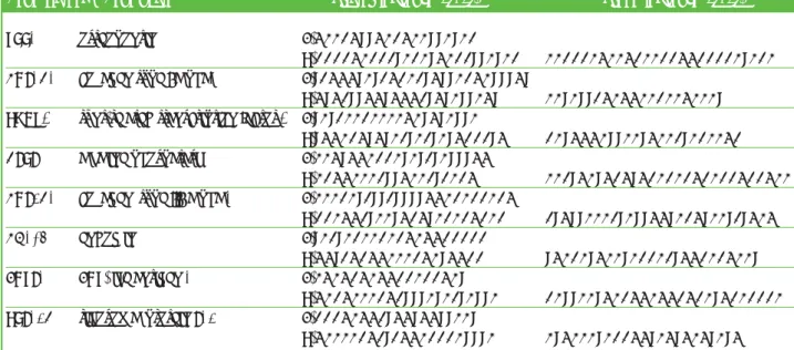
Forward and reverse primers and probes for the selected genes were designed using primer express software (Applied Biosystems, Foster City, CA, USA) and are listed in Table 1.
All PCR reactions were performed in a 20 µl volume using the ABI PRISM 7500 (Applied Biosystems, Foster City, CA, USA). Each reaction contained 10 µl 2X TaqMan universal PCR master mix (Applied Biosystems, Foster City, CA, USA), 400 nM concentration of each primer and 200 nM of the
Table 1Primer and probes used in real time PCR.
SPP1 osteopontin F-GCCAGTTGCAGCCTTCTCA
R-AAAAGCAAATCACTGCAATTCTCA CCAAACGCCGACCAAGGAAAACTCAC
COL1A1 collagen type I alpha1 F-TAGGGTCTAGACATGTTCAGCTTTGT
R-GTGATTGGTGGGATGTCTTCGT CCTCTTAGCGGCCACCGCCCT
RUNX2 runt-related transcription factor 2 F-TCTACCACCCCGCTGTCTTC
R-TGGCAGTGTCATCATCTGAAATG ACTGGGCTTCCTGCCATCACCGA
ALPL alkaline phospatasi F-CCGTGGCAACTCTATCTTTGG
R-CAGGCCCATTGCCATACAG CCATGCTGAGTGACACAGACAAGAAGCC
COL3A1 collagen, type III, alpha 1 F-CCCACTATTATTTTGGCACAACAG
R-AACGGATCCTGAGTCACAGACA ATGTTCCCATCTTGGTCAGTCCTATGCG
CD105 endoglin F-TCATCACCACAGCGGAAAAA
R-GGTAGAGGCCCAGCTGGAA TGCACTGCCTCAACATGGACAGCCT
FOSL1 FOS-like antigen 1 F-CGCGAGCGGAACAAGCT
R-GCAGCCCAGATTTCTCATCTTC ACTTCCTGCAGGCGGAGACTGACAAAC
RPL13A ribosomal protein L13 F-AAAGCGGATGGTGGTTCCT
R-GCCCCAGATAGGCAAACTTTC CTGCCCTCAAGGTCGTGCGTCTG
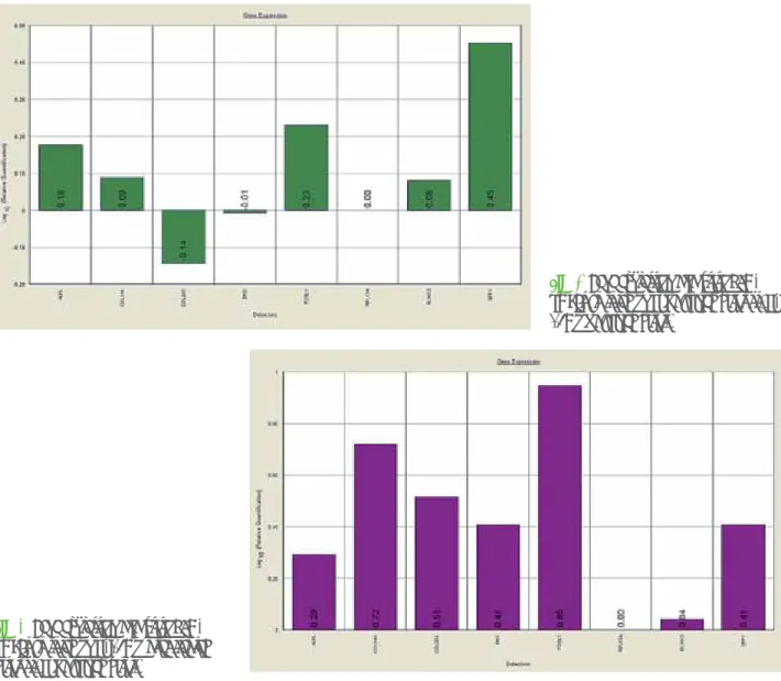
FOSL1 and SPP1, which increase 20% or more. Also COL1A1, and RUNX2 were up-regulated but a lower level. Instead COL3A1 was down-regulated (Fig. 2) like ENG. Quantification was done with the delta/delta calculation method (see M&M section). It was not possible to apply the ANOVA test as we performed biological replication (i.e. we performed several different wells treated in the same time in the same way). In Test 2 (Ti-15Mo laser treated disks vs. Ti machined disks) all investigated genes were up-regulated (Fig. 3).
DISCUSSION
The success of most current orthopedic and dental implants depends on osseointegration. The desire and necessity to increase osseointegration has led to the development of new materials and implant designs (17).
Osteoblast response to Ti implants depends not only on the chemistry of the implant but also on the physical properties of the implant surface, such as microtopography and roughness. The nature of the surrounding bone, hard tissue formation around probe, and cDNA. The amplification profile was
initiated by 10-minute incubation at 95°C, followed by two-step amplification of 15 seconds at 95°C and 60 seconds at 60°C for 40 cycles. All experiments were performed including non-template controls to exclude reagents contamination. PCRs were performed with two biological replicates.
RESULTS
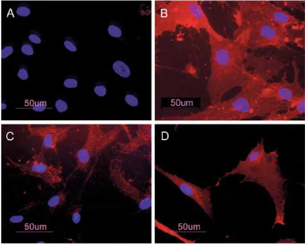
BM-hMSCs were characterized by immuno-fluorescence. The cell surfaces were positive for mesenchymal stem cell marker, CD105, CD90 and CD73 and negative for markers of hematopoietic origin, CD34 (Fig. 1).
Transcriptional expressions of several osteoblast-related genes (ALPL, COLIA1, COL3A1, ENG, FOSL1RUNX2, SPP1) and mesenchymal stem cells marker (ENG) were examined after 7 days of culture on Ti-machined disks, Ti-15Mo machined disks and Ti-15Mo laser treated disks.
In Test 1 (comparison between Ti-15Mo machined disks vs. Ti machined disks) quantitative real-time RT–PCR showed a significant induction of ALPL,
Fig. 1 BM-hMSCs by indirect immunofluorescence (Rhodamine). Cultured cells were positive for the mesenchymal stem cell marker CD73 (A), CD90 (C), CD105 (D) and negative for the hematopoietic markers CD34 (A). Nuclei were stained with DAPI. Original magnification x 40
ability to differentiate, under adequate stimuli, into several mesenchymal lineages, including osteoblasts (19). In the present study, BM-hMSCs were isolated and characterized by morphology and immunophenotype. Isolated BM-hMSCs showed fibroblast-like morphology and were positive for MSC surface molecules (CD90, CD105, CD73) and negative for markers of hematopoietic progenitors (CD34). After 7 days of culture the expression levels of osteodifferentiation genes were measured by relative quantification methods using real-time RT–PCR. In Test 1 (comparison between Ti-15Mo machined disks and Ti-machined disks) quantitative real-time RT–PCR showed that ALPL was significantly up-regulated in treated stem cells with respect to control. ALPL is widely used as a marker of osteoblasts differentiation, an increase in ALPL expression should be associated with osteoblast differentiation. Ti-15Mo machined disks induce significant up-regulation of SPP1 too. This gene encodes osteopontin, which is a phosphoglycoprotein of bone matrix and it is the implants is profoundly influenced by the surface of
the implant (18). Implants physically and chemically interact with the tissues where they are placed and induce the production of systemic mediators that have substantial effects on tissues at more distant sites (18). Titanium is a great material for inducing osseointegration, while a number of other materials, such as stainless steel, tend to promote fibrous tissue formation. This metal is one of the most biocompatible implant materials used in clinics today. Titanium alloys are superior to pure titanium in strength, and thus have replaced pure titanium in many orthopaedic devices (18).
In order to analyze how BM-hMSCs respond to Ti-15Mo alloy with surface modified by laser beam irradiation, in the present study changes in expression of bone related marker genes (RUNX2, SPP1, COLIA1, COL3A1, ALPL and FOSL1) and mesenchymal stem cells marker (ENG) were investigated by real-time RT–PCR. Mesenchymal stem cells are defined as self-renewable, multipotent progenitors cells with the
Fig. 2 Gene expression analysis of BM-hMSCs cultured on Ti machined disks vs. Ti-15Mo machined disks.
Fig. 3 Gene expression analysis of BM-hMSCs cultured on Ti-15Mo laser treated disks vs. Ti machined disks.
most representative non collagenic component of extracellular bone matrix (20). Osteopontin is actively involved in bone resorption processes directly by ostoclasts (21). Osteopontin produced by osteoblasts shows high affinity to the molecules of hydroxylapatite in extracellular matrix and it is chemo-attractant to osteoclasts (22).
FOSL1, another significant up-regulated gene in treated stem cells, encodes for Fra-1, a component of the dimeric transcription factor activator protein-1 (Ap-1), which is composed mainly of Fos (c-Fos, FosB, Fra-1 and Fra-2) and Jun proteins (c-Jun, JunB and JunD). AP-1 sites are present in the promoters of many developmentally regulated osteoblast genes, including alkaline phosphatase, collagen I, osteocalcin. McCabe et al. (23) demonstrated that differential expression of Fos and Jun family members could play a role in the developmental regulation of bone-specific gene expression and, as a result, may be functionally significant for osteoblast differentiation. Kim et al. (24), studying the effect of a new anabolic agents that stimulate bone formation, find that this gene is activated in the late stage of differentiation, during the calcium deposition.
ENG (CD105), a surface marker used to define a bone marrow stromal cell population capable of multilineage differentiation (25), was not affected in treated BM-hMSCs when compared to control at 7 days. Ti-15Mo machined disks also modulate the expression of genes encoding for collagenic extracellular matrix proteins like collagen type 1·1 (COL1A1). COL1A1 was up-regulated as compared to the control when exposed to Ti-15Mo machined disks. Instead, COL3A1 was down regulated, probably because this gene is activated in the late stage of differentiation and is related to extracellular matrix synthesis. Its expression did not have significant change in treated cells respect to control after 7 day of treatment. The trascriptional factor RUNX2 was up-regulated but a lower level. This gene is a key transcriptional modulator of osteoblast differentiation that plays a fundamental role in osteoblast maturation and homeostasis. RUNX2-null mice despite normal skeletal patterning have no osteoblasts and consequently bone tissue. RUNX2 at the early stage of embryogenesis determines the osteoblast lineage from multipotent mesenchymal stem cells (26).
In Test 2 (Ti-15Mo laser treated disks vs. Ti machined disks) all investigated genes were up-regulated (Fig. 3). This results showed that Ti machined disks have higher oseoinductive properties with respect to Ti-15Mo machined disks.
The present study showed the effect of laser modification on Ti-15Mo alloys on BM-hMSCs in the early differentiation stages. Specifically, Ti-15Mo alloy seemed to be inducer of osteogenesis on human stem cells. By comparing Test 1 and Test 2, machined and
laser-surfaced Ti-15Mo alloys respectively, it was detected that COL1A1, COL3A1, FOSL1 and ENG sensibly increased their expression, whereas RUNX2, ALPL and SPP1 expression remain substantially unchanged. This study demonstrates that Ti-15Mo surface roughness affects osteoblast proliferation and gene expression.
CONCLUSION
The laser surface modification allowed obtaining physical-chemical properties and morphology suitable for the use as dental and orthopedics implants. In conclusion, the present study demonstrated that laser treated Ti-15Mo alloys are promising materials for implants application.
ACKNOWLEDGEMENTS
This work was partially supported by Fondazione Cassa di Risparmio di Ferrara, Ferrara, Italy.
The authors are grateful to FAPESP for scholarships (Proc. 04/11751-8 and 08/04867-0) and grants (Proc. 2005/04050-6) that made this work possible. This work was partially supported by the National Research Council (C.N.R.), Rome, Italy, by the Ministry of Education, University and Research (M.I.U.R.), Rome, Italy.
REFERENCES
1. Oliveira NTC, Aleixo G, Caram R, Guastaldi AC. Development of Ti-Mo alloys for biomedical applications: Microstructure and electrochemical characterization. Mater Sci Eng A 2007;452-453:727-731.
2. Oliveira NT, Guastaldi AC. Electrochemical stability and corrosion resistance of Ti-Mo alloys for biomedical applications. Acta Biomater 2009;5(1):399-405.
3. Traini T, Mangano C, Sammons RL, Mangano F, Macchi A, Piattelli A. Direct laser metal sintering as a new approach to fabrication of an isoelastic functionally graded material for manufacture of porous titanium dental implants. Dent Mater 2008 Nov;24(11):1525-1533. 4. Lopez MF, Gutierrez A, Jimenez JA. In vitro corrosion
behaviour of titanium alloys without vanadium. Electrochim Acta 2002;47(9):1359-1364.
5. Kuroda D, Niinomi M, Morinaga M, Kato Y, Yashiro T. Design and mechanical properties of new ‚ type titanium alloys for implant materials. Mater Sci Eng A 1998;243:244-249.
6. Oliveira NTC, Guastaldi AC, Piazza S, Sunseri C. Photo-Electrochemical Investigation of Anodic Oxide Films on as-cast Ti-Mo Alloys. I. Anodic behaviour and effect of Alloy Composition. Electrochim. Acta 2009;54:1395-1402.
force microscopy analysis of different surface treatments of Ti dental implant surfaces. Appl Surf Sci 2004;233:29-34.
8. MacDonald DE, Rapuano BE, Deo N, Stranick M, Somasundaran P, Boskey AL. Thermal and chemical modification of titanium-aluminum-vanadium implant materials: effects on surface properties, glycoprotein adsorption, and MG63 cell attachment. Biomaterials 2004;25(16):3135-3146.
9. Rivera-Denizard O, Diffoot-Carlo N, Navas V, Sundaram PA. Biocompatibility studies of human fetal osteoblast cells cultured on gamma titanium aluminide. J Mater Sci Mater Med 2008;19(1):153-158.
10. Götz HE, Müller M, Emmel A, Holzwarth U, Erben RG, Stangl R. Effect of surface finish on the osseointegration of laser-treated titanium alloy implants. Biomaterials 2004;25(18):4057-4064.
11. Marticorena M, Corti G, Olmedo D, Guglielmotti MB, Duhalde S. Laser surface modification of Ti implants to improve osseointegration. J Phys Conf Ser 2007;59:662-665.
12. Gaggl A, Schultes G, Müller WD, Kärcher H. Scanning electron microscopical analysis of laser-treated titanium implant surfaces--a comparative study. Biomaterials 2000;21(10):1067-1073.
13. Braga FJC, Marques RFC, Filho EdA, Guastaldi AC. Surface modification of Ti dental implants by Nd:YVO4 laser irradiation. Appl Surf Sci 2007;253:9203-9208.
14. Cho SA, Jung SK. A removal torque of the laser-treated titanium implants in rabbit tibia. Biomaterials 2003;24(26):4859-4863.
15. Oliveira NTC, Biaggio SR, Piazza S, Sunseri C, Di Quarto F. Photo-electrochemical and impedance investigation of passive layers grown anodically on titanium alloys. Electrochim Acta 2004;49:4563-4576.
16. Livak KJ, Schmittgen TD. Analysis of relative gene expression data using real-time quantitative PCR and the 2(-Delta Delta C(T)) Method. Methods 2001;25(4):402-408. 17. Zhang H, Ahmad M, Gronowicz G. Effects of transforming growth factor-beta 1 (TGF-beta1) on in vitro mineralization of human osteoblasts on implant materials. Biomaterials 2003;24(12):2013–2020.
18. Kim HJ, Kim SH, Kim MS, Lee EJ, Oh HG, Oh WM, Park SW, Kim WJ, Lee GJ, Choi NG, Koh JT, Dinh DB, Hardin RR, Johnson K, Sylvia VL, Schmitz JP, Dean DD. Varying Ti-6Al-4V surface roughness induces different early morphologic and molecular responses in MG63 osteoblast-like cells. J Biomed Mater Res A 2005;74(3):366–373.
19. Alhadlaq A, Mao JJ. Mesenchymal stem cells: isolation and therapeutics. Stem Cells Dev 2004;13(4):436-448. 20. McKee MD, Farach-Carson MC, Butler WT, Hauschka PV,
Nanci A. Ultrastructural immunolocalization of noncollagenous (osteopontin and osteocalcin) and plasma (albumin and alpha 2HS-glycoprotein) proteins in rat bone. J Bone Miner Res 1993;8(4):485-496.
21. Dodds RA, Connor JR, James IE, Rykaczewski EL, Appelbaum E, Dul E, Gowen M. Human osteoclasts, not osteoblasts, deposit osteopontin onto resorption surfaces: an in vitro and ex vivo study of remodeling bonee. J Bone Miner Res 1995;10(11):1666-1680. 22. Ohtsuki C, Kamitakahara M, Miyazaki T. Bioactive
ceramic-based materials with designed reactivity for bone tissue regeneration. J R Soc Interface 2009 Jun 6;6 Suppl 3:S349-360.
23. McCabe LR, Banerjee C, Kundu R, Harrison RJ, Dobner PR, Stein JL, Lian JB, Stein GS. Developmental expression and activities of specific fos and jun proteins are functionally related to osteoblast maturation: role of Fra-2 and Jun D during differentiation. Endocrinology 1996;137(10): 4398-4408.
24. Kim JM, Lee SU, Kim YS, Min YK, Kim SH. Baicalein stimulates osteoblast differentiation via coordinating activation of MAP kinases and transcription factors. J Cell Biochem 2008 Aug 1;104(5):1906-17.
25. Jin HJ, Park SK, Oh W, Yang YS, Kim SW, Choi SJ. Down-regulation of CD105 is associated with multi-lineage differentiation in human umbilical cord blood-derived mesenchymal stem cells. Biochem Biophys Res Commun 2009 Apr 17;381(4):676-681.
26. Ducy P, Starbuck M, Priemel M, Shen J, Pinero G, Geoffroy V, Amling M, Karsenty G. A Cbfa1-dependent genetic pathway controls bone formation beyond embryonic development. Genes Dev 1999 Apr 15;13(8):1025-1036.