Propionibacterium acnes
Attilio Fabbretti,aCheng-Guang He,aEleonora Gaspari,bSonia Maffioli,bLetizia Brandi,aRoberto Spurio,aMargherita Sosio,b Daniela Jabes,bStefano Donadiob
Laboratory of Genetics, School of Biosciences and Veterinary Medicine, University of Camerino, Camerino, Italya
; Naicons Srl, Milan, Italyb
A chemical derivative of the thiopeptide GE2270A, designated NAI003, was found to possess a substantially reduced antibacte-rial spectrum in comparison to the parent compound, being active against just a few Gram-positive bacteria. In particular, NAI003 retained low MICs against all tested isolates of Propionibacterium acnes and, to a lesser extent, against Enterococcus faecalis. Furthermore, NAI003 showed a time- and dose-dependent killing of both a resistant and a clindamycin-sensitive P. acnes isolate. Gel shift experiments indicated that, like the parent compound, NAI003 retained the ability to bind to elongation factors Tu (EF-Tus) derived from Escherichia coli, E. faecalis, or P. acnes, albeit with reduced efficiency. In contrast, EF-Tus derived from the NAI003-insensitive Staphylococcus aureus or Streptococcus pyogenes did not bind this compound. These results were confirmed by in vitro studies using a hybrid translation system, which indicated that NAI003 can inhibit most efficiently protein synthesis driven by the P. acnes EF-Tu. P. acnes mutants resistant to NAI003 were isolated by direct plating. With one exception, all analyzed strains carried mutations in the tuf gene, encoding EF-Tu. Because of its selective effect on P. acnes in comparison to resident skin flora, NAI003 represents a promising candidate for the topical treatment of acne, which has already completed a phase 1 clinical study.
A
cne vulgaris, a complex disease of the pilosebaceous units of the face and upper trunk, is the most common skin condition seen by physicians. Although it affects almost 100% of adolescents to various degrees and generally wanes as adolescence ends, the disease may persist into adulthood, and it has been estimated that more than 17 million people in the United States are affected by acne (1,2). At least four factors contribute to the development of acne: increased sebum production by the sebaceous glands, follic-ular hyperkeratinization, colonization of the sebaceous follicles with Propionibacterium acnes, and inflammation (3,4).Inflammatory acne is the result of the host response to P. acnes, a pleomorphic, anaerobic rod belonging to the phylum
Actinobac-teria. P. acnes, a component of the normal skin flora, is a usually
harmless commensal largely incapable of tissue invasion or seri-ous infection. The organism catabolizes sebaceseri-ous triglycerides, using the glycerol moiety as a carbon source without catabolizing the fatty acids. While this species has been advocated to play a fundamental role in the etiogenesis of acne (5,6), a significant controversy remains as to how important a role P. acnes plays (4, 7). Recent metagenomic studies have confirmed it to be the major inhabitant of pilosebaceous units (8). Nonetheless, P. acnes is a normal skin commensal not necessarily associated with acne, al-though a correlation between disease and P. acnes phylotypes has been proposed (8). It is also worth pointing out that
Staphylococ-cus epidermidis can produce catabolites that inhibit P. acnes
growth (9), highlighting the importance of an appropriate balance of the microflora for a healthy skin. On the basis of the above considerations, a reduction in the population of P. acnes without affecting other commensal flora may constitute a plausible ap-proach to an effective therapy of acne. However, no antibacterial agent possessing such an antibacterial spectrum has been de-scribed so far.
The thiopeptide GE2270A 1, produced by the actinomycete
Planobispora rosea, is extremely active against numerous
Gram-positive pathogens, including methicillin-resistant strains of
Staphylococcus aureus and vancomycin-resistant Enterococcus spp.
(10). It has been demonstrated that GE2270A binds to domain II of elongation factor Tu (EF-Tu), making contacts with residues 215 to 230, 256 to 264, and 273 to 277 (11). These interactions alter the conformation of EF-Tu, so as to increase its electrophoretic mobility (12) and to inhibit EF-Tu–GTP–aminoacyl-tRNA (aa-tRNA) ternary complex formation. As a result, GE2270A in-terferes selectively with the functioning of the elongation factor in protein synthesis.
During a program aimed at generating analogs of GE2270A by semisynthesis, we observed that one of the derivatives, designated NAI003, showed a surprisingly restricted antibacterial spectrum, mostly limited to P. acnes. Here, we report the antibacterial prop-erties of this compound, investigate the molecular basis for its reduced activity against other Gram-positive bacteria, and pro-vide epro-vidence that NAI003 targets EF-Tu in P. acnes.
MATERIALS AND METHODS
Chemistry. GE2270A (5 g, 3.87 mmol), prepared as described previously (10), was dissolved in dioxane-water-formic acid (10:1:1) and left over-night at 80°C under stirring. After cooling, the solvent was evaporated and concentrated to dryness, and the resulting residue was incubated for 1 h at
Received 28 December 2014 Returned for modification 4 February 2015 Accepted 11 May 2015
Accepted manuscript posted online 18 May 2015
Citation Fabbretti A, He C-G, Gaspari E, Maffioli S, Brandi L, Spurio R, Sosio M, Jabes D, Donadio S. 2015. A derivative of the thiopeptide GE2270A highly selective against Propionibacterium acnes. Antimicrob Agents Chemother 59:4560 –4568.
doi:10.1128/AAC.05155-14.
Address correspondence to Attilio Fabbretti, [email protected], or Stefano Donadio, [email protected].
Copyright © 2015, American Society for Microbiology. All Rights Reserved.
doi:10.1128/AAC.05155-14
on July 17, 2015 by guest
http://aac.asm.org/
room temperature in 0.5 M sodium carbonate. The reaction mixture was then diluted with cold water and brought to pH 6.5 with HCl. The thio-peptides were extracted from the aqueous phase with ethyl acetate and then precipitated from the concentrated organic phase by adding petro-leum ether. The resulting solid (1.3 g, 1 mmol) was dissolved in dimeth-ylformamide (DMF) (10 ml), and 4-amino-N-benzylpiperidine (1.2 mmol), triethanolamine (TEA) (1.4 mmol), and diphenyl phosphorazi-date (1.2 mmol) were added at 0°C. The temperature was allowed to rise to room temperature, and stirring was continued for about 4 h. The reaction mixture was acidified to pH 3 with 1 N HCl and then diluted with water to complete precipitation of the product. The wet solid was dried in air and then purified by flash chromatography (silica gel 60, 230 to 400 mesh; ASTM; Merck) with elution with 3 to 5% methanol in chloroform. Frac-tions containing NAI003 were pooled and evaporated to dryness as a white solid. The data are as follows:1H nuclear magnetic resonance (NMR), dimethyl sulfoxide (DMSO)-d60.86 (d, 3H); 0.90 (d, 3H); 1.91 to 1.70 (n, 2H); 2.26 to 2.05 (m, 2H); 2.60 (s, 3H); 2.91 to 2.69 (m, 4H); 3.40 (s, 3H); 3.51 (br s, 2H); 3.95 to 3.75 (m, 2H); 4.30 (dd, 1H); 4.99 (s, 2H); 7.41 to 7.18 (m, 2H); 8.28 (s, 1H); 8.45 (s, 1H); 8.66 (s, 1H).
Bacterial strains and media. Bacterial strains were from public collec-tions (ATCC) or from the Naicons collection of pathogenic strains (des-ignated by “L” or “ND” prefixes). In particular, the P. acnes strains with an “L” or “ND” prefix were independent clinical isolates collected before 2003 and in 2011, respectively. Brucella broth (BB) and brucella agar (BA), Middlebrook 7H9 broth (7H9B), Mueller-Hinton broth (MHB), Todd-Hewitt broth (THB), and Wilkins-Chalgren agar (WCA) were from Difco Laboratories (Detroit, MI, USA). Cation-adjusted MHB (CAMHB), prepared by adding 20 mg/ml CaCl2and 10 mg/ml MgCl2to MHB, was used for all aerobic bacteria except for streptococci, which were grown in THB; Corynebacterium sp., which was grown in CAMHB with 5% lysed horse blood; and Mycobacterium smegmatis, which was grown in 7H9B supplemented with 0.5 g/liter Tween 80 and 10% oleic acid-albu-min-dextrose-catalase (OADC) enrichment (BD, MD, USA). P. acnes was grown in BB or BA supplemented with 5% lysed horse blood, 5g/ml hemin, and 1g/ml vitamin K1(supplemented BB). All liquid media contained 0.02% bovine serum albumin, unless otherwise noted. All cul-tures were grown at 37°C.
Determination of MICs. MICs were determined by the broth mi-crodilution methodology (13,14); bacteria were inoculated at 5⫻ 105 CFU/ml. Assays were performed in sterile 96-well microtiter plates with round-bottomed wells. Plates were read after 20 to 24 h (aerobic bacteria) or 72 h (Mycobacterium smegmatis; P. acnes, anaerobic atmosphere). GE2270A and its derivatives were dissolved in dimethyl sulfoxide. Clin-damycin and erythromycin (Sigma-Aldrich) were dissolved in water and in 95% ethanol, respectively. Appropriate dilutions were made with the required culture medium immediately before testing.
Time-kill assays. P. acnes colonies from 72-h BA plates were resus-pended in BB at approximately 1⫻ 105to 2⫻ 105CFU/ml and incubated for 24 h in supplemented BB. Then, 0.5 ml of the culture was added to fresh 5 ml supplemented BB in glass vials with plastic caps containing the desired concentration of antibiotic. For this experiment, a series of anti-biotic solutions were made at 100⫻ the desired final concentration in either DMSO (for NAI003) or water (for clindamycin). In all experiments, an extra vial, supplemented with just 50l DMSO, was used as a growth control. Cultures were incubated at 37°C, and anaerobic conditions were achieved with 1:25 (vol/vol) Oxyrase (Oxyrase Inc., Mansfield, OH, USA). At different time points, 0.2-ml aliquots were withdrawn and serially di-luted 1:10 with 0.9% NaCl, and 25l of each dilution was spread onto two to four BA plates. Colonies were counted after 48 to 72 h at 37°C under an anaerobic atmosphere and average values used in time-kill curves (varia-tions in colony numbers were usually within⫾30% of average counts).
Resistance analysis. The occurrence of spontaneous resistant mutants of P. acnes ATCC 6922 was determined by plating 0.1 ml of a stationary-phase culture on WCA containing NAI003 or GE2270A at 1 or 10g/ml. Plates were incubated in an anaerobic atmosphere, with colonies scored
after 5 and 12 days. Colonies emerging on the plates were cultured in BB, centrifuged, and stored at⫺80°C in nutrient broth supplemented with 20% glycerol for MIC determination and analysis of the tuf genes, encod-ing EF-Tu. From selected strains, a cell lysate was made by heatencod-ing at 95°C for 5 min and used for PCR amplifying the entire tuf gene with primers 5=-GTGGCAAAGGCCAAGTTCG-3= and 5=-TCACTTGATGATCTTGG TGACTC-3=. PCR products were directly sequenced (Primm Srl, Mi-lan, Italy). The sequenced segment from the parental strain ATCC 6922 was found to be identical to the tuf gene from P. acnes KPA171202 (15) (GenBankNC_006085.1).
Biochemical methods. Buffers used were the following: buffer A, 25 mM Tris-HCl (pH 7.1), 5% glycerol, 700 mM NaCl, 6 mM -mercapto-ethanol, 0.1 mM phenylmethylsulfonyl fluoride (PMSF), 0.1 mM benz-amidine; buffer B, 25 mM Tris-HCl (pH 7.1), 300 mM NaCl, 5% glycerol, 20 mM imidazole, 6 mM-mercaptoethanol, 0.1 mM PMSF, and 0.1 mM benzamidine; buffer C, 25 mM Tris-HCl (pH 7.1), 300 mM NaCl, 5% glycerol, 300 mM imidazole, 6 mM-mercaptoethanol, 0.1 mM PMSF, 0.1 mM benzamidine; buffer D, 20 mM Tris-HCl (pH 7.1), 100 mM NH4Cl, 5% glycerol, 6 mM-mercaptoethanol, 0.1 mM EDTA; buffer E, 10 mM Tris-HCl (pH 7.7), 10 mM magnesium acetate, 60 mM NH4Cl.
Cloning, overexpression, and purification of EF-Tus. The strategy used to clone the tuf genes from genomic DNA of S. aureus (ATCC 25923), Enterococcus faecalis (ATCC 29922), Streptococcus pyogenes (ATCC 700294), and P. acnes (ATCC 6919) was essentially as reported by Zeng (16). Briefly, four different phosphorylated primers (Table 1) carry-ing NcoI and BamHI restriction sites were used in two separate DNA amplification reactions. Equal volumes of the two PCR mixtures were combined and subjected to a 5-min denaturation at 95°C and a 15-min renaturation at room temperature. Of the resulting final products, 25% are expected to carry NcoI and BamHI cohesive ends, which can be ligated with the appropriately restricted expression vector pETM11. The ligation products were then transformed into the Escherichia coli BL21 strain.
E. coli BL21(DE3)pLysS cells harboring the pETM11 derivatives were grown in LB medium at 37°C up to an optical density at 600 nm (OD600) of 0.7, when 1 mM isopropyl--D-1-thiogalactopyranoside was added. After 4 h at 37°C, the cells were harvested by centrifugation at 5,000 rpm for 10 min at 4°C and the pellet, resuspended in buffer A, was stored at ⫺80°C. After thawing, the cells were lysed by sonication, and the resulting cell extract was cleared by centrifugation at 13,000 rpm for 45 min at 4°C. The cell extract was subsequently loaded on a 5-ml nickel-nitrilotriacetic TABLE 1 Oligonucleotide primers used for sticky-end PCR
tufA gene source Sequence (5=-3=)a
S. aureus F: CATGGCAAAAGAAAAATTCGATCGTTC R: CTTATTTAATGATTTCAGTAACAACGC F: GCAAAAGAAAAATTCGATCGTTCTAAAG R: GATCCTTATTTAATGATTTCAGTAACAACG E. faecalis F: CATGGCAAAAGAAAAATTTGACCGTTC R: CTTATTTAATGATTTCAGTAACAACGC F: GCAAAAGAAAAATTTGACCGTTCTAAATC R: GATCCTTATTTAATGATTTCAGTAACAACG S. pyogenes F: CATGGCAAAAGAAAAATACGATCGTAG R: GATCCTTAAGCTTCGTTTCTGAAACG F: GCAAAAGAAAAATACGATCGTAGTAAAC R: CTTAAGCTTCGATTTCTGAAACGATACC P. acnes F: CATGGCAAAGGCCAAGTTCGAGCGG R: GATCCTCACTTGATGATCTTGGTGACTCG F: GCAAAGGCCAAGTTCGAGCGGACC R: CTCACTTGATGATCTTGGTGACTCGACC a
F, forward; R, reverse. The methodology employed (16) requires two forward and two reverse primers for each amplification.
on July 17, 2015 by guest
http://aac.asm.org/
acid (Ni-NTA) chromatographic column equilibrated in buffer A. The column was initially washed with 10 bed volumes of buffer A, followed by an additional wash with 5 bed volumes of buffer B. EF-Tu was eluted from the column with buffer C. Fractions containing EF-Tu were dialyzed to reduce salt concentration against buffer C containing 100 mM NaCl (without imidazole) and digested by tobacco etch virus (TEV) protease (enzyme/substrate ratio, 1:40) overnight at 20°C, with 20% extra TEV protease added for an additional 3 to 4 h. The reaction mixture was again loaded on the Ni-NTA resin, and the cleaved protein, without the His tag, was recovered in the flowthrough of the column. EF-Tu was concentrated by centrifugation at 4,000 rpm for 60 min at 4°C in Amicon Ultra-3 cen-trifugal filter devices, and after dialysis at 4°C against buffer D, it was stored at⫺80°C in small aliquots.
In vitro tests. 30S and 50S ribosomal subunits, 027-IF2Cp(A) mRNA,
EF-G, and E. coli cell extract were prepared as described previously (17, 18). In vitro mRNA translation driven by 027-IF2Cp(A) mRNA was car-ried out as described previously (17). Native polyacrylamide (12%) gel electrophoresis of EF-Tu preincubated with 1 mM GTP in the presence of increasing concentrations (0.1, 0.5, 1, 5, 10, 50, and 100M) of GE2270A or NAI003 was performed in Tris-glycine buffer (pH 8.3) as described previously (11).
Poly(U) hybrid translation system. Poly(U) translation was carried out in 30l of buffer E containing 3 mM phosphoenolpyruvate, 0.05 g/ml pyruvate kinase, 1 mM GTP, 0.15 g/l poly(U), 10 M [3 H]Phe-tRNA, and 0.2M E. coli 30S, 50S, and EF-G. Twenty picomoles of puri-fied EF-Tu (from E. coli, S. aureus, E. faecalis, S. pyogenes, or P. acnes) was added to the mixture after a 5-min incubation at room temperature in the presence of increasing concentrations of GE2270A or NAI003. After 30 min at 37°C, the level of poly(U) translation was quantified from the amount of acid-insoluble [3H]Phe-tRNA incorporated.
RESULTS
Synthesis of NAI003. The oxazolidine-linkedL-Ser-L-Pro-NH2
side chain of GE2270A undergoes, on acid treatment, an N-O acyl shift, forming a diketopiperazine ester that can be easily removed by mild basic treatment (19,20). The newly formed carboxylic acid 2 can thus be effectively amidated, as for example with 4-ami-no-N-benzylpiperidine, to afford NAI003 (Fig. 1).
Antibacterial spectrum of NAI003. The parent compound GE2270A is highly active against Gram-positive bacteria but not effective against Gram-negative species (10).Table 2shows the activity of GE2270A against Staphylococcus, Streptococcus, and
En-terococcus spp., with MICs in the ranges ofⱕ0.015 to 0.25, 0.06 to
2, and 0.008 to 0.015g/ml, respectively. The acid derivative 2 main-tains substantially the same spectrum as GE2270A, although MICs were 32- to 64-fold and 4- to 16-fold higher against staphylococci and streptococci, respectively (Table 2). Surprisingly, NAI003 had lost potency against most of the tested strains: measurable MICs were observed against Enterococcus spp., with MICs in the 0.5- to 16-g/ml range, and against Mycobacterium smegmatis. Interestingly, in the latter case, compounds 1, 2, and 3 were found to be equally active, with MICs of 4 to 8g/ml (Table 2). It should be noted that all four Corynebacterium strains tested in this experiment were equally insensitive to the three compounds, suggesting that this genus may be insensitive to this class of thiopeptides (Table 2).
We then wondered whether, among the pathogenic
Actinobac-teria, some species might have retained sufficient sensitivity to
NAI003. Preliminary experiments indicated that P. acnes was suf-ficiently sensitive to this compound to warrant further investiga-tion. The results of these evaluations are reported inTable 3, where it can be seen that Propionibacterium spp., including P.
acnes, were highly sensitive to NAI003, with MIC values in the
range of 4 to 250 ng/ml. In contrast to most bacterial species in Table 2, P. acnes appears to be equally sensitive to all of the three thiopeptides tested in this study (Table 3). Among the tested P.
acnes isolates, one strain was highly resistant to clindamycin and
erythromycin, an increasingly frequent phenotype among P. acnes isolates (21). As expected from their different targets, NAI003 and GE2270 retained activity against this strain.
Killing effect on P. acnes. To our knowledge, a single study has been performed to evaluate the effect of topical treatment on the number of P. acnes cells on the forehead of healthy volunteers: a reduction of approximately 1 log CFU was observed after 1 or 2 weeks of daily applications of 1% clindamycin (22,23).
We thus wondered whether NAI003 had any effect in vitro on the number of viable P. acnes cells in comparison with clindamy-cin. To this end, we tested the effect of the two antibiotics when added to exponentially growing cells. The results are illustrated in Fig. 2A. Under the experimental conditions used, P. acnes reached stationary phase (108CFU/ml) after approximately 48 h, with an apparent duplication time of 4 to 5 h. When these cultures were exposed to clindamycin at 1⫻, 10⫻, or 100⫻ the MIC, a slow decline in viable counts was observed during the first 48 h, fol-FIG 1 Synthetic route to NAI003. DPPA, diphenyl phosphorazidate.
on July 17, 2015 by guest
http://aac.asm.org/
lowed by an apparent faster decline afterwards, resulting in viable counts below the detection limit of the assay (around 20 CFU/ml). In contrast, the NAI003 killing rate was relatively constant with time, leading after 6 days to a 2- and 3-log reduction in viable counts at 10⫻ and 100⫻ the MIC. In the presence of 1⫻ the MIC, after an approximately 24-h lag, the strain was eventually able to grow, albeit at a reduced rate (Fig. 2A).
A similar experiment was then performed with strain ND063311, which is resistant to clindamycin (MIC, 64g/ml) but equally sensitive to NAI003 (Table 2). While NAI003 could be
used again at 1⫻, 10⫻, and 100⫻ the MIC, we could test clinda-mycin only at 1⫻ (i.e., 64 g/ml) and 4⫻ (i.e., 256 g/ml) the MIC. The results are shown inFig. 2B. Neither clindamycin nor NAI003 at 1⫻ the MIC had a significant effect on the viable counts of the strain but simply resulted in growth retardation and 1-log-lower CFU per milliliter at stationary phase. At higher concentra-tions, NAI003 was able to decrease the number of viable counts in a dose-dependent manner, with again a 3-log reduction seen after 6 days. Interestingly, the behaviors of NAI003 and clindamycin at intermediate concentrations (10⫻ and 4⫻ the MIC, respectively) TABLE 2 MICs of GE2270A, compound 2, and NAI003 against selected Gram-positive bacteria
Species and strain
MIC (g/ml) of antibiotic:
GE2270A Compound 2 NAI003 Vancomycin
Staphylococcus aureus ATCC 29213 ⱕ0.015 0.5 ⬎128 1 ND041009 ⱕ0.015 0.5 ⬎128 1 ND018107 ⱕ0.015 1 ⬎128 0.25 ND018107 0.03 1 ⬎128 0.5 ND055710 0.03 1 ⬎128 0.5 ND055910 0.03 0.5 ⬎128 1 Staphylococcus epidermidis ND006607 ⱕ0.015 1 ⬎128 1 ND015007 0.125 2 ⬎128 2 ND020708 0.03 0.5 ⬎128 2 ND021208 0.03 2 ⬎128 2 ND024908 ⱕ0.015 1 ⬎128 2 ND025708 0.03 1 ⬎128 2 ND026508 0.06 4 ⬎128 1 Staphylococcus capitis ND004307 0.03 2 ⬎128 2 ND018807 0.25 8 ⬎128 2 Streptococcus agalactiae ND050210 2 ⬎128 ⬎128 0.5 L310 2 32 ⬎128 0.5 Streptococcus pyogenes L1303 0.5 2 ⬎128 0.5 L800 0.5 4 ⬎128 0.5
Streptococcus pneumoniae ATCC 49619 0.06 0.5 ⬎128 0.25 Enterococcus faecalis VanS L559 0.008 NTa 0.5 0.5 VanA L560 0.008 NT 0.5 ⬎128 Enterococcus faecium VanS L568 0.015 NT 16 2 VanA L569 0.015 NT 4 ⬎128 Mycobacterium smegmatis mc2155 4 8 4 NT Micrococcus luteus L108 ⱕ0.015 1 ⬎128 0.125 Corynebacterium jeikeium L2508 ⬎128 ⬎128 ⬎128 1 L2645 ⬎128 ⬎128 ⬎128 0.5 Corynebacterium sp. L689 ⬎128 ⬎128 ⬎128 1 L691 ⬎128 ⬎128 ⬎128 0.5 aNT, not tested.
on July 17, 2015 by guest
http://aac.asm.org/
Downloaded from
were similar, with some decrease in viable counts up to 3 to 4 days, followed by a tendency of viable counts to rebound (Fig. 2B).
In conclusion, these experiments indicate that NAI003 at 100⫻ the MIC (i.e., 1.5g/ml) can decrease the number of viable counts to similar extents for both a clindamycin-sensitive (strain ND062711) and a clindamycin-resistant (ND063111) strain. In contrast, killing by clindamycin can occur against the sensitive strain at lower MIC multiples and concentrations (i.e., 0.06g/ ml) than those for NAI003. However, with the clindamycin-resis-tant strain tested here, 256g/ml clindamycin led to a lower re-duction in viable counts than 1.5g/ml NAI003.
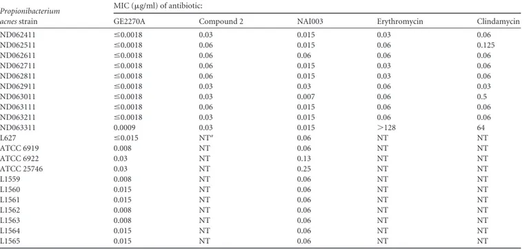
NAI003 binds to and inhibits the function of EF-Tu from Escherichia coli. To confirm that the molecular target of NAI003 is EF-Tu, the assay described by Anborgh and Parmeggiani was initially used (12). This test takes advantage of the different mo-bilities of free E. coli EF-Tu–GTP and of the EF-Tu–GTP– GE2270A complex in native polyacrylamide gel electrophoresis (12,24).
The results shown inFig. 3indicate that NAI003 is indeed able to alter the electrophoretic mobility of E. coli EF-Tu, thereby con-firming that this elongation factor is also the target of this mole-cule. It is also interesting that the difference in the electrophoretic TABLE 3 MICs of GE2270A, compound 2, and NAI003 against P. acnes
Propionibacterium acnes strain
MIC (g/ml) of antibiotic:
GE2270A Compound 2 NAI003 Erythromycin Clindamycin
ND062411 ⱕ0.0018 0.03 0.015 0.03 0.06 ND062511 ⱕ0.0018 0.06 0.015 0.06 0.125 ND062611 ⱕ0.0018 0.06 0.06 0.06 0.06 ND062711 ⱕ0.0018 0.06 0.015 0.03 0.06 ND062811 ⱕ0.0018 0.06 0.015 0.03 0.06 ND062911 ⱕ0.0018 0.03 0.03 0.06 0.03 ND063011 ⱕ0.0018 0.03 0.007 0.06 0.5 ND063111 ⱕ0.0018 0.06 0.015 0.06 0.06 ND063211 ⱕ0.0018 0.03 0.015 0.06 0.06 ND063311 0.0009 0.03 0.015 ⬎128 64 L627 ⱕ0.015 NTa 0.06 NT NT ATCC 6919 0.008 NT 0.06 NT NT ATCC 6922 0.03 NT 0.13 NT NT ATCC 25746 0.03 NT 0.25 NT NT L1559 0.008 NT 0.06 NT NT L1560 0.015 NT 0.06 NT NT L1561 0.015 NT 0.06 NT NT L1562 0.008 NT 0.06 NT NT L1563 0.008 NT 0.06 NT NT L1564 0.015 NT 0.06 NT NT L1565 0.015 NT 0.06 NT NT aNT, not tested.
FIG 2 Killing kinetics of P. acnes. Effect of NAI003 (closed symbols, solid lines) and clindamycin (empty symbols, dashed lines) on the viability of P. acnes, using the clindamycin-sensitive ND062711 (A) and clindamycin-resistant ND06311 (B) isolates. Compounds were added at 1⫻ MIC (triangles), 10⫻ MIC (circles), or 100⫻ MIC (squares). In panel B, clindamycin was used at only 1⫻ (open triangles) and 4⫻ (open diamonds) the MIC. Growth controls are shown for both panels by a thick dashed line.
on July 17, 2015 by guest
http://aac.asm.org/
mobilities between free and antibiotic-bound EF-Tu is smaller for NAI003 than for GE2270A (Fig. 3B). This finding suggests that the interaction of the two antibiotics with EF-Tu induces different conformational changes of the elongation factor. From the gel shift experiments, it can be surmised that low concentrations (0.5 to 1M) of GE2270A increase the mobility of more than 50% of EF-Tu, while greater concentrations (approximately 10M) of the derivative are required to shift the same amount of protein. In addition, from the gel shift experiments it can be observed that low concentrations (0.5 to 1M) of GE2270A increase the mobility of EF-Tu by more than 50%, while greater concentrations (approx-imately 10M) of the derivative are required to shift the same amount of protein.
GE2270A is known to inhibit in vitro protein synthesis in an E.
coli-based cell-free system. When the effect of NAI003 was
com-pared to that of GE2270A in an E. coli in vitro translation system programmed with the 027-IF2Cp(A) mRNA (17), both antibiot-ics proved to interfere with protein synthesis but to different ex-tents: whereas GE2270A caused 50% inhibition of protein synthe-sis at a 10M concentration, a 10-fold-higher concentration of NAI003 was necessary to achieve 25 to 30% inhibition only (Fig. 3C).
From the results of the in vitro translation assays and from those obtained with the gel shift experiments, it is possible to deduce that both GE2270A and NAI003 form a stable interaction with elongation factor EF-Tu that causes an altered electropho-retic mobility of the protein in a native gel. However, the chemical modification of GE2270A which gives rise to NAI003 reduces by almost 1 order of magnitude the affinity of the latter antibiotic for EF-Tu and also reduces its efficiency as a protein synthesis inhib-itor in E. coli (Fig. 3C).
Effect of NAI003 on different EF-Tus. To understand whether the difference in the antibacterial spectra of GE2270A and NAI003 might correlate with differences in affinities for the corresponding EF-Tus, in vitro experiments were carried out with EF-Tu purified from various organisms, namely, the NAI003-insensitive S. aureus and S. pyogenes, the moderately sensitive E. faecalis, and the highly sensitive P. acnes. To do so, the tuf genes encoding the EF-Tus from these organisms were amplified and cloned in the expression
vector pETM-11, following essentially the method described by Zeng (16). This procedure and the presence of an N-terminal oligohistidine tail afforded sufficient amounts of protein from the four microorganisms after affinity chromatography. After purifi-cation, the oligohistidine tail was removed by treatment with the TEV protease and the affinities of these factors for NAI003 and for GE2270A were compared by gel shift assays as described above.
As seen in Fig. 4, the different EF-Tus bind GE2270A with approximately the same affinities, at least within the resolution limits of the experimental technique used, but display appreciable differences in their capacities to bind NAI003. Indeed, whereas the FIG 3 Effects of GE2270A and NAI003 on the electrophoretic mobility of EF-Tu and on in vitro translation. (A) Migration on native polyacrylamide gel of E. coli EF-Tu (preincubated with GTP) in the presence of increasing concentrations (from left to right, 0.1, 0.5, 1, 5, 10, 50, and 100M) of GE2270A (lanes 2 to 8) or NAI003 (lanes 9 to 15). (B) Electrophoretic mobility difference between EF-Tu–GTP alone (lane 1) and in the presence of 1M GE2270A (lane 2) or of 10 M NAI003 (lane 3). The two arrows indicate the different migrations of EF-Tu in complex with GE2270A or with NAI003. (C) Inhibition by GE2270A () or NAI003 () in a protein synthesis system based on an E. coli extract programmed with 027-IF2Cp(A) mRNA.
FIG 4 Effect of GE2270A and NAI003 on the electrophoretic mobilities of different EF-Tus. Migration on native polyacrylamide gels of EF-Tu from E. coli (A), S. aureus (B), P. acnes (C), and S. pyogenes (D) in the presence of increasing concentrations (1, 4, 19, 50, and 100M, respectively) of GE2270A (lanes 2 to 6) or of NAI003 (lanes 7 to 11).
on July 17, 2015 by guest
http://aac.asm.org/
proteins from E. coli and P. acnes can clearly bind NAI003, as judged from their different electrophoretic mobilities in the pres-ence of increasing concentrations of the antibiotic, the same be-havior was not observed for the factors from S. aureus and S.
pyogenes, whose mobilities are hardly affected by the presence of
NAI003.
The electrophoretic assay used in the experiments shown above yields qualitative but not sufficiently quantitative data to permit an estimation of the binding constants of the antibiotics for the various types of EF-Tu. In fact, the altered mobility is caused by the net electrical charge displayed on the protein surface as a result of antibiotic-induced conformational change. Different ligands can induce different changes on EF-Tu, and some of these alterations may not trigger variations of the electrical charge. Fur-thermore, the lack of a mobility shift does not constitute an abso-lute proof of the lack of an interaction between the elongation factors and the antibiotic. For these reasons, in vitro translation tests were carried out to detect a possible inhibition of EF-Tu function. Since EF-Tu is involved in the elongation stage of pro-tein synthesis, these experiments were performed in a poly(U)-dependent poly(Phe) synthesis test, a translational assay that does not depend upon the complex initiation pathway of protein syn-thesis (17). The tests were carried out using a hybrid system in which ribosomes and EF-G purified from E. coli were incubated with [3H]Phe-tRNA and a poly(U) template in the presence of an
appropriate energy-regenerating system and of the appropriate EF-Tu protein.
When increasing amounts of EF-Tu originating from the se-lected bacteria were added to the heterologous cell-free system, the response was similar to that observed upon the addition of the homologous E. coli EF-Tu. The addition of 10 pmol of E. coli EF-Tu promoted the incorporation of⬃90 pmol of [3H]Phe in a hot-acid-insoluble product while the incorporation promoted by the same amount of EF-Tu from P. acnes, S. aureus, and E. faecalis was⬃40 pmol. The only exception was found with S. pyogenes EF-Tu, whose efficiency was about 15% of that of E. coli (Fig. 5A). These preliminary data indicate that the EF-Tus from the selected bacteria are compatible with the E. coli components in the heter-ologous translation system. In turn, these data indicate that this type of test can be used to identify EF-Tu inhibitors, even in the
case of the less efficient S. pyogenes EF-Tu-dependent system, and to compare the inhibitions by GE2270A and by NAI003 on the different EF-Tus.
The graph presented inFig. 5Bshows that protein synthesis was fully inhibited by GE2270A independently of the source of EF-Tu, with a 50% inhibitory concentration (IC50) between 0.2
and 0.5M. With the exception of translation in the presence of S.
pyogenes EF-Tu, at⬃1 M GE2270A inhibition reached 100%
(Fig. 5B). The results obtained in the same test with NAI003 indi-cate that at⬃1 M this antibiotic caused ⬃25 to 30% inhibition when translation depended upon E. coli or E. faecalis EF-Tu but had no effect on the systems containing S. aureus or S. pyogenes EF-Tus, which are only slightly inhibited at very high NAI003 concentrations (Fig. 5C). On the other hand, the translational systems dependent on P. acnes EF-Tu proved to be the most sen-sitive to NAI003 inhibition, translation being reduced by⬎50% at ⬃1 M NAI003 (Fig. 5C). These findings are overall consistent with the conclusions drawn from the data presented inFig. 4and allow for the conclusion that the replacement of the C-terminal moiety in GE2270A with the 4-amino-N-benzylpiperidine moiety present in NAI003 results in a weaker or different interaction of the latter molecule with EF-Tu and a general reduction of its in-hibitory activity. However, this same chemical modification re-sulted in a molecule endowed with an increased selectivity in the inhibition of EF-Tu from P. acnes.
Resistance to NAI003 is due to mutations in P. acnes EF-Tu. The frequency of resistance to this class of thiopeptides was eval-uated in parallel experiments with NAI003 and GE2270A. When
P. acnes was plated on medium containing 1 or 10g/ml
antibi-otic, colonies appeared at frequencies of 6.3⫻ 10⫺11or 3.1⫻ 10⫺11, respectively, for GE2270A and at frequencies of 1.5⫻ 10⫺10 and 9.4⫻ 10⫺11, respectively, for NAI003.
Twelve independent NAI003risolates were evaluated and
char-acterized for the possible presence of mutations in the tuf gene, encoding EF-Tu, as well as for their susceptibilities to GE2270A, NAI003, and compound 2. With one exception, all resistant col-onies harbored one mutation in the tuf gene for a total of 8 inde-pendent changes at 6 different positions (Table 4). Some of the observed mutations (G260R and G260C) occur at a residue (G257 in E. coli and Bacillus subtilis EF-Tu) previously reported to confer FIG 5 Effects of GE2270A and NAI003 on in vitro protein synthesis. (A) Poly(U)-dependent incorporation of [3H]Phe in a hot-trichloroacetic acid-insoluble
product in the presence of increasing concentrations of different EF-Tus. The amounts of Phe incorporated with E. coli EF-Tu are indicated on the right ordinate, while those obtained with EF-Tu from P. acnes, S. aureus, E. faecalis, or S. pyogenes are indicated on the left ordinate. (B and C) Effects of GE2270A (B) and NAI003 (C) on in vitro translation with different EF-Tus. Symbols for EF-Tu are as follows: black circles, E. coli; red diamonds, P. acnes; purple squares, E. faecalis; green inverted triangles, S. aureus; turquoise triangles, S. pyogenes.
on July 17, 2015 by guest
http://aac.asm.org/
GE2270A resistance through the G257S substitution (11,25,26). Interestingly, in the three-dimensional (3D) structure of the com-plex between Thermus thermophilus EF-Tu, GTP, and GE2270A, a Gln residue (equivalent to Q99 in the P. acnes protein) makes direct contact with the antibiotic. Consistently, three independent
P. acnes isolates carry the Q99H mutation. Finally, two
indepen-dent mutations (N276K and N276S) were observed at an Asn residue (equivalent to N273 in the E. coli protein) that also makes direct contact with the antibiotic (11). When transposed to the crystal structure of T. thermophilus EF-Tu, the other observed mutations (Table 4) map to domain I (H68Q and V81A) or II (M263T), although they do not appear to be directly involved in antibiotic binding.
In terms of resistance levels, some mutations (i.e., V81A, G260C, and G260R) confer elevated protection to NAI003, while mutant strains harboring the other mutations exhibit intermedi-ate resistance (Table 4). Remarkably, although the different mu-tations afforded an increased resistance to GE2270 or to com-pound 2 with respect to the parental strain, none of them was sufficient to confer resistance levels equivalent to those achieved against NAI003 (Table 3). It should be noted, however, that we sequenced the tuf gene only and the existence of additional muta-tions elsewhere in the genome cannot be ruled out. Indeed, one mutant (L1015R86) did not show any mutation in the tuf gene, despite its high level of resistance to NAI003 (Table 4). Thus, further studies will be necessary to understand the full range of mutations conferring resistance to NAI003.
DISCUSSION
The results presented here demonstrate that NAI003 binds to the EF-Tu from P. acnes with an affinity comparable to that of GE2270A and that its binding alters the electrophoretic mobility to the same extent as that observed with GE2270A. Furthermore, the translation test performed demonstrates that NAI003 inhibits
in vitro P. acnes EF-Tu-dependent translation to a level similar to
that obtained with GE2270. That EF-Tu is the target of NAI003 in
vivo is confirmed by the observation that most spontaneous
resis-tant muresis-tants of P. acnes carry mutations in the tuf gene, corre-sponding to residues already established to confer resistance to or to bind GE2270A (11).
GE2270A is a better inhibitor of P. acnes EF-Tu than its deriv-ative NAI003. Consistently, GE2270A MICs tend to be lower than those of NAI003. Nonetheless, we were surprised to observe that mutations in EF-Tu that confer complete resistance to NAI003 in
vivo, some of which correspond to residues previously
demon-strated to be critical for GE2270A binding, afford only partial pro-tection to GE2270A in P. acnes. Further studies will be necessary to explain this observation.
We have demonstrated that the lack of sensitivity of many Gram-positive bacteria to NAI003 is correlated with a lower inhi-bition of their EF-Tus by this antibiotic. In this respect, the ob-served extent of EF-Tu inhibition (Fig. 5C) well correlates with the MIC values for the cognate microorganism (Table 1). It should be noted that GE2270A is able to effectively inhibit all EF-Tus tested so far, with natural resistance documented only in the producer strain Planobispora rosea (25) and in the Corynebacterium species tested here (Table 2). This is consistent with the fact that EF-Tu is a highly conserved protein with limited variation among diver-gent bacterial taxa. Despite these features, GE2270A has been con-verted by a relatively simple chemical modification into a deriva-tive with a dramatically reduced antibacterial spectrum. This has resulted from the fortuitous occurrence of two factors: the highly reduced susceptibility of EF-Tus from the three Firmicutes species analyzed here and the lack of permeability of E. coli (and presum-ably of other Gram-negative bacteria) to this class of thiopeptides, which protect an otherwise partially sensitive EF-Tu. In light of these results, it would be interesting to conduct further experi-ments aimed at elucidating the 3D structure of the P. acnes EF-Tu complexed with NAI003 and compare it to the EF-Tu–GE2270A complex. This would provide insights into the nature of the dif-ferent EF-Tu conformations induced by the two antibiotics and enable predictive models of which bacterial strains may be sensi-tive to NAI003 or to other GE2270A derivasensi-tives.
NAI003 was found to be equally active against 21 independent
P. acnes strains, including one highly resistant to clindamycin and
erythromycin (Table 3). Additional testing confirmed that this GE2270A derivative retains activity against several independent clindamycin- and erythromycin-resistant P. acnes clinical isolates (D. Jabes, unpublished data). This observation is not surprising, TABLE 4 Genotypes and phenotypes of P. acnes NAI003rmutants
Strain tuf mutationa
EF-Tu MIC (g/ml) of antibiotic:
Substitutionb Domainc NAI003 GE2270A Compound 2
ATCC 6922 0.015 0.007 ⱕ0.06 L1015R13 C207A H68Q I 8 0.015 0.5 L1015R51 T245C V81A I 32 ⱕ0.06 1 L1015R6 G300T Q99H I 8 0.015 1 L1015R5 G300T Q99H I 2 0.007 0.5 L1015R7 G300T Q99H I 1 0.003 0.25 L1015R24 G781C G260R II ⬎128 0.125 0.25 L1015R8 G781T G260C II 32 0.125 2 L1015R96 T791C M263T II 4 ⱕ0.06 4 L1015R73 A830G N276S II 1 0.007 0.125 L1015R72 A830G N276S II 2 0.007 0.25 L1015R21 C831G N276K II 1 0.03 0.25 L1015R86 None None 128 0.25 4
aNucleotide numbering is based on tuf sequence from P. acnes KPA171202 (15). b
Amino acid numbering is based on EF-Tu sequence from P. acnes KPA171202 (15).
cAs defined by Parmeggiani et al. (11).
on July 17, 2015 by guest
http://aac.asm.org/
since clindamycin and erythromycin resistance in P. acnes mostly involves mutation in 23S RNA (27–29), a site unrelated to the interaction of EF-Tu with the ribosome.
Antibacterial agents with an extremely narrow spectrum hold the promise of curing infections with minimal disturbance of commensal flora. This might be particularly true for topical appli-cations, where a diverse and body-specific flora is expected to contribute to skin health (30–32). The compound previously des-ignated BIK 0376 has already completed a clinical study in healthy volunteers as a topical application (32,33). A clinical study will be necessary to establish its efficacy.
ACKNOWLEDGMENTS
This work was partially supported by a grant from Regione Lombardia (Progetto ATP).
We thank Gabriella Romanò and Paola Guglierame for early contri-butions to this project and Cristina Brunati for performing the time-kill assays. We are also grateful to Claudio O. Gualerzi for valuable advice and insights into this work.
REFERENCES
1. Bergfeld W. 1995. The evaluation and management of acne: economic considerations. J Am Acad Dermatol 5:S52–S56.
2. Bhate K, Williams HC. 2013. Epidemiology of acne vulgaris. Br J Der-matol 168:474 – 485.http://dx.doi.org/10.1111/bjd.12149.
3. Johnson BA, Nunley JR. 2000. Topical therapy for acne vulgaris. Postgrad Med 107:69 – 80.
4. Shaheen B, Gonzalez M. 2013. Acne sans P. acnes. J Eur Acad Dermatol Venereol. 27:1–10.http://dx.doi.org/10.1111/j.1468-3083.2012.04516.x. 5. Webster GF, Leyden JJ, Musson RA, Douglas SD. 1985. Susceptibility of
Propionibacterium acnes to killing and degradation by human neutrophils and monocytes in vitro. Infect Immun 49:116 –121.
6. Beylot C, Auffret N, Poli F, Claudel JP, Leccia MT, Del Giudice P, Dreno B. 2014. Propionibacterium acnes: an update on its role in the pathogenesis of acne. J Eur Acad Dermatol Venereol 28:271–2788.http: //dx.doi.org/10.1111/jdv.12224.
7. Dessinioti C, Katsambas AD. 2010. The role of Propionibacterium acnes in acne pathogenesis: facts and controversies. Clin Dermatol 28:2–7.http: //dx.doi.org/10.1016/j.clindermatol.2009.03.012.
8. Fitz-Gibbon S, Tomida S, Chiu BH, Nguyen L, Du C, Liu M, Elashoff D, Erfe MC, Loncaric A, Kim J, Modlin RL, Miller JF, Sodergren E, Craft N, Weinstock GM, Li H. 2013. Propionibacterium acnes strain populations in the human skin microbiome associated with acne. J Inves-tig Dermatol 133:2152–2160.http://dx.doi.org/10.1038/jid.2013.21. 9. Wang Y, Kuo S, Shu M, Yu J, Huang S, Dai A, Two A, Gallo RL, Huang
CM. 2014. Staphylococcus epidermidis in the human skin microbiome mediates fermentation to inhibit the growth of Propionibacterium acnes: implications of probiotics in acne vulgaris. Appl Microbiol Biotechnol 98:411– 424.http://dx.doi.org/10.1007/s00253-013-5394-8.
10. Goldstein BP, Berti M, Ripamonti F, Resconi A, Scotti R, Denaro M. 1993. In vitro antimicrobial activity of a new antibiotic, MDL 62,879 (GE2270 A). Antimicrob Agents Chemother 37:741–745.http://dx.doi .org/10.1128/AAC.37.4.741.
11. Parmeggiani A, Krab IM, Okamura S, Nielsen RC, Nyborg J, Nissen P. 2006. Structural basis of the action of pulvomycin and GE2270 A on elon-gation factor Tu. Biochemistry 45:6846 – 6857.http://dx.doi.org/10.1021 /bi0525122.
12. Anborgh PH, Parmeggiani A. 1991. New antibiotic that acts specifically on the GTP-bound form of elongation factor Tu. EMBO J 10:779 –784. 13. NCCLS. 2012. Methods for dilution antimicrobial susceptibility tests for
bacteria that grow aerobically, 9th ed. Approved standard M7-A9. NC-CLS, Wayne, PA.
14. NCCLS. 2012. Performance standards for antimicrobial susceptibility testing, 22nd informational supplement. Approved standard M100-S22. NCCLS, Wayne, PA.
15. Brüggemann H, Henne A, Hoster F, Liesegang H, Wiezer A,
Strittmat-ter A, Hujer S, Dürre P, Gottschalk G. 2004. The complete genome sequence of Propionibacterium acnes, a commensal of human skin. Science 305:671– 673.http://dx.doi.org/10.1126/science.1100330.
16. Zeng G. 1998. Sticky-end PCR: new method for subcloning. Biotech-niques 25:206 –208.
17. Brandi L, Fabbretti A, Milon P, Carotti M, Pon CL, Gualerzi CO. 2007. Methods for identifying compounds that specifically target translation. Methods Enzymol 431:229 –267.http://dx.doi.org/10.101 6/S0076-6879(07)31012-4.
18. Milon P, Konevega AL, Peske F, Fabbretti A, Gualerzi CO, Rodnina MV. 2007. Transient kinetics, fluorescence, and FRET in studies of initi-ation of transliniti-ation in bacteria. Methods Enzymol 430:1–30.http://dx.doi .org/10.1016/S0076-6879(07)30001-3.
19. Tavecchia P, Gentili P, Kurz M, Sottani C, Bonfichi R, Selva E, Lociuro S, Restelli E, Ciabatti R. 1995. Degradation studies of antibiotic MDL 62,879 (GE2270A) and revision of the structure. Tetrahedron 51:4867– 4890.
20. LaMarche MJ, Leeds JA, Dzink-Fox J, Gunderson K, Krastel P, Mem-mert K, Patane MA, Rann EM, Schmitt E, Tiamfook S, Wang B. 2011. 4-Aminothiazolyl analogues of GE2270A: antibacterial lead finding. J Med Chem 54:2517–2521.http://dx.doi.org/10.1021/jm101602q.
21. Humphrey S. 2012. Antibiotic resistance in acne treatment. Skin Therapy Lett 17:1–3.
22. Leyden J, Kaidbey K, Levy SF. 2001. The combination formulation of clindamycin 1% plus benzoyl peroxide 5% versus 3 different formulations of topical clindamycin alone in the reduction of Propionibacterium acnes. An in vivo comparative study. Am J Clin Dermatol 2:263–266.http://dx .doi.org/10.2165/00128071-200102040-00007.
23. Gans EH, Kligman AM. 2002. Comparative efficacy of clindamycin and benzoyl peroxide: in-vivo suppression of Propionibacterium acnes. J Der-matol Treat 13:107–110.http://dx.doi.org/10.1080/09546630260199451. 24. Anborgh PH, Parmeggiani A. 1993. Probing the reactivity of the
GTP-and GDP-bound conformations of elongation factor Tu in complex with the antibiotic GE2270 A. J Biol Chem 268:24622–24628.
25. Sosio M, Amati G, Cappellano C, Sarubbi E, Monti F, Donadio S. 1996. An elongation factor Tu (EF-Tu) resistant to the EF-Tu inhibitor GE2270 in the producing organism Planobispora rosea. Mol Microbiol 22:43–51. http://dx.doi.org/10.1111/j.1365-2958.1996.tb02654.x.
26. Zuurmond AM, Martien de Graaf J, Olsthoorn-Tieleman LN, van Duyl BY, Mörhle VG, Jurnak F, Mesters JR, Hilgenfeld R, Kraal B. 2000. GE2270A-resistant mutations in elongation factor Tu allow productive aminoacyl-tRNA binding to EF-Tu.GTP.GE2270A complexes. J Mol Biol 304:995–1005.http://dx.doi.org/10.1006/jmbi.2000.4260.
27. Ross JI, Eady EA, Cove JH, Ratyal AH, Cunliffe WJ. 1997. Clinical resistance to erythromycin and clindamycin in cutaneous propionibacte-ria is associated with mutations in 23S rRNA. Antimicrob Agents Che-mother 41:1162–1165.
28. Ross JI, Snelling AM, Eady EA, Cove JH, Cunliffe WJ, Leyden JJ, Collignon P, Dréno B, Reynaud A, Fluhr J, Oshima S. 2001. Phenotypic and genotypic characterization of antibiotic-resistant Propionibacterium acnes isolated from acne patients attending dermatology clinics in Europe, the USA, Japan and Australia. Br J Dermatol 144:339 –346.http://dx.doi .org/10.1046/j.1365-2133.2001.03956.x.
29. Oprica C, Löfmark S, Lund B, Edlund C, Emtestam L, Nord CE. 2005. Genetic basis of resistance in Propionibacterium acnes strains isolated from diverse types of infection in different European countries. Anaerobe 11: 137–143.http://dx.doi.org/10.1016/j.anaerobe.2005.01.005.
30. Grice EA, Segre JA. 2011. The skin microbiome. Nat Rev Microbiol 9:244 –253.http://dx.doi.org/10.1038/nrmicro2537.
31. Oh J, Byrd AL, Deming C, Conlan S, NISC Comparative Sequencing Program, Kong HH, Segre JA. 2014. Biogeography and individuality shape function in the human skin metagenome. Nature 514:59 – 64.http: //dx.doi.org/10.1038/nature13786.
32. Butler MS. 2005. Natural products to drugs: natural product derived compounds in clinical trials. Nat Prod Rep 22:162–195.http://dx.doi.org /10.1039/b402985m.
33. Donadio S, Maffioli S, Monciardini P, Sosio M, Jabes D. 2010. Sources of novel antibiotics—aside the common roads. Appl Microbiol Biotech-nol 88:1261–1267.http://dx.doi.org/10.1007/s00253-010-2877-8.