Received: July 29, 2017. Accepted: October 16, 2017. Pre-published: October 27, 2017.
©2018 Ferrata Storti Foundation
Material published in Haematologica is covered by copyright. All rights are reserved to the Ferrata Storti Foundation. Use of published material is allowed under the following terms and conditions:
https://creativecommons.org/licenses/by-nc/4.0/legalcode. Copies of published material are allowed for personal or inter-nal use. Sharing published material for non-commercial pur-poses is subject to the following conditions:
https://creativecommons.org/licenses/by-nc/4.0/legalcode, sect. 3. Reproducing and sharing published material for com-mercial purposes is not allowed without permission in writing from the publisher.
Correspondence:
[email protected] Ferrata Storti FoundationHaematologica
2018
Volume 103(1):116-125
doi:10.3324/haematol.2017.177162Check the online version for the most updated information on this article, online supplements, and information on authorship & disclosures: www.haematologica.org/content/103/1/116
M
antle cell lymphoma and other lymphoma subtypes often
spread to the bone marrow, and stromal interactions mediated
by focal adhesion kinase frequently enhance survival and drug
resistance of the lymphoma cells. To study the role of focal adhesion
kinase in mantle cell lymphoma, immunohistochemistry of primary
cases and functional analysis of mantle cell lymphoma cell lines and
pri-mary mantle cell lymphoma cells co-cultured with bone marrow stromal
cells (BMSC) using small molecule inhibitors and RNAi-based focal
adhesion kinase silencing was performed. We showed that focal
adhe-sion kinase is highly expressed in bone marrow infiltrates of mantle cell
lymphoma and in mantle cell lymphoma cell lines. Stroma-mediated
activation of focal adhesion kinase led to activation of multiple kinases
(AKT, p42/44 and NF-
κB), that are important for prosurvival and
prolif-eration signaling. Interestingly, RNAi-based focal adhesion kinase
silenc-ing or inhibition with small molecule inhibitors (FAKi) resulted in
block-age of targeted cell invasion and induced apoptosis by inactivation of
multiple signaling cascades, including the classic and alternative NF-
κB
pathway. In addition, the combined treatment of ibrutinib and FAKi was
highly synergistic, and ibrutinib resistance of mantle cell lymphoma
could be overcome. These data demonstrate that focal adhesion kinase
is important for stroma-mediated survival and drug resistance in mantle
cell lymphoma, providing indications for a targeted therapeutic strategy.
Inhibition of focal adhesion kinase overcomes
resistance of mantle cell lymphoma to ibrutinib
in the bone marrow microenvironment
Martina Rudelius,1,2Mathias Tillmann Rosenfeldt,1 Ellen Leich,1
Hilka Rauert-Wunderlich,1Antonio Giovanni Solimando,3 Andreas Beilhack,3
German Ott4 and Andreas Rosenwald1
1Institute of Pathology, University of Würzburg and CCC-Mainfranken, Würzburg; 2Institute of Pathology, University of Duesseldorf; 3Medizinische Klinik II, University
Hospital of Würzburg; 4Department of Clinical Pathology, Robert-Bosch-Krankenhaus,
and Dr. Margarete Fischer-Bosch Institute of Clinical Pharmacology, Stuttgart, Germany ABSTRACT
Introduction
Mantle cell lymphoma (MCL) is an aggressive B-cell lymphoma with a poor prog-nosis, and a significant number of patients relapse after treatment.1 Promising
results can be achieved in relapsed or refractory MCL with ibrutinib, a small mole-cule inhibitor of Bruton tyrosine kinase (BTK), with a significant improvement in progression-free survival. However, despite this, primary resistance to ibrutinib occurs in one-third of all patients. Acquired secondary resistance has also been described.2-4 Although some mechanisms of resistance, such as activation of the
alternative NF-αB signaling pathway,5mutations in the BTK binding site and
oth-ers6 have been identified, most mechanisms of ibrutinib resistance remain unclear,
and multiple mechanisms are likely to be involved.
In several B-cell malignancies, stromal interactions support cell survival, and it has been shown that in MCLs bone marrow (BM) stromal interaction can increase drug resistance.7 Over 90% of MCL patients have extranodal manifestations, and
especially the aggressive blastoid variant of MCL is characterized by bone marrow involvement. Homing to the BM requires the expression of adhesion molecules on the lymphoma cells and intact intracellular signaling, with the classic and alterna-tive NF-κB signaling pathway being some of the major components.7
Recently, focal adhesion kinase (FAK), a major signaling molecule that functions downstream of integrins and that translates signals from the extracellular matrix,8,9
stud-ies have demonstrated that FAK can enhance cell prolifer-ation, survival and migration in response to stromal inter-action.10,11
Therefore, we chose to study the role of FAK in BM stroma-mediated enhancement of MCL proliferation and survival. We identified FAK inhibition as a possible mech-anism of restoring the ibrutinib response, which makes it an attractive target for combination treatment, especially in patients who present with BM involvement.
Methods
Primary cases and cell lines
Thirty primary MCL cases [10 typical MCLs, 10 MCLs of the blastoid variant, and 10 paired typical MCL samples of BM infil-trates and extramedullary infilinfil-trates (lymph node or gastro-intesti-nal tract)] were selected from the files of the Institute of Pathology, University of Wuerzburg, Germany. The cases were classified according to the World Health Organization (WHO) classification as typical MCL or as blastoid variant. All human specimens were processed after informed consent in compliance with the institu-tional review board of the Faculty of Medicine of the University of Wuerzburg, Germany, and conformed to the principles set out in the WMA Declaration of Helsinki and the Department of Health and Human Services Belmont Report.
Nine well-characterized and widely used MCL cell lines were used in this study: Granta 519, Z138C, HBL-2, REC-1, JEKO, MINO, MAVER, JVM-2 and UPN-1. BM stromal cells (BMSC) were isolated from BM samples from patients as previously described.12 For co-culture experiments, BMSC were plated overnight, and after confirming the confluence of the stroma layer, medium was replaced by 5x10⁵MCL cells in RPMI-1640. Drugs
were added after 4 hours (h) of incubation and ibrutinib was pre-incubated for 30 minutes (min) before addition of VS-6063.
Immunoreagents and inhibitors
The following antibodies were used for immunoblotting and immunohistochemistry: FAK, pFAK (Tyr397), pPaxillin (Tyr118), pAKT (Ser473), actin, p-p42/44 (Tyr202/204), pGSK3β (Ser9), pIκBα (Ser32/36), IKKα, pIKKα/β (Ser176/180), p52, cleaved cas-pase-3, anti-mouse and anti-rabbit IgG horseradish peroxidase (HRP)-linked from Cell Signaling (Beverly, MA, USA). Cyclin D1 was obtained from Thermo Scientific (Waltham, MA, USA); c-Myc was from Abcam (Cambridge, UK). Immunodetection was performed with the DAKO REAL detection kit (DAKO GmbH, Hamburg, Germany).
The following inhibitors and immunoreagents were used: VS-6063 (Selleckchem, Muenchen, Germany), ibrutinib (Selleckchem, Muenchen, Germany), and rhCXCL-12 (R&D Systems, Wiesbaden, Germany).
Western blot analysis, immunoprecipitation and
immunohistochemistry
Western blot analysis, immunoprecipitation and immunohisto-chemistry were performed as previously described.13
microRNA sequences, plasmid constructs and lentiviral
transduction
A short hairpin oligonucleotide directed against FAK was designed. The sense strand of the FAK-microRNA sequence was TGCTGTTGACAGTGAGCGAAGCGATTATGTTAGAGATAT AGTGAAGCCACAAGATGTATATCTAACATATAATCGCTCT GCCTACTGCCTCGGA. FAK oligonucleotide and a control microRNA, which lacks complementary sequences in the human
genome, was cloned into lentiviral Tet-regulated expression vector (pRRLT3GmiR-E, a kind gift from J. Zuber13 to MTR).
Molecular targets for therapy test and combination
treatment
The molecular targets for therapy (MTT) test was performed as previously described.13 Synergism was assessed by the Chou-Talalay method,14calculating combination index (CI) values using CalcuSyn software (Biosoft, Cambridge, UK).
Invasion assay
Matrigel-coated nucleopore filter inserts in a 24-well transwell chamber (Corning Biocoat, New York, USA) were used for the invasion assays. Cells (treated or untreated with VS-6063) were seeded at a density of 40,000 cells/well into the upper part of the Matrigel-coated filter, and RPMI-1640 supplemented with 10% heat-inactivated fetal bovine serum and rhCXCL-12 (R&D Systems, Wiesbaden, Germany) (1 ng/mL) was added to the lower part. After 24 h, the cells that had migrated through the Matrigel and the 8-μm pore-size membrane were fixed, stained, and count-ed under a light microscope.
Statistical analysis
Continuous variables and categorical variables were compared by t-test or Fisher exact test. All reported P-values were two-sided; P<0.05 was considered statistically significant.
Results
FAK is highly expressed in bone marrow infiltrates of
MCL and in MCL cell lines
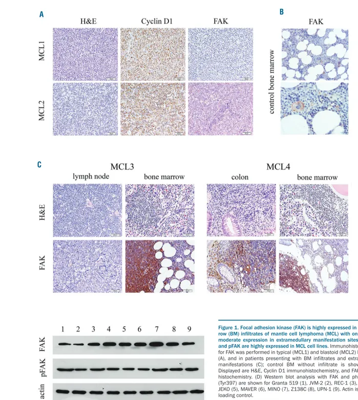
We first examined the expression of FAK in primary MCL samples by immunohistochemistry. Cases selected were lymph node infiltrates of MCL that were classified according to the WHO as 10 typical MCLs and 10 MCLs of the blastoid variant. They showed only mild (n=14) or no (n=5) expression of FAK, with only one typical case dis-playing high FAK expression. FAK expression did not cor-relate with the WHO classification (typical or blastoid) (Figure 1A).
As FAK can be induced and activated by integrin and extracellular matrix signaling,8,15 we next compared FAK
expression in BM infiltrates versus an extramedullary (lymph node or gastro-intestinal tract) manifestation of MCL. We performed immunohistochemistry of 10 paired samples (medullary and extramedullary infiltrates) of typ-ical MCLs. All BM infiltrates were characterized by high FAK expression, whereas only one infiltrate in the colon showed high expression of FAK. All other extramedullary infiltrates displayed no or only weak staining of FAK (Figure 1C). Thus, high FAK expression correlated with infiltration of the BM (P=0.0001).
We then evaluated FAK expression in MCL cell lines. We performed western blot analysis of 9 well-character-ized MCL cell lines (Granta 519, Z138C, REC-1, HBL-2, JEKO, MAVER, MINO, JVM-2, UPN-1). All cell lines showed clear FAK expression with the highest levels of FAK expression in MINO and HBL-2. They also showed high expression of pFAK (Tyr397), which is the autophos-phorylation and binding site for Src family kinase mem-bers (PI3K, PLCα) (Figure 1D).
FAK expression and activation in MCL can be induced
by CXCL12
co-oper-ation between chemokines, adhesion molecules, ligands and their receptors expressed by stromal and lymphoma cells. MCLs express G-protein-coupled chemokine recep-tors such as CXCR4 and CXCR5 that bind CXCL12.16
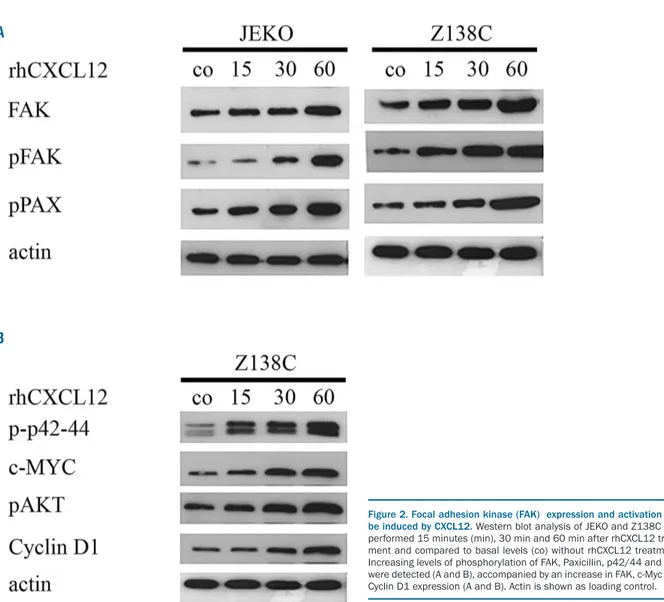
Hence, we determined whether FAK expression and acti-vation could be induced by CXCL12. We cultured two dif-ferent MCL cell lines, JEKO and Z138C, in the presence of 1 ng/mL rhCXCL12. Western blot analysis was performed after 15, 30 and 60 min. Expression of total FAK and pFAK (Tyr397) increased after incubation with rhCXCL12. In addition, rhCXCL12 induced increased phosphorylation of the direct downstream target of FAK, Paxillin, confirm-ing FAK activation (Figure 2A).
Focal adhesion kinase can mediate cell proliferation and
survival in many types of solid and non-solid tumors17 and,
therefore, we analyzed downstream targets affected by FAK signaling in MCL. Indeed, FAK expression and activa-tion led to phosphorylaactiva-tion and activaactiva-tion of several downstream targets. An increase in the phosphorylation of p42/44 and AKT, as well as a constant upregulation of c-MYC and Cyclin D1, could be observed (Figure 2B).
Co-culture with BMSC activates FAK signaling in MCL
cell lines and primary MCL-cells
We could observe a high FAK expression in BM infil-trates, and incubation of MCL cell lines with rhCXCL12 led to FAK upregulation and activation. As CXCL12 is highly expressed by BMSCs,18,19 we determined whether
Figure 1. Focal adhesion kinase (FAK) is highly expressed in bone mar-row (BM) infiltrates of mantle cell lymphoma (MCL) with only weak to moderate expression in extramedullary manifestation sites, and FAK and pFAK are highly expressed in MCL cell lines. Immunohistochemistry for FAK was performed in typical (MCL1) and blastoid (MCL2) MCL cases (A), and in patients presenting with BM infiltrates and extramedullary manifestations (C); control BM without infiltrate is shown in (B). Displayed are H&E, Cyclin D1 immunohistochemistry, and FAK immuno-histochemistry. (D) Western blot analysis with FAK and phospho-FAK (Tyr397) are shown for Granta 519 (1), JVM-2 (2), REC-1 (3), HBL-2 (4), JEKO (5), MAVER (6), MINO (7), Z138C (8), UPN-1 (9). Actin is shown as loading control.
A
C
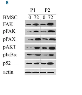
co-culture of MCL with BMSCs could activate FAK signal-ing. Co-culture of the MCL cell lines Z138C and JEKO with BMSC resulted in FAK upregulation. Western blot analysis revealed a steady increase of FAK and phospho-Paxillin after 8, 24 and 48 h. FAK activation led to the phosphorylation and activation of multiple downstream targets, such as p42/44 and AKT, and to the upregulation of Cyclin D1 (Figure 3A). Interestingly, we could observe interactions of FAK with the NF-κB signaling pathway. FAK activation resulted in an increase in the phosphoryla-tion of IKKα as shown by the immunoprecipitation of IKKα and detection with pIKKα/β. (To our knowledge, there is no phospho-specific IKKα antibody commercially available). IKKα is an important component of the canon-ical and non-canoncanon-ical NF-κB pathway.20,21 Indeed,
phos-phorylation of IkBα led to the phosphos-phorylation of IkBα and to an increase in p52 in MCL cell lines (Figure 3C). To rule out cell culture artefacts due to the use of MCL cell lines, we repeated the experiment with primary MCL cells. Western blot analysis showed similar results with an upregulation and activation of FAK after 72 h accompa-nied by an increase in p52 and increase in the phosphory-lation of IkBα and AKT (Figure 3B).
FAK silencing suppresses multiple signaling pathways
and FAK is essential for CXCL12 induced activation of
several downstream targets
Our results indicate that incubation with rhCXCL12 or co-culture with BMSCs can activate FAK and several downstream targets in MCLs. Although the downstream targets studied are well-established FAK targets, it is pos-sible that these proteins could have been activated through alternative kinases. Therefore, we studied the effect of FAK silencing on the activation of downstream targets.
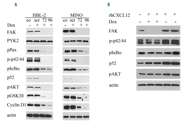
We used a Tet-inducible microRNA system in the two MCL cell lines HBL-2 and MINO with high constitutive FAK expression and activation. Western blot analysis showed that, after 72 and 96 h of incubation with doxi-cyclin in both cell lines, FAK expression was completely abolished, whereas expression of the kinase PYK2 with high homology to FAK did not change. As expected, FAK downregulation dramatically decreased or resulted in a complete loss of activation or phosphorylation of the downstream targets Paxillin, p42/44, IkBα, p52, AKT and GSK3β, as well as in downregulation of Cyclin D1 (Figure 4A).
Figure 2. Focal adhesion kinase (FAK) expression and activation can be induced by CXCL12. Western blot analysis of JEKO and Z138C was performed 15 minutes (min), 30 min and 60 min after rhCXCL12 treat-ment and compared to basal levels (co) without rhCXCL12 treattreat-ment. Increasing levels of phosphorylation of FAK, Paxicillin, p42/44 and AKT were detected (A and B), accompanied by an increase in FAK, c-Myc and Cyclin D1 expression (A and B). Actin is shown as loading control. A
Interestingly, incubation with doxycyclin and silencing of FAK also reduced the upregulation of phosphorylation of AKT, IkBα and p42/44 and upregulation of p52 expres-sion after rhCXCL12 treatment, as shown by western blot analysis for HBL-2 (Figure 4B). This result clearly demon-strates that FAK participates in CXCL12-mediated activa-tion of the AKT, p42/44 and NF-κB signaling pathways.
FAK inhibition leads to inhibition of the alternative and
classical NF-
κB signaling pathway
Silencing of FAK resulted in the inhibition of multiple downstream targets. There are several FAK-inhibitors available that are currently being evaluated for use in clin-ical trials for the treatment of solid tumors.22,23 Therefore,
we determined whether we could achieve a blockage of the downstream targets of FAK in MCL with 6063. VS-6063 is a 2nd-generation inhibitor that is in clinical trials for
solid tumors and that targets the FAK/PYK2 kinase domain in an (ATP)-competitive, reversible way.24 We
were especially interested in the regulation of the NF-κB signaling pathway, as this pathway was highly activated by co-culture with BMSCs. We performed experiments with different MCL cell lines with activation of the classic or alternative NF-κB signaling pathway. We chose the MINO and HBL-2 cell lines, which are characterized by the activation of the classic NF-κB signaling pathway, and which showed high expression levels of FAK in our west-ern blot analysis. In addition, we chose the cell line Z138C, with a TRAF2 mutation. The TRAF2 mutation leads to an activation of the alternative NF-κB pathway, so
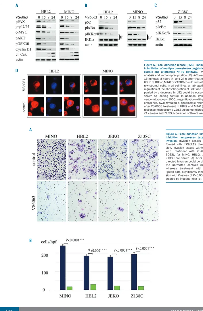
Z138C is characterized by the activation of the classic and alternative NF-κB signaling pathway. The cell lines were co-cultured with BMSCs and treated with VS-6063 (100 nM). After 15 min, 8 h, and 24 h, the cells were harvested and western blots were performed. VS-6063 treatment inhibited the phosphorylation of Paxillin, p42/44, AKT and GSK3β. We also observed a downregulation of the total protein levels of Cyclin D1 and c-MYC after 15 min and an increase in cleaved caspase-3 after 8 h (Figure 5A). In each cell line, even in Z138C (carrier of a TRAF2 muta-tion), an inhibition of the activation of the classic and alternative NF-κB signaling pathway could be observed (Figure 5B and C). There was a clear inhibition of the phosphorylation of IKKα (shown by immunoprecipita-tion) and IkBα as components of the classic NF-κB signal-ing pathway. In addition, downregulation of p52 indicated the inhibition of the alternative NF-κB signaling pathway. We confirmed the western blot results with immunofluorescence microscopy. HBL-2 and MINO were treated with VS-6063 (100 nM) for 8 h, the cells were fixed and immunofluorescence staining for p65 was performed. After VS-6063 treatment, the p65 subunit accumulated in the cytoplasma and decreased in the cell nucleus in both cell lines (Figure 5D).
FAK inhibition results in suppression of targeted cell
invasion
One major escape mechanism of lymphoma cells treat-ed with chemotherapy is homing to the BM niche. For this reason, we analyzed whether treatment with VS-6063
Figure 3. Co-culture with bone marrow stromal cells (BMSC) activates FAK signaling in mantle cell lymphoma (MCL) cell lines and primary MCL cells.Western blot analysis was per-formed 8 hours (h), 24 h, 48 h and 72 h after co-culture with BMSCs. An increase in phosphorylation of FAK, AKT, p42/44 and Paxcillin, as well as an increase in expression level of FAK and Cyclin D1 could be observed in Z138C and JEKO (A) and primary MCL cells (P1 and P2) (B). In addition, activation of the classic and alternative NF-κB signaling pathway (phospho-IκBα, phos-pho-IκKα and p52) could be detected (B and C), shown by west-ern blot analysis and immunoprecipitation (IP).
A
B
could inhibit the targeted migration of MCL cells. VS-6063 treatment (100 nM) inhibited the invasion of the MCL cell line MINO, HBL-2, JEKO and Z138C towards medium supplemented with rhCXCL12. After 24 h almost no inva-sion could be observed in the VS-6063-treated group, whereas untreated cells showed a high invasion towards rhCXCL12 (P=0.0001) (Figure 6).
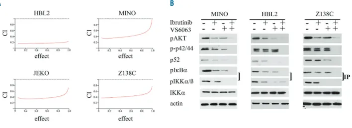
FAK inhibition acts highly synergistically with ibrutinib
treatment
In the treatment of refractory MCL, promising results can be achieved with ibrutinib. However, activation of the alternative NF-κB signaling pathway can lead to primary or secondary resistance to ibrutinib. As FAK inhibition resulted in the suppression of the classic and alternative NF-κB signaling pathway, we questioned whether resist-ance to ibrutinib could be overcome with VS-6063 treat-ment, and whether ibrutinib and VS-6063 treatment are synergistic. We chose four MCL cell lines whose resist-ance to ibrutinib is known: JEKO and MINO (which are responsive to ibrutinib), Z138C (with a TRAF2 mutation), and HBL-2 (which are resistant to ibrutinib).5Cells
co-cul-tured with BMSCs were treated with escalating doses of ibrutinib and VS-6063 (0-1 µM) for 48 h and MTT-assays were performed. We observed a highly synergistic effect in all four MCL cell lines with CI values less than 0.6 (Figure 7A) (ED50, ED75, ED90) and resistance to ibrutinib could be overcome by combination treatment.
Combination of VS-6063 and ibrutinib leads to
complete abrogation of the NF-
κB signaling pathway
We observed a high synergistic effect by combination treatment (ibrutinib and VS-6063) of different MCL cell lines. As ibrutinib and VS-6063 can both inhibit the classic and alternative NF-κB signaling pathway, we performed western blot analysis to study the effect on this pathway by the various treatments (VS-6063 or ibrutinib alone (100 nM) or in combination (10 nM). After 48 h of single or combination treatment, western blot assays were per-formed (Figure 7B). As expected, single treatment with ibrutinib or VS-6063 resulted in the downregulation of the phosphorylation of IkBα, AKT and p42/44, whereas com-bination treatment resulted in complete abrogation of the phosphorylation. In addition, treatment with VS-6063 alone or in combination with ibrutinib led to the down-regulation or complete loss of p52 expression. Therefore, combination treatment resulted in the complete inhibition of the classic and alternative NF-κB signaling pathways.
Discussion
Mantle cell lymphoma and other lymphoma subtypes frequently spread to the BM, and stromal interactions often lead to enhanced survival and drug resistance.16,25-27
Therefore, targeting deregulated kinases activated by the microenvironment has emerged as a promising strategy
Figure 4. Focal adhesion kinase (FAK) silencing suppresses multiple signaling pathways and FAK is essential for CXCL12 induced activation of several downstream targets. A tet-inducible miroRNA system was used to functionally knock-down FAK by treatment with doxycyclin (Dox). Western blot analysis of HBL-2 and MINO was performed 72 or 96 hours after addition of doxycyclin and compared to basal levels without doxycyclin (co) and microRNA with a scramble sequence (scr). A clear downregulation of FAK and multiple downstream targets (phospho-Paxicillin, phospho-p42/44, phospho-IκBα, p52, phospho-AKT, phospho-GSK3β and Cyclin D1) could be observed, whereas PYK2 levels remained stable (A). In addition, pre-incubation of HBL-2 with doxycyclin resulted in blockage of the CXCL12-induced-upreg-ulation of phospho-p42/44, phospho-AKT, phospho-IkBα and p52, as shown by western blot analysis (B).
Figure 5. Focal adhesion kinase (FAK) inhibition results in inhibition of multiple downstream targets including the classic and alternative NF-κB pathway. Western blot analysis and immunoprecipitation (IP) (A-C) was performed 15 minutes, 8 hours (h) and 24 h after treatment with VS-6063 of HBL-2, MINO or Z138C co-cultured with bone mar-row stromal cells. In all cell lines, an abrogation or down-regulation of the phosphorylation of IκBα and IKKα accom-panied by a decrease in p52 could be observed. Actin is shown as loading control. In addition, immunofluores-cence microscopy (1000x magnification) with p65 (red flu-orescence, Cy3) revealed a cytoplasmic retention of p65 after VS-6063 treatment in HBl-2 and MINO (D). For fluo-rescence microscopy a ZEISS Apotome microscope, ZEISS Z1 camera and ZEISS acquisition software was used.
Figure 6. Focal adhesion kinase (FAK) inhibition suppresses targeted cell invasion. Invasion assays were per-formed with rhCXCL12 directed inva-sion. Invasion assays without (co) or with treatment with VS-6063 (VS-6063), for MINO, HBL-2, JEKO and Z138C are shown (A). After 24 hours, directed invasion could be observed in the untreated controls (blue bars) whereas treatment with VS-6063 (green bars) significantly inhibited inva-sion with P-values of P<0.0001, as cal-culated by Student t-test (B).
A B C A B D P<0.0001*** P<0.0001*** P<0.0001*** P<0.0001***
for the treatment of lymphomas. It is well known that integrin-mediated signaling cascades play important roles in cell adhesion and interaction with the microenviron-ment.28-30 FAK kinase is activated in response to
chemokines, cytokines or ligand-binding, and plays a key role in integrin signaling.8,9,11,31 Here, we show that FAK
was highly expressed in BM infiltrates of MCLs with only weak to moderate expression in lymph node infiltrates and moderate to high expression in MCL cell lines, consis-tent with previous studies demonstrating weak or moder-ate expression of FAK in the majority (63%) of lymph node infiltrates of MCLs.32 Earlier studies of MCLs showed
that FAK could be activated by BCR or Hedgehog-signal-ing.33,34 A recently published paper35 demonstrated that
FAK expression in MCL is regulated by SOX11, and FAK expression leads to invasion of MCL and homing to the BM. As we observed high FAK expression, especially in BM infiltrates, we investigated whether FAK could also be activated by stromal interaction and integrin signaling in MCL. Indeed, FAK activation and expression could be induced in MCL by incubation with rhCXCL12 or co-cul-ture with BMSC. Incubation with rhCXCL12 or co-culco-cul-ture with BMSCs resulted in a significant increase in FAK, and in an increase in the phosphorylation of the autophospho-rylation site of FAK and an increase in the phosphoryla-tion of Paxillin, a direct downstream target of FAK.
In addition to cell adhesion and cytoskeletal reorganiza-tion, FAK mediates proliferation and survival sig-nals.8,9,11,36,37 Here, we show, that FAK activation by
rhCXCL12 or co-culture with BMSCs leads to the activa-tion of AKT and p42/44, two kinases that are important for cell proliferation and survival. In addition, the classic and alternative NF-κB pathway was activated, as shown by the phosphorylation of pIkBα, IKKα and by an increase in p52 expression. Previous studies in MCL, CLL and solid tumors emphasize the importance of the NF-κB signaling pathway in stromal-lymphoma interactions, supporting long-term expansion and drug resistance.7,38-40 In our
exper-iments, FAK was essential for the activation of multiple downstream targets, including the classic and alternative
NF-κB signaling cascade, as FAK silencing resulted in a nearly complete abrogation of the activation of AKT, p44/42, pIkBα, and IKKα or in downregulation of p52. This is in line with Balsas et al.35 who were also able to
demonstrate a blockage of the PI3K/AKT pathway after FAK inhibition.
These data are clinically highly relevant, given that there are several FAK inhibitors22,24 currently being tested in
patients. We chose the 2nd-generation inhibitor VS-6063 to
perform the inhibition experiments, as this targets the FAK kinase domain with a better pharmacodynamics pro-file than first-line inhibitors. In MCL, enhanced activation of several downstream targets (AKT, c-Myc and p42/44) by co-culture with BMSCs could be blocked by the inhibi-tion of FAK with VS-6063. In addiinhibi-tion, the downregula-tion of Cyclin D1, which is up-regulated in MCLs by the t(11;14)(q13;q32) translocation could be achieved after 8-24 h. This effect could be due to reactivation of GSK3β kinase, which targets Cyclin D1 for degradation41-43 After
24 h, we observed an increase in cleaved caspase-3 as an indicator that cells underwent apoptosis, which makes FAK an attractive target in MCLs. As previously shown in combination with sorafenib treatment,33 inhibition of FAK
also resulted in a dramatic decrease in targeted cell inva-sion, underlining its key role in tissue microenvironmental regulation of lymphoma dissemination to the BM.
Mesenchymal stromal cells protect MCL from apoptosis through the activation of the classic and alternative NF-κB signaling pathways.7These pathways have gained great
attention as promising new therapeutic targets in lym-phoma with activated BCR/NF-κB pathway,44-47 and with
available BTK-inhibitors such as ibrutinib, a significant efficacy could be achieved in the treatment of refractory or relapsed MCL.2,3,48 However, somatic mutations in
NF-κB regulatory genes can confer resistance to ibrutinib treatment in MCLs.5,49 Interestingly, FAK inhibition
result-ed in the inhibition of the classic and alternative NF-κB signaling pathways not only in the MCL cell lines MINO and HBL2, but also in Z138C, which has a known TRAF2 mutation and is characterized by the upregulation of p52.5
Figure 7. Focal adhesion kinase (FAK) inhibition acts highly synergistic with ibrutinib treatment.JEKO, HBL-2, MINO and Z138C co-cultured with bone marrow stro-mal cells (BMSCs) were treated with escalating doses of ibrutinib and VS-6063 alone or in combination, and MTT-assays were performed after 48 hours. Combination index (CI) values calculated were less than 0.6 indicating a high synergistic effect for all cell lines. In addition, western blot analysis after 48 hours of single treatment with ibrutinib or VS-6063 demonstrates decreased phosphorylation of downstream targets (AKT, p42/44, IkKα and IKKα) and complete inhibition by combination treatment.
This result could be achieved by abrogation of the phos-phorylation of IKKα, which has been previously described as a substrate of FAK.50 IKKα is a component of both the
classic and the alternative NF-κB pathway by functioning within an IkB kinase complex to directly phosphorylate the negative regulator IkB and by facilitating the cleavage of p100 to p52/Rel B.20,21Combined treatment with
ibruti-nib and FAK inhibition turned out to be highly synergistic, and ibrutinib resistance in HBL2 and Z138C could be over-come by complete inhibition of the alternative and classic NF-κB signaling pathway. This supports previously pub-lished data35 demonstrating that FAK confers
cell-adhe-sion-mediated drug resistance and contributes to a more aggressive phenotype.
In conclusion, our results provide evidence that FAK modulates the migratory and prosurvival signals mediated by the microenvironment in MCL. Furthermore, FAK inhi-bition, especially in combination with ibrutinib, may rep-resent a promising approach to treat patients with advanced MCL presenting with BM involvement.
Acknowledgments
The excellent technical assistance of Sabine Roth and Barbara Leibbrandt is gratefully acknowledged.
Funding
This work was supported in part through a grant to M.T.R. (DFG-FOR2314) from the German Research Foundation (DFG).
References
1. Dreyling M, Ferrero S. The role of targeted treatment in mantle cell lymphoma: is transplant dead or alive? Haematologica. 2016;101(2):104-114.
2. Dreyling M, Jurczak W, Jerkeman M, et al. Ibrutinib versus temsirolimus in patients with relapsed or refractory mantle-cell lym-phoma: an international, randomised, open-label, phase 3 study. Lancet. 2016;387 (10020):770-778.
3. Wang ML, Rule S, Martin P, Goy A, et al. Targeting BTK with Ibrutinib in relapsed or refractory mantle-cell lymphoma. N Engl J Med. 2013;369 (6):507-516.
4. Martin P, Maddocks K, Leonard JP, et al. Postibrutinib outcomes in patients with mantle cell lymphoma. Blood. 2016; 127(12):1559-1563.
5. Rahal R, Frick M, Romero R, et al. Pharmacological and genomic profiling identifies NF-κB-trageted treatment strate-gies for mantle cell lymphoma. Nat Med. 2014;20(1):87-92.
6. Chiron D, Di Liberto M, Martin P, et al. Cell-cycle reprogramming for PI3K inhibition overrides a relapse-specific C481S BTK mutation revealed by longitudinal function-al genomics in mantle cell lymphoma. Cancer Discov. 2014;4(9):1022-1035. 7. Medina DJ, Goodell L, Glod J, et al.
Mesenchymal stromal cells protect mantle cell lymphoma cells from spontaneous and drug-induced apoptosis through secretion of B-cell activating factor and activation of the canonical and non-canonical nulear factor κB pathways. Hematologica. 2012; 97(8):1255-1263.
8. Desgrosellier JS, Cheresh DA. Integrins in cancer: biological implications and thera-peutic opportunities. Nat Rev Cancer. 2010;10(1):9-22.
9. Guan JL. Integrin signaling through FAK in the regulation of mammary stem cells and breast cancer. IUBMB Life. 2010;62(4):268-276.
10. Mitra SK, Schlaepfer DD. Integrin-regulated FAK-Src signaling in normal and cancer cells. Curr Opin Cell Biol. 2006;18(5):516-523. 11. Schaller MD. Cellular functions of FAK
kinases: insight into molecular mechanisms and novel functions. J Cell Sci. 2010; 123(7):1007-1013.
12. Stühmer T, Zöllinger A, Siegmund D, et al. Signaling profile and antitumour activity of
the novel Hsp90 inhibitor NVP-AUY922 in multiple myeloma. Leukemia. 2008; 22(8):1604-1612.
13. Fellmann C, Hoffmann T, Sridhar V, et al. An optimized microRNA backbone for effective single-copy RNAi. Cell Rep. 2013;5(6):1704-1713.
14. Chou TC, Talalay P. Quantitative analysis of dose-effect relationships: The combined effects of multiple drugs or enzyme inhibitors. Adv Enzyme Regul. 1984;22:27-55.
15. Hanks SK, Calalb M, Harper MC, et al. Focal adhesion protein-tyrosine phospho-rylated in response to cell attachment to fibronectin. Proc Natl Acad Sci USA. 1992; 89(18):8487-8491.
16. Kurtova AV, Tamayo A, Ford RJ, et al. Mantle cell lymphoma cells express high levels of CXCR4, CXCR5, and VLA-4 (CD49d): importance for interactions with the stromal microenvironment and specific targeting. Blood. 2009;113(19):4604-4613. 17. Gabarra-Niecko V, Schaller MD, Dunty JM.
FAK regulates biological processes impor-tant for the pathogenesis of cancer. Cancer Metastasis Rev. 2003;22(4):359-374. 18. Alsayed Y, Ngo H, Rummels J, et al.
Mechanisms of regulation of CXCR4/SDF-1 (CXCLCXCR4/SDF-12)-dependent migration and homing in multiple myeloma. Blood. 2007; 109(7):2708-2717.
19. Jakubikova J, Cholujova D, Hideshima T, et al. A novel 3D mesenchymal stem cell model of the multiple myeloma bone mar-row niche: biologic and clinical applica-tions. Oncotarget. 2016;7(47):77326-77341. 20. Karin M, Yamamoto Y, Wang QM. The IKK NF-kappa B system: a treasure trove for drug development. Nat Rev Drug Discov. 2004;3(1):17-21.
21. Kendellen MF, Bradford J, Lawrence CL, et al. Canonical and non-canonical NF-kappaB signaling promotes breast cancer tumor-initiating cells. Oncogene. 2014; 33(10):1297-1305.
22. Golubovskaya VM. Targeting FAK in human cancer: from finding to first clinical trials. Front Biosci. 2014;1(19):687-706. 23. Jones SF, Siu LL, Bendell JC, et al. A phase I
study of VS-6063, a second-generation fokal adhesion kinase inhibitor, in patients with advanced solid tumors. Invest New Drugs. 2015;33(5):1100-1107.
24. Infante JR, Camidge D, Mileshkin LR, et al. Safety, pharmacokinetic, and
pharmacody-namic phase I dose-escalation trial of PF-00562271, an inhibitor of focal adhesion kinase, in advanced solid tumors. J Clin Oncol. 2012;30(13):1527-1533.
25. Burger JA, Ford R. The microenvironment in mantle cell lymphoma: cellular and molecular pathways and emering targeted therapies. Semin Canc Biol. 2011;21(5):308-312.
26. Mraz M, Zent CS, Church AK, et al. Bone marrow stromal cells protect lymphoma B-cells from Rituximab-induced apoptosis and trageting integrin alfa-4-beta-1 (VLA-4) with Natalizumab can overcome its resist-ance. Br J Haematol. 2011;155(1):53-64. 27. Burger JA, Ghia P, Rosenwald A, et al. The
microenvironment in mature B-cell malig-nancies: a target for new treatment strate-gies. Blood. 2009;114(16):3367-3375. 28. Faraldo MM, Deugnier M, Lukashev M, et
al. Pertubation of beta 1-integrin function alters the development of murine mamma-ry gland. EMBO J. 1998;17(8):2139-2147. 29. Yamada KM, Aota S, Akiyama SK, et al.
Mechanisms of fibronectin and integrin functions during cell adhesion and migra-tion. Cold Spring Harb Symp Quant Biol. 1992;57:203-212.
30. Jones JI, Doerr M, Clemmons DR. Cell migration: interactions among integrins, IGFs and IGFBPs. Prog Growth Factor Res. 1995;6(2-4):319-327.
31. Shen Y, Schaller MD. Focal adhesion target-ing: the critical determinat of FAK regula-tion and substrate phosphorylaregula-tion. Mol Biol Cell. 1999;10(8):2507-2518.
32. Ozkal S, Paterson J, Tedoldi S, et al. Focal adhesion kinase (FAK) expression in normal and neoplastic lymphoid tissues. Pathol Res Pract. 2009;205(11):781-788.
33. Xargay-Torrent S, Lopez-Guerra M, Montraveta A., et al. Sorafenib inhibits cell migration and stroma-mediated borte-zomib resistance by interfering B-cell receptor signaling and protein translation in mantle cell lymphoma. Clin Cancer Res. 2013;19(3):586-597.
34. Zhang H, Chen Z, Neelapu SS, et al. Hedgehog inhibitors selectively target cell migration and adhesion of mantle cell lym-phoma in bone marrow microenviron-ment. Oncotarget. 2016;7(12):14350-14365. 35. Balsas P, Palomero J, Eguileor A, et al. SOX11 promotes tumor protective microenvironment interactions through CXCR4 and FAK regulation in mantle cell
lymphoma. Blood. 2017;130(4):501-513. 36. You D, Xin J, Volk A, Wei W, et al. FAK
mediates a compensatory survival signal parallel to PI3K-AKT in PTEN-null T-ALL cells. Cell Rep. 2015;10(12):2055-2068. 37. Itoh S, Maeda M, Shimada S, et al. Role of
expression of focal adhesion kinase in pro-gression of hepatocellular carcinoma. Cancer Res. 2004;10(8):2812-2807. 38. Lutzny G, Kocher T, Schmidt-Supprian M,
et al. Protein kinase c-ß-dependent activa-tion of NF-kB in stromal cells is indispensa-ble for the survival of chronic lymphocytic leukemia B cells in vivo. Cancer Cell. 2013; 23(1):77-92.
39. Wu XB, Liu Y, Wang GH, et al. Mesenchymal stem cells promote colorec-tal cancer progression through AMPK/mTOR- mediated NF-kB activation. Sci Rep. 2016;19(6):21420.
40. Koliaraki V, Pasparakis M, Kollias G. IKKß in intestinal mesenchymal cells promotes initiation of colitis-associated cancer. J Exp
Med. 2015;212(13):2235-2251.
41. Rudelius M, Pittaluga S, Nishizuka S, et al. Constitutive activation of Akt contributes to the pathogenesis and survival of mantle cell lymphoma. Blood. 2006;108(5):1668-1676.
42. Yu XJ, Han QB, Wen ZS, et al. Gambogenic acid induces G1 arrest via GSK3ß-depen-dent cyclin D1 degradation and triggers autophagy in lung cancer cells. Cancer Lett. 2012;322(2):185-194.
43. Diehl JA, Cheng M, Roussel MF, et al. Glycogen synthase kinase-3beta regulates cyclin D1 proteolysis and subcellular local-ization. Genes Dev. 1998;12(22):3499-3511. 44. Yang Y, Shaffer AL, Emre NC, et al. Exploiting synthetic lethality for the thera-py of ABC diffuse large B cell lymhoma. Cancer Cell. 2012;21(6):723-737.
45. Jares P, Colomer D, Campo E. Molecular pathogenesis of mantle cell lymphoma. J Clin Invest. 2012;122(10):3416-3423. 46. Pham LV, Tamayo AT, Yoshimura LC, et al.
Inhibition of constitutive NF-kappa B acti-vation in mantle cell lymphoma B cells leads to induction of cell cycle arrest and apoptosis. J Immunol. 2003;171(1):88-95. 47. Rossi D, Deaglio S, Dominguez-Sola D, et
al. Alteration of BIRC3 and multiple other NF-kB pathway genes in splenic marginal zone lymphoma. Blood. 2011;118(18): 4930-4934.
48. Wang ML, Blum KA, Martin P, et al. Long-term follow-up of MCL patients treated with single-agent ibrutinib: updated safety and efficacy results. Blood. 2015; 126(6):739-745.
49. Bea S, Valdes-Mas R, Navarro A, et al. Landscape of somatic mutations and clonal evolution in mantle cell lymphoma. Proc Natl Acad Sci USA. 2013;110(45):18250-18255.
50. Dwyer SF, Gao L, Gelman IH. Identification of novel focal adhesion kinase substrates: role of FAK in NFkB signaling. Int J Biol Sci. 2015;11(4):404-410.