European Journal of Heart Failure (2018) 20, 427–430
VIEWPOINT
doi:10.1002/ejhf.1071Left ventricular ejection fraction and heart
failure: an indissoluble marriage?
Donato Mele
1*
, Marianna Nardozza
1, and Roberto Ferrari
1,21Centro Cardiologico Universitario and LTTA Centre, University of Ferrara, Italy; and2Maria Cecilia Hospital, GVM Care & Research, E.S. Health Science Foundation, Cotignola, RA, Italy
Introduction
Left ventricular ejection fraction (LVEF) is the most commonly used instrumental parameter to evaluate patients with heart failure (HF). The major strength of LVEF is that it is a universally known and accepted parameter, not only by cardiologists but also by all physicians. In addition, LVEF is relatively easy to obtain, as it can be determined using any cardiac imaging technique. A further advantage is that in clinical practice LVEF can be evaluated visually on echocardiographic images, which means rapidly, especially at a patient’s bedside or at home by means of portable echo scanners. Although all these features undoubtedly favour the use of LVEF in HF patients, there are also limitations. For example, LVEF does not correlate with patient symptoms, a normal LVEF does not exclude nor imply diastolic dysfunction, and LVEF cannot be used as a substitute for cardiac output.
The aim of this article is to briefly review the role of LVEF in HF patients and then discuss a different view on how to catego-rize these patients instrumentally based on a pathophysiological approach.
Left ventricular ejection fraction
as a classifier in heart failure
patients
Based on LVEF values, HF patients have been classified by the European Society of Cardiology (ESC) into three groups: those with preserved LVEF [≥50%, HF with preserved ejec-tion fracejec-tion (HFpEF)], mid-range LVEF [40–49%, HF with mid-range ejection fraction (HFmrEF)] and reduced LVEF [<40%, HF with reduced ejection fraction (HFrEF)], with implications for pharmacological and device treatment and prognosis.1
The meta-analysis of the MAGGIC group (39 372 patients from 30 studies) showed that LVEF can stratify HF prognosis in the
*Corresponding author. Cardiology Unit, University Hospital of Ferrara, Viale Aldo Moro 8, 44024 Cona, Ferrara, Italy. Tel: +39 0532 239058, Fax: +39 0532 239048, Email: [email protected]
...
...
HFrEF group2: the lower the LVEF, the worse the risk of mortality
(both for all and cardiovascular causes). This meta-analysis shows a better prognosis for HFpEF compared with HFrEF, but it is also true that once above 50% there is no relationship between LVEF and mortality, e.g. the risk of death is the same for a value of 60% or 70%. HFpEF patients have been reported to have an in-hospital mortality of 2.3–5%, a 1-year mortality rate of 22–29% and a 5-year mortality rate of 65%.3 However, in clinical trials,
cardiovascular causes accounted for more deaths in HFrEF than in HFpEF patients (83% and 70%, respectively).3
The HFpEF diagnosis is often challenging. Therefore, when LVEF is normal, the ESC guidelines comprise an advanced work-up for objective demonstration of structural and/or functional alterations of the heart as the underlying cause for the clinical presentation.1
Key structural alterations are increased left ventricular (LV) mass index and left atrial volume index, whereas main functional alter-ations are increased E/e’ ratio and e’.1
What about HFmrEF patients? They represent about one-fifth of the HF population.4,5HFmrEF pathogenesis has been reported to be similar to that of HFrEF and different from true HFpEF:6this
was related to the increased prevalence of coronary artery disease in HFmrEF and HFrEF, in contrast with the lower prevalence in HFpEF.6However, the resemblance of HFmrEF to HFrEF or HFpEF
varies in different studies and mostly depends on the examined case series. Also, both HFrEF and HFpEF patients may become HFmrEF patients due to variations in LVEF, making it unclear whether HFmrEF represents a transitional status between HFpEF and HFrEF or an independent entity of HF.7 The improvement of LVEF from
HFrEF to HFmrEF (or HFpEF) may portend a better prognosis.7
Finally, because the three HF categories are separated by only a few LVEF percentage points, the error and observer variability of standard echocardiography, which is the most widely used cardiac imaging technique in HF, may compromise accurate identification of borderline patients.8
In the light of these considerations, the use of LVEF to differen-tiate HF patients has both advantages and disadvantages. Because
© 2018 The Authors
428 Viewpoint
LVEF may change from one HF category to the other, with con-sequent prognostic variation, its modifications over time should probably be taken into account as well as the aetiology, which might be key to the relationship between LVEF and prognosis, as discussed below.
Left ventricular ejection fraction
and underlying cardiac disease
The classification of HF based on LVEF1applies indistinctly to allpatients with HF and is not differentiated according to the aeti-ology of the cardiac disease, which, in turn, is important in con-ditioning prognosis. In this regard Felker et al.9showed that, even
after adjustments for haemodynamic variables, patients with car-diomyopathy due to ischaemic heart disease have a worse sur-vival than patients with non-ischaemic causes of cardiomyopathy. LVEF is also an inadequate surrogate for the underlying myocar-dial phenotype predisposing to malignant arrhythmias and is a poor indicator to stratify the risk of sudden cardiac death.10
Thus, it is the pathological substrate which, in many cases, con-stitutes the major prognostic determinant and LVEF may have a different prognostic significance according to the underlying HF aetiology.
Left ventricular ejection fraction
as a pump index
LVEF is the ratio of LV stroke volume (SV), which is the volume ejected from the left ventricle, to end-diastolic volume (EDV). The LVEF value, therefore, depends closely and inversely on LVEDV. In other words, for the same SV, LVEF will be greater if EDV is smaller and vice versa. Thus, LVEF is unable to predict the magnitude of SV, which is the true indicator, when indexed for body size (SVi) and multiplied by heart rate (cardiac index), of the anterograde pump function. Only if EDV is known it can be understood whether the blood pumped forward, that is the SV, is preserved or reduced, whereas LVEF on its own is insufficient. Unfortunately, LVEF is often interpreted as an iso-lated parameter, especially when it is visually assessed on cardiac images.
A typical example of mismatch between LVEF and SV is a dilated left ventricle with a preserved SV: in this case LVEF can be reduced, even markedly, with SV being normal. The opposite is the case of paradoxical aortic stenosis: here, even if LVEF is normal, SV is reduced because the LV cavity volume is small. It is evident that the addition of SV evaluation to that of LVEF improves the assessment of pump dysfunction (type and severity): in fact, a reduced LVEF associated with low cardiac output is worse than a decreased LVEF with preserved cardiac output.
These examples reinforce the concept that LVEF alone cannot adequately describe LV pump function. In other words, HFpEF does not necessarily mean HF with preserved pump function as well as HFrEF does not necessarily mean reduced LVSVi or cardiac index. ...
...
...
Left ventricular ejection fraction,
neuro-hormonal compensation
and cardiac remodelling
Neuro-hormonal compensation, remodelling and structural changes of the heart occur in HF. In particular, compensatory mechanisms may bring to structural changes and maladaptive remodelling. LVEF grossly separates the HF universe in two different populations, responding or not responding to the neuro-hormonal modulation, with consequent reduction or not of maladaptive remodelling. However, LVEF is not the most precise indicator of remodelling, as other metrics, such as LV volumes and mass, relate more closely to prognosis and to the impact of therapy. For instance, in patients with myocardial infarction, LV end-systolic volume further stratifies the patient’s risk within groups with various degrees of LV dysfunction defined by LVEF.11
Therefore, the distinction of HFrEF or HFpEF could be only a step of a process leading to a better individual pathophysiological characterization of HF patients.
A different perspective
Let’s look at HF from a pathophysiological perspective. According to the HF definition:
‘a complex clinical syndrome that results from any structural or
functional impairment of ventricular filling or ejection of blood’,12
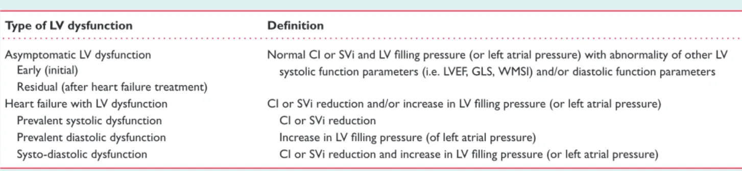
a reduced cardiac output and an increased LV filling pressure are the two key elements. HFrEF and HFpEF do not match with this pathophysiological view. In fact, as we pointed out previously, a reduced LVEF does not necessarily imply a decreased cardiac output but may be associated with increased filling pressure; conversely, a normal LVEF may be associated with reduced LV ejection and/or augmented LV filling pressure. Therefore, for a better understanding of HF patients both cardiac output and LV filling pressure (or left atrial pressure) should be determined in addition to LVEF. They could be categorized as shown in Table 1.
It should also be considered that a number of patients may have normal cardiac output and LV filling pressure (or left atrial pressure) but alteration of LVEF or other indices of LV systolic function (global longitudinal strain, wall motion score index, etc.) and/or mild alteration of LV diastolic function parameters (E/A ratio, E/e’ ratio, e’, etc.). This condition may occur at an early stage of a cardiac disease without HF or after the HF diagnosis as a result of efficacious treatments (Table 1).
This pathophysiological approach to HF could potentially be helpful to the clinician because it is expected to correlate better with symptoms than LVEF and may be used to confirm clinical diagnosis of HF. However, validation is needed for prognosis and to guide HF therapy.
From a practical point of view, both anterograde pump func-tion and LV filling pressure (or left atrial pressure) can be assessed using Doppler echocardiography. Temporelli et al.13 showed that
cardiac output evaluated by Doppler echocardiography correlates and agrees well with haemodynamic measurements (r = 0.94), also © 2018 The Authors
Viewpoint 429
Table 1 Heart failure and types of left ventricular dysfunction Type of LV dysfunction Definition
. . . . Asymptomatic LV dysfunction Normal CI or SVi and LV filling pressure (or left atrial pressure) with abnormality of other LV
systolic function parameters (i.e. LVEF, GLS, WMSI) and/or diastolic function parameters Early (initial)
Residual (after heart failure treatment)
Heart failure with LV dysfunction CI or SVi reduction and/or increase in LV filling pressure (or left atrial pressure)
Prevalent systolic dysfunction CI or SVi reduction
Prevalent diastolic dysfunction Increase in LV filling pressure (of left atrial pressure)
Systo-diastolic dysfunction CI or SVi reduction and increase in LV filling pressure (or left atrial pressure)
CI, cardiac index; GLS, global longitudinal strain; LV, left ventricular; LVEF, left ventricular ejection fraction; SVi, stroke volume index; WMSI, wall motion score index. Normal value of CI is>2.5 L/min/m2and of SVi>34 mL/m2.
after loading manipulation (r = 0.87).13 The method has
accept-able reproducibility for clinical application13,14 and is already in current clinical practice, since it is commonly used to calculate anterograde SV when assessing effective aortic valve area in aortic stenosis, mitral regurgitant fraction and Qp/Qs ratio in atrial septal defect.
Regarding LV filling pressure, Ritzema et al.15 evidenced that
single and serial Doppler echocardiographic evaluations can reliably detect raised directly measured left atrial pressure in ambulant subjects with compensated chronic HF. Recently, Andersen et al.16
showed that Doppler echocardiographic assessment of LV filling pressure is feasible and accurate and, when combined with clinical data, leads to a more accurate diagnosis of HF, regardless of LVEF. Other techniques, especially magnetic resonance and nuclear imaging, can be used to accurately evaluate LV function but they cannot be applied extensively to all the HF patients.
The pathophysiological approach shown in Table 1 also has limitations. Only functional evaluations have been included in this approach, which does not consider structural aspects. This approach apparently does not incorporate the concept of dis-ease progression. However, an advanced disdis-ease is expected to worsen systolic and/or diastolic LV dysfunction, thus the combined assessment of both LV functions could be a better approach to dis-ease progression than LVEF alone. It is unknown whether cardiac output and LV filling pressure have a more stable behaviour than LVEF, which may vary over time from one group to another of the ESC classification. Aetiology affects prognosis regardless of cardiac output9 and should be taken into account as for LVEF. Finally, in
some cases, resting cardiac output and LV filling pressure could not adequately characterize patients and a stress assessment could potentially provide more information, as shown with other indices of LV systolic function.17
Conclusions
LVEF is currently the most commonly used cardiac imaging param-eter in HF patients and is utilized to guide therapy in patients with HFrEF. However, LVEF alone cannot adequately categorize all HF patients and should be integrated in a more comprehensive approach. Aetiology should be considered as a key determinant of prognosis. Evaluation of both cardiac output and LV filling pressure ...
...
could be used to improve characterization of LV function and help HF diagnosis. Such a pathophysiological approach to HF classifica-tion may also be a promising area of clinical research. The time has arrived to start reconsidering the indissolubility of HF and LVEF. Conflict of interest: none declared.
References
1. Ponikowski P, Voors AA, Anker SD, Bueno H, Cleland JG, Coats AJ, Falk V, González-Juanatey JR, Harjola VP, Jankowska EA, Jessup M, Linde C, Nihoy-annopoulos P, Parissis JT, Pieske B, Riley JP, Rosano GM, Ruilope LM, Rus-chitzka F, Rutten FH, van der Meer P. 2016 ESC Guidelines for the diag-nosis and treatment of acute and chronic heart failure: The Task Force for the diagnosis and treatment of acute and chronic heart failure of the Euro-pean Society of Cardiology (ESC). Developed with the special contribution of the Heart Failure Association (HFA) of the ESC. Eur J Heart Fail 2016;18: 891–975.
2. Pocock SJ, Ariti CA, McMurray JJ, Maggioni A, Køber L, Squire IB, Swed-berg K, Dobson J, Poppe KK, Whalley GA, Doughty RN; Meta-Analysis Global Group in Chronic Heart Failure. Predicting survival in heart failure: a risk score based on 39 372 patients from 30 studies. Eur Heart J 2013;34: 1404–1413.
3. Butler J, Fonarow GC, Zile MR, Lam CS, Roessig L, Schelbert EB, Shah SJ, Ahmed A, Bonow RO, Cleland JG, Cody RJ, Chioncel O, Collins SP, Dunnmon P, Filippatos G, Lefkowitz MP, Marti CN, McMurray JJ, Misselwitz F, Nodari S, O’Connor C, Pfeffer MA, Pieske B, Pitt B, Rosano G, Sabbah HN, Senni M, Solomon SD, Stockbridge N, Teerlink JR, Georgiopoulou VV, Gheorghiade M. Developing therapies for heart failure with preserved ejection fraction: current state and future directions. JACC Heart Fail 2014;2:97–112.
4. Chioncel O, Lainscak M, Seferovic PM, Anker SD, Crespo-Leiro MG, Harjola VP, Parissis J, Laroche C, Piepoli MF, Fonseca C, Mebazaa A, Lund L, Ambrosio GA, Coats AJ, Ferrari R, Ruschitzka F, Maggioni AP, Filippatos G. Epidemiology and one-year outcomes in patients with chronic heart failure and preserved, mid-range and reduced ejection fraction: an analysis of the ESC Heart Failure Long-Term Registry. Eur J Heart Fail 2017;19:1574–1585.
5. Löfman I, Szummer K, Dahlström U, Jernberg T, Lund LH. Associations with and prognostic impact of chronic kidney disease in heart failure with preserved, mid-range, and reduced ejection fraction. Eur J Heart Fail 2017;19:1606–1614. 6. Vedin O, Lam CSP, Koh AS, Benson L, Teng THK, Tay WT, Braun OÖ, Savarese
G, Dahlström U, Lund LH. Significance of ischemic heart disease in patients with heart failure and preserved, midrange, and reduced ejection fraction: a nationwide cohort study. Circ Heart Fail 2017;10:e003875.
7. Rastogi A, Novak E, Platts AE, Mann DL. Epidemiology, pathophysiology and clinical outcomes for heart failure patients with a mid-range ejection fraction.
Eur J Heart Fail 2017;19:1597–1605.
8. Mele D, Campana M, Sclavo M, Seveso G, Aschieri D, Nesta F, D’Aiello I, Ferrari R, Levine RA. Impact of tissue harmonic imaging in patients with distorted left ventricles: improvement in accuracy and reproducibility of visual, manual and automated echocardiographic assessment of left ventricular ejection fraction. Eur
J Echocardiogr 2003;4:59–67.
9. Felker GM, Thompson RE, Hare JM, Hruban RH, Clemetson DE, Howard DL, Baughman KL, Kasper EK. Underlying causes and long-term survival in
© 2018 The Authors
430 Viewpoint
patients with initially unexplained cardiomyopathy. N Engl J Med 2000;342: 1077–1084.
10. Priori S, Blomstrom-Lundqvist C, Mazzanti A, Blom N, Borggrefe M, Camm J, Elliott PM, Fitzsimons D, Hatala R, Hindricks G, Kirchhof P, Kjeldsen K, Kuck KH, Hernandez-Madrid A, Nikolaou N, Norekvål TM, Spaulding C, Van Veldhuisen DJ. 2015 ESC Guidelines for the management of patients with ventricular arrhythmias and the prevention of sudden cardiac death: The Task Force for the Management of Patients with Ventricular Arrhythmias and the Prevention of Sudden Cardiac Death of the European Society of Cardiology (ESC). Eur Heart
J 2015;36:2793–2867.
11. White HD, Norris RM, Brown MA, Brandt PW, Whitlock RM, Wild CJ. Left ventricular end-systolic volume as the major determinant of survival after recovery from myocardial infarction. Circulation 1987;76:44–51.
12. Yancy CW, Jessup M, Bozkurt B, Butler J, Casey DE Jr, Drazner MH, Fonarow GC, Geraci SA, Horwich T, Januzzi JL, Johnson MR, Kasper EK, Levy WC, Masoudi FA, McBride PE, McMurray JJ, Mitchell JE, Peterson PN, Riegel B, Sam F, Stevenson LW, Tang WH, Tsai EJ, Wilkoff BL. 2013 ACCF/AHA guideline for the management of heart failure: executive summary: a report of the American College of Cardiology Foundation/American Heart Association Task Force on Practice Guidelines. Circulation 2013;128:e240–327. ...
13. Temporelli PL, Scapellato F, Eleuteri E, Imparato A, Giannuzzi P. Doppler echocardiography in advanced systolic heart failure. A non-invasive alternative to Swan-Ganz catheter. Circ Heart Fail 2010;3:387–394.
14. Lewis JF, Kuo LC, Nelson JG, Limacher MC, Quinones MA. Pulsed Doppler echocardiographic determination of stroke volume and cardiac output: clin-ical validation of two new methods using the apclin-ical window. Circulation 1984;70:425–431.
15. Ritzema JL, Richards AM, Crozier IG, Frampton CF, Melton IC, Doughty RN, Stewart JT, Eigler N, Whiting J, Abraham WT, Troughton RW. Serial Doppler echocardiography and tissue Doppler imaging in the detection of elevated directly measured left atrial pressure in ambulant subjects with chronic heart failure. JACC
Cardiovasc Imaging 2011;4:927–934.
16. Andersen OS, Smiseth OA, Dokainish H, Abudiab MM, Schutt RC, Kumar A, Sato K, Harb S, Gude E, Remme EW, Andreassen AK, Ha JW, Xu J, Klein AL, Nagueh SF. Estimating left ventricular filling pressure by echocardiography. J Am
Coll Cardiol 2017;69:1937–1948.
17. Dini FL, Mele D, Conti U, Ballo P, Citro R, Menichetti F, Marzilli M. Peak power output to left ventricular mass: an index to predict ventricular pumping performance and morbidity in advanced heart failure. J Am Soc Echocardiogr 2010;23:1259–1265.
© 2018 The Authors