Università degli Studi di Ferrara
DOTTORATO DI RICERCA IN
BIOCHIMICA, BIOLOGIA MOLECOLARE E BIOTECNOLOGIE
CICLO XXIII
COORDINATORE Prof. Francesco Bernardi
ACTIVATING TRANSCRIPTION FACTOR 4 (ATF4) IS
UPREGULATED BY HUMAN HERPESVIRUS 8
INFECTION, INCREASES VIRUS REPLICATION AND
PROMOTES VIRUS PROANGIOGENIC PROPERTIES
Settore Scientifico Disciplinare MED/07
Dottoranda Tutore
Dott.ssa Grigolato Jessica Prof. Di Luca Dario
_______________________________ _____________________________
(firma) (firma)
Correlatore Prof.ssa Caselli Elisabetta _______________________________
(firma)
INDEX
ABSTRACT page 3
RIASSUNTO page 4
INTRODUCTION
page 5
1. Herpesviruses: general features page 5
1.1. Herpesviruses classification page 5
1.2. Herpesviruses structure page 7
1.3. Herpesviruses genome page 8
1.4. Herpesviruses replication page 9
2. Human herpesvirus 8 (HHV-8) page 12
2.1. HHV-8 structure page 12
2.2. HHV-8 genome page 13
2.3. HHV-8 replication cycle page 15
2.4. HHV-8 transactivator genes page 19
2.5. HHV-8 epidemiology and transmission page 22
2.6. HHV-8 pathogenesis page 23
2.7. HHV-8 and angiogenesis page 27
3. The cellular activating transcription factor 4 (ATF4) page 29
AIM OF THE RESEARCH page 33
METHODS page 35
1. Cells page 35
3. DNA extraction page 38
4. RNA extraction and retrotranscription page 39
5. PCR and rtPCR page 39
6. Cell transfection page 40
7. Reporter assay page 41
8. Real time PCR page 41
9. Western blotting page 42
10. ELISA page 43
11. Immunofluorescence page 43
12. HHV-8 infection page 43
13. Analysis of in vitro cord formation capability page 44
14. Small interfering RNA (siRNA) studies page 44
15. Statistical analyses page 44
RESULTS page 45
1. HHV-8 infection increases ATF4 expression page 45
2. ATF4 increases HHV-8 replication page 49
3. ATF4 does not activate HHV-8 promoters page 52
4. ATF4 activates the MCP-1 promoter and induces MCP-1 secretion page 54 5. ATF4 induces capillary-like structure formation in endothelial cells page 56 6. ATF4 silencing impairs HHV-8 replication and induction of proangiogenic properties
page 57
DISCUSSION page 61
MEETINGS AND PUBLICATION LISTS page 63
ABSTRACT
Previous results from our group demonstrate that human herpesvirus 8 (HHV-8) triggers proangiogenic behavior in endothelial cells by promoting transcriptional activation and secretion of monocyte chemoattractant protein 1 (MCP-1), through activation of Nuclear Factor κB (NF-κB). However, inhibition of NF-κB still results in partial MCP-1 induction and consequent capillary-like structure formation, suggesting the involvement of another transcriptional pathway. Here we describe that HHV-8 infection upregulates cellular activating transcription factor 4 (ATF4), a member of the CREB family involved in the cell response to ER stress. Upregulation of ATF4 promotes HHV-8 infection, whereas its silencing decreases virus replication, transcription and antigen expression. Furthermore, ATF4 silencing decreases virus-induced MCP-1 production, as well as viral induction of tube-like structures in endothelial cells. We also show that ATF4 per se activates the MCP-1 promoter and induces proangiogenic properties in transfected endothelial cells. The elucidation of molecular mechanism involved in this process will result in a better understanding of the angiogenic process, its involvement in cancer and will help in designing novel therapies to reduce growth and vascularisation of Kaposi’s sarcoma.
RIASSUNTO
Risultati precedentemente ottenuti nel nostro gruppo hanno dimostrato che l’herpesvirus umano di tipo 8 (HHV-8) induce proprietà proangiogeniche nelle cellule endoteliali mediante l’induzione di MCP-1 (proteina chemiotattica monocitica) in seguito all’attivazione di NF-kB (fattore nucleare kB).
Tuttavia, mutazioni o delezioni dei siti di legame per NF-kB localizzati nella regione promotrice di MCP-1, risultano in una parziale inibizione di MCP-1, virus indotto, e nello sviluppo dell’angiogenesi, suggerendo l’implicazione di altre vie trascrizionali.
In questo lavoro è stato analizzato il fattore di attivazione trascrizionale ATF4, dal suo ruolo centrale nella risposta cellulare dovuta allo stress, fino alla sua implicazione nell’angiogenesi. I risultati mostrano che HHV-8 aumenta l’espressione di ATF4, il quale a sua volta promuove l’infezione da HHV-8. ATF4 induce, inoltre, la produzione di MCP-1 e proprietà proangiogeniche nelle cellule endoteliali.
Il silenziamento di ATF4, al contrario, diminuisce la replicazione virale e inibisce la produzione di MCP-1 indotta dal virus e quindi l’induzione di strutture tubulari tipiche dell’angiogenesi.
Si può dunque evincere che ATF4 gioca un ruolo importante nella replicazione di HHV-8 e nell’angiogenesi indotta dal virus.
La comprensione dei meccanismi molecolari implicati in questo processo, potranno contribuire a chiarire i processi angiogenici indotti dal virus e potranno pertanto essere utili nel disegno di nuove terapie per ridurre lo sviluppo dei tumori virus associati.
INTRODUCTION
1. Herpesviruses: general features
1.1.Herpesviruses classification
More than 100 herpesviruses have been discovered, all of them are double-stranded DNA viruses that can establish latent infections in their respective hosts. Eight herpesviruses infect humans. The Herpesviridae family is subdivided into three subfamilies: the Alpha-, Beta-, or
Gammaherpesvirinae1 (Fig.1).
Alphaherpesvirinae are defined by a wide cellular host range, short viral reproductive cycle,
rapid growth in culture, high cytotoxic effects, and the ability to establish latency in sensory ganglia. Human alpha-herpesviruses includes herpes simplex viruses 1 and 2 (1 and HSV-2) and varicella zoster virus (VZV), respectively officially designated human herpesviruses 1, 2, and 3.
Betaherpesvirinae have a more restricted host range with a longer reproductive viral cycle and
slower growth in culture. Infected cells show cytomegalia (enlargement of the infected cell). Latency is established in secretory glands, lymphoreticular cells, and in other tissues, such as the kidneys and others. In humans, the group includes human cytomegalovirus (HCMV or herpesvirus 5) and roseoloviruses (causing the disease roseola infantum in children) including human herpesviruses 6A and 6B (HHV-6A and -6B) and human herpesvirus 7 (HHV-7).
Gammaherpesvirinae in vitro replication occurs in lymphoblastoid cells, but lytic infections may
occur in epithelial and fibroblasts for some viral species in this subfamily. Gammaherpesviruses are specific for either B or T cells with latent virus found in lymphoid tissues (Table 1). Only two human gammaherpesviruses are known, human herpesvirus 4, or Epstein-Barr virus (EBV), and human herpesvirus 8, referred to as HHV-8 or Kaposi's sarcoma-associated herpesvirus (KSHV). The gammaherpesviruses subfamily contains two genera that include both the gamma-1 or Lymphocryptovirus (LCV) and the gamma-2 or Rhadinovirus (RDV) virus genera. EBV is the only Lymphocryptovirus and HHV-8 is the only Rhadinovirus discovered in humans. LCV are found only in primates but RDV can be found in both primates and subprimate mammals. RDV DNAs are more diverse across species and are found in a broader range of mammalian species.
HHV-8 has sequence homology and genetic structure similar to another RDV, Herpesvirus
saimiri (HVS). The T-lymphotropic Herpesvirus saimiri establishes specific replicative and
persistent infection in different primate host species, causes fulminant T-cell lymphoma in its primate host and can immortalize infected T-cells.
Table 1. Properties of herpesviruses.
1.2.Herpesviruses structure
Herpesviruses have a central toroidal-shaped viral core containing a linear double stranded DNA. This DNA is embedded in a proteinaceous spindle2. The capsid is icosadeltahedral (16 surfaces) with 2-fold symmetry and a diameter of 100-120 nm that is partially dependent upon the thickness of the tegument. The capsid has 162 capsomeres.
The herpesvirus tegument, an amorphorous proteinaceous material that under EM lacks distinctive features, is found between the capsid and the envelope; it can have asymmetric distribution. Thickness of the tegument is variable depending on the location in the cell and varies among different herpesviruses3.
The herpesvirus envelope contains viral glycoprotein protrusions on the surface of the virus. As shown by EM there is a lipid trilaminar structure derived from the cellular membranes.
Glycoproteins protrude from the envelope and are more numerous and shorter than those found on other viruses (Fig.2).
Figure 2. Representation of a herpesvirus virion.
1.3. Herpesviruses genome
Herpesviruses genomes studied to date range in size from 130 to 235 kbp.
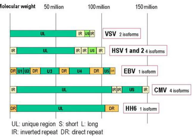
Herpesviruses DNA is characterized by two unique components, unique long (UL) and unique small (US) regions, each flanked by identical inverted repeat sequences. Herpesviruses genome also contains multiple repeated sequences.
All known herpesviruses have capsid packaging signals at their termini4. The majority of genes contain upstream promoter and regulatory sequences, an initiation site followed by a 5' nontranslated leader sequence, the open reading frame (ORF) itself, some 3' nontranslated sequences, and finally, a polyadenylation signal. Gene overlaps are common, whereby the promoter sequences of antisense strand (3') genes are located in the coding region of sense strand (5') genes; ORFs can be antisense to one another (Fig.3).
Figure 3. Genomic organization of some herpesviruses. HSV, VZV and CMV have inverted repeated sequences.
Proteins can be embedded within larger coding sequences and yet have different functions. Most genes are not spliced and therefore are without introns and sequences for noncoding RNAs are present.
Herpesviruses code for genes that synthetize proteins involved in establishment of latency, production of DNA, and structural proteins for viral replication, nucleic acid packaging, viral entry, capsid envelopment, for blocking or modifying host immune defences, and for transition from latency to lytic growth. Although all herpesviruses establish latency, some (e.g., HSV) do not necessarily require latent protein expression to remain latent, unlike others (e.g., EBV and HHV-8).
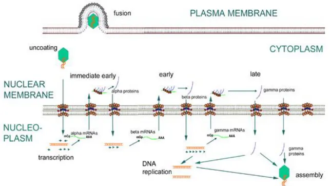
1.4.Herpesviruses replication
Herpesviruses can establish lytic or latent infection. The lytic stage is divided in different phases:
1. Binding to the cell surface. As many other viruses, cell tropism is determined by the availability of the correct receptor on the surface of the cell. Binding occurs between the herpesvirus envelope glycoproteins and cell receptors.
2. Envelope fusion with the plasma membrane, mediated by surface virus glycoproteins. 3. Uncoating: degradation of the tegument in the cytoplasm is followed by transport of the
nuclear membrane, where virus DNA is introduced in the cell nucleus through pores of the nuclear membrane.
4. Transcription. This is a very complex process, as expected considering the large size of viral genome. Viral DNA replicates by circularization followed by production of concatemers and cleavage of unit-length genome during packaging. The herpesvirus lytic replicative phase can be divided into four stages (Fig. 4):
α or immediate early (IE), requiring no prior viral protein synthesis. The genes expressed in this stage are involved in transactivating transcription from other viral genes.
β or early genes (E), whose expression is independent of viral DNA synthesis.
Following the E phase, γ1 or partial late genes are expressed in concert with the beginning of viral DNA synthesis.
γ2 or late genes (L), where protein expression is totally dependent upon synthesis of viral DNA and expression of structural genes encoding for capsid proteins and envelope glycoproteins occurs.
Herpesvirus DNA is transcribed to RNA by cellular RNA polymerase I. The neo-formed viral mRNAs block cellular protein synthesis and activate the replication of viral DNA. Herpesviruses encode their own DNA-dependent DNA polymerase and other enzymes and proteins necessary to replication, such as ori-Lyt (replication start for the lytic phase), major DNA binding protein (MDBP) and origin DNA binding protein (OBP). Herpesviruses can alter their environment by affecting host cell protein synthesis and host cell DNA replication, immortalizing the host cell, and altering the host's immune responses (e.g., blocking apoptosis, cell surface MHC I expression, modulation of the interferon pathway).
5. Assembly: capsids are assembled in the nucleus.
6. Maturation and egress: the viral particles bud through the inner lamella of the nuclear membrane which has been modified by the insertion of viral glycoproteins and leave the cell via the exocytosis pathway (Fig.5).
In the latent phase, the virus genome depends on the host replication machinery and replicates as closed circular episome. Latency typically involves the expression of only a few latency specific genes. Generally, most infected host cells harbour latent virus, as in the case of HHV-8: when KS tissue or HHV-8 infected cultured cells are analyzed, the virus is latent in majority of infected cells. Different signals such as inflammation and immunosuppression may cause the virus to enter into a new lytic phase.
2. Human herpesvirus 8 (HHV-8)
Human herpesvirus 8 (HHV-8), also known as Kaposi’s sarcoma associated herpesvirus (KSHV), is a member of the Rhadinovirus genus in the gamma-herpesvirus subfamily, first detected in 1994 in a patient affected by Kaposi sarcoma (KS)5, a neoplasm of endothelial origin. Since then, HHV-8 has been identified as the etiologic agent of all epidemiologic forms of KS, including classical, endemic African, iatrogenic and AIDS types. In addition, HHV-8 has been implicated in the pathogenesis of other neoplastic disorders affecting immunocompromised hosts: primary effusion lymphoma (PEL, a rare form of B-cell lymphoma)6, multicentric Castleman disease (MCD, a B-cell lymphoproliferative disease)7, other lymphoproliferative disorders affecting patients infected with HIV8, and neoplastic complications in patients after transplantation9.
2.1. HHV-8 structure
HHV-8 has the typical morphology of the herpesviruses.
The envelope contains proteins of cellular origin and virus-specific glycoproteins, such as gB, gM, gH and K8.1.
The tegument is an amorphous asymmetric proteinaceous layer between envelope and capsid and contains proteins encoded by ORFs 19, 63, 64, 67, 75.
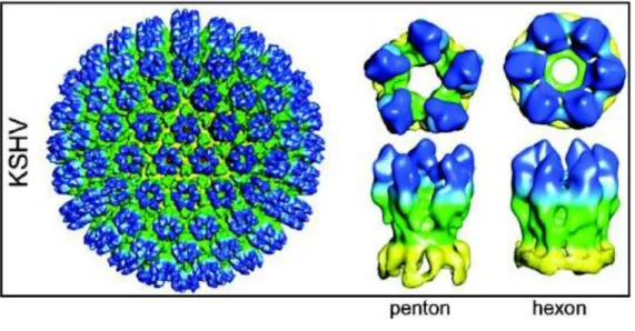
Each capsid, 125 nm in diameter, contains 12 pentons and 150 hexons which are interconnected by 320 triplexes. These capsomers or structural components are arranged in a icosahedral lattice with 20 triangular faces. Each asymmetric unit (one-third of a triangular face) of the capsid contains one-fifth of a penton at the vertex (Fig. 6). Several proteins are involved in capsid
ORF65 and a protease encoded by ORF17. Hexons and pentons contain 5 or 6 MCPs, and triplexes contain ORF62 monomer and ORF26 dimer10.
The core contains the linear double stranded DNA.
Figure 6. Representation of the three-dimensional structures of gammaherpesvirus capsids. The capsid maps of KSHV
is shown as shaded surfaces colored according to particle radius and viewed along an icosahedral three-fold axis. The resolution of the KSHV capsid maps is 24 Å. The right two columns are detailed comparisons of a penton and an hexon, which were extracted computationally from map and shown in their top and side views.
2.2. HHV-8 genome
In the viral capsid, HHV-8 DNA is linear and double stranded, but upon infection of the host cell and release from the viral capsid, it circularizes. Reports of the length of the HHV-8 genome have been complicated by its numerous, hard-to-sequence, terminal repeats. Renne et al.11 reported a length of 170 kilobases (Kb) but Moore et al.12 suggested a length of 270 Kb after analysis with clamped homogeneous electric field (CHEF) gel electrophoresis.
Base pair composition on average across the HHV-8 genome is 59% G/C; however, this content can vary in specific areas across the genome.
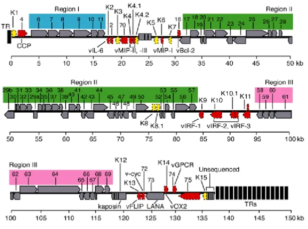
HHV-8 possesses a long unique region (LUR) of approximately 145 Kb, containing all the known ORFs (open reading frame), flanked by terminal repeats (TRs) (Fig.7). Varying amounts of TR lengths have been observed in the different virus isolates. These repeats are 801 base pairs
in length with 85% G/C content, and have packaging and cleavage signals. The LUR is similar to HVS and at least 66 ORFs have homology with the HSV genes. New genes are still being discovered through transcription experiments with alternative splicing. A "K" prefix denotes no genetic homology to any HVS genes (K1–K15).
Figure 7. HHV-8 genome. The genome consists of a long unique region (145 kb) encoding for over 80 ORFs,
surrounded by terminal repeats regions.
HHV-8 possesses approximately 26 core genes, shared and highly conserved across the alpha-,
beta-, and gammaherpesviruses. These genes are in seven basic gene blocks, but the order and
orientation can differ between subfamilies. These genes include those for gene regulation, nucleotide metabolism, DNA replication (polymerase ORF9 and thymidin kinase ORF21), and virion maturation and structure (envelope glycoproteins: ORF8, ORF22, ORF38).
HHV-8 encodes several ORFs homologous to cellular genes (at least 12), not shared by other human herpesviruses13. These genes seem to have been acquired from human cellular cDNA as evidenced by the lack of introns. Some retain host function, or have been modified to be constitutively active; an example of this is the viral cyclin-D gene14. Cellular homologs related to known oncogenes have been identified in HHV-8, including genes encoding viral Bcl-2
(ORF16), cyclin D (ORF72), interleukin-6 (K2), G-protein-coupled receptor (ORF74), and ribonucleotide reductase (ORF2).
Other genes have homologues in other members of the RDV genus, such as v-cyclin (ORF72), latency-associated-nuclear antigen (LANA, ORF73), viral G-protein coupled receptor (ORF74). A number of other genes encoding for capsid protein have been identified, including ORF25, ORF26, and ORF65. In addition to virion structural proteins and genes involved in virus replication, HHV-8 has genes and regulatory components (e.g. ORFs K3, K4 and K5) that interact with the host immune system, presumably to counteract cellular host defenses15.
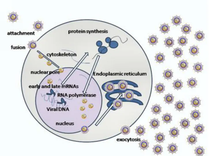
2.3. HHV-8 replication cycle
Like other herpesviruses, HHV-8 genome structure and gene expression pattern varies depending on the replication state. The lytic phase consists of different steps:
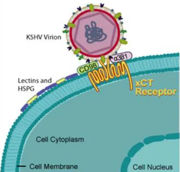
1. Binding to the cell surface (Fig. 8) mediated by glycoproteins B and K8.1, encoded by ORF8 and K8.116. HHV-8 can use multiple receptor for infection of target cells, and these receptors differ according to the cell type. HHV-8 utilizes the ubiquitous cell surface heparan sulphate (HS) proteoglycan to bind several target cells (e.g. B lymphocytes). Glycoprotein B also interacts with the host cell surface α3-β1-integrin, a heterodimeric receptor containing transmembrane subunits. Another cellular receptor used is the dentritic cell specific intracellular adhesion molecule-3 (ICAM-3) for the binding to the myeloid dendritic cells and macrophages. Moreover, HHV-8 utilizes the transporter protein xCT for entry into cells (but not into the B cells); xCT molecule is a part of the membrane glycoprotein CD98 complex.
Fig. 8. KSHV binding to the cell surface.
2. Fusion between envelope and plasma membrane (Fig. 9). The binding between cellular receptors and HHV-8 glycoproteins leads to induction of the host signal cascades critical for maintenance of viral gene expression, such as protein kinase C (PKC), phosphatidylinositol 3-kinase (PI3K), and nuclear factor kB (NF-kB). In fact, HHV-8 reprogrammes the elements of host cell transcriptional machinery that are involved in regulating a variety of processes (apoptosis, cell cycle regulation, signalling, inflammatory response and angiogenesis).
3. Uncoating: tegument degradation by cellular enzymes and viral capsid transfer to the nuclear membrane. Viral DNA enters in the nucleus.
4. Transcription: viral genome initiates transcription in the nucleus and regulator genes are transcribed by host RNA polymerase. mRNAs are translated in virus-specific proteins, able to block cellular synthesis and to start viral replication. Lytic gene expression begins with transcription of immediate-early (IE) genes that regulate the synthesis of other viral genes. Some of the genes transcribed in this step are: ORF6, coding the major DNA binding protein (MDBP), ORFs 9, 56, 59, encoding the DNA polymerase and ORFs 40, 41, 44 (helicase/primase complex). Expression of IE genes occurs independent of viral replication, and afterwards, early and late genes are expressed. The early genes (E) expression is activated by IE genes within 24 hours after infection or viral reactivation. Early genes encode proteins involved in viral DNA replication, nucleotides metabolism, virus assembly. Some early genes are: K2-5, T1.1, ORFs2, 41, 59, 70, 74. Expression of late genes (L) begins after viral DNA replication. They encode structural proteins involved in virus assembly, such as glycoproteins B and H (ORF8 and ORF22), capsid proteins (ORFs 25, 26), the small viral capsid antigen (ORF65).
5. Assembly: transcription of genes coding structural proteins and production of viral particle. ORFs26 and 29 proteins are responsible for capsid assembly and viral DNA packaging.
6. Maturation and egress: the viral particles bud through the inner lamella of the nuclear membrane which has been modified by the insertion of viral glycoproteins and leave the cell via exocytosis (Fig. 10).
Figure 10. Lytic KSHV lifecycle in BCBL-1 cells.
After initial infection, HHV-8 may establish lifelong latency. Throughout latency, viral gene expression is tightly regulated and only a few viral genes are expressed. The latent HHV-8 genome is circularized by joining of GC rich terminal repeats (TRs) at the ends of the viral genome to form an extrachromosomal circular episome17. The latency associated nuclear antigen (LANA) regulates episome replication by host cell machinery18. LANA is a phosphoprotein expressed in latently infected cells and promotes the maintenance of latency by associating with the ORF50 promoter19 or binding cellular factors which normally interact with ORF50. HHV-8 infection can be reactivated from latency and the lytic gene expression may restart.
2.4. HHV-8 transactivator genes
Two immediate-early genes play a key role in the reactivation from latent phase to lytic phase: ORF50 and ORF57.
ORF50
ORF50 is an immediate early gene whose product is the major transcriptional transactivator and his activity is required for viral reactivation by all known chemical inducer (e.g. tetradecanoyl phorbol acetate, TPA). The ORF50 gene is rapidly expressed, within 2 to 4 h after induction. ORF50 belongs to the family of R transactivators, highly conserved among herpesviruses and is related to immediate-early transcriptional activator proteins of other gammaherpesviruses, such as ORF50a encoded by Herpesvirus saimiri and Rta encoded by the BRLF1 ORF of Epstein-Barr virus20.
During latency, ORF50 expression is repressed; however, ORF50 may be activated by physiological conditions, such as hypoxia, or by pharmaceutical agents and the activation triggers the start of the lytic replication cascade.
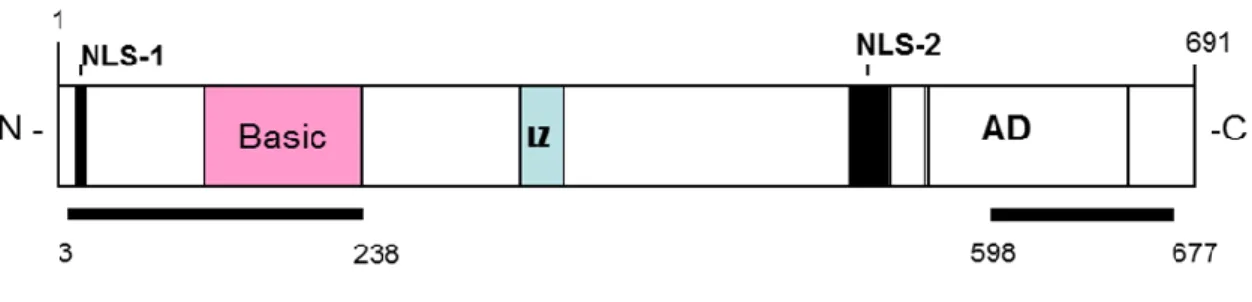
The genomic sequence of ORF50 is characterized by 5 exons and 4 introns (Fig.11) and transcribes an mRNA of 3,6 Kb. The transcript initiates at position 71560, 23 nts downstream a potential TATA box; its first AUG is located at position 71596.
Figure 11. Schematic representation of ORF50 protein.
The ORF50 transcript encodes a 691 aa protein (110 kDa) located in the nucleus during the latent phase for the presence of two nuclear localization signals (NLSs). The N-terminus 272 amino acids of ORF50 binds independently to HHV-8 promoters and mediates sequence-specific DNA binding; N-terminal region is followed by a leucine zipper domain. The C-terminal domain contains multiple charged amino acids alternated with repeated bulky hydrophobic residues, a
primary structure conserved in many eukaryotic transcriptional activation domains. The C-terminus is sufficient to activate transcription when targeted to promoters with a heterologous DNA binding domain21.
This region contains four overlapping domains termed activation domains (AD1, AD2, AD3, AD4), sharing significant homology to the R proteins encoded by other gamma-herpesviruses. Analysis of the ORF50 amino acids sequence reveals multiple sites of phosphorilation, including a C-terminal region rich in serines and threonines, and 20 other consensus sites for phosphorilation by serine-threonine kinase and protein kinase C (PKC).
R response elements (RREs) have been identified within several lytic gene promoters. The response element contains a 12-bp palindrome with additional sequences flanking the palindrome which are also required for both DNA binding and activation by ORF50.
The ORF50 protein binds directly to this palindromic sequence, and the N-terminal 272 aa is sufficient for binding in vitro. ORF50 can directly transactivate the early gene promoters22. The ORF50-responsive promoters include the following: ORF 6 (single-stranded DNA binding protein), ORF21 (thymidine kinase [TK]), ORF57 (posttranscriptional activator), ORF59 (DNA polymerase associated processivity factor), K8 (K-bZip), K9 (viral interferon response factor), K12 (kaposin), and nut-1 or PAN or T1.1 (polyadenylated nuclear RNA)23.
Furthermore, recent studies suggest autoactivation of ORF50 by interaction with the cellular protein octamer-1 (oct-1) and an intact octamer element that is located approximately 200 bp upstream of the ORF50 transcription start site24.
Expression of ORF50 reactivates viral lytic cycle in cells containing the virus in a latent phase25, and is also able to activate heterologous viral promoters such as LTR HIV, synergizing with Tat26. This molecular transactivation increases cellular susceptibility to HIV infection and could have clinical consequences in patients co-infected with HHV-8 and HIV.
The constitutive expression of ORF50 in stable clones increases expression of several cellular transcription factors, including activating transcriptional factor-4 (ATF4) (Unpublished data).
ORF57
ORF57 is a lytic gene expressed between 2 and 4 h after activation of the lytic phase, immediately following the appearance of ORF50 transcripts but prior to most early mRNAs. ORF57 is homologous to known posttranscriptional regulators in other herpesviruses. One of these, ICP27 of HSV is a regulator whose functions include downregulation of intron containing transcripts and upregulation of some late messages. ICP27 is essential for lytic viral replication, is required for inhibition of host cell splicing and shuttles from the nucleus to the cytoplasm to
promote the export of intronless viral RNAs27. The other gammaherpesviruses, EBV and
herpesvirus saimiri, also encode ICP27 homologs.
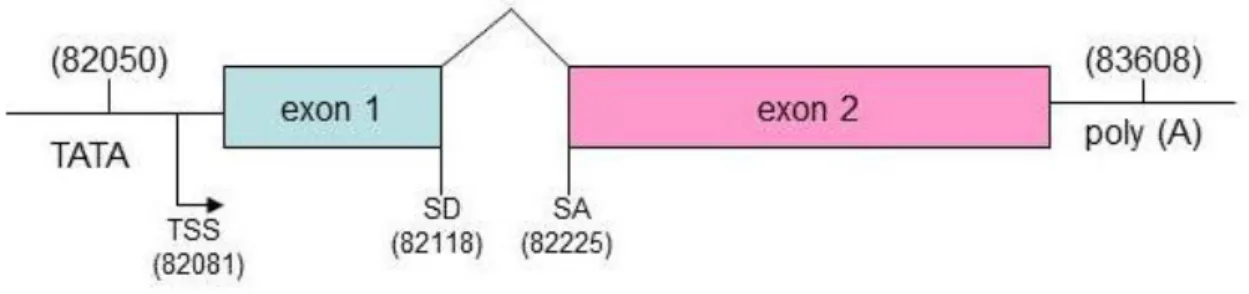
ORF57 gene is positioned in a unique long region of the HHV-8 genome and is flanked by ORF56 (primase) and K9 (viral interferon regulatory factor, vIRF) genes. ORF56 and ORF57 have their own promoter to initiate transcription, but they share the same polyadenilation signal downstream of ORF57.
ORF57 contains two coding exons and a single 108 bp intron (Fig.12). The exon 1 is relatively small, 114 nts, and has four ATG codons, clustered within a region of 33 nucleotides, in frame with each other and with the first exon and separated from it by a single stop codon. The intron is 109 nts in size and contains consensus splice donor and acceptor sites. The exon 2 is about 1,4 kb long. The transcriptional start site (TSS) is located at nt 82003 and polyA signal starts at nt 83608. A TATA box 24 bp is identified upstream the TSS as well as several consensus transcription factor binding sites (NF-kB, AP-1, Oct-1), and at least four R responsive elements involved in the transcriptional activation by ORF50.
Figure 12. Schematic representation of ORF57 mRNA.
ORF57 expression is highly dependent on ORF50, and a RRE in the ORF57 promoter is responsible for ORF57 binding.
ORF57 is expressed predominantly in the nucleus and nuclear localization is driven by three independent nuclear localization signals (NLS) that form a cluster in the N-terminal.
ORF57 encodes a protein of 455 aa residues.
Analyses of amino acid sequence reveals several structural and functional motifs. The N-terminus contains a long stretch of arginine residues, two separate RGG-motifs, which are
typical of RNA-binding proteins and four serine/arginine dipeptides, characteristic of SR proteins, the major cellular splicing factors. The three NLSs overlap the arginine rich region. The C terminus of ORF57 is enriched in leucine residues and contains a leucine zipper motif, typical of cellular transcriprion factors. The C-terminus also contains the zinc-finger-like motif. ORF57 promotes the expression of HHV-8 intronless genes, including several viral early and late genes, such as ORF59, T1.1 (PAN or nut-1), gB, MPC. ORF59 is an early gene encoding a viral DNA polymerase processivity factor involved in viral DNA replication. T1.1 is a non-coding RNA that accumulates at unusually high levels in the nucleus of lytically infected cells. ORF50 and ORF57 have a synergic activity that is promoter specific: expression of some promoters that are upregulated by ORF50 can be synergistically enhanced by coexpression with ORF57. This synergy results from a post-translational enhancement of the transcriptional activity of ORF50. ORF57 transactivates specific viral promoters in synergy with ORF50, such as promoters of T1.1, ori-Lyt and Kaposin. ORF57 interacts with ORF50 via its N-terminal region and the central region of ORF50.
2.5. HHV-8 epidemiology and transmission
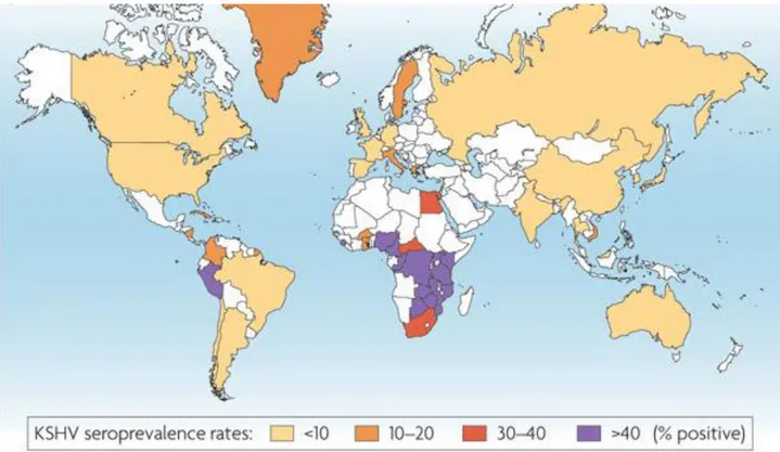
The serologic prevalence of HHV-8 infection has been explored in most continents worldwide and in different populations with different levels of risk of HHV-8 infection. It should be noted that the comparisons of prevalence are limited by the fact that either antibodies to latent or lytic HHV-8 antigens were detected. Several studies have confirmed that there is a low seroprevalence in central and northern Europe, North America and most of Asia, intermediate prevalence in the Middle East and Mediterranean, and high prevalence in southern Africa (Fig.13).
Fig. 13. KSHV seroprevalence rates.
The virus, first thought to be transmitted only sexually, is now also considered transmissible through low risk or more casual behaviours. Important risk factors for transmission of the virus are a spouse's seropositivity and maternal seropositivity. Of all anatomic sites, HHV-8 DNA is found most frequently in saliva, which also has higher viral concentrations than other secretions28. For this reason, it has been hypothesized that saliva could be the route of casual transfer of infectious virus among family members.
Other possible transmissions are blood-borne and organ transplantation.
2.6. HHV-8 pathogenesis
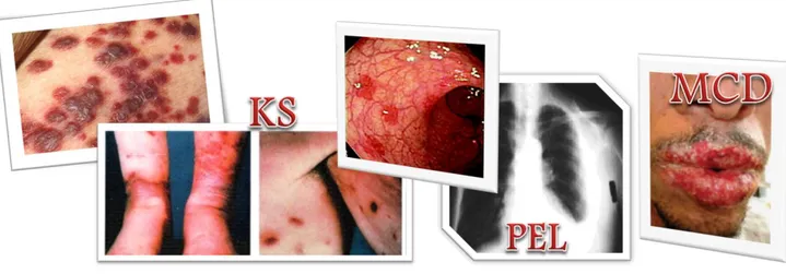
Human herpesvirus 8 is associated with proliferative disorders including Kaposi’s sarcoma (KS), multicentric Castleman disease (MCD), primary effusion lymphoma (PEL), and other lymphoadenopathologies (Fig. 14).
Fig. 14. Representation of proliferative disorders associated to KSHV.
HHV-8 induces the formation of neoplasias in natural or experimental hosts, and reactivation from latency is essential for this activity.
Latent viral proteins, such as vFLIP and LANA, inactivate tumour suppressors and block apoptosis29.
However, lytic replication is also important for transmission of the virus in the population and in the pathogenesis of KS. HHV-8 vIL-6 is highly expressed during the lytic cycle, and promotes cellular growth and angiogenesis, while protecting against apoptosis30. Additional evidence for the importance of lytic replication includes the fact that inhibition of active HHV-8 replication by gancyclovir reduces the incidence of HHV-8 in HIV-infected individuals31.
Kaposi’s sarcoma
KS was first described by Moritz Kaposi in the 1870s32 and was described as an aggressive tumour affecting patients younger than those currently observed. For all epidemiological forms of KS, the tumour presents as an highly vascularised neoplasm that can be polyclonal, oligoclonal, or monoclonal. Its antigenic profile suggests either endothelial, lymphoendothelial, or macrophage origins. All forms of KS lesions contain a variety of cell types, including endothelium, extravasated erythrocytes, infiltrating inflammatory cells, and characteristic “spindle” cells of endothelial origin33
(Fig.15). The spindle cells express both endothelial and macrophagic markers.
Fig. 15. Cellular transformation in Kaposi’s sarcoma.
Extensive and aberrant neoangiogenesis in KS lesions is accompanied by elevated levels of many cytokines, including basic fibroblast growth factor (bFGF), interleukin-1 (1), 6, IL-8, platelet-derived growth factor (PDGF), tumour necrosis factor (TNF), gamma interferon
(IFN-), and vascular endothelial growth factor (VEGF). Many of these cytokines are secreted by spindle cells, are essential for spindle cell viability in culture, and are themselves proangiogenic (Fig. 16).
HIV infection increases the risk for development of KS, and therefore, the incidence of KS has increased substantially during the HIV pandemic, particularly in younger HIV-infected patients34.
Microscopic cellular transformation in Kaposi’s sarcoma
Macroscopic cellular transformation in Kaposi’s sarcoma
Figure 16. Representation of Kaposi’s sarcomagenesis.
Four forms of Kaposi’s sarcoma are known, differentiated on clinical parameters and epidemiology:
Classic KS: is an indolent tumour affecting the elderly population, preferentially men, in Mediterranean countries. The lesions tend to be found in the lower extremities and the disease, due to its non-aggressive course, usually does not kill those afflicted.
AIDS-KS: in the context of the acquired immunodeficiency syndrome (AIDS), KS is the most common malignancy and is an AIDS defining illness35. AIDS-KS is a more aggressive tumour than classic KS and can disseminate into the viscera with a greater likelihood of death. It presents more often multifocally and more frequently on the upper body and head regions. Endemic KS: HHV-8 was prevalent in Africa prior to the HIV epidemic. Prior to HIV coinfections, endemic KS affected men with an average age of 35 and very young children36. HIV coinfection has raised the prevalence of KS significantly in Africa, where endemic KS is found more often in women and children than in other areas of the world37.
Iatrogenic KS: Immunosuppression, as that occurring in transplant recipients, is known to facilitate reactivation of herpesviruses and therefore transplant patients under immunosuppressive therapy can develop KS. Withdrawal of the therapy can cause the KS to regress38.
Multicentric Castleman disease
MCD is a rare polyclonal B-cell angiolymphoproliferative disorder. Most of the B-cells in the tumour are not infected with HHV-8, and the HHV-8 infected cells are primarily located in the mantel zone of the lymphatic follicle. It is thought that uninfected cells are recruited into the tumour through HHV-8 paracrine mechanisms, such as vIL-6, a known growth factor for the tumour. More than 90% of AIDS patients with MCD are HHV-8 positive, whereas MCD in the context of no HIV infection has a HHV-8 prevalence of approximately 40%39.
Primary effusion lymphoma
First identified as a subset of body-cavity-based lymphomas (BCBL), PELs contain HHV-8 DNA sequences6. These lymphomas are distinct from malignancies that cause other body cavity effusions. PEL cell lines have 50–150 copies of HHV-8 episomes per cell40.
2.7. HHV-8 and angiogenesis
The effects of acute HHV-8 infection on endothelial cell functions, induction of angiogenesis, and triggering of inflammatory processes are still largely unknown.
In our laboratory, we demonstrated that HHV-8 selectively triggers the expression and secretion of high levels of monocyte chemoattractant protein 1 (MCP-1). We also found that this event is accompanied by virus-induced capillary-like structure formation at a very early stage of acute infection41 (Fig.17).
Figure 17. HUVEC monolayer not infected or infected with HHV-8 41.
The MCP-1 expression is controlled by the nuclear factor kB (NF-kB), that induces the expression of chemokines promoting cell migration and angiogenesis, such as IL-8 and vascular endothelial growth factor (VEGF), several matrix metalloproteinases (MMPs) that promote tumour invasion of surrounding tissue. NF-kB is a critical regulator of the immediate early response to HHV-8, playing an important role in promoting inflammation, in the control of cell proliferation and survival, and in the regulation of virus replication. Several studies show that transfection of different HHV-8 genes results in NF-kB activation in different cell types, and in our laboratory we demonstrated that HHV-8 acute infection induces NF-kB activation in endothelial cells. The human MCP-1 gene contains 2 NF-kB-binding sites in the enhancer region. The kB binding sites are required for TPA-induced expression. In HHV-8 infection we observed that the NF-kB pathway is involved in the enhancement of MCP-1 expression and is required for maximal production of the chemokine. However, mutations in both NF-kB sites in the enhancer region did not result in the complete loss of promoter induction in HHV-8 infected cells, and inhibitors of NF-kB do not prevent MCP-1 activation following HHV-8 infection suggesting that at least another signalling pathway may be involved in the control of MCP-1 expression in the course of acute HHV-8 infection.
3. The cellular activating transcription factor 4 (ATF4)
ATF4 (also called cAMP responsive element binding 2, CREB2) belongs to the ATF/CREB family of transcription factors that represent a large group of basic region-leucine zipper (bZip) proteins. The basic region of the bZIP protein interacts with DNA, and they dimerize by their leucine zipper domains forming homodimers, heterodimers or both42 (Fig. 18).
CREB/ATF family members include ATF1 (also known as TREB36), CREB/CREM, CREB314 (also known as Aibzip or Atce1), CREB-H, ATF2 (also known as CRE-BP1), ATF3, ATF4, ATF6, ATF7, B-ATF and ATFX (also known as ATF5).
ATF4 gene is in chromosome 22 at the cytogenetic band 22q13.1, located at 38,241,069– 38,243,191 bp, with a genomic size of 2122 and is constitutively expressed in many cells.
The structure of human ATF4 mRNA includes three short open reading frames (uORFs) in the 5’UTR that precede the functional coding sequence43
and are out of frame with the main protein-coding region. The organization of the 5’UTR uORFs in ATF4 is essential for the response of ATF4 to stress such as ER stress and hypoxia.
ATF4 protein consists of 351 amino acids. The protein is structured into several domains/motifs that are essential for ATF4 homo/heterodimerization and DNA binding. A transcriptional activation domain has been located at the N-terminus of ATF444.
The mammalian ATF4 can form a homodimer, and heterodimers with members of the AP-1 and C/EBP family of proteins, including Fos and Jun45, and several C/EBP proteins46. ATF4 has a very short half-life of about 30-60 minutes.
ATF4 has several interacting partners, which include p30047, RNA polymerase II subunit RPB348, ZIP kinase, a serine/threonine kinase, which mediates apoptosis49, HTLV1 transactivator Tax, which activates the expression of viral mRNA through a three 21 bp repeat enhancer located within the HTLV-1 LTR50. Tax transactivates the HTLV-1 promoter via the Tax responsive elements that contain the consensus ATF/CRE core sequence. ATF4 enhances the ability of Tax to transactivate the HTLV-1 promoter. The numerous dimerization and interaction partners determine the diverse functions of ATF4.
ATF4 can function as a transcriptional activator, as well as a repressor. It is a stress responsive gene, which is upregulated by several factors/stressors, including oxygen deprivation (hypoxia/anoxia), amino acid deprivation, endoplasmic reticulum stress (ER stress), oxidative stress, and by the growth factor heregulin51.
In mammalian cells, hypoxia/anoxia and perturbation of ER homeostasis induces a complex transcriptional program and triggers a reduction in protein translation (UPR: unfolded protein response). A central mediator of this translational response to anoxia is phosphorylation of the eukaryotic initiation factor 2 (eIF2) by PERK protein kinase. Although the phosphorylation of eIF2 results in global translational reduction, it specifically increases the translation of ATF4 mRNA. The various stress signals integrate in a common pathway of increased translation of ATF4, which subsequently ensures supply of amino acids for protein biosynthesis and protects cells against oxidative stress, by modulating a number of genes involved in mitochondrial function (e.g., Lon mitochondrial protease homologue), amino acid metabolism and transport (e.g., asparagine synthetase), as well as in redox chemistry (e.g., NADH cytochrome B5 reductase homolog). As a result of metabolic and ER stress that activate the PERK pathway of translational inhibition, ATF4 initiates a feedback regulatory loop to ensure the transient nature of protein synthesis inhibition. ATF4 induces GADD34 transcription, a component of the phosphatase complex that dephosphorylates eIF-2alpha.
Some of the genes that are induced by ATF4 include receptor activator of nuclear factor-kappa B (RANK) ligand (RANKL), osteocalcin, E-selectin, VEGF, Gadd153, gadd34, asparagine-synthesase, TRB3, and several genes involved in mitochondrial function, amino acid metabolism and redox chemistry52 (Fig. 19).
Figure 19. Several events cause cellular stress. The ER protein kinase (PERK) is activated by homodimerization
and autophosphorylation to phosphorylate eIF2α, thereby reducing the rate of mRNA translation and the biosynthetic protein-folding load on the ER. eIF2α phosphorylation paradoxically increases translation of ATF4 mRNA to produce a transcription factor that activates expression of genes encoding protein chaperones, ERAD machinery, enzymes that reduce oxidative stress, and functions in amino acid biosynthesis and transport.
One important stress factor relevant to cancer progression is hypoxia and more extremely, anoxia. Tumour hypoxia/anoxia is associated with a more aggressive clinical phenotype, and ATF4 protein has been observed to be in much greater levels in primary human tumours compared to normal tissues53.
ATF4 induces VEGF and E-selectin which may be associated with increased metastasis. Since ATF4 protein has shown to be present at greater levels in cancer compared to normal tissue, and it is upregulated by signals of the tumour microenvironment such as hypoxia/anoxia, oxidative
stress, and ER stress, it could potentially serve as a specific target in cancer therapy. As a target ATF4 is attractive because it is also potentially involved in angiogenesis and adaptation of cancer cells to hypoxia/anoxia, which are major problems in cancer progression. The induction of VEGF (vascular endothelial growth factor) has a key role in angiogenesis, and preliminary study in our laboratory demonstrate that transfection of ATF4 induces capillary-like structure formation in vitro.
Many viruses have been shown to induce ER stress and activate the UPR: these include three members of the flavivirus, C hepatitis and HCMV. Viral infection induces the cell response to stress, which should lead to an attenuation of viral replication. However, some aspects of the UPR can be regulated and limited by the virus. For example, infection with HCMV (betaherpesvirus) induces the UPR by regulating specifically three signalling pathways: PERK, ATF6 (activating transcription factor 6) and Ire-154.
Cells infected with this betaherpesvirus show an increase in ATF4 protein levels, leading to the activation of genes involved in metabolism and redox reactions and helping the virus to maintain a cellular environment permissive to infection.
Studies demonstrate that also HHV-8 can interact with ATF4. LANA, the latency associated nuclear antigen, encoded by ORF73, represses the transcriptional activation activity of ATF4 and the interaction requires the bZIP domain of ATF4. Repression by LANA is independent from the DNA-binding ability of ATF455.
AIM OF THE RESEARCH
Angiogenesis, the formation of new blood vessels, is the hallmark of cancer, and aggressiveness of primary tumours depends on the novo angiogenesis. A large number of proangiogenic factors have been identified, including well known cytokines with predominant angiogenic activity, such as VEGF.
Activating Transcription Factor 4 is a central factor used by the cell to respond to multiple, seemingly divergent stresses, including nutrient depletion, oxidative stress, UV irradiation, reductive stress, exposure to toxins, hypoxia.
The detailed molecular events controlling stress response have not been fully elucidated, but it is known that ATF4 is a common downstream target that integrates signalling from different stimuli. This pathway is collectively referred to as the Integrated Stress Response (ISR) and consequent unfolded protein response (UPR). Several conditions can cause protein misfolding, such as altered metabolic conditions including anoxia and hypoxia, expression of mutant proteins and infection by viruses, including hepatitis C virus, Japanese encephalitis virus, bovine diarrhoea virus, adenoviruses and human Cytomegalovirus. ATF4 is also related to hypoxia tolerance and tumour progression, is overexpressed in tumours and it can crosstalk with NFkB in controlling VEGF-dependent tumour angiogenesis. In particular, ATF4 cooperates with NFkB in activating the signalling cascade that ultimately increases VEGF expression, associated to different grades of breast cancer. Similarly, in a human medulloblastoma cell line treated with UPR inducers, ATF4 was shown to bind to VEGF gene, although its contribution to VEGF transcription appeared to be fairly modest. In spite of these evidences, a direct role of ATF4 in the induction of proangiogenic properties in primary endothelial cells is not yet well defined, and the observations reported so far are mainly direct. The recent observation that silencing of ATF4 impairs monocyte chemotactic protein 1 (MCP-1) transcription in aortic endothelial cells, is particularly interesting in the context of the role of MCP-1 as a potent mediator of the angiogenesis induced by human herpesvirus 8 infection.
HHV-8, also known as KS-associated herpesvirus (KSHV), is the causative agent of Kaposi’s sarcoma (KS), and has been implicated as a major agent in the development of primary effusion lymphomas (PEL), multicentric Castleman’s disease and lymphoproliferative disorders affecting HIV-infected patients. Despite the name, KS is not a true sarcoma but a “vascular” neoplasm, containing abnormally dense and irregular blood vessels, that fill with blood cells, giving the tumour its characteristic bruise-like appearance. Several morphologic variants of KS are known, but all the described forms are characterized by angiogenesis and contain tumour cells of endothelial
origin with a characteristically abnormal elongated shape, called spindle cells. Inflammatory cells, including lymphocytes, plasma cells and dendritic cells, are also present in KS lesions. The majority of neoplastic cells in KS support a latent HHV-8 infection and virus lytic replication, required for neoplastic development, is limited to a small number of cells. Several HHV-8 gene products, including homologues of cellular oncogenes, cytokines, chemokines and chemokine receptors, have been related to endothelial growth and transformation.
We recently reported that HHV-8 activates NFkB in endothelial cells, promotes transcriptional activation and secretion of MCP-1, and induces MCP-1-dependent angiogenesis. However mutation or deletions involving NFkB binding sites located in the MCP-1 enhances region resulted only in a partial inhibition of HHV-8 induced MCP-1 transcription and angiogenesis, suggesting the involvement of at least another transcriptional pathway.
These observations prompted us to investigate the role of ATF4 in inducing neo-angiogenesis in primary human endothelial cells, with particular focus on a possible crosstalk with MCP-1 in the induction of the proangiogenic phenotype. Our results show that ATF4 has a direct role in promoting neo-angiogenesis in endothelial cells. In fact, the overexpression of ATF4 induces capillary-like structure formation, and activates directly the enhancer-less promoter of MCP-1, inducing the release of MCP-1.
We investigated also the possible relationship between ATF4 and HHV-8 induced angiogenesis. We found that in vitro HHV-8 infection increases ATF4 expression, most likely through the action of the major viral transactivator ORF50, and this upregulation is directly associated to virus proangiogenic potential, possibly by the induction of MCP-1 production.
These data provide new insights into the mechanism underlying the regulation of angiogenic process, both as a consequence of cellular stress conditions or virus infection, and might help to understand the basis of tumour angiogenesis, opening new therapeutic regimens for the management of tumours such as Kaposi’s sarcoma.
METHODS
1. Cells
For angiogenesis studies human umbilical vein endothelial cells (HUVECs) were used56. HUVECs were cultured in EGM-2 medium (BioWhittaker, Walkersville, MD), using collagen coated flasks (Biocoat Collagene, BD Biosciences, Bedford, MA). All the experiments were performed on third to fifth passages HUVECs.
For the infections and transfection studies the following human cell types were used: HUVECs, human microdermal endothelial cells (HMECs), 293 epithelial cell line and Jurkat T cell line. HMECs were obtained from derma, immortalized by infection with a replication-defective adeno-5/SV40 virus as previously described57, and cultured in EGM-2 medium. The human 293 cell line (ATCC CRL-1573) was grown in complete DMEM medium plus 10% inactivated fetal calf serum (FCS). The Jurkat T cell line (ATCC TIB-152) was grown in complete RPMI medium plus 10% FCS. The PEL-derived BC-3 cell line58, used for HHV-8 production, was maintained in RPMI medium (Gibco, Invitrogen, Carlsbad, CA) supplemented with 2 mM L-glutamine, 100 U/ml of penicillin, and 100 µg/ml of streptomycin (complete medium) and 20% FCS41.
All cells were cultured at 37°C in the presence of 5% CO2.
2. Plasmids
Transfection experiments were performed using recombinant plasmids pCR-50sp, PR57, pGL-PR50, pGL-PRT1.1, pGLM-PRM, pGLM-ENH, pGLM-MA1MA2, pRL-SV40 and pCG-ATF4. Spliced forms of ORF50 were cloned in the expression vector pCR3.1-Uni (Invitrogen) in our laboratory25. Briefly, spliced genes were obtained from TPA-activated BCBL-1 cells by specific retrotranscription of polyA RNA followed by PCR amplification. Amplified fragments were sequenced to verify their integrity and were then inserted into the vector pCR3.1-Uni to obtain the recombinant plasmid pCR-50sp.
Fig. 19. Schematic representation of eukaryotic expression vector pCR3.1-Uni, utilized for cloning of genes ORF50
and ORF57.
HHV-8 gene promoters were cloned in the reporter vector pGL3-Basic in this laboratory56, containing the Firefly luciferase gene cloned under the transcriptional control of HHV-8 promoters. The MCP-1 promoter-luciferase constructs (provided by Dr. T. Yoshimura)57 contain the proximal promoter (pGLM-PRM), the proximal promoter and the distal enhancer (pGLM-ENH), the proximal promoter and the distal enhancer in which both NF-kB sites are mutated (pGLM-MA1MA2).
Fig. 20. Schematic representation of reporter vector pGL3-Basic, utilized for cloning of HHV-8 promoters genes.
pRL-SV40 vector (Promega) was used as an internal transcriptional control, and contains the Renilla luciferase gene cloned under the transcriptional control of the SV40 virus promoter.
pCG-ATF4 plasmid contains the ATF4 gene cloned under the transcriptional control of the CMV promoter, and was a kind gift of Dr. T. Hai51.
Fig. 19. Schematic representation of expression vector pCG-AFT4.
3. DNA extraction
Genomic DNA was extracted from 106 cell samples.
Cells were lysed in 500µL lysis buffer (10 mM Tris-HCl pH 8.0, 10 mM disodium EDTA pH 8.0, 10 mM NaCl, 0.6% SDS and 100 µg/ml proteinase K) and incubated at 37°C for a minimum time of 4 hours. After three cycles of phenol:chloroform:isoamyl alcohol (25:24:1) extractions, DNA was recovered by ethanol precipitation, suspended in sterile water, and RNase A was added to a final concentration of 100 µg/ml. Following 1 hour incubation at 37°C, RNase A was removed by phenol extraction. After precipitation with ethanol, DNA pellets were dissolved in sterile water and stored at -20°C until PCR or qPCR analysis.
4. RNA extraction and retrotranscription
Total RNA was extracted with RNAzol B (Tel-Test), following the protocol provided by the manufacturer. Briefly, 106 cell samples were lysed with RNAzol, then, chloroform in ratio 1:5 was added. The mixture was centrifuged 20 minutes at 8500 x g and the aqueous phase was collected and RNA precipitated with isopropanol. After two washes with 75% ethanol and DNAse treatment (4 U/mg RNA, 3 x 20 min. at room temperature), RNA was precipitated with isopropanol 20 minutes at 8500 x g at 4°C. RNA quality was checked by electrophoretic analysis on a 0.8% agarose gel. The absence of contaminating DNA was checked by PCR amplification of human β-actin gene before retrotranscription (RT-). Negative PCR results for β-β-actin ensured that the RNA sample was completely free from DNA sequences. First strand cDNA synthesis was carried out with MuLV reverse transcriptase and random hexamer primers (Applied Biosystems), following the manufacturer’s instructions, retrotranscribing 2 µg of total RNA from all the samples. The mixture was incubated for 1 hour at 42°C. Efficiency of retrotranscription was assessed by analysis of dilutions of cDNA with PCR specific for human β-actin gene (RT+).
5. PCR and rtPCR
The presence and the level of transcription of HHV-8 were analyzed by PCR and reverse transcription PCR (rtPCR) amplification of the ORF26 and ORF50 genes. PCR amplification was performed using 100 ng total DNA or 200 ng total cDNA extracted from infected cells. Amplification of the housekeeping β-actin gene was used as a control. The transcription of ATF4 was analysed by rtPCR amplification of the ATF4 gene. Specific primers and PCR conditions are described in Table 2. The absence of contaminating DNA was checked by PCR amplification of human β-actin gene before retrotranscription. Negative PCR results for β-actin ensured that the RNA sample was completely free from DNA sequences, and that positive amplification after retrotranscription was positively associated to viral transcripts.
Particular care was taken to avoid sample-to-sample contamination: different rooms and dedicated equipment were used for DNA extraction and processing, for PCR set-up and gel analyses, all pipette tips had filters for aerosol protection.
GENES PRIMERS SEQUENCES AMPLICONS ORF50 ORF50-Forw ORF50-Rev 5'-TTGGTGCGCTATGTGGTCTG-3‘ 5'-GGAAGGTAGACCGGTTGGAA-3' 420 bps ORF26 ORF26-Forw ORF26-Rev 5'-GCCGAAAGGATTCCACCAT-3‘ 5'-TCCGTGTTGTCTACGTCCAG-3' 232 bps ATF4 ATF4-Forw ATF4-Rev 5’-GTGGCCAAGCACTTCAAACC-3’ 5’-GGAATGATCTGGAGTGGAGG-3’ 414 bps β-actin HACT-Forw HACT-Rev 5’-TCACCCACACTGTGCCCATCT-3’ 5’-GACTACCTCATGAAGATCCTCAC-3’ 674 bps
GENES CONDITIONS CYCLES [Mg
Cl2]
ORF50 94°C 5min
94°C 30 sec, 57°C 1 min, 72°C 1 min + ext. 3 sec/cycle 72°C 10 min, 4°C >>> 1 35 1 2mM ORF26 94°C 5min
94°C 1min, 57°C 1 min, 72°C 1 min + ext. 3 sec/cycle 72°C 10 min, 4°C >>> 1 45 1 1.5 mM ATF4 94°C 5min
94°C 1min, 57°C 1 min, 72°C 1 min + ext. 3 sec/cycle 72°C 10 min, 4°C >>> 1 35 1 2mM β-actin 94°C 5min
94°C 1min, 57°C 1 min, 72°C 1 min + ext. 3 sec/cycle 72°C 10 min, 4°C >>> 1 30 1 1.25 mM
Table 2. Primer sets and thermal conditions used for PCR and rtPCR reactions.
6. Cell transfections
Jurkat, BC-3 and BCBL-1 cells were seeded 24 hours before transfection to obtain optimal cellular density (106 cells/mL) and 106 cell samples were transfected with 1 µg of plasmid DNA by nucleofection (Nucleofector, Amaxa), following the manufacturer’s instructions. This method permits to obtain high efficiency of transfection, carrying the exogenous DNA into the cells and directly into the nucleus. Efficiency of transfection determined in parallel samples by transfection with pMax- GFP plasmid (Amaxa).
HeLa and 293 cells were seeded in 24-well plates 24 hours prior to transfection to obtain optimal confluence, then were transfected with 1µg of plasmid DNA by GeneJuice Transfection Reagent (Novagen), based on polyamine formulation, following the manufacturer’s instructions.
7. Reporter assay
The Luciferase assay was performed using the Dual-Glo Luciferase Assay System (Promega). Luciferase and Renilla were used as co-reporters to measure gene expression. In order to generate luminescence Firefly luciferase requires luciferin, ATP, magnesium and molecular oxygen. Renilla luciferase requires coelenterazine and molecular oxygen.
Cells were seeded into 24-well plates 24 hours prior to transfection, then, they were co-transfected with the reporter constructs, pRL-SV40, and with the expression plasmids. 48 hours post-transfection, Dual-Glo Luciferase Reagent was added to each well in ratio 1:1 with the medium and cells were transferred to 96-well plate. After at least 10 minutes of incubation, the firefly luminescence was measured. Following the first measurement, an equal volume of Dual-Glo Stop&Glo Reagent was added and after 10 minutes incubation, the Renilla luminescence was measured.52
The results were expressed as Luciferase activity, normalized using the Renilla emission.
8. Real time PCR
Real time PCR or quantitative PCR (qPCR) was used to determine the amount of ORF26 and ATF4 genes. β-actin housekeeping gene was used as internal control in order to normalize the results. qPCR using the TaqMan probes was performed using the 7300 ABI Prism (Applied Biosystems). The DNA or cDNA copy numbers were quantified by comparison with a 10-fold serial dilutions of known concentrations of pCG-ATF4 or pCR-ORF26 plasmids. The concentration of the standard DNA was assessed by spectrophotometer and plasmid copy numbers were calculated to prepare standard curves (107 to 102 copies).
Amplifications were carried out in a 50 µL total volume containing 25 µL 2X TaqMan Universal PCR Master Mix (Applied Biosystems), 5 µL of primer and probes, and 20 µL template. The target genes were amplified using TaqMan FAM-labelled probe at the 5’-end and TAMRA quencher at the 3’-end. The β-actin housekeeping gene was simultaneously amplified using a 5’-VIC-labelled
probe. Primers and probes used are shown in Table 3. Concentrations of the primers and probes were 900 nM of each primer and 200 nM of each probe.
The reaction conditions for all templates were 10 minutes at 95°C for enzyme activation, and 40 cycles with 15 seconds at 95°C for DNA denaturation and 1 minute at 60°C for annealing and extension. Fluorescence data were collected in the primer elongation phase at 60°C.
To normalise the data, the cycle threshold (Ct) values of the housekeeping gene were subtracted from the target gene Ct value of the sample.
GENES PRIMERS &
PROBES SEQUENCES ORF26 H8-Forward H8-Reverse H8-Probe 5'-CTCGAATCCAACGGATTTGAC-3’ 5'-TGCTGCAGAATAGCGTGCC-3'
5’-(6Fam) CCATGGTCGTGCCGCAGCA (Tamra)-3’
ATF4 ATF4-Forward ATF4-Reverse ATF4-Probe
5’-CCCCCCTAGTCCAGGAGACT-3’ 5’-CTGGGAGATGGCCAATTGG-3’
5’-(6Fam) ATAAGCAGCCCCCCCAGACGG (Tamra)-3’
β-actin β-ACT-Forward β-ACT-Reverse β-ACT-Probe
5'-TCACCCACACTGTGCCCATCT-3' 5'-GACTACCTCATGAAGATCCTCAC-3'
5’-(Vic) ATGCCCTCCCCCATGCCATCCTGCGT (Tamra)-3’
Table 3. Primer and probe sets used for real time PCR quantifications.
9. Western blotting
Cell samples (1-2*106) were harvested and centrifuged 2 minutes at 8000 x g. Pellets were suspended in lysis buffer (6,7 M urea, 10 mM Tris-HCl pH 6.8, 5 mM DTT, 1% SDS, 10% glycerol, protease inhibitors cocktail) and debris was eliminated by centrifugation. 6X loading buffer was added to 20 µg cellular total lysated, and the mixture was subjected to 10% SDS-PAGE electrophoresis. Following electrophoresis, proteins were transferred in the Hibond-P PVDF-membrane (Amersham) in an electroblotter (Biorad). After transfer, the PVDF-membrane was probed with the rabbit anti-ATF4 antibody (SantaCruz Biotechnology) 1:200 diluted, 90 minutes at room
temperature. After three washes, the membrane was incubated with the anti-rabbit HRP conjugated antibody (SantaCruz Biotechnology) 1:1000 diluted for 90 minutes at room temperature. After three washes the antibody was revealed by luminol staining (SuperSignal West Pico Chemiluminescent Substrate, Pierce). ATF4 molecular weight is 38 kDa.
10. ELISA
For MCP-1 release analysis, supernatants from HHV-8 infected, mock-infected or transfected HUVECs were collected at different times after infection, cleared by centrifugation to eliminate cell debris, and stored at -80 °C. After thawing, samples were analyzed for the presence of MCP-1 using a specific quantitative ELISA kit (Biosource International, Camarillo, CA), following manufacturer’s instructions. Each sample was tested in triplicate.
11. Immunofluorescence
Antigen expression of HHV-8 was evaluated by immunofluorescence (IFA), performed on 5x10⁴ infected cells fixed with cold methanol/acetone (50:50), and incubated with antibodies directed against lytic antigen (ORFK8.1) and nuclear antigen associated to latency (ORF73), as already described (Caselli et al., 2007). After primary antibody incubation, secondary antibodies (anti-mouse IgG, fluorescein conjugated, Sigma) were added and slides were mounted for fluorescence observation using a Nikon Eclipse TE2000-S microscope.
12. HHV-8 infection
HHV-8 inoculum was obtained from BC-3 cell line stimulated with TPA (20 ng/ml) for 72 h, and virions were purified by density centrifugation using Optiprep self-forming gradients (Sentinel, Milan, Italy), as previously described56. Cell-free purified virions were suspended in PBS containing 1% BSA, and stored at -80 °C until use. Virus stock was quantitated by real-timePCR TaqMan assays (qPCR) as already described41, and contained 5x10⁵ copies of viral DNA/µl. Prior to use, virus stock was treated for 10 min at room temperature with DNase-I and RNase-A, to eliminate free viral nucleic acids potentially present in the preparation. Infective titers were
determined by counting the number of cells positive for the HHV-8 K8.1 protein by immunofluorescence at 36 h after infection, as previously described41.
For in vitro infection experiments HUVEC, HMEC, 293 or Jurkat cells were seeded at optimal density 24 h before infection, and subsequently infected as already described41,56,59. Briefly, 0.5 x 10⁶ cells were washed once with PBS and infected with 10 µl of purified virus preparation (i.e. 5x10⁶ copies of viral DNA, corresponding to a m.o.i. 10:1). After 3 h of absorption at 37°C, the viral inoculum was removed, cells were washed with PBS and incubated in complete medium for the designed times. Cell samples were harvested at specific time points for subsequent analyses. Control samples were uninfected or mock-infected with UV-inactivated HHV-8, obtained by exposing the purified inoculum under UV light (200 mJ per cm2) for 30 min41.
13. Analysis of in vitro cord formation capability
In vitro capillary-like structure formation assay was performed using an extracellular basement membrane extract (BME; Cultrex; Trevigen, Gaithersburg, MA), as described41. Uninfected, infected or transfected HUVECs suspended in EGM-2 medium were seeded on solidified BME (5x10⁴ cells per well) and incubated at 37 °C. Tube formation on BME was analyzed at different times after cell seeding by optical microscopy.
14. Small interfering RNA (siRNA) studies
For silencing studies, HUVECs plated at optimal density were transfected with 100 nM (1.35 μg/ml) of siRNA targeting human ATF4 or of unrelated Negative Control siRNA (Ambion), using the RNAiFect technique (Qiagen) and following manufacturer’s instructions. Effective knockdown of ATF4 transcription was checked by RT-qPCR, using the gene expression assay described above (Applied Biosystems).
15. Statistical analyses
RESULTS
1. HHV-8 infection increases ATF4 expression
The effect of HHV-8 infection on ATF4 expression was studied in different human cell types, including human primary endothelial cells (HUVECs, as representative of natural targets of HHV-8
in vivo), immortalized microdermal endothelial cells (HMEC), epithelial cells (293 cell line) and T
lymphocytes (Jurkat cell line). As previously reported by us, HHV-8 supports a productive infection in all these cell types, followed by the establishment of latency at 5-10 days post infection (d.p.i.)41,56,59,70 .Efficiency of infection ranged between 15% (293 and Jurkat T cells) and 20% (HMEC and HUVEC), as measured by virus K8.1 antigen expression by immunofluorescence (IFA). Control samples were uninfected or mock-infected with UV-inactivated HHV-8.
HHV-8 infected and control cells were analysed for ATF4 expression at 8, 24, 48 and 72 hours post infection (h.p.i.) by quantitative real time PCR after retrotranscription (RT-qPCR) and Western blot. The results (Fig. 1) showed that HHV-8 infection induces a substantial increase of ATF4 transcription and translation, compared to levels detected in uninfected or mock infected cells. Transcriptional increase was present as early as 8 h.p.i. and peaked at 24 h.p.i., when levels of ATF4 mRNA were 10 times higher compared to controls. The increase persisted at 48 and 72 h.p.i., although slowly declining. ATF4 protein increased consistently and accumulated till 72 h.p.i., when protein amount was 6.9 fold compared to mock-infected samples.