Activating mutations and translocations in the guanine
exchange factor VAV1 in peripheral T-cell lymphomas
Francesco Abatea,1, Ana C. da Silva-Almeidab,1, Sakellarios Zairisa, Javier Robles-Valeroc, Lucile Couronned,e,f, Hossein Khiabaniana, S. Aidan Quinnb, Mi-Yeon Kimb, Maria Antonella Laginestrag, Christine Kimb, Danilo Fioreh, Govind Bhagati, Miguel Angel Pirisj, Elias Campok, Izidore S. Lossosl,m, Olivier A. Bernardn,o,p, Giorgio Inghiramih, Stefano Pilerig, Xosé R. Busteloc,q, Raul Rabadana,r,2, Adolfo A. Ferrandob,i,s,2, and Teresa Palomerob,i,2
aDepartment of Systems Biology, Columbia University Medical Center, New York, NY 10032;bInstitute for Cancer Genetics, Columbia University, New York, NY 10032;cCentro de Investigación del Cáncer, Consejo Superior de Investigaciones Científicas-University of Salamanca, 37007 Salamanca, Spain; dDepartment of Adult Hematology, Necker Hospital, 75015 Paris, France;eINSERM UMR163, Centre National de la Recherche Scientifique Équipe de Recherche Labellisée 8254, Institut Imagine, 75015 Paris, France;fParis Descartes University, 75006 Paris, France;gHemopathology Unit, European Institute of Oncology, 20139 Milan, Italy;hDepartment of Pathology and Laboratory Medicine, Weill Cornell Medical College, New York, NY 10065;iDepartment of Pathology and Cell Biology, Columbia University Medical Center, New York, NY 10032;jInstituto de Formación e Investigación, Hospital Universitario Marqués de Valdecilla, 39008 Santander, Spain;kDepartment of Pathology, Hospital Clinic, 08036 Barcelona, Spain;lDivision of Hematology-Oncology, Sylvester Comprehensive Cancer Center, Miami, FL 33136;mDepartment of Molecular and Cellular Pharmacology, University of Miami, Miami, FL 33101; nINSERM U1170, 94805 Villejuif, France;oUniversité Paris-Sud, 91405 Orsay, France;pGustave Roussy, 94805 Villejuif, France;qCentro de Investigacion Biomedica en Red de Cancer (CIBERONC), Spain;rDepartment of Biomedical Informatics, Columbia University Medical Center, New York, NY 10032; andsDepartment of Pediatrics, Columbia University Medical Center, New York, NY 10032
Edited by Seishi Ogawa, Kyoto University, Kyoto, Japan, and accepted by Editorial Board Member Tadatsugu Taniguchi December 5, 2016 (received for review June 3, 2016)
Peripheral T-cell lymphomas (PTCLs) are a heterogeneous group of non-Hodgkin lymphomas frequently associated with poor prognosis and for which genetic mechanisms of transformation remain incom-pletely understood. Using RNA sequencing and targeted sequencing, here we identify a recurrent in-frame deletion (VAV1Δ778–786) gen-erated by a focal deletion-driven alternative splicing mechanism as well as novelVAV1 gene fusions (VAV1-THAP4, VAV1-MYO1F, and VAV1-S100A7) in PTCL. Mechanistically these genetic lesions result in increased activation of VAV1 catalytic-dependent (MAPK, JNK) and non–catalytic-dependent (nuclear factor of activated T cells, NFAT) VAV1 effector pathways. These results support a driver oncogenic role for VAV1 signaling in the pathogenesis of PTCL.
peripheral T-cell lymphoma
|
VAV1|
mutation|
gene fusionP
eripheral T-cell lymphomas (PTCLs) are malignant and highly aggressive hematologic tumors arising from mature postthymic T cells (1). The diagnosis of PTCL includes diverse lymphoma subgroups, altogether accounting for about 15% of all non-Hodgkin lymphomas (2, 3). Despite much effort in developing reliable di-agnostic markers, the diagnosis of PTCLs is challenging, and 20 to 30% of cases are diagnosed as PTCL-NOS (not otherwise speci-fied). This heterogeneous and poorly defined group constitutes one of the most aggressive forms of non-Hodgkin lymphoma, in which limited response to intensified chemotherapy and high relapse rates result in a dismal 5-y overall survival rate of 20 to 30% (4, 5). Moreover, a paucity of information on driver oncogenes activated in PTCL-NOS hampers the development of targeted therapies in this aggressive lymphoma subgroup.TheVAV1 protooncogene encodes a guanine nucleotide exchange factor (GEF) and adaptor protein with crucial signaling roles in protein tyrosine kinase-regulated pathways (6). Structurally, VAV1 contains a calponin homology domain and an acidic domain in the N terminus followed by a GEF catalytic active core consisting of a cen-tral Dbl homology domain, pleckstrin homology domain, and C1 domain (6). Finally, the C-terminal region of VAV1 contains three Src homology domains in an SH3-SH2-SH3 arrangement (6). The GEF activity of VAV1 stimulates the transition of RAC1 and RHOA small GTPases from their inactive (GDP-bound) to the active (GTP-bound) configuration (6–8). In addition, the adap-tor function of VAV1 mediates activation of the nuclear facadap-tor of activated T cells (NFAT) in synergy with signals from antigenic receptors in lymphoid cells (6, 8–13). In basal conditions, un-phosphorylated VAV1 adopts an inactive closed configuration in which the N-terminal calponin homology and acidic domains and
the C-terminal SH3 (C-SH3) domain block access of small GTPases to the catalytic core and limit the noncatalytic activities of the protein (6, 14, 15). Activation of VAV1 by transmembrane and cytosolic protein kinases reverses these intramolecular inhibitory interactions by promoting an open active configuration associated with phos-phorylation in the acidic, C1 finger, and C-SH3 domains (6, 14, 15). VAV1 is specifically expressed in hematopoietic tissues, and plays key roles in lymphocyte development and function (8). VAV1 is essential for T-cell receptor (TCR)-mediated cytoskeletal reorganization, cytokine secretion, proliferation, and survival (8, 12). Thus,Vav1-deficient mice show a partial block in thymic de-velopment at the CD4−CD8−double-negative to CD4+ CD8+ double-positive transition, defective positive selection, and im-paired negative selection, which altogether point to a major role for VAV1 in TCR signaling (16, 17). Biochemically, mouseVav1 knockout T cells fail to elicit TCR-induced intracellular Ca2+flux and to activate MAP/ERK pathway and NF-κB signaling (18–21). Consistently, the function of mature T-cell populations is also defective in the absence of Vav1, with reduced TCR-induced
Significance
Guanine nucleotide exchange factorVAV1 encodes an adaptor and signal transduction factor with important roles in T-cell re-ceptor signaling. This study identifies activatingVAV1 recurrent mutations andVAV1 fusions in peripheral T-cell lymphomas, di-rectly establishing an oncogenic role for constitutive VAV1 signal-ing in the pathogenesis of this disease.
Author contributions: H.K., X.R.B., R.R., A.A.F., and T.P. designed research; A.C.d.S.-A., J.R.-V., L.C., M.-Y.K., C.K., and T.P. performed research; M.A.L., D.F., G.B., M.A.P., E.C., I.S.L., O.A.B., G.I., and S.P. contributed new reagents/analytic tools; F.A., A.C.d.S.-A., S.Z., H.K., S.A.Q., X.R.B., R.R., A.A.F., and T.P. analyzed data; and A.A.F. and T.P. wrote the paper.
The authors declare no conflict of interest.
This article is a PNAS Direct Submission. S.O. is a Guest Editor invited by the Editorial Board.
Data deposition: The next-generation sequencing data reported in this paper have been deposited in the database of Genotypes and Phenotypes (dbGaP),https://www.ncbi.nlm. nih.gov/(accession no.phs000689.v1.p1) and the Sequence Read Archive at the National Center for Biotechnology Information,https://www.ncbi.nlm.nih.gov/sra/(accession nos.
SRP029591,PRJNA255877, andSRP057085). 1F.A. and A.C.d.S.-A. contributed equally to this work.
2To whom correspondence may be addressed. Email: [email protected], [email protected], or [email protected].
This article contains supporting information online atwww.pnas.org/lookup/suppl/doi:10. 1073/pnas.1608839114/-/DCSupplemental.
proliferation and cytokine secretion (8, 22, 23). Similarly, VAV1-null human JURKAT T cells show impaired TCR-induced calcium flux, IL-2 transcription, and NF-κB activation, as well as decreased TCR-induced JNK and NFAT signaling (24).
Here we report the identification and functional character-ization of recurrent activating mutations and gene fusions in VAV1 in PTCL.
Results
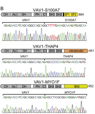
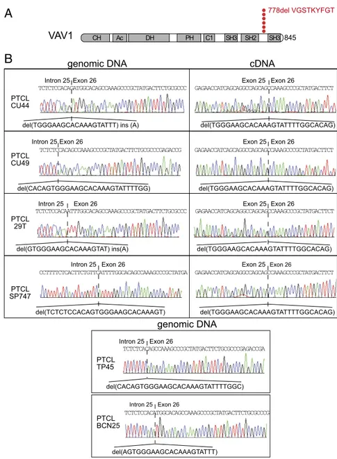
Identification of VAV1 Mutations and Gene Fusions in PTCL. To identify new genetic drivers responsible for T-cell transformation and potential targets for therapy in PTCL, we performed a sys-tematic analysis of genetic alterations using RNA-sequencing (RNA-seq) data from a cohort of 154 PTCL samples, including 41 PTCL-NOS, 60 angioimmunoblastic T-cell lymphoma (AITL), 17 natural killer/T-cell lymphoma (NKTCL), and 36 anaplastic large T-cell lymphoma (ALCL) tumors (25–27) (Dataset S1). These analyses confirmed a high prevalence of RHOA, TET2, IDH2, and DNMT3A mutations in AITL (25, 26, 28) and the re-current presence of fusion transcripts involving theALK1 gene, including NPM1-ALK1, TRAF1-ALK, and TPM3-ALK, and STAT3 activating mutations in ALCL (27) (SI Appendix, Fig. S1 and Dataset S2). However, the most notable finding of these analyses was the identification of gene fusions and novel recurrent mutations involving the VAV1 protooncogene. Specifically, we identified three different fusion transcripts encoding proteins in which the C-terminal SH3 domain of VAV1 is replaced by the calycin-like domain of THAP4 (in two cases), the SH3 domain of MYO1F, or the EF domains of S100A7 (Fig. 1,SI Appendix, Figs. S1 and S2, andDataset S3). Reverse-transcription PCR amplifi-cation and DNA sequencing validated the expression of each of theseVAV1 chimeric mRNAs in all samples analyzed (Fig. 1). In addition, we identified two PTCL cases harboring a novel in-tragenicVAV1 in-frame deletion, r.2473_2499del, which results in the loss of nine amino acids (p.Val778_Thr786del) in the linker region between the SH2 and C-terminal SH3 domains of the VAV1 protein (Fig. 2 andSI Appendix, Figs. S3 and S4).
To further explore the prevalence and mechanisms ofVAV1 mutations in PTCL, we performed targeted genomic DNA se-quencing ofVAV1 in a panel of 126 PTCL samples. Genomic DNA sequencing of the two index RNA-seq cases harboring the r.2473_2499del mutation revealed the presence of focal genomic deletions inVAV1 involving the 3′ end of intron 25 and extending into exon 26 (g.81269_81294del and g.81275_81302del) (Figs. 2B and 3A and SI Appendix, Fig. S4). In addition, we identified additional three cases harboring similar focal genomic deletions involving theVAV1 intron 25–exon 26 boundary (g.81275_81301del, g.81279_81296indelA, and g.81279_81298del) and one additional case with a mutation resulting in the loss of 19 nt at the 5′ end of exon 26 but preserving the intron 25–exon 26 AG splice acceptor sequence (g.81280_81298indelA) (Figs. 2B and 3A andSI Appendix, Fig. S4). cDNA-sequencing analysis in three of our additional mutated cases for which RNA was available, including case PTCL CU44, in which the deletion spared the canonical exon 26 splice acceptor site, revealed that in all cases these mutations resulted in activation of a cryptic splice acceptor site in exon 26 and the consequent expression of misspliced transcripts containing the r.2473_2499del (p.Val778_Thr786del)VAV1 mutation (Fig. 2B). Notably, analysis of VAV1 exon 26 sequences proximal to this cryptic splice acceptor site uncovered the presence of an exonic splicing silencer element (29), which is disrupted or completely lost in allVAV1 intron 25–exon 26 indel mutated cases analyzed (Fig. 3). Altogether, PTCL VAV1 intron 25–exon 26 deletions activate a crypticVAV1 exon 26 splice acceptor site by disrupting the corresponding intron 25–exon 26 canonical splice acceptor sequence (5/6 cases) and removing an exon 26 exonic splicing si-lencer (6/6 cases). In addition to removing these splicing regula-tory elements, these focal deletions reconfigure the architecture
of the intron 25–exon 26 boundary by placing the intron 25 poly-pyrimidine tract immediately distal to the alternative exon 26 AG splice acceptor site (6/6 cases) (Fig. 3C and D). Additionally, our mutation analyses also identified three nonrecurrent point mutations resulting in amino acid substitutions in the Dbl homol-ogy (p.His337Tyr), C1 finger (p.Glu556Asp), and C-terminal SH3 domains (p.Arg798Pro) of VAV1 (SI Appendix, Figs. S1 and S5and Datasets S2andS4).
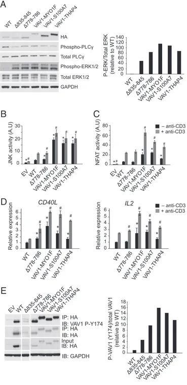
VAV1 Fusion Proteins Induce Increased VAV1 Signaling. Given the prominent role of VAV1 in T-cell activation and to explore the functional consequences of PTCL-associated VAV1 mutations and gene fusions, we analyzed the effect of these genetic alter-ations on lymphocyte signaling. Recent reports have demonstrated that the C-terminal SH3 domain of VAV1 contributes to intra-molecular inhibition of VAV protein family members (14). Con-sidering that our fusions and intragenic deletion mutants specifically affect the region containing the C-SH3 domain of the protein, we used the VAV1 Δ835–845 deletion mutant, which lacks the C-SH3 domain, as positive control in our experiments. To avoid interference with endogenous VAV1, JURKAT cells lacking endogenous expression of VAV1 protein (Jurkat J.VAV1) were infected with lentiviral constructs forVAV1 Δ778–786 and the VAV1-MYO1F, VAV1-S100A7, and VAV1-THAP4 fusions as well as the empty vector,VAV1 wild type, and VAV1 Δ835–845 con-trols. Analysis of signaling events downstream of VAV1 demonstrated
VAV1
VAV1-THAP4
941 CH Ac DH PH C1 SH3 SH2 nitrobindinVAV1-MYO1F
952 CH Ac DH PH C1 SH3 SH2 SH3VAV1-S100A7
884 CH Ac DH PH C1 SH3 SH2 EF1 EF2A
B
VAV1 THAP4 AGAGAACCATCAGCAGGCCAGCAGAGCCCCCCAAGATGAACCCAGTG AGAGAACCATCAGCAGGCCAGCAGGCTTTTTGAAAGCAAAGATGAGCAA VAV1 S100A7 VAV1 MYO1F AGAGAACCATCAGCAGGCCAGCAGAGCCTACGCGGAAGGGAATGGCC 845 CH Ac DH PH C1 SH3 SH2 SH3Fig. 1. VAV1 fusion genes in PTCL. (A) Schematic representation of the domain structure of the VAV1 protein. (B) Schematic representation of the domain structures of the VAV1-S100A7, VAV1-THAP4, and VAV1-MYO1F fusion proteins. Ac, acidic domain; C1, C1 domain; recognition motif for diacylglycerol and phorbol esters, atypical; CH, calponin homology domain; DH, DBL homology; EF, pseudo-EF hand domain; nitrobindin, nitrobindin domain; PH, pleckstrin ho-mology domain; SH2, Src hoho-mology 2 domain; SH3, Src hoho-mology 3 domain.
MEDI
CAL
SCIENC
increased phosphorylation in ERK1/2 but not in PLCγ1 in JURKAT J.Vav1 cells expressing VAV1 Δ778–786, and the VAV1-MYO1F, VAV1-S100A7, and VAV1-THAP4 fusions, compared with wild-type VAV1 (Fig. 4A). Additionally, we also analyzed the impact of the p.His337Tyr, p.Glu556Asp, and p.Arg798Pro missense mutations identified in our study, as well as that of p.E157Lys, p.Lys404Arg, p.Gln498Lys, and p.Met501Arg VAV1 missense mutations iden-tified in adult T-cell lymphoma (ATL) (4), on the phosphorylation and activation of signaling pathways downstream of VAV1 (SI Appendix, Fig. S5). These analyses revealed weaker and variable effects of these mutations, with only VAV1 p.Gln498Lys showing clear increased ERK1/2 phosphorylation and modestly higher levels of phospho-PLCγ1 (SI Appendix, Fig. S5).
Analysis of JNK signaling in AP1 reporter assays, a functional readout of VAV1 catalytic-dependent functions downstream of RAC1, showed marked increased JNK activation in JURKAT cells expressing the MYO1F, S100A7, and VAV1-THAP4 fusions (Fig. 4B). Notably, this effect was primarily in-dependent of TCR stimulation with anti-CD3 supporting that PTCL-associated VAV1 fusion proteins adopt a constitutively active configuration. In contrast, expression of the VAV1Δ778–786 mutant protein induced only minor increases in JNK activation
compared with wild-type VAV1, even after anti-CD3 stimulation (Fig. 4B). Next, and to explore noncatalytic VAV1 activity, we analyzed the effects of VAV1Δ778–786 mutant and fusion proteins in NFAT reporter assays in JURKAT cells (Fig. 4C). In these ex-periments, expression of VAV1-MYO1F, VAV1-S100A7, and VAV1-THAP4 fusions induced increased NFAT activity, which was further increased upon anti-CD3 stimulation (Fig. 4C). In contrast, VAV1Δ778–786 expression induced NFAT responses similar to those elicited by expression of wild-type VAV1 (Fig. 4C). Consistently, VAV1-MYO1F, VAV1-S100A7, and VAV1-THAP4 fusions strongly increased transcription of CD40L and IL-2 NFAT target genes, in basal conditions and after stimulation with anti-CD3 in JURKAT J.VAV1 cells, whereas expression of VAV1Δ778–786 resulted in only a modest increase in gene ex-pression (Fig. 4D).
VAV1 Δ778–786 and VAV1 Fusions Induce an Open Active VAV1 Configuration.The inhibitory role of VAV1 C-terminal SH3 do-main involves its folding over to the N-terminal catalytic and pleckstrin homology domains, which occludes the access of VAV effector factors to the catalytic GEF domain (14). Thus, we postulated that the loss of the C-terminal SH3 domain in the
CH Ac DH PH C1 SH3 SH2 SH3 845 VAV1 778del VGSTKYFGT
A
PTCL CU44 PTCL CU49 PTCL 29T PTCL SP747genomic DNA cDNA
Exon 26 Intron 25
Exon 26 Intron 25
del(TGGGAAGCACAAAGTATTT) ins (A)
del(TCTCTCCACAGTGGGAAGCACAAAGT) del(GTGGGAAGCACAAAGTAT) ins(A) del(CACAGTGGGAAGCACAAAGTATTTTGG) Exon 26 Exon 25 del(TGGGAAGCACAAAGTATTTTGGCACAG) Exon 26 Exon 25 del(TGGGAAGCACAAAGTATTTTGGCACAG) Exon 26 Exon 25 del(TGGGAAGCACAAAGTATTTTGGCACAG) Exon 26 Exon 25 del(TGGGAAGCACAAAGTATTTTGGCACAG) genomic DNA PTCL TP45 del(CACAGTGGGAAGCACAAAGTATTTTGGC) Exon 26 Intron 25 del(AGTGGGAAGCACAAAGTATTT) Exon 26 Intron 25 PTCL BCN25
B
Exon 26 Intron 25 Exon 26 Intron 25 TCTCTCCACAGATGGCACAGCCAAAGCCCGCTATGACTTCTGCGCCC TCTCTCCACAGCCAAAGCCCGCTATGACTTCTGCGCCCGAGACCG TCTCTCCACAATTTGGCACAGCCAAAGCCCGCTATGACTTCTGCGCCC CCTTTTCTCACTTCTGTTCATTTTGGCACAGCCAAAGCCCGCTATGA TCTCTCACAGCCAAAGCCCGCTATGACTTCTGCGCCCGAGACCGA TCTCTCCACATGGCACAGCCAAAGCCCGCTATGACTTCTGCGCCCG GAGAACCATCAGCAGGCCAGCAGCCAAAGCCCGCTATGACTTCT GAGAACCATCAGCAGGCCAGCAGCCAAAGCCCGCTATGACTTCT GAGAACCATCAGCAGGCCAGCAGCCAAAGCCCGCTATGACTTCT GAGAACCATCAGCAGGCCAGCAGCCAAAGCCCGCTATGACTTCTFig. 2. Recurrent VAV1Δ778–786 mutation in PTCL. (A) Schematic representation of the domain struc-ture of the VAV1 protein indicating the location of the VAV1 p.778delVGSTKYFGT (VAV1 Δ778–786) mutation. Each red circle is indicative of a PTCL mu-tant sample. (B) Genomic DNA and cDNA sequences corresponding to the intron 25–exon 26 genomic DNA and exon 25–exon 26 cDNA boundaries, re-spectively, in PTCL samples harboring the VAV1 p.778delVGSTKYFGT (VAV1Δ778–786) mutation.
VAV1-MYO1F, VAV1-S100A7, and VAV1-THAP4 fusions would result in VAV1 activation via loss of these inhibitory intramolecular interactions. Moreover, we proposed that the re-moval of nine amino acids proximal to the C-terminal SH3 do-main in the VAV1Δ778–786 mutant protein could also limit the inhibitory role of this domain by favoring an open VAV1
conformation. To test this hypothesis, we analyzed the levels of Tyr174 phosphorylation, a regulatory posttranslational modifica-tion indicative of an active VAV1 open configuramodifica-tion (7, 15), in VAV1 wild type, VAV1 Δ835–845, VAV1 Δ778–786, and the VAV1-MYO1F, VAV1-S100A7, and VAV1-THAP4 fusions or control empty vector. Consistent with the loss of the inhibitory role of the VAV1 C-terminal SH3 domain, immunoprecipitation of HA-tagged VAV1 proteins with anti-HA antibody in these cells, followed by immunoblotting with an antibody recognizing phospho-Y174, showed high levels of phosphorylation in VAV1-MYO1F, VAV1-S100A7, and VAV1-THAP4 fusions (Fig. 4E). Similarly, we also observed increased levels of VAV1 Y174 phosphorylation in the VAV1 Δ778–786 mutant protein (Fig. 4E) compared with VAV1 wild-type controls. These results support that the PTCL-associated VAV1Δ778–786 mutation and in particular the VAV1-MYO1F, VAV1-S100A7, and VAV1-THAP4 fusion proteins can adopt an open configuration even in the absence of TCR stimu-lation and mechanistically implicate the loss or impairment of the inhibitory role of the C-terminal SH3 domain of VAV1 in the pathogenesis of PTCL (Fig. 4E). Similar results were also obtained in HEK293T cells in the context of VAV1 signaling activated by FYN kinase expression (SI Appendix, Fig. S6).
Discussion
Initially identified as a protooncogene isolated in a gene transfer screen for oncogenes with the ability to transform NIH 3T3 fi-broblasts (30), the oncogenic activity of the original VAV1 clone resulted from an artificially generated N-terminal deletion driv-ing increased VAV1 activation (31). A pathogenic role for the VAV family of signaling factors in cancer has been proposed primarily based on their deregulated expression in solid tumors and hematological malignancies (32). In addition, recent geno-mic profiling analyses of ATL have revealed the presence of recurrent point mutations in VAV1 in this disease (33), and VAV1 gene fusions have recently been implicated in one case of ATL and more broadly in PTCL (34).VAV1 mutations identified in ATL result mostly in single-amino acid substitutions involving the acidic, pleckstrin homology, C1 finger, and C-terminal SH3 domains of the VAV1 protein (33). In addition, both the VAV1 gene fusion found in ATL (VAV1-TRIP10) (33) and those reported in two cases of PTCL-NOS (VAV1-MYO1F and VAV1-GSS) (34) involve the loss of the C-terminal SH3 do-mains of VAV1. The recurrent pattern ofVAV1 mutations and gene fusions found in PTCL (34) and ATL (33) support a gain-of-function mechanism. Moreover, the identification here of additional VAV1 genetic alterations in PTCL, including a novel recurrent in-frame deletion resulting in the loss of amino acids 778–786 in the linker region between the SH2 and C-terminal SH3 domains of VAV1, further supports a pathogenic role for VAV1 signaling in T-cell transformation. However, constitutive genetic loss of Vav1 is associated with the development of ag-gressive T-cell lymphoblastic lymphomas in aged mice (35, 36), probably as a result of deregulated oncogenic pathways activated in response to defective Vav1 signaling (37).
Functional characterization of PTCL-associated VAV1 fusion proteins revealed increased levels of VAV1 activation, implicat-ing the loss of C-terminal SH3 domain-mediated VAV1 regulation in PTCL transformation. In this context, it is worth noting that an artificially generated VAV1 mutant protein devoid of the C-terminal SH3 domain, VAV1Δ835–845, shows increased VAV1 signaling (14). Moreover, the recurrentVAV1 Δ778–786 mutation and PTCL- and ATL-associated VAV1 missense mutations ana-lyzed here also behaved as a gaof-function allele, eliciting in-creased levels of VAV1 effector pathway activation. However, no C-terminal protein-truncating mutations inVAV1 were identified in our patient cohort, suggesting a role for the MYO1F, S100A7, and THAP4 domains fused to VAV1 in the oncogenic activity of VAV1-MYO1F, VAV1-S100A7, and VAV1-THAP4, respectively. ACA
A
B
A K A R Y D F K E P E K R T I S R P A V G S T K Y F G T A K A R Y D Exon 26 Exon 25 VAV1 wild type VAV1 g.81275_81301del Intron 25Splicing Canonical splice acceptor site
Intraexonic splice acceptor site deleted segment
Exonic splicing silencer Polypyrimidine tract
D
AGTATTTTG F K E P E K R T I S R P AC
P-score 9 6 0 3 -6 -3 I-score ESE cutoff ESS cutoff VAV1 exon26 Z-Score ctcacttctgttctctctccac...TGGCACAGCCAAAGCCCGCTATGACTTCTGCGCC ctcacttctgttctctctccacag...ATGGCACAGCCAAAGCCCGCTATGACTTCTGCGCC ctcacttctgttctctctc...CACAGCCAAAGCCCGCTATGACTTCTGCGCC ctcacttctgttctctctccaca...ATTTGGCACAGCCAAAGCCCGCTATGACTTCTGCGCC ctcacttctgttc...ATTTTGGCACAGCCAAAGCCCGCTATGACTTCTGCGCC CU44 BCN25 CU49 29T SP747 ctcacttctgttctctctcTP45 ...ACAGCCAAAGCCCGCTATGACTTCTGCGCC ctcacttctgttctctctccacagTGGGAAGCACAAAGTATTTTGGCACAGCCAAAGCCCGCTATGACTTCTGCGCC Wild type Exonic splicing silencer (ESS) Exonic splicing silencer (ESS) Splice acceptor site Cryptic alternative splice acceptor site Intron 25TGGGAAGCACAAAGTATTTTGGC GCC...
V G S T K Y F G T A... yyyyyyyypyyyyypyyyyyyyyyypyppag Exon26 Exon25 (...) ...CCAGCAG ...P A Polypyrimidine tract Exonic splicing silencer (ESS) Splice acceptor site Cryptic alternative splice acceptor site
Fig. 3. VAV1 intron 25–exon 26 deletion–induced missplicing and VAV1 Δ778– 786 expression. (A) Genomic DNA sequences for PTCLs with verified intron 25– exon 26 indel mutations. Deleted genomic DNA sequences are indicated with orange dotted lines. Inserted nucleotides are indicated in red. Intron 25 nu-cleotides are shown in lowercase letters. Exon 26 nunu-cleotides are indicated in capital letters. (B) VAV1 exon 26 splicing sequencer analysis. ESE, exonic splicing enhancer; ESS, exonic splicing silencer. P scores indicate the Z value for sequence over/underrepresentation in internal noncoding exons vs. pseudo exons. I scores indicate the Z value for sequence over/underrepresentation in internal non-coding exons vs. 5′ UTRs of intronless genes. Underrepresented octamers are assigned negative Z scores. Nine nucleotides corresponding to two overlapping octamers, GTATTTAT and TATTTATG, with strong exonic splicing silencer scores are boxed in orange. (C) Scheme of genomic DNA of wild-type VAV1 intron 25– exon 26 boundary indicating the intron 25 polypyrimidine tract and splice ac-ceptor sequence (green box), exon 26 exonic splicing silencer (orange box), and the cryptic alternative splice acceptor site activated in PTCL samples harboring VAV1 intron 25–exon 26 indel mutations (blue box). Partial sequences of exons 25 and 26 (capital letters) and intron 25 (lowercase letters) are shown. (D) Schematic representation of a representative VAV1 intron 25–exon 26 mutation (g.81275_81301del) and the consequent missplicing-induced deletion (r.2473_2499del) and protein product (p.Val778_Thr786del; VAV1Δ835–845).
MEDI
CAL
SCIENC
In VAV1-S100A7, the C-terminal SH3 domain of VAV1 is re-placed by the full length of S100A7, a calcium-binding epidermal protein with proposed antibacterial and chemoattractant roles (38). More interestingly, the VAV1-MYO1F fusion replaces the C-terminal SH3 domain of VAV1 by the also C-terminally located SH3 domain ofMYO1F, an actin-interacting motor protein (39). Remarkably, a recent report identified an additional VAV1-MYO1F fusion in a PTCL sample (34), which further supports a possible role of the MYO1F SH3 domain in promoting the activity of the VAV1-MYO1F oncoprotein. Finally, theVAV1-THAP4 fusion re-places the C-terminal SH3 domain of VAV1 with the C-terminal nitrobindin domain of THAP4 (40). The recurrent finding of the VAV1-THAP4 fusion in two independent samples in our cohort supports that, in addition to removing the C-terminal SH3 domain on VAV1, the incorporation of theβ-barrel heme-Fe(III)–binding nitrobindin domain of THPA4 may play an active role in the ac-tivity of the VAV1-THAP4 oncoprotein. Animal models with se-lective expression ofVAV1 mutations and gene fusions will facilitate the analysis of the specific oncogenic roles and mechanisms of these genetic alterations in T-cell transformation.
The apparent predominance ofVAV1 point mutations in ATL compared with the more frequent occurrence of gene fusions and focal indel mutations in PTCL-NOS may be reflecting dif-ferent mutagenic mechanisms or, alternatively, context-relevant specific functions of the resulting VAV1 oncoproteins in these diseases. Of note, the VAV1 Δ778–786 mutation involves a unique mechanism that couples genomic disruption of theVAV1 intron 25–exon 26 boundary with a new splicing event triggered by the coordinated loss of an intraexonic splice silencer in exon 26 and the disruption of the canonical AG intron 25–exon 26 splice acceptor site. Somatic mutations involving splicing sites are a common mechanism of tumor suppressor gene inactivation in cancer, where disruption of splicing donor and acceptor site sequences frequently results in intron retention or exon skipping events and expression of aberrant transcripts containing pre-mature stop codons (41). However, missplicing mutations can also result in oncogene activation. Thus, splicing site mutations in theMET oncogene promote exon 14 skipping and consequent expression of mutant oncogenic forms of MET with increased stability and prolonged signaling upon HGF stimulation (42). Similarly, mutations driving missplicing of the distal coding region of NOTCH1 into the 3′ UTR of this gene result in expression of C-terminally truncated forms of NOTCH1 with increased stability and prolonged signaling in chronic lymphocytic leukemia (43). In the case of VAV1Δ778–786, an in-frame missplicing of exon 25 into a cryptic intraexonic splice acceptor motif in exon 26 gener-ates mRNAs with an in-frame deletion and the expression of a gain-of-function VAV1 oncoprotein. These findings call for careful interpretation of the functional consequences of cancer-associated splice site mutations in genomic studies.
In all, our identification of recurrent activating events affect-ingVAV1 in PTCL-NOS and AITL supports an important driver role for druggable effector signaling pathways downstream of VAV1 in T-cell transformation. These results warrant the com-prehensive evaluation of the prevalence and clinical impact of VAV1 genetic alterations on extended cohorts of homogeneously treated PTCL patients.
Materials and Methods
DNA and RNA samples from PTCL biopsies were obtained with informed consent in a multiinstitutional setting. Studies were conducted under the su-pervision of the Columbia University Medical Center Institutional Review Board.
Genomic Analyses. Mutational analysis of VAV1 was performed by targeted resequencing using microfluidics PCR (Access Array System; Fluidigm) followed by sequencing of amplicon libraries in a MiSeq instrument (Illumina). We identified variants that differed from the reference genome using the SAVI algorithm (statistical algorithm for variant identification) based on coverage Input EV WT Δ778-786VAV 1-MYO1F VAV 1-S100A7 VAV 1-TH AP 4 Δ835-845 IP: HA ΙΒ: VAV1 P-Y174 IP: HA ΙΒ: HA IB: HA IB: GAPDH P-V A V1 (Y174)/total V A V1 (relative to WT) WT Δ835-845Δ778-786 VAV1-MYO1FVAV 1-S100A7 VAV 1-TH AP 4 0 2 4 6 8 10 12 14 16 18 – anti-CD3 + anti-CD3 0 1 2 3 4 IL2 WT Δ778-786 VA V1-MYO1F VA V1-S100A7 VAV 1-T HA P4 Relative expression # # # # * * * 5 6 0 10 20 30 JNK activity (A.U) EVWT EV Δ778-786 VAV 1-MYO1F VAV 1-S100A7 VAV 1-TH AP 4 *# * * * # # # * # NF A T activity (A.U) 0 20 40 60 80 *# * * # # * WT Δ778-786 VAV 1-MYO1F VAV 1-S100A7 VAV 1-TH AP4 CD40L # # # # * * * * 0 1 2 3 4 5 6 WT Δ778-786 VA V1-MYO1F VA V1-S100A7 VAV 1-T HA P4 Relative expression – anti-CD3 + anti-CD3 # GAPDH Total ERK1/2 Phospho-ERK1/2 Total PLCγ Phospho-PLCγ HA WT Δ835-845Δ778-786VAV1-MYO1FVAV 1-S100A7 VAV 1-TH AP4 WT Δ835-845Δ778-786 VAV 1-MYO1F VAV 1-S100A7 VAV 1-TH AP4 0 20 40 60 80 100 120 140 P-ERK/T o tal ERK (relative to WT)
A
B
C
E
D
Fig. 4. VAV1 fusions and the VAV1Δ778–786 mutation induce increased VAV1 signaling in T cells. (A) Analysis of ERK1/2 and PLCγ1 phosphorylation in J.VAV1 cells upon expression of VAV1 fusion proteins or the VAV1Δ778–786 in-tragenic deletion mutant. The VAV1Δ835–845 C-terminal SH3 domain deletion mutant is used as positive control for VAV1 activity. (B and C) Reporter assays analyzing JNK (B) and NFAT activity (C) in JURKAT cells expressing wild-type, mutant, or fusion VAV1 proteins in basal conditions and upon stimulation with anti-CD3. A.U., arbitrary units. (D) Quantitative RT-PCR analysis of CD40L and IL2 NFAT target genes in JURKAT cells expressing wild-type, mutant, or fusion VAV1 proteins in basal conditions and upon stimulation with anti-CD3. (E) Immuno-precipitation/Western blot analysis of VAV1 Tyr174 phosphorylation in J.VAV1 cells upon expression of wild-type and mutant or fusion VAV1 proteins shows increased activated phosphorylation of VAV1 mutants and fusion proteins compared with wild-type control. Bar graphs in B–D show mean values, and error bars represent the SD. Values are indicative of results in triplicate samples in a representative example of three independent experiments. P values were calculated using two-tailed Student’s t test; *P < 0.05 relative to nonstimulated wild-type VAV1-expressing cells;#P< 0.05 relative to stimulated wild-type VAV1-expressing cells. EV, empty vector; IB, immunoblotting; IP, immunoprecipitation.
depth and frequency (44). Candidate variants were independently validated by targeted deep sequencing. We analyzed Illumina HiSeq paired-end RNA-seq data from 154 PTCL samples and identified variants using GATK Hap-lotypeCaller (45) and Bcftools v1.2 (46) with standard parameters, and gene fusions using the ChimeraScan algorithm (47) and the Pegasus pipeline (48).
In Vitro Studies. We performed Western blot detection using standard pro-cedures with the following antibodies: HA (Roche; 11867423001; 1:500), Vav1 phospho-Y174 (Abcam; ab76225; 1:1,000), Vav1 DH domain (49) (1:10,000 di-lution), GAPDH (Cell Signaling Technology; 5174; 1:5,000), ERK (Santa Cruz Biotechnology; SC-271270; 1:250), phospho-p44/p42 (Cell Signaling Technolo-gies; 4377; 1:1,000), PLC gamma1 (E-12) (Santa Cruz Biotechnology; SC-7290; 1:250), phospho-PLC gamma1 (Tyr783) (Cell Signaling Technologies; 2821; 1:1,000), andα-tubulin (Calbiochem; CP06; 1:2,000). We ran immunoprecipi-tation analyses on cleared cell lysates using EZview Red Anti-HA Affinity Gel (Sigma; E6779) and analyzed them by SDS/PAGE and immunoblotting.
For NFAT and JNK activation assays, we coelectroporated JURKAT cells with JUN (pFR-Luc, pFA2-c-Jun) or NFAT (pNFAT-luc, pRL-SV40) luciferase reporter vectors together with VAV1 wild type or mutant expression con-structs plus a Renilla luciferase internal normalization control.
We induced TCR stimulation with antibodies against human CD3 (Cal-biochem; 217570; UCHT1 clone) and determined luciferase activity with the Dual-Luciferase Assay System (Promega).
Statistical Analyses. Analyses of significance were performed using Student’s t test assuming equal variance. Continuous biological variables were assumed to follow a normal distribution. A P value of<0.05 was considered to indi-cate statistical significance.
Detailed information regarding the reagent assembly and assay conditions can be found inSI Appendix, Materials and Methods.
ACKNOWLEDGMENTS. This work was supported by National Cancer Insti-tute R01 CA197945-01 (to T.P.), Leukemia & Lymphoma Society (LLS) Awards TRP-6507-17 (to T.P.) and TRP-6163-12 (to A.A.F.), a Herbert Irving Compre-hensive Cancer Center interprogrammatic pilot project grant (to A.A.F. and R.R.), and Grant 10007 from the Italian Association for Cancer Research (AIRC) (to S.P.). O.A.B. was supported by Institut National du Cancer (INCa), 2013-1-PL BIO-09, INCa-DGOS-INSERM 6043, equipe labellisée Ligue Natio-nale Contre le Cancer (LNCC), and INCa-DGOS-INSERM 6043. X.R.B.’s work was supported by grants from the Spanish Ministry of Economy and Com-petitiveness (RD12/0036/0002, SAF2012-31371, and SAF2015-64556-R), World-wide Cancer Research (14-1248), and Ramón Areces Foundation. A.C.d.S.-A. was supported by a Lady Tata Memorial Trust fellowship and an LLS Special Fellowship Award. S.Z. was supported by a TL1 personalized medicine fellow-ship (5TL1TR000082). L.C. was funded by a postdoctoral grant from Institut Multi Organismes Cancer and INCa. M.-Y.K. was funded by an LLS post-doctoral fellowship.
1. de Leval L, Gaulard P (2011) Pathology and biology of peripheral T-cell lymphomas. Histopathology 58(1):49–68.
2. Rüdiger T, Müller-Hermelink HK (2002) [WHO-classification of malignant lymphomas]. Radiologe 42(12):936–942. German.
3. Armitage JO (2015) The aggressive peripheral T-cell lymphomas: 2015. Am J Hematol 90(7):665–673.
4. Savage KJ, Ferreri AJ, Zinzani PL, Pileri SA (2011) Peripheral T-cell lymphoma—Not otherwise specified. Crit Rev Oncol Hematol 79(3):321–329.
5. Federico M, et al. (2013) Clinicopathologic characteristics of angioimmunoblastic T-cell lymphoma: Analysis of the International Peripheral T-Cell Lymphoma Project. J Clin Oncol 31(2):240–246.
6. Bustelo XR (2014) Vav family exchange factors: An integrated regulatory and func-tional view. Small GTPases 5(2):9.
7. Crespo P, Schuebel KE, Ostrom AA, Gutkind JS, Bustelo XR (1997) Phosphotyrosine-dependent activation of Rac-1 GDP/GTP exchange by the vav proto-oncogene prod-uct. Nature 385(6612):169–172.
8. Tybulewicz VL (2005) Vav-family proteins in T-cell signalling. Curr Opin Immunol 17(3):267–274.
9. Wu J, Katzav S, Weiss A (1995) A functional T-cell receptor signaling pathway is re-quired for p95vav activity. Mol Cell Biol 15(8):4337–4346.
10. Kuhne MR, Ku G, Weiss A (2000) A guanine nucleotide exchange factor-independent function of Vav1 in transcriptional activation. J Biol Chem 275(3):2185–2190. 11. Zhou Z, et al. (2007) The calponin homology domain of Vav1 associates with
cal-modulin and is prerequisite to T cell antigen receptor-induced calcium release in Jurkat T lymphocytes. J Biol Chem 282(32):23737–23744.
12. Saveliev A, et al. (2009) Function of the nucleotide exchange activity of Vav1 in T cell development and activation. Sci Signal 2(101):ra83.
13. Reynolds LF, et al. (2002) Vav1 transduces T cell receptor signals to the activation of phospholipase C-gamma1 via phosphoinositide 3-kinase-dependent and -independent pathways. J Exp Med 195(9):1103–1114.
14. Barreira M, et al. (2014) The C-terminal SH3 domain contributes to the intramolecular inhibition of Vav family proteins. Sci Signal 7(321):ra35.
15. Yu B, et al. (2010) Structural and energetic mechanisms of cooperative autoinhibition and activation of Vav1. Cell 140(2):246–256.
16. Turner M, et al. (1997) A requirement for the Rho-family GTP exchange factor Vav in positive and negative selection of thymocytes. Immunity 7(4):451–460.
17. Kong YY, et al. (1998) Vav regulates peptide-specific apoptosis in thymocytes. J Exp Med 188(11):2099–2111.
18. Fischer KD, et al. (1998) Vav is a regulator of cytoskeletal reorganization mediated by the T-cell receptor. Curr Biol 8(10):554–562.
19. Costello PS, et al. (1999) The Rho-family GTP exchange factor Vav is a critical trans-ducer of T cell receptor signals to the calcium, ERK, and NF-kappaB pathways. Proc Natl Acad Sci USA 96(6):3035–3040.
20. Holsinger LJ, et al. (1998) Defects in actin-cap formation in Vav-deficient mice im-plicate an actin requirement for lymphocyte signal transduction. Curr Biol 8(10): 563–572.
21. Reynolds LF, et al. (2004) Vav1 transduces T cell receptor signals to the activation of the Ras/ERK pathway via LAT, Sos, and RasGRP1. J Biol Chem 279(18):18239–18246. 22. Tarakhovsky A, et al. (1995) Defective antigen receptor-mediated proliferation of B
and T cells in the absence of Vav. Nature 374(6521):467–470.
23. Zhang R, Alt FW, Davidson L, Orkin SH, Swat W (1995) Defective signalling through the T- and B-cell antigen receptors in lymphoid cells lacking the vav proto-oncogene. Nature 374(6521):470–473.
24. Cao Y, et al. (2002) Pleiotropic defects in TCR signaling in a Vav-1-null Jurkat T-cell line. EMBO J 21(18):4809–4819.
25. Palomero T, et al. (2014) Recurrent mutations in epigenetic regulators, RHOA and FYN kinase in peripheral T cell lymphomas. Nat Genet 46(2):166–170.
26. Yoo HY, et al. (2014) A recurrent inactivating mutation in RHOA GTPase in an-gioimmunoblastic T cell lymphoma. Nat Genet 46(4):371–375.
27. Crescenzo R, et al.; European T-Cell Lymphoma Study Group, T-Cell Project: Pro-spective Collection of Data in Patients with Peripheral T-Cell Lymphoma and the AIRC 5xMille Consortium“Genetics-Driven Targeted Management of Lymphoid Malig-nancies” (2015) Convergent mutations and kinase fusions lead to oncogenic STAT3 activation in anaplastic large cell lymphoma. Cancer Cell 27(4):516–532.
28. Sakata-Yanagimoto M, et al. (2014) Somatic RHOA mutation in angioimmunoblastic T cell lymphoma. Nat Genet 46(2):171–175.
29. Fairbrother WG, Chasin LA (2000) Human genomic sequences that inhibit splicing. Mol Cell Biol 20(18):6816–6825.
30. Katzav S, Martin-Zanca D, Barbacid M (1989) vav, a novel human oncogene derived from a locus ubiquitously expressed in hematopoietic cells. EMBO J 8(8):2283–2290. 31. Katzav S, Cleveland JL, Heslop HE, Pulido D (1991) Loss of the amino-terminal
helix-loop-helix domain of the vav proto-oncogene activates its transforming potential. Mol Cell Biol 11(4):1912–1920.
32. Katzav S (2015) Vav1: A Dr. Jekyll and Mr. Hyde protein—Good for the hematopoietic system, bad for cancer. Oncotarget 6(30):28731–28742.
33. Kataoka K, et al. (2015) Integrated molecular analysis of adult T cell leukemia/lym-phoma. Nat Genet 47(11):1304–1315.
34. Boddicker RL, et al. (2016) Integrated mate-pair and RNA sequencing identifies novel, targetable gene fusions in peripheral T-cell lymphoma. Blood 128(9):1234–1245. 35. Dumont C, et al. (2009) Rac GTPases play critical roles in early T-cell development.
Blood 113(17):3990–3998.
36. Ruiz S, Santos E, Bustelo XR (2009) The use of knockout mice reveals a synergistic role of the Vav1 and Rasgrf2 gene deficiencies in lymphomagenesis and metastasis. PLoS One 4(12):e8229.
37. Oberley MJ, Wang DS, Yang DT (2012) Vav1 in hematologic neoplasms, a mini review. Am J Blood Res 2(1):1–8.
38. Eckert RL, Lee KC (2006) S100A7 (psoriasin): A story of mice and men. J Invest Dermatol 126(7):1442–1444.
39. Crozet F, et al. (1997) Cloning of the genes encoding two murine and human cochlear unconventional type I myosins. Genomics 40(2):332–341.
40. Bianchetti CM, Blouin GC, Bitto E, Olson JS, Phillips GN, Jr (2010) The structure and NO binding properties of the nitrophorin-like heme-binding protein from Arabidopsis thaliana gene locus At1g79260.1. Proteins 78(4):917–931.
41. Diederichs S, et al. (2016) The dark matter of the cancer genome: Aberrations in regulatory elements, untranslated regions, splice sites, non-coding RNA and synony-mous mutations. EMBO Mol Med 8(5):442–457.
42. Kong-Beltran M, et al. (2006) Somatic mutations lead to an oncogenic deletion of Met in lung cancer. Cancer Res 66(1):283–289.
43. Puente XS, et al. (2015) Non-coding recurrent mutations in chronic lymphocytic leu-kaemia. Nature 526(7574):519–524.
44. Trifonov V, Pasqualucci L, Tiacci E, Falini B, Rabadan R (2013) SAVI: A statistical al-gorithm for variant frequency identification. BMC Syst Biol 7(Suppl 2):S2. 45. McKenna A, et al. (2010) The Genome Analysis Toolkit: A MapReduce framework for
analyzing next-generation DNA sequencing data. Genome Res 20:1297–1303. 46. Narasimhan V, et al. (2016) BCFtools/RoH: a hidden Markov model approach for
de-tecting autozygosity from next-generation sequencing data. Bioinformatics 32(11): 1749–1751.
47. Iyer MK, Chinnaiyan AM, Maher CA (2011) ChimeraScan: A tool for identifying chi-meric transcription in sequencing data. Bioinformatics 27(20):2903–2904. 48. Abate F, et al. (2014) Pegasus: A comprehensive annotation and prediction tool for
detection of driver gene fusions in cancer. BMC Syst Biol 8:97.
49. Zugaza JL, et al. (2002) Structural determinants for the biological activity of Vav proteins. J Biol Chem 277(47):45377–45392.
MEDI
CAL
SCIENC