• Non ci sono risultati.

Bleeding after renal biopsy resulting from a persisting left cardinal vein.

N/A
N/A
Protected

Academic year: 2021

Condividi "Bleeding after renal biopsy resulting from a persisting left cardinal vein."

Copied!
2
0
0

Testo completo

(1)Nephrol Dial Transplant (1999) 14: 185–186. Nephrology Dialysis Transplantation. Case Report. Bleeding after renal biopsy resulting from a persisting left cardinal vein Mario Meola1, Massimiliano Barsotti2, Ester Morelli2, Adamasco Cupisti2 and Giuliano Barsotti2 1Scuola Superiore ‘S. Anna’, Pisa and 2Dipartimento di Medicina Interna, Universita` di Pisa, Pisa, Italy. Key words: colour-Doppler sonography; renal biopsy; renal haematoma; vascular abnormalities. Introduction Ultrasound guidance has made the percutaneous renal biopsy (PRB) a relatively safe technique in skilled hands [1]. However, this procedure is not totally free of risk [2–5]. Sonography is used to locate the kidney pole or, more properly, to continuously guide the needle toward the kidney pole [3,6,7]. Two main procedures are described to perform PRB using ultrasound guidance, the first consisting in the introduction of the needle by a fixed support at 45° or coaxial probe, while in the second, the needle is introduced free-handedly and the progression of the needle tip is followed toward the kidney pole on continuous sonography scans [3]. An area of about 3 cm2 of the posterolateral region of lower pole of the native kidney (or anterolateral region of the upper pole of transplanted kidney) represents the anatomical and relatively safe target of PRB, to avoid accidental laceration of caliceal or vascular structures. The number and severity of complications following PRB have become minimal today, and consist mainly of haematuria or clinically silent perirenal haematoma. We describe a case of severe post-biopsy bleeding caused by accidental puncture of a renal vascular abnormality undetectable by B-mode sonography and diagnosed only after kidney biopsy. This suggests the clinical usefulness of the colour-coded Doppler examination before kidney biopsy procedure to prevent complications related to unusual vascular abnormalities.. Case A 21-year-old female was admitted for a relapsing nephrotic syndrome. Past history: at 16 years of age, onset of nephrotic Correspondence and offprint requests to: Mario Meola MD, Clinica Medica I, Department of Internal Medicine, University of Pisa, Via Roma 67, I-56100 Pisa, Italy.. syndrome due to focal segmental glomerulosclerosis, unresponsive to steroids and immunosuppressive therapy. Then symptomatic and dietary treatment was commenced [8]. Some months later a complete remission of the disease occurred until 1 month before the admission. At physical examination ankle oedema was detected. Blood pressure was 110/70 mmHg, body weight 48 kg and mass body index 18.1 kg/m2. Biochemistry showed serum creatinine 1.1 mg/dl, creatinine clearance 65 ml/min, proteinuria 9 g/24 h; serum total cholesterol 410 mg/dl, triglycerides 350 mg/dl, total protein 4.4 g/dl, albumin 1.8 mg/dl; WBC count 8800, RBC 5.030.000, haemoglobin 14.8 g/dl, haematocrit 43.9%. Prothrombin time, PTT, bleeding time, and tourniquet test were within the normal ranges. Sonography scanning showed large kidneys with reduced echo density. The maximum diameter was 115 mm and corticomedullary thickness 21 mm. Perirenal adipose capsule, lumbar wall muscles and subcutaneous fat tissue were not well visualized. Renal biopsy was performed free-handedly with an automated device and continuous ultrasound guidance without any apparent trouble (one attempt, one sample of 15 mm length, adequate for histological analysis). A few minutes after biopsy, the patient complained of lumbar and hypogastric pain, cold sweat and hypotension. Systolic blood pressure decreased to 80 mmHg. Peritoneal signs developed during the following 2 h. Abdominal X-ray excluded subdiaphragmatic air, but sonography showed the presence of fluid in the pouch of Douglas and a hyperechoic thickening of the left perirenal anterior region, indicating the formation of clinically significant perirenal haematoma. Haematocrit decreased and two blood transfusions were needed. Twenty-four hours after biopsy, sonography confirmed the presence of fluid in the pouch of Douglas and the presence of a perirenal haematoma of about 60–70 ml in the anterior perirenal region. Colour-coded Doppler evaluation showed a small longitudinal vessel in front of the haematoma ( Figure 1). Duplex analysis showed a slow but continuous flow modulated by breathing ( Figure 2). This vessel was demonstrable in the supramesocolic region of the inferior vena cava after a retroaortic pathway (retroperitoneal left cardinal vein). No surgical procedure was needed.. © 1999 European Renal Association–European Dialysis and Transplant Association.

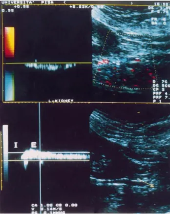
(2) 186. Fig. 1. ( Upper): Colour-Doppler mapping on longitudinal posterior scan. Evidence of perirenal haematoma (asterisk) and of the anomalous vessel in front of it (arrow). (Lower): Colour-Doppler after resolution of the haematoma. The anomalous vessel is closely located near the lower pole of the kidney.. M. Meola et al.. Fig. 2. ( Upper): The duplex analysis shows the slow but continuous flow of the vessel. (Lower): This venous flow is modulated by breathing in (I ) and breathing out ( E ). .. In conclusion, colour-coded Doppler evaluation of perirenal vasculature is useful to reveal rare abnormalities, as the presence of a left cardinal vein or other abnormal lumbar vessels.. Comment The present case shows that haematoma can occur even if the execution of renal biopsy is technical correct. In our case, severe bleeding was caused by accidental puncture of an abnormal venous vessel undetectable by B-mode standard scan. It was only subsequently revealed using the colour-coded Doppler sonography. In the adult, the presence of this vessel is infrequent. Anatomical studies in animals and humans showed that the persistence of right posterior cardinal vein, left supracardinal vein, or both right and left posterior cardinal veins are the most common types of anatomical variants, while the persistence of isolated left posterior cardinal vein occurs far less frequently [9]. This case suggests that the evaluation of the renal or perirenal vessels should be included in the evaluation of native kidney before a renal biopsy is performed. Colour-coded Doppler demonstration of vascular abnormalities should reduce the risk of bleeding complications.. References 1. Health and Public Policy Committee, American College of Physicians, Philadelphia. Clinical competence in percutaneous renal biopsy. Ann Intern Med 1988; 108: 301–303 2. Madaio MP. Renal biopsy. Kidney Int 1990; 38: 529–543 3. Meola M, Barsotti G, Cupisti A et al. Free-hand ultrasoundguided renal biopsy: report of 650 consecutive cases. Nephron 1994; 67: 425–430 4. Diaz-Buxo JA, Donadio JV Jr. Complications of percutaneous renal biopsy: an analysis of 1000 consecutive biopsies. Clin Nephrol 1975; 4: 223–227 5. Wickre CG, Golper TA. Complications of percutaneous needle biopsy of the kidney. Am J Nephrol 1982; 2: 173–178 6. Birnholz JC, Balakuntalam SK, Corwin HL. An improved technique for ultrasound guided percutaneous renal biopsy. Kidney Int 1985; 27: 80–82 7. Yoshimoto M, Fujisawa S, Sudo M. Percutaneous renal biopsy well-visualised by orthogonal ultrasound application using linear scanning. Clin Nephrol 1988; 30: 106–610 8. Barsotti G, Cupisti A, Morelli E et al. A special, supplemented ‘Vegan’ diet for nephrotic patients. Am J Nephrol 1991; 11: 380–385 9. McClure CFW, Butler EG. The development of the vena cava inferior in man. Am J Anat 1925; 35: 331–383 Received for publication: 29.7.98 Accepted in revised form: 9.9.98.

(3)

Figura

Fig. 2. ( Upper): The duplex analysis shows the slow but continuous flow of the vessel

Riferimenti

Documenti correlati

In Fig 5b, invece, si osserva che, per il tubo incollato di lato 40mm la risposta della simulazione è più rigida nel primo tratto elastico a causa sia della cedevolezza

All'organismo sovrebbe inoltre spettare il compito di valutare ex ante l'idoneità delle misure proposte dal Governo, in primo luogo in sede di definizione della

La rivista è edita dalla «Fondazione Spadolini Nuova Antologia» – costituita con decreto del Presidente della Repubblica, Sandro Pertini, il 23 luglio 1980, erede universale di

Alcune più recenti applicazioni ed esemplificazioni del disegno informa- tizzato, sembrano infatti quasi ricondu- cibili alla ricerca di un rinnovato legame con

The simulation and measurement of topological insulators in different quantum simulators is a very active field and different techniques allow one to char- acterize the

Three groups of isolated perfused hearts from adult Wistar rats underwent 30-min ischemia and 120-min reperfusion (I/R, Group 1), or were post-conditioned by brief ischemic

The introduction of dedicated clinical pathways, integrated care process management for COVID-19 patients and case managers are seen as reasonable healthcare practices by

We found that, in regular one-dimensional models, the confidence distribution obtained from the bias corrected log-likelihood ratio is third– order equivalent to the unique