Alma Mater Studiorum – Università di Bologna
DOTTORATO DI RICERCA IN
SCIENZE BIOTECNOLOGICHE E FARMACEUTICHE
Ciclo XXX
Settore Concorsuale: 03/D1
Settore Scientifico Disciplinare: CHIM/11
Dynamics of the infant microbiomes onset: exploring
the gut and the oral microbiota in full-term and
pre-term infants in the frame of mother’s milk microbial
ecosystem
Presentata da:
Sara Quercia
Coordinatore Dottorato
Supervisore
Prof. Santi Mario Spampinato
Prof. Patrizia Brigidi
1 Table of contents 1 2 Abstract ... 2 3 Introduction ... 3 4 Project outline ... 7 5 Experimental procedures ... 8 6
Total bacterial DNA extraction from complex matrix ... 8 7
16 rRNA gene amplification and sequencing ... 8 8
Bioinformatics ... 9 9
Part A: The bacterial ecosystem of mother’s milk and infant’s mouth and gut ...10 10
Introduction...10 11
Subjects recruitment...11 12
Experimental procedure and statistics ...12 13
Results and discussion...12 14
Conclusions...25 15
Part B: Dynamics of the infant microbiomes onset: exploring the gut and the oral microbiota in full-term and 16
pre-term infants in the frame of mother’s milk microbial ecosystem...26 17
Introduction...26 18
Subjects recruitment...27 19
Experimental procedure and statistics ...30 20
Results and discussion...30 21
Comparison between full-term and moderately to late pre-term...39 22
Conclusion and further perspectives ...47 23
Isolation and characterization of probiotic bacteria from human breast milk ...49 24 References ...50 25 26 27 28 29 30 31 32
2 Abstract
33 34
The gut microbiota assembly during the very first days of life plays a pivotal role in the education of
35
the immune system and in the building of a healthy status later in life. Besides the mode of deliver y,
36
also the feeding type and the gestational age have an impact on the gut microbiota composition. For
37
this reason, by means of next-generation sequencing of the 16S rRNA gene on Illumina MiSeq, we
38
characterized and compared the intestinal bacterial community in 2 cohorts of infants, 1 constituted
39
by 36 healthy breast-fed infants born full-term and the other constituted by 21 infants born moderate
40
to late pre-term (32 to 37 weeks) receiving different types of feeding (mother breast milk, human
41
breast milk from donor and formula). The first cohort was sampled at 20th day of life, whilst the 42
second one was sampled longitudinally from birth to 30th day of life. In addition, also the infant ’s 43
saliva and the mother’s milk were sampled and sequenced.
44
The characterization of the 3 ecosystems in full-term infants led to the hypothesis that the mother
45
milk, together with the microorganisms that reside in the baby’s mouth, may act as seeding
46
community and may participate to infant gut microbiota assembly. On the other hand, in the
47
moderately-to late pre-term cohort, the extreme diversity of the infant’s clinical history provokes a
48
tremendous inter-individual variability. Nevertheless, milk and saliva microbiological structure s
49
resembled the ones of full-term cohort. The gut microbiota instead presented a very different
50
composition and it is plausible that its establishment is strongly influenced by the infant’s clinica l
51
history and environmental bacteria than the mutual relationship with the mother.
52 53
3
Introduction
5455
Almost 150 years have passed since Il'ja Il'ič Mečnikov theorized that health could be enhanced by
56
manipulating the gut microbiota (GM). Where do we stand today? Many studies have been carried
57
out to investigate the characteristics the intestinal community and of the other microbial ecosystems
58
that inhabit human body. Our organism contains up to 27 sites where a microbial community can be
59
found (Costello et al., 2009). The gastrointestinal tract is fully colonized by microorganisms, starting
60
from the mouth down to the colon. The oral cavity represents the first access to the gastrointest ina l
61
tract and it communicates perpetually with the external environment. In the mouth, many different
62
ecosystems can be found: salivary, gingival, lingual and mucosal. The salivary microbiota (SM) of a
63
healthy adult is scarcely biodiverse and it is mainly constituted by genus Streptococcus (species S.
64
salivarius and S. mitis), but also the genera Neisseria, Rothia and Prevotella are present (Yun-ji K im
65
et al., 2016; Zaura et al., 2014). Yet the most dense and biodiverse of these communities present in
66
the GIT resides in the colon, where it reaches the concentration of 1012 CFU/g of luminal content. 67
When the GM is described at a compositional level, only a limited number of phyla is found:
68
Firmicutes, Actinobacteria, Bacteroidetes, Proteobacteria, and Verrucomicrobia, with Firmicutes and
69
Bacteroidetes accounting for up to 90% of the ecosystem (Costello et al., 2009). On the contrary, if
70
we look at a lower phylogenetic level, the biodiversity explodes, reaching more than 1000 species
71
(Qin et al., 2010). The microbial community that resides in our gut is very unique, describing a
72
personal fingerprint. The GM provides the host with metabolic functions, such as the digestion of
73
complex polysaccharides, production of vitamins, cofactors and other secondary metabolites
74
(Bäckhed et al., 2004). There is a wide range of metabolites produced that depend on the
75
macronutrient ingested. The endpoint of polysaccharide fermentation is mainly represented by
short-76
chain fatty acids (SCFAs), namely acetic, propionic and butyric. They participate in the pathway that
77
regulates appetite and play a role in host nutrition and energy homeostasis, controlling energy
78
production and storage (Russel et al., 2013). On the other hand, when amino acids are metabolized
79
by the GM, we obtain indolic and phenolic compounds, together with methylamines, which are linked
80
to obesity, type 2 diabetes and hepatic steatosis (Lin et al., 2017).
81
Many researches have also demonstrated how crucial the GM is in educating the immune system
82
starting from the very beginning of life, by modulating the generation of an equilibrium between anti-
83
and pro-inflammatory response when exposed to bacteria (Berrington et al., 2013). An impaired GM
84
leads to a defective communication with the immune system, causing persisting effect on host
85
physiology later in life even after GM resembling (Arrieta et al., 2014).
4
The gut colonization was thought to start during delivery, but recently Aagard and colleagues
87
demonstrated that microbial DNA belonging to the species Escherichia coli, Bacteroides spp.,
88
Neisseria lactamica, Staphylococcus epidermidis and Propionibacterium acnes is found in the
89
placenta (Aagard et al., 2014). Nevertheless, the mode of delivery, together with gestational age at
90
birth and type of nutrition, play a pivotal role in the bacterial establishment at early stage. The most
91
favourable condition for the infant health status is being born at term with vaginal delivery and being
92
fed exclusively with maternal breast milk. However, statistics says that in Italy in 2015, 34.1% of the
93
delivery were C-sections (Italian Health Ministry Data). Vaginal-delivered infant GM is different
94
from the C-section one (Dominguez-Bello et al., 2010; Gritz and Bhandari, 2015): while vaginall
y-95
born infants have a gut microbial composition enriched in Bacteroides, Bifidobacterium,
96
Parabacteroides, Escherichia and Shigella (Bäckhed et al., 2015) and they present also bacteria
97
deriving from maternal vaginal microbial community, such as Lactobacillus and Prevotella species
98
(Gritz and Bhandari, 2015), infants born via C-section are instead colonized by epidermal and
99
environmental species, namely Clostridium, Staphylococcus, Propionobacterium, and
100
Corynebacterium and, when they are compared to the vaginally-born, they have lower levels of
101
anaerobes, in particular Bacteroides and Bifidobacterium (Brugman et al., 2015). These differences
102
though smooth over with the introduction of milk. Newborns can be fed with maternal breast milk,
103
human breast milk from donor and formula. Human breast milk is thought to be the most beneficia l
104
for the baby’s health and also for the GM establishment (Agostoni et al., 2009). It contains a mixture
105
of nutrients and molecules, such as immunoglobulins (IgA), carbohydrates, fatty acids and lactoferr in
106
(Jain and Walker, 2014). Among the glucidic fraction, the human milk oligosaccharides (HMOs)
107
represent one of the most investigated, because of its capacity to selectively stimulate the growth of
108
specific bacteria in the infant gut. Human milk has not only a prebiotic function, but it has been
109
suggested that could act also as microbes source, being enriched in Streptococcaceae,
110
Bifidobacteriaceae and Staphylococcaceae (Biagi et al., 2017). The origin of these bacteria has not
111
been elucidated yet. An enteromammary pathway has been suggested by Perez and colleagues (2006)
112
and they support the idea that fragments of DNA, antigens and microbial protein may be transported
113
via blood by dendritic cells to the mammary gland and be secreted in the milk. Throughout the milk,
114
the mother starts to educate the immature immune system of the baby to the late formation of a
115
microbial community. Another theory explains the presence of bacteria as a contamination coming
116
from the infant’s mouth, that inoculate milk duct during suction (Biagi et al., 2017). Infants receiving
117
nourishment from the mother display a GM structure dominated by species of Bifidobacterium and,
118
when compared to formula- fed infants, show lower relative abundance of Enterobacteriaceae.
119
Formula-fed ones also show the presence of Escherichia coli, Clostridium difficile, Bacteroides,
5
Prevotella, and Lactobacillus (Jost et al., 2012). C-section is a more common practice in premature
121
birth, when it is essential to give birth to the foetus due to diseases affecting either the mother or the
122
baby, as well as uterine infections or breakage of the amniotic sac. A baby born premature is usually
123
described according to the gestational age, that is the weeks dating from the first day of the mother's
124
last menstrual period (NIH, US library of medicine). The World Health Organization has drawn up a
125
classification to define newborns according to gestational age:
126
• extremely pre-term (<28 weeks);
127
• very pre-term (28 to <32 weeks);
128
• moderate to late pre-term (32 to <37 weeks);
129
• full-term (37 to 40 weeks).
130
It is estimated that every year in Italy around 40.000 babies are born pre-term (Italian Society of
131
Neonatology). Pre-term infants display a different gut colonization when compared to full term ones
132
(Arboleya et al., 2012) and it is influenced not only by the mode of delivery (mainly C-section) and
133
nutrition (mainly with a mixture of human breast milk and formula), but also by an impaired feeding
134
process, due to the delay in the full establishment of coordinated latch, suckling, swallowing and
135
breathing (Mizuno et al., 2003), incomplete oesophageal peristalsis (Staiano et al., 2007) and altered
136
gastric emptying (Riezzo et al., 2000). These newborns are exposed to many complications, such as
137
necrotizing enterocolitis (NEC) and sepsis (Gregory et al., 2016). NEC is characterized by intestina l
138
inflammation that can lead to tissue necrosis and sepsis. Its onset appears to be multifactorial: gut
139
immaturity, intestinal damage or injury, enteral feeding and impaired bacterial colonization play a
140
role, although the exact pathogenesis remains unidentified. The role of GM establishment in NEC has
141
been investigated by a meta-analysis carried out by Pammi and colleagues (2017) and they
142
highlighted that the onset of the disease is predated by an increase of Proteobacteria and a concomitant
143
decrease of Firmicutes and Bacteroidetes. Moreover, maternal breast milk administration is
144
considered protective against NEC onset (Meinzen-Derr et al., 2009).
145
Although many researches focus on GM features in very and extremely pre-term infants, moderately
146
to late pre-term is considered a neglected category, because poorly investigated. Even if they have a
147
much lower risk of medical complications than more premature infants, they still experience higher
148
rates of infant morbidity and mortality, as well as higher risks of childhood disabilities
(Shapiro-149
Mendoza et al., 2012) when compared to full term babies.
150
For these reasons it is important to focus the research on this group, describing their gut ecosystem
151
from birth until weaning, in the frame of milk microbial composition. Finally, in order to understand
152
how much these babies differ from the normal healthy situation, it is crucial to compare them with
153
full term vaginally delivered babies, who are exclusively breastfed. This approach allows the
6
identification of the bacterial genera considered “health-promoting” that are lacking in the GM of
155
pre-term infants, paving the way to the development of an individual probiotic intervention to restore
156
the ecosystem.
157 158
7
Project outline
159160
It’s becoming always clearer that disturbances that strike the GM in newborns affect health status
161
later in life. Many factors can interfere with a correct GM establishment, such as gestational age,
162
mode of delivery and type of feeding. According to gestational age, we can divide the newborns in 4
163
categories (WHO definition):
164
• extremely pre-term (<28 weeks)
165
• very pre-term (28 to <32 weeks)
166
• moderate to late pre-term (32 to <37 weeks).
167
• full-term (37 to 40 weeks)
168
Being born full-term with vaginal delivery and being fed exclusively with mother breast milk is
169
considered the most desirable condition for health and for a correct GM establishment. For this
170
reason, the first part of this research work was addressed to the characterization of the gut microbia l
171
community of 36 healthy, vaginally delivered and full-term babies, who were exclusively breastfed
172
at 20th day of life. In addition, also saliva microbiota from the baby and breast milk from the mother 173
were analysed, in order to understand the relationship between the infant GM and the mother milk
174
microbial ecosystem, in the frame of the mouth community, which acts as mandatory connection.
175
Consequently, our focus moved to infants who are born between 32 to 37 weeks (moderate to late
176
pre-term). This group is considered a neglected one, because yet poorly investigated. According to
177
the OMS report on pre-term births drawn up in 2012, the total number of premature infants was of
178
131,296,785 and the moderate to late pre-term category accounted for the 84.3%. These newborns do
179
not have the typical clinical progress of full term babies, having high probability of developing many
180
complications, in particular necrotizing enterocolitis (NEC) and sepsis. Nevertheless, their
181
complication onset rate is smaller when compared to the very and extremely pre-term ones. GM
182
assembly appears to play a pivotal role in the development of such complications and for this reason
183
we decided to characterize the gut microbial community establishment in a cohort of 21 neonates
184
sampled longitudinally. From these babies, in addition to faecal samples, also a saliva swab was taken,
185
together with an aliquot of mother’s milk (when present) at different timepoints, from birth until
186
weaning, collecting a total number of 348 samples. The microbial community of all these samples
187
was sequenced using Illumina platform, then characterized and described from a phylogenetic point
188
of view; moreover, the impact of breastfeeding was examined. Finally, the comparison between full
189
term and pre-term babies at 20 days of life was performed in order to decipher the differences between
190
these 2 conditions.
191 192
8
Experimental procedures
193194
Total bacterial DNA extraction from complex matrix
195 196
Total bacterial DNA was extracted from feces using the DNeasy Blood and Tissue Kit (QIAGEN ,
197
Hilden, Germany) with a modified protocol (Yu and Morrison, 2004). Briefly, 250 mg of stool
198
samples were resuspended in 1 mL of lysis buffer (500 mM NaCl, 50 mM Tris–HCl pH 8, 50 mM
199
EDTA and 4% SDS) and treated with 3 beads-beating steps in FastPrep instrument (MP Biomedica ls,
200
Irvine, CA) at 5.5 movements per sec for 1 min and kept in ice among treatments. Samples were
201
centrifuged at full speed for 5 min at 4°C, then 260 μl of 10M ammonium acetate were added and the
202
samples incubated for 5 min in ice. After 10 min of centrifugation at full speed at 4°C the supernatants
203
were collected and 1 volume of isopropanol added. Samples were mixed and incubated in ice for 30
204
min. DNA was collected by 15 min of centrifugation at full speed at 4°C and the pellet washed with
205
70% ethanol. The pellet was then resuspended in 100 μl of TE buffer and RNA and proteins removed
206
treating the samples respectively with 2 μl of DNase-free RNase (10 mg/ml) for 15 min at 37°C and
207
15 μl of proteinase K at 70°C for 10 min. DNA was further purified using QIAamp Mini Spin columns
208
(QIAGEN) following the manufacturer’s instructions. For milk samples, the same protocol described
209
above for fecal samples was applied, preceded by the centrifugation of 2 ml of sample at full speed
210
for 10 min at 4°C. For DNA extraction from oral swabs, the cotton swab was suspended in 500 μl of
211
PBS, vortexed for 1min and sonicated for 2 min. These 2 steps were repeated twice, then 2 cycles of
212
bead-beating with FastPrep at 5.5 movements per sec for 1min, with 200 mg of glass beads, were
213
applied. Cotton residues were removed and the debris pelleted by centrifugation at 9000g for 5 min.
214
The supernatant was discarded and the pellet resuspended in 180 μl of enzymatic lysis buffer
215
(QIAGEN). Samples were then treated according to the DNeasy Blood&Tissue kit (QIAGEN )
216
instructions, following the protocol for Gram positive bacteria. Extracted DNAs were quantified
217
using the NanoDrop ND-1000 spectrophotometer (NanoDrop Technologies, Wilmington, DE).
218
219
16 rRNA gene amplification and sequencing
220 221
For each sample, the V3-V4 region of the 16S rRNA gene was PCR amplified in 25 μl final volume
222
containing 5 μl of microbial DNA (diluted to 5 ng/μl for fecal samples, undiluted for milk and oral
223
swab), 2X KAPA HiFi HotStart ReadyMix (KAPA Biosystems, Resnova, Rome, Italy), and 200nM
224
of S-D-Bact-0341-b-S-17/S-D-Bact-0785-a-A-21 primers (Klindworth et al., 2013) carrying Illumi na
9
overhang adapter sequences. PCR conditions set up as follows: initial denaturation at 95°C for 3
226
minutes, 25 cycles of denaturation at 95°C for 30 seconds, annealing at 55°C for 30 seconds, and
227
extension at 72°C for 30 seconds, and a final extension step at 72°C for 5 minutes. PCR amplico ns
228
were purified with a magnetic bead-based clean-up system (Agencourt AMPure XP; Beckman
229
Coulter, Brea, CA). Indexed libraries were prepared by limited-cycle PCR using Nextera technology
230
and further cleaned up with AMPure XP magnetic beads (Beckman Coulter). Libraries were pooled
231
at equimolar concentrations (4nM), denatured and diluted to 6 pmol/L before loading onto the MiSeq
232
flow cell. Sequencing on Illumina MiSeq platform was performed by using a 2×300 bp paired end
233
protocol, according to the manufacturer’s instructions (Illumina, San Diego, CA).
234 235
Bioinformatics
236 237
Data analysis is performed using a pipeline combining PANDAseq (paired-end assembler for
238
Illumina sequences) (Masella et al., 2012) and QIIME (Quantitative Insights Into Microbial Ecology)
239
(Caporaso et al., 2010). High-quality reads are filtered and then clustered into operational taxonomic
240
units (OTUs) at a 0.97 similarity threshold using UCLUST (Edgar, 2010). Taxonomy is assigned
241
using the RDP (Ribosomal Database Project) classifier against Greengenes database (May 2013
242
release) and the chimera filtering is performed by discarding all singleton OTUs. Alpha diversity is
243
measured using the Chao1, observed species and Shannon index metrics. Beta diversity was estimated
244
by computing Bray-Curtis distances. The distance matrix obtained was used for principal coordinates
245
analysis (PCoA) and plotted using the rgl and vegan packages of R.
246 247
10
Part A: The bacterial ecosystem of mother’s milk and infant’s mouth and gut
248249
Biagi Elena, Quercia Sara, Aceti Arianna, Beghetti Isadora, Rampelli Simone, Turroni Silvia, Faldella Giacomo,
250
Candela Marco, Brigidi Patrizia, Corvaglia Luigi
251 252
Published on Frontiers in microbiology in 2017
253 254
Introduction
255 256
The microbiota of individuals with whom a human being has direct and frequent contacts contributes
257
in shaping its microbial communities (Song et al., 2013; Stahringer et al., 2012). This is even more
258
true in the case of breastfed infants and their mothers, where the microbial ecosystems of the latter
259
are the most relevant sources of colonizing microbes for the former (Arrieta et al., 2014). The
260
progressive building of the infants’ microbiota, especially for what concerns the gut ecosystem, is a
261
crucial proceeding for educating their immune system to the delicate balance between tolerance and
262
reactivity that is needed to maintain health throughout the entire human life (Arrieta et al., 2015;
263
Honda and Littman, 2016; Lynch and Pedersen, 2016). Consequently, the understanding of the
264
colonization dynamics of the infant’s gut microbiota is not only fascinating from the ecological point
265
of view, but also incredibly relevant for clinical immunology (Arrieta et al., 2015; Honda and
266
Littman, 2016).
267
The infant’s gut microbiota is a highly dynamic community that is progressively and continuo usly
268
shaped during the first days of life, with nutrition (breast vs. formula feeding) being among the most
269
relevant drivers for its composition (Gritz and Bhandari, 2015). With its estimated 3 log CFU/ml of
270
bacterial concentration (Jost et al., 2014), human breast milk is listed among the first sources of
271
microbes for the infant’s gut ecosystem, together with the mother’s skin, mouth and vaginal tract, in
272
case of vaginal delivery (Mueller et al., 2015). Research struggles to give a conclusive demonstrat io n
273
for the origin of the bacteria recovered in human milk: even if a controversial “bacterial
entero-274
mammary pathway” has been proposed (Rodríguez, 2014), contamination by the surrounding skin
275
microbiota and other environmental sources might also occur. Indeed, facultative anaerobic or
276
prevalently aerobic species are the major colonizers of the human milk ecosystem: Streptococcus and
277
Staphylococcus are the most frequently isolated and abundant bacterial groups in milk samples,
278
together with skin-derived or environmental contaminants (i.e. Propionibacterium and genera of the
279
Enterobacteriaceae family) (Fitzstevens et al., 2016). However, well-known intestinal probiotic
280
bacteria (i.e. Bifidobacterium and Lactobacillus) are often retrieved by both molecular and
11
cultivation-based studies (Fitzstevens et al., 2016). Next generation sequencing also allowed the
282
detection of obligate anaerobic, gut-associated genera, such as Bacteroides, Blautia, Dorea, and
283
Faecalibacterium (Jost et al., 2013); if alive, these bacteria could act as pioneers in the infant gut for
284
the construction of the adult gut microbiota, which will begin to settle down at weaning (Rodrígue z,
285
2014).
286
In this scenario of microbial exchange between mother and child, the baby’s mouth is unavoidab ly
287
involved, being the obligate transition point for the milk to reach the gastrointestinal tract. The oral
288
microbiota is a well-characterized portion of the human microbiome. It is usually dominated by
289
Streptococcus and Staphylococcus in healthy, breastfed term infants; aerobic or facultative anaerobic
290
bacterial taxa, such as Gemella, Actinomyces, and Veillonella, act as later and minor colonize rs
291
(Sampaio-Maya and Monteiro-Silva, 2013). The mouth is a particularly exposed ecosystem,
292
anatomically open to the external environment and continuously in contact with air, food, and water.
293
For these reasons, this ecosystem needs to cope with chemical, physical and mechanical fluctuatio ns.
294
The mouth of healthy individuals is not routinely found to be colonized by non-oral microorganis ms,
295
possibly because exogenous bacteria lack in specific adhesins and receptors that would enable them
296
to bind to oral surfaces, or are excluded by immune mechanisms (Wade, 2013). On the contrary,
297
evidences of seeding of the baby’s gut by the oral microbiome have been provided (Costello et al.,
298
2013; Ding and Schloss, 2014).
299
In this frame, it is crucial to include the bacteria inhabiting the infant’s oral cavity in the complex
300
mechanism of bacterial transfer between the mother’s milk microbiota and the infant’s gut ecosystem.
301
Indeed, the oral ecosystem might contribute in seeding the gut both directly, through deglutition, and
302
indirectly, by contaminating the mother’s milk ducts, during suction.
303
In an attempt to decipher the relationship between the mother’s milk ecosystem and the infant ’s
304
microbiome, we analyzed, to our knowledge for the first time, the microbial composition of oral, gut
305
and milk ecosystems in a small, yet very homogeneous, cohort of 36 healthy mother-infant pairs. By
306
limiting the influence of confounding variables (e.g. delivery mode, gestational age), our find ings
307
shed some light on the relevance of bacterial sharing between these ecosystems.
308 309
Subjects recruitment
310 311
The Nursery of S. Orsola-Malpighi Hospital in Bologna, Italy, recruited mother-infant pairs meeting
312
the following criteria: (i) vaginal delivery at term (≥37 weeks gestation), (ii) exclusive breastfeeding
313
during the sampling period, (iii) no antibiotic/probiotic exposure of either the mother or the infant
12
during pregnancy, intrapartum or postnatally. Infants who had or developed clinical conditions that
315
required hospitalization were excluded.
316
Written informed consent was obtained, in accordance with the Declaration of Helsinki, from each
317
mother before the mother-infant pair was discharged from the nursery (48-72 hours after deliver y).
318
Follow-up visits at 20 days of life were scheduled in order to obtain a neonatal fecal sample, two
319
neonatal oral swabs (before and after breastfeeding), and a fresh mother milk sample. Feces were
320
collected from diapers using a standard sterile collection tube. Milk samples were collected with the
321
aid of a breast pump into sterile plastic tubes; prior to collection, mothers were asked to wash the
322
nipple and mammary areola with soap and water. Oral samples were obtained by gently swabbing a
323
sterile cotton-tipped applicator on the inside of the infant’s cheek. Samples were immediate l y
324
delivered to the laboratory using cold packs, then split into aliquots ready for DNA extraction and
325
frozen within few hours from collection. Samples were thawed in batches for processing. All samples
326
were processed within 4 months of receipt. Demographic and clinical data were recorded in a specific
327
case report form. The study was approved by the ethics committee of the S. Orsola-Malpighi Hospital
328
in Bologna (study protocol 53/2014/U/Tess). Methods were carried out in accordance with the
329
approved guidelines.
330 331
Experimental procedure and statistics
332 333
Samples processing were conducted as described in “Experimental procedure”. Statistics was
334
performed using R software (https://www.r-project.org/) and the libraries vegan and made4.
335
Weighted and unweighted UniFrac distances were used for Principal Coordinates Analyses (PCoA),
336
and the significance of separation was tested by permutational multivariate analysis of variance using
337
the function “adonis” of the vegan package, after testing for homogeneity of dispersion using the
338
function “betadisper”. Wilcoxon test was used to assess significant differences between two groups
339
of samples; adaptations for paired samples were used when necessary. Kruskal-Wallis test was used
340
for multiple comparisons, followed by Tukey post-hoc test when appropriate. P values were corrected
341
for multiple comparisons using the Benjamini-Hochberg method. P<0.05 was considered as
342
statistically significant. Correlation between datasets was tested by using the Kendall method.
343 344
Results and discussion
345 346
13
Thirty-six mother-infant pairs were included in the study. All infants were vaginally delivered and
347
exclusively breastfed. Neither infants nor mothers had received any antibiotics or probiotics until the
348
sampling date. One-hundred-forty-three samples were collected 20 days after delivery: 36 mother’s
349
milk samples, 36 infant’s fecal samples, and 71 infants’ oral swabs (35 pairs of pre and post
350
breastfeeding samples plus one unpaired pre-breastfeeding sample).
351
The extracted bacterial DNA was phylogenetically characterized by 16S rRNA gene (V3-V4 region)
352
Illumina sequencing. A total of 1,475,619 high-quality reads was obtained with a mean of
353
10,319±3,364 reads per sample. Rarefaction curves obtained with Shannon and Chao1 metrics
354
approximated the saturation level after 3,000 reads. Reads were clustered in 7,524 operational
355
taxonomic units (OTUs) at 97% of identity. OTU table and taxa summary tables at family and genus
356
level are available as Supplementary Tables.
357 358 359
14
M, mother’s milk ecosystem; Opre, infant’s oral ecosystem before breastfeeding; Opost, infant’s oral ecosystem after breastfeeding; F, infant’s fecal ecosyst em.
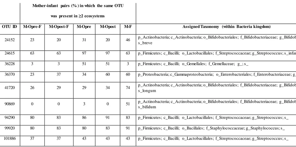
Table A1. OTU sharing among the bacterial ecosystem of the infant’s mouth and feces, and the mother’s milk. The OTUs most frequently (>40% of the mother-infant pairs)
shared by at least two bacterial ecosystems are shown, with assigned taxonomy. Only OTUs present at a relative abundance >0.1% were considered. Assigned taxonomy is reported using Greengenes syntax.
Mother-infant pairs (% ) in which the same OTU was present in ≥2 ecosystems
OTU ID M-Opre-F M-Opost-F M-Opre M-Opost M-F Assigned Taxonomy (within Bacteria kingdom)
24152 23 20 31 20 46 p_Actinobacteria; c_Actinobacteria; o_Bifidobacteriales; f_Bifidobacteriaceae; g_Bifidobacterium;
s_breve
24615 63 63 97 97 63 p_Firmicutes; c_Bacilli; o_Lactobacillales; f_Streptococcaceae; g_Streptococcus; s_infantis
36228 3 3 51 51 3 p_Firmicutes; c_Bacilli; o_Gemellales; f_Gemellaceae; g_; s_
36370 23 37 34 60 60 p_Proteobacteria; c_Gammaproteobacteria; o_Enterobacteriales; f_Enterobacteriaceae; g_; s_
41720 26 29 29 34 74 p_Actinobacteria; c_Actinobacteria; o_Bifidobacteriales; f_Bifidobacteriaceae; g_Bifidobacterium;
s_longum
90869 0 0 3 0 51 p_Actinobacteria; c_Actinobacteria; o_Bifidobacteriales; f_Bifidobacteriaceae; g_Bifidobacterium;
s_bifidum
94290 80 83 86 91 83 p_Firmicutes; c_Bacilli; o_Lactobacillales; f_Streptococcaceae; g_Streptococcus; s_
99920 80 83 80 83 91 p_Firmicutes; c_Bacilli; o_Bacillales; f_Staphylococcaceae; g_Staphylococcus; s_
15
A PCoA based on unweighted UniFrac distance showed that the microbiota of infants’ oral swabs, infants’ feces and mothers’ milk clustered separately (Figure A1.A), as expected being the resident communities of three distinct body districts that are different for pH, oxygen levels, and nutrients availability. Adonis test confirmed that the reported separation was significant, even if this result needs to be taken into account cautiously since the test for homogeneity of dispersion (funct io n betadisper) returned that milk samples had a significantly different dispersion compared to the other groups of samples. When weighted UniFrac distances were used for PCoA (Figure A1.B), fecal samples overlapped with milk samples on PCo1. Thus, the difference between milk and fecal ecosystems was better explained by unweighted metrics, hinting that it might reside in fractions of the microbial communities that are exclusive of one of the two ecosystems (Lozupone et al., 2007). Fecal samples also showed higher dispersion, indicating higher variability in the most abundant species of the ecosystem, with respect to oral and milk communities.
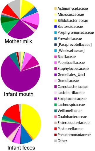
The oral microbiome was the least diverse of the considered ecosystems (Shannon diversity index, mean ± standard deviation (SD), 2.3±0.6; Figure A1.C), largely dominated by Streptococcaceae (average relative abundance (rel. ab.), 69.8%) (Figure A2), with Streptococcus being the dominant genus in 94% of samples, confirming the known literature on this ecosystem (Zaura et al., 2014; Lif Holgerson et al., 2015; Hendricks- Muñoz et al., 2015; Davè et al., 16). Also confirming the large amount of knowledge on the topic (Muller et al., 2015; Jost et al., 2012), the fecal microbiota of breastfed infants at 20 days of life was dominated by Bifidobacteriaceae (average rel. ab., 38.2%), with Bifidobacterium being the dominant genus in 67% of samples. Fecal microbiota also included relevant average abundances of Enterobacteriaceae (15.4%), Streptococcaceae (13.9%), Bacteroidaceae (9.5%), Staphylococcaceae (5.4%), and Lactobacillaceae (4.8%) (Figure 2).
18
Figure A1. Diversity in the bacterial ecosystem of the mother’s milk, and infant’s feces and mouth.
PCoA based on unweighted (A) and weighted (B) UniFrac distances of the microbiota of mother’s milk (light blue), infant feces (yellow), and infants mouth (pink). Samples are identified by filled circles. In both PCoA first and second principal components (PCo1 and PCo2) are plotted. The percentage of variance in the dataset explained by each axis is reported. Box and whiskers distribution of the Shannon α-diversity index (C), intra-group unweighted UniFrac distances (D), and intra-group weighted UniFrac distances (E), calculated for milk (light blue), fecal (yellow) and oral (pink) samples. Significant differences between datasets are indicated, as calculated using Tukey post -hoc test after Kruskal-Wallis test for multiple comparisons (*, P≤0.001; **, P≤0.0001).
Figure A2. Average composition of the bacterial community in mother’s milk and infant’s feces and mouth. For
each group of samples, a pie chart based on the average relative abundance (%) at family level is plotted. Bacterial familie s with relative abundance ≥0.2% in at least 10% of the samples are depicted. Colors for each family are reported in the legend.
Both oral and fecal microbiota of infants showed higher unweighted UniFrac distances within group (0.80±0.03 and 0.80±0.04, respectively) compared to milk (0.68±0.04, Kruskal-Wallis test, P<0.0001). When the weighted UniFrac metric was considered, the within-group distances obtained
19
for the fecal samples remained the highest (0.5±0.2), whereas those calculated for the oral samples became the lowest (0.14±0.06 Kruskal-Wallis test, P<0.0001). Since weighted UniFrac measure is better suited to detect differences in abundance even when the overall groups of organisms that are present in each sample remain the same (Lozupone et al., 2007), these observations suggest that the variability between oral samples might reside in subdominant species that are not highly conserved among samples.
The average microbiota profile obtained for breast milk was significantly more diverse (Shannon diversity index = 4.9±1.1; Figure 1C) than both infant’s feces and oral swabs (3.0±0.7 and 2.3±0.6, respectively; Kruskal-Wallis test, P<0.0001); interestingly, according to the unweighted UniFrac metric, the variability among milk samples was the lowest (0.68±0.03; Kruskal-Wallis test P<0.0001; Figure A1.D), and remained significantly lower than that of fecal samples when the weighted UniFrac distances were computed (0.25±0.07; P<0.0001; Figure A1.E). In other words, the milk ecosystem of the 36 enrolled mothers was richer and more similarly composed among samples (in terms of bacterial species) than the fecal or mouth ecosystem of their children, suggesting that the milk duct might act as an environmental filter allowing for the survival and proliferation of the same bacterial species in most individuals (a “niche-based” community assembly, according to Costello et al. (2012). As expected (Fitzstevens et al., 2016), the milk ecosystem phylogenetic structure showed a slight dominance of Streptococcaceae (average rel. ab., 24.5%), with Streptococcus being the dominant genus in 53% of samples, but also a considerable representation of the typically infant fecal family Bifidobacteriaceae (11.2%, with Bifidobacterium being the dominant genus in 19% of samples) and Staphylococcaceae, which is instead a common skin and mouth inhabitant (Belkaid and Segre, 2014) (11.1%, with Staphylococcus being the dominant genus in 11% of samples) (Figure 2). Confirmi ng previous studies (Jost et al., 2014; Jost et al., 2013) , and supporting the hypothesis of a possible link between the milk microbiota and the gut ecosystem of the mother, also anaerobic bacterial families that are commonly found in the adult human intestine, such as Lachnospiraceae, Ruminococca ceae and Bacteroidaceae, were present with an average rel. ab. of 10.3, 5.4 and 4.4%, respectively. Specifically designed studies are required to investigate if these bacteria are indeed alive in the milk ecosystem, as well as the ecological importance of this putative bacterial migration from the mother’s gut to the milk duct. The milk microbiota was the only one showing a few genera that were present in more than half of the subjects but never retrieved in the other two ecosystems, partly confirmi ng the observations of Jost et al. (2014). In particular, sequences assigned to the genus Ralstonia were detected at a relative abundance >0.1% in 83% of the milk samples (average rel. ab., 0.67%), and in none of the infant’s fecal or oral samples; similar trends were shown by the genus Sediminibacterium
20
(detected in 61% of the milk samples, average rel. ab., 0.15%) and unclassified members of the Flavobacteriaceae family (detected in 50% of the milk samples, average rel. ab., 0.16%).
No correlation between the relative abundance of each family or genus in the three different ecosystems was found, after P values correction, with the exception of the abundance of the subdominant family Lactobacillaceae, whose values were positively correlated in saliva and feces of the same infant (Kendall tau = 0.61, P = 0.05).
According to our observations, the passage of the milk through the mouth affects the composition of the oral microbiota in each infant. Indeed, samples from the same subjects were rarely plotted closer to each other than to samples taken from other babies on the PCoA based on unweighted UniFrac distance (Figure A3), even if the multivariate analysis showed no significant separation between the two groups of samples (before and after breastfeeding). Significant differences were not found comparing the genus-level profiles of the samples before and after breastfeeding. However, it was possible to notice that 77% of the enrolled babies showed higher coordinate values on the PCo2 axis in the post-breastfeeding sample than in the pre-breastfeeding one. PCo2 was found to account for more variation in data than expected by random chance (broken-stick eigenvalue = 1.07, actual eigenvalue = 1.17). Indeed, the difference between pre- and post-breastfeeding PCo2 coordinates was found significant (paired Wilcoxon test, P = 0.02), meaning that it could be possible to find a common trend in the small changes occurring in the infant’s mouth ecosystem after the mother’s milk passage, in the frame of the individual microbiota structure. Pursuing the identification of these small changes, we found that the coordinate values on the PCo2 axis of each samples were significantly (P<0.01) and positively correlated to the relative abundance of a few bacterial families, which were found averagely more represented in the breast milk than in the infant’s oral ecosystem, such as Lachnospiraceae (Kendall tau, 0.49; average rel. ab.: 0.07% in infant’s mouth, 1.3% in infant’s feces, and 10.3% in mother’s milk), Ruminococcaceae (Kendall tau, 0.46; average rel. ab.: 0.04% in infant’s mouth, 0.1% in infant’s feces, and 5.4% in mother’s milk), Oxalobacteriaceae (Kendall tau, 0.40; average rel. ab.: 0.29% in infant’s mouth, 0% in infant’s feces, and 0.72% in mother’s milk), and Bacteroidaceae (Kendall tau, 0.48; average rel. ab.: 0.09% in infant’s mouth, 9.5% in infant’s feces, and 4.4% in mother’s milk).
21
Figure A3. Relationship between pre- and post-breastfeeding infant oral microbiota. PCoA based on unweighted
UniFrac distances of the microbiota of the infant’s mouth sampled before (empty circles) and after (filled circles) breastfeeding. Samples from the same subject are connected by a black line. The first and second principal components (PCo1 and PCo2) are plotted. The percentage of variance in the dataset explained by each axis is reported.
Aiming at exploring the possibility of passage of bacteria from one ecosystem to another, we focused our attention to the OTUs shared between two or three samples taken from the same mother-infa nt pair. It is important to remember that 16S rRNA gene-based characterization does not allow for strain-level analysis. However, the sharing of the same OTUs might give indications on what species could be interesting to further explore (and possibly identify to the strain level) using culture-based techniques and/or metagenomics. Filtering for the OTUs accounting for at least 0.1% of the ecosystem diversity (number of normalized sequences per sample), a mean of 4.5 (considering pre-breastfeeding oral samples, range 2-10) and 4.7 (considering post-pre-breastfeeding oral samples, range 1-11) OTUs were shared between the three ecosystems (Figure A4). Among those more frequent ly shared (Table 1) we found OTUs assigned to Staphylococcus spp. (shared by the three samples in 80% [pre-breastfeeding] and 83% [post-breastfeeding] of pairs, and by feces and milk only in 91% of cases), Streptococcus spp. (shared by the three samples in 80% [pre-breastfeeding] and 83% breastfeeding] of pairs, and by feces and oral samples only in 86% [pre-breastfeeding] and 91% [post-breastfeeding] of cases), and Streptococcus infantis (shared by the three samples in 63% of pairs, and by feces and milk only in 97% of cases). Interestingly, the Streptococcus OTUs found to be preserved
22
among two or three ecosystems were also the dominant ones in all the infant’s oral samples: indeed, one or a couple of these OTUs generally accounted for the totality of the Streptococcaceae populatio n and, in most cases, for the dominant portion of the entire ecosystem, confirming previous findings on the adult’s oral microbiota (Li et al., 2013). Even if the genera Streptococcus and Staphylococcus have been recognized as universally predominant in the human milk by a recent systematic review (Fitzstevens et al., 2016), the mechanisms of their colonization of the milk ducts are not explained. The very high abundance of Streptococcus in the baby’s mouth that we report in the present study, together with the identity between the dominant Streptococcus OTUs in the infant’s mouth and those detected in their mothers’ milk, bring us to suggest that the infant’s mouth could have a seeding effect on the milk duct resident community during suction.
Milk and infant’s oral microbiota also shared the presence of a OTU assigned to unclassified members of Gemellaceae family, in 51% of mother-infant pairs; indeed, Gemella is another known major core genus in both adult and infant’s oral mucosa (Costello et al., 2013; Zaura et al., 2014; Hendricks-Muñoz et al., 2015).
Most interestingly, the majority of the OTUs shared between mother’s milk and infant’s feces, but not present in infant’s mouth, was assigned to members of the Bifidobacterium genus, well-known inhabitants of the gut microbiota of breastfed infants (Arrieta et al., 2014) In particular, OTUs assigned to Bifidobacterium breve, Bifidobacterium bifidum, and Bifidobacterium longum were shared by 46%, 51%, and 74% of the milk and fecal samples taken from the same mother-infant pair (Table 1), supporting the hypothesis that the mother’s milk may act as a reservoir of pioneer probiotic bacteria for the baby’s gut microbiome (Jost et al., 2013). These bacteria are necessary for the degradation of human milk oligosaccharides (HMO) and are boosted in the infant’s gut by the continuous refueling of these energy source (Mueller et al., 2015). It was not surprising to find that bifidobacteria were almost absent in the infant’s oral ecosystem (average rel. ab., 0.4%), probably due to the aerobic environment provided by the baby’s mouth; however, thanks to their known abilit y to tolerate oxygen exposure (Bottaccini et al., 2014), they could be able to survive the transit io n through the oral cavity without actively colonizing it.
Our study has the limitations of a 16S-based molecular characterization, namely the possible biases deriving from the DNA extraction method, PCR amplification, and OTU assignment algorithm (Schloss and Westcott, 2011; Walker et al., 2015), as well as the failure in discriminating between DNA deriving from live and actively proliferating bacteria and DNA fragments from dead cells. However, the homogeneity of our cohort, as well as the inclusion of oral samples from the infants before and after breastfeeding, led to interesting and useful observations that add knowledge to the complex, and still to be disentangled, topic of the microbiome assembly.
23
In particular, we observed a very limited number of shared OTUs and reported no correlation between the abundances of bacterial families or genera among the mother’s milk, the child’s mouth and the child’s gut. These findings seem to support the hypothesis that, for most of the inhabiting species, the process of microbiota assembly in different infant’s body sites and in the mothers’ milk ducts is driven more by local adaptation than by true immigration of bacteria from other ecosystems, according to the metacommunity theory depicted by Costello et al. (2012).
On the contrary, an interesting behavior was observed for OTUs assigned to the genera Bifidobacterium, Streptococcus and Staphylococcus, which constitute a relevant fraction of the infant gut and mouth ecosystem. Indeed, among the OTUs assigned to these genera, a few were retrieved as dominant or very abundant in the majority of the infants and were also shared by the corresponding mother’s milk. Even if the sharing of the same OTUs cannot be considered as a proof of transmissio n, the colonization of both the mother’s milk and infant’s feces by the same Bifidobacterium OTUs seems to sustain the hypothesis that the human milk is among the sources for the baby’s gut inoculation of this bacterial group. At the same time, it does not constitute a proof that live bacteria can be translocated through an entero-mammary pathway (Rodríguez, 2014). Indeed, more recent observations (Meadow et al., 2015) seem to imply that bacteria do not need to be transported through complex enteric mechanisms to migrate from one human ecosystem to other body sites or, possibly, to other individuals, but just to be “emitted” in the microbial cloud that surrounds each individual. The very frequent retrieval of the same Streptococcus and Staphylococcus OTUs in the majority of the infants, as well as in their mothers’ milk microbiota, is also an intriguing observation, because this consistency of behavior among the enrolled subjects might call for the existence of a biologica l or ecological role for these bacteria during the infant’s microbiota assembly. A streptococcal and/or staphylococcal migration from one ecosystem to another cannot be proven by our results, but the very high abundance of Streptococcus spp. in the oral ecosystem leads us to speculate that the baby’s mouth might be the among the sources of contamination of both the infant’s gut ecosystem, via deglutition, and mother’s milk ducts, during suction. This will need to be proven by cultivation-based studies, where strains can be isolated and fully characterized, as well as by studies with a longitud ina l layout for all the considered ecosystems. Moreover, since all the enrolled infant were born in the same hospital, it cannot be excluded that the frequent and abundant retrieval of the same Streptococcus and Staphylococcus OTUs might be linked to the contact with the same environment in the very first days of life; this observation could yet strengthen the importance of the living environment in determini ng the human microbiome composition.
24
Figure A4. OTU sharing between mother’s milk, and infant’s fecal and oral bacterial ecosystems. Venn diagrams
25
infant’s feces (yellow) and the infant’s mouth (pink), the latter sampled before (A) and after (B) breastfeeding. The total number of OTUs for each ecosystem is reported in the boxes outside the circles, expressed as mean and range (in brackets).
Conclusions
The assembly dynamics of the infant’s gut ecosystem are a topic of huge interest for human immunology and microbiology (Lynch and Pedersen, 2016). Indeed, the existence of a crucial window of time in which the microbiota contributes to the education of the infant’s immune system has been demonstrated (Arrieta et al., 2015; Honda and Littman, 2016). Our study highlights that bacterial communities in other body sites could be involved in the early phases of the gut microbiota assembly. Even if the specific conditions (pH, oxygen level, nutrient availability) of the infant’s gut seem to be the most relevant filter impacting on its final phylogenetic structure and abundance profile, other bacteria-colonized districts and/or the bacteria-coated body surfaces of the mother might act as a reservoir of seeding species, among which those ecologically necessary and intestinally-adaptab le will be selected.
26
Part B: Dynamics of the infant microbiomes onset: exploring the gut and the oral
microbiota in full-term and pre-term infants in the frame of mother’s milk
microbial ecosystem
Introduction
The early gut microbial community establishment in infants during delivery and the very first days after birth has been shown to contribute in building a solid healthy status for the following age later in life (Arrieta et al., 2014). The gestational age at birth is one of the first factor that affects the intestinal colonization (Gregory et al., 2016, Microbiome). Moreover, sooner the infant is born, higher is the risk of complication onset. Normal duration of pregnancy settles on 40 weeks, but infants born between 37 and 40 weeks are in any case considered full-term. For what concerns preterm birth, the World Health Organization has drawn up a classification based on gestational age:
• extremely preterm (<28 weeks); • very preterm (28 to <32 weeks);
• moderate to late preterm (32 to <37 weeks);
In 2012, in the face of 131,296,785 global births, 15,000,000 infants were born preterm. Of these, 84.3% belong to the category “moderate to late preterm” (OMS report, Born to soon, 2012). Preterm births can be provoked by maternal or foetal infections and health conditions of the mother (ascribable to nutrition, psychological stress or anxiety, smoking, age) (Ruiz et al., 2016). The common procedure for delivery in preterm birth is C-section, in order to avoid the stress linked to the deliver y process to the foetus (Italian Society of Gynecologics and Ostetricians, Gestione del parto pretermine). C-section leaves a very peculiar signature on gut microbial community, characterized by a higher relative abundance of Clostridium, Staphylococcus, Propionibacterium and Corynebacterium with respect to vaginally-delivered infants (Dominguez-Bello et al., 2010). While the gut microbial colonization in full-term infants has been extensively investigated (Jost et al., 2012; Arrieta et al., 2015; Honda and Littman, 2016; Lynch and Pedersen, 2016), focus has been moved also on the preterm ones only recently (Gregory et al., 2016; Arboleya et al., 2011; Arboleya et al., 2017). Among the 3 categories of preterm infants, the moderately to late preterm one is the less examined, although it is the most frequent type of premature birth in the world. These infants are subjected to many complications, (such as necrotising enterocolitis and sepsis) and, even their clinica l history is often less complex compared to the extremely and very preterm ones, they have to be followed carefully, because they often develop more critical conditions than full-term babies. Interestingly, the GM has been shown to be implicated in the onset of these complications (Pammi et
27
al., 2017). The gut microbial community of moderately to late preterm babies has been analysed through 16S rDNA Illumina sequencing in a few papers (Gregory et al., 2016; Sherman et al., 2016), with a limited sample size (Berrington et al., 2013). The additional obstacle to these studies is the tremendous number of variables affecting the clinical history of these subjects. Indeed, they are often administered with antibiotics and probiotics, together with surfactants. Moreover, until the infant cannot feed itself from the maternal breast, she/he is nourished using enteral nutrition, with a mixture of maternal breast milk (when available), human breast milk from donor and formula. Breast milk is considered the best choice for infants’ health and it contributes to GM shaping not only as a source of prebiotic nutrients (above all human milk oligosaccharides, HMOs), but also as a putative supply of bacterial cells (especially microorganisms belonging to the families of Streptococcaceae, Paenibacillaceae, Lachnospiraceae, Bifidobacteriaceae and Pasteurellaceae) (Biagi et al., 2017). These bacteria maybe are among the first colonizers of infant’s gut, but the vertical transmissio n mechanism is still scarcely depicted, especially considering that also mother’s skin microbiota and infant’s oral microbial community are involved in this process. Infant oral cavity is a forced route for the milk to cross during feeding. Moreover, during suction, it also acts as collector of the mother’s skin microbial cells. The oral microbiota has been extensively described and we know that healthy infant salivary community is constituted mainly by microorganisms belonging to the family of the Streptococcaceae (with Streptococcus being the dominant genus), Staphylococcaceae, Paenibacillaceae and Veillonellaceae (Biagi et al., 2017).
In order to shed light in the intestinal colonization process in moderately to late preterm infants, we characterize through Illumina sequencing stool sample collected from 21 infants, besides oral swab samples and milk samples from the mothers, having, in our knowledge for the first time, a highly comprehensive vision of the bacterial ecosystems that surround this neglected preterm category.
Subjects recruitment
Sixteen mother-infant pairs (16 mothers and 21 infants) were recruited in the Nursery of S. Orsola-Malpighi Hospital in Bologna, Italy. Five of the 16 recruited infants were twin pairs, of which 1 pair monochorionic monoamniotic and 4 pairs monochorionic biamniotic. Inclusion criteria matched the WHO definition for moderate to late preterm, according to which gestational age must be included between 32 and 37 weeks. Exclusion criteria were the necessity or preference of the mother to use formula milk instead of human breast one (both maternal and from donor). Anthropometric and clinical characteristics of the infants recruited are reported in Table B1. Milk samples from the
28
mother, faecal samples and oral swabs from the infant were collected at birth and at 4, 7, 14, 21 and 30 days after birth.
Faeces were collected from diapers using a standard sterile collection tube. Milk samples were collected with the aid of a breast pump into sterile plastic tubes; prior to collection, mothers were asked to wash the nipple and mammary areola with soap and water. Oral samples were obtained by gently swabbing a sterile cotton-tipped applicator on the inside of the infant’s cheek. All samples were immediately delivered to the laboratory and stored at -80°C until analyses. Demographic and clinical data were recorded in a specific case report form. All participants signed a written consent form. The study was approved by the ethics committee of the S. Orsola Malpighi Hospital in Bologna (study protocol 53/2014/U/Tess). The study was conducted according to the principles expressed in the Declaration of Helsinki. Methods were carried out in accordance with the approved guidelines.
29
Gestational age at delivery 33.4±1.0
Weight at birth (g) 1787.3±444.9
Female and male 13/21 (61.9%) and 8/21 (38.1%)
Vaginal delivery 2/21 (9.5%)
C-section 19/21 (90.4%)
Twin 10/21 (47.6%)
Sepsis 4/21 (19.0%)
NEC 2/21 (9.5%)
Probiotic administration (Reuflor) 17/21 (80.9%)
Surfactant administration 3/21 (14.2%)
Antibiotic administration 14/21 (66.6%)
of which ampicillin 11/14 (78.6%)
of which ampicillin+a mikacin 3/14 (21.4%)
Antimycotic administration (fluconazole) 1/21 (4.8%)
Enteral feeding 21/21 (100%)
Range start enteral feeding (days) 1-5 (mean 1.4± SD 1)
Feeding
maternal breast milk+ formula 12/21 (57.1%) human breast milk from donor+formula 1/21 (4.7%) human breast milk from donor+maternal breast
milk
6/21 (28.5%)
human breast milk from donor 2/21 (9.5%)
Breastfeeding 15/21 (71.4%)
Range start breastfeeding (days) 7-45 (mean 16.6 ± SD 9.9)
30
Table B1. Anthropometric and clinical characteristics of the pairs mother-infant recruited. Descriptive
characteristics of pre-term infants, together with the drug therapy , probiotics administration and the feeding type are reported for each subject recruited (21 infants and 16 mothers)
Experimental procedure and statistics
Samples were processed as described in “Experimental procedure”. Statistics was performed using RStudio software version 1.0.136 running on R software 3.1.3 (https://www.r-project.or g/), implemented with the libraries vegan and made4. Relative abundance filtering was performed keeping the genera showing a minimum abundance of 2% in at least 10% of the samples. Weighted and unweighted UniFrac and Bray-Curtis distances were used for Principal Coordinates Analyses (PCoA), and the significance of separation was tested by permutational multivariate analysis of variance using the function “Adonis” of the vegan package. Wilcoxon test was used to assess significant differences between two groups of samples. Correlation between datasets was verified computing Kendall correlation coefficient.
Results and discussion
In this research work we characterized the GM community and the salivary microbiota in 21 moderate to late preterm infants, from birth until the 30th day of life, through the Illumina sequencing of 16S
rDNA. Moreover, the microbial community of the maternal milk (when available) was described. We sequenced a total of 348 sample (138 stools, 69 milk samples and 141 salivary samples), obtaining a number of 11,810,468 high quality reads with a mean of 66,713.2069 ± SD 271,834.7874 reads per sample. Reads were clustered in 46,404 operational taxonomic units (OTUs) at 97% of identity.
31
Figure B1. Area plot representing the faecal microbial ecosystems in all subjects across time. Graphs represent the relative abundance at family level for
all the subjects analysed from birth to the 30th day after birth. Each infant is associated with a number and the twin pairs are reported with the same number followed by the letters “a” or “b”. Black vertical bar indicates the breastfeeding starting day.