REGULAR ARTICLE
Murine platelet production is suppressed by S1P release in the
hematopoietic niche, not facilitated by blood S1P sensing
Hira Niazi,1,2,* Nesrine Zoghdani,1,2,* Ludovic Couty,1,2Alexandre Leuci,2,3Anja Nitzsche,1,2Maria L. Allende,4Boubacar Mariko,1,2,5 Rameez Ishaq,6,7Yetki Aslan,8Pierre Hadrien Becker,9,10Salom ´e L. Gazit,1,2Sonia Poirault-Chassac,2,3Benoit Decouture,2,3
Veronique Baudrie,1,2Erica De Candia,11Mari Kono,4Ammar Benarab,1,2Pascale Gaussem,2,3Pierre-Louis Tharaux,1,2Jerold Chun,12 Sylvain Provot,8Najet Debili,6Patrice Therond,9,10Richard L. Proia,4Christilla Bachelot-Loza,2,3and Eric Camerer1,2
1INSERM U970, Paris Cardiovascular Research Centre, Paris, France;2Department of Medicine, Universit ´e Paris-Descartes, Sorbonne Paris Cit ´e, Paris, France;3INSERM U1140, Facult ´e de Pharmacie, Paris, France;4Genetics of Development and Disease Branch, National Institute of Diabetes and Digestive and Kidney Diseases, National Institutes of Health, Bethesda, MD;5College of Agronomy and Veterinary Science, University of S ´egou, S ´egou, Mali;6INSERM Unit ´e Mixte de Recherche 1170, Universit ´e Paris-Saclay & Gustave Roussy, Villejuif, France;7Gustave Roussy Cancer Campus, Universit ´e Paris Diderot, Paris, France;8INSERM, Hˆopital Lariboisi `ere–Centre Viggo Petersen, Paris, France;9Assistance Publique–H ˆopitaux de Paris, H ˆopital Bic ˆetre, Service de Biochimie, Le Kremlin Bic ˆetre, France;10Universit ´e Paris-Sud, ´Equipes d’Accueil 7357 Lipides: syst `emes analytiques et biologiques, Unit ´e de Formation et de Recherche de Pharmacie, Chˆatenay-Malabry, France;11Servizio Malattie Emorragiche e Trombotiche, Polo Oncologia ed Ematologia, Istituto di Medicina Interna, Universit `a Cattolica del Sacro Cuore, Rome, Italy; and12Neuroscience Drug Discovery, Sanford Burnham Prebys Medical Discovery Institute, La Jolla, CA
Key Points
• The vascular S1P gradient is dispensable for platelet formation in mice.
• Instead, local S1P production restrains megakaryopoiesis via S1P1and can further suppress platelet production via S1P2 when deregulated.
The bioactive lipid mediator sphingosine 1-phosphate (S1P) was recently assigned critical roles in platelet biology: whereas S1P1receptor-mediated S1P gradient sensing was reported to be essential for directing proplatelet extensions from megakaryocytes (MKs) toward bone marrow sinusoids, MK sphingosine kinase 2 (Sphk2)–derived S1P was reported to further promote platelet shedding through receptor-independent intracellular actions, and platelet aggregation through S1P1. Yet clinical use of S1P pathway modulators includingfingolimod has not been associated with risk of bleeding or thrombosis. We therefore revisited the role of S1P in platelet biology in mice. Surprisingly, no reduction in platelet counts was observed when the vascular S1P gradient was ablated by impairing S1P provision to plasma or S1P degradation in interstitialfluids, nor when gradient sensing was impaired byS1pr1 deletion selectively in MKs. Moreover, S1P1expression and signaling were both undetectable in mature MKs in situ, and MKS1pr1 deletion did not affect platelet aggregation or spreading. WhenS1pr1 deletion was induced in hematopoietic progenitor cells, platelet counts were instead significantly elevated. Isolated global Sphk2 deficiency was associated with thrombocytopenia, but this was not replicated by
MK-restrictedSphk2 deletion and was reversed by compound deletion of either Sphk1 or S1pr2, suggesting that this phenotype arises from increased S1P export and S1P2activation secondary to redistribution of sphingosine to Sphk1. Consistent with clinical observations, we thus observe no essential role for S1P1in facilitating platelet production or activation. Instead, S1P restricts megakaryopoiesis through S1P1, and can further suppress thrombo-poiesis through S1P2when aberrantly secreted in the hematopoietic niche.
Introduction
Sphingosine-1-phosphate (S1P) is a lipid mediator that plays critical roles in the homeostasis of vascular and immune systems.1Although most of its functions are mediated by 5 cognate G protein-coupled receptors (S1P1-5; encoded by S1pr1-5), S1P also plays intracellular receptor-independent roles.1 Submitted 21 January 2019; accepted 17 April 2019. DOI 10.1182/
bloodadvances.2019031948.
*H.N. and N.Z. contributed equally to this study.
For original data, please contact [email protected]. The full-text version of this article contains a data supplement.
Sphingosine is phosphorylated to S1P by sphingosine kinases (Sphk) 1&2.2,3Compound deletion of Sphk1&2 results in a tissue-wide loss of S1P, isolated Sphk1 deletion in a;50% reduction in plasma S1P and complete loss of red blood cell (RBC) S1P, and Sphk2 deletion in a paradoxical doubling of plasma S1P and near-complete loss of megakaryocyte (MK)/platelet S1P.4-9 While RBCs and endothelial cells continuously export S1P to plasma and lymph through transporters Mfsd2b and spinster 2, respec-tively, S1P is actively broken down and removed from interstitial fluids by S1P lyase (encoded by Sgpl1) and lipid phosphatases.10-15 Platelets store abundant S1P, roughly equivalent to the plasma pool, that can be exported by Mfsd2b on activation.6,11,14
Combined with a short half-life, tight control of export and degradation thus allows the maintenance of a steep gradient of S1P between blood and interstitial fluids that is used by hematopoietic cells to gauge their proximity to blood and lymph.10,16 When sensed by S1P1, S1P drives lymphocytes into circulation by activation of Gai and Rac; when sensed by S1P2, it confines lymphocytes within germinal centers through Ga12/13 and RhoA.17-19 Plasma membrane receptor expres-sion thereby dictates how a cell responds to S1P. S1P1is internalized once cells reach the circulation and are exposed to receptor-saturating S1P levels, and reexpressed after cells are attracted back to S1P-poor environments by other chemokines.10The clinically approved multiple sclerosis drug fingolimod (AKA FTY720, Gilenya) induces immunosuppression by disruption of S1P1-mediated gradient sensing. Once phosphorylated by Sphk2, fingolimod acts as a functional antagonist of S1P1, first activating and then rapidly desensitizing the receptor.10,16 Fingolimod also targets S1P3-5.1
MKs were recently proposed to use S1P1 to direct proplatelet (PP) extensions along the S1P gradient toward bone marrow sinusoids, and for subsequent platelet shedding, as schemati-cally presented in Figure 1A.20,21A parallel, MK-intrinsic receptor-independent role for S1P was proposed in platelet shedding (Figure 1A),22 and platelet-derived S1P and S1P1have been further implicated in the amplification of platelet aggregation during arterial thrombosis.7S1P4is also involved in terminal dif-ferentiation of MKs, although S1P4-deficient animals have normal platelet counts.23 Together, these studies position S1P as a key player in platelet production and function. Fingolimod and S1P1-selective modulators are being explored for the treat-ment of a range of disease conditions, some of which are as-sociated with risk of hemorrhage and thrombosis.1,24 Sphks and S1P lyase are also emerging as potential drug targets for treatment of cancer, pulmonary hypertension, bone loss, and sickle cell disease.24-26Should it be a concern that these drugs also target S1P production and signaling in MKs and platelets? In this regard, it is reassuring that case reports of thrombocy-topenia and bleeding in patients with MS receiving fingolimod are rare, and that pilot trials with fingolimod for ischemic and hemorrhagic stroke have not revealed an increase in bleeding propensity.27,28Whether this reflects on the complex mecha-nisms of action of fingolimod or if experimental studies have overestimated the role of S1P1 in platelet production and function is unclear.
The role for S1P gradient sensing in lymphocyte trafficking was demonstrated by rendering either interstitial fluids S1P-rich by
inhibiting S1P lyase, or blood and lymph S1P-poor by tissue-specific Sphk1&2 deletion.10A key role for S1P1was suggested using S1P1-modulating drugs and established by hematopoietic or lymphocyte selective deletion of S1pr1.10,29,30Using similar tools to interrogate the role of S1P in platelet biology, we fail to confirm a critical role for the S1P gradient and S1P1in platelet production or function, consistent with clinical observations. We instead reveal that S1P1 signaling in the hematopoietic compartment restrains megakaryopoiesis, and that aberrant S1P2activation can further suppress platelet production when S1P is released in the hema-topoietic niche.
Materials and methods
Generation and validation of conditional (f) and global (2) knock-outs of Sphk1, Sphk2, S1pr1, and Sgpl1 (encoding Sphk1, Sphk2, S1P1, and S1P lyase, respectively), plasma S1Pless mice (Sphk1f/2:2f/2:Mx1Cre1), platelet S1Pless mice (Sphk1f/2:2f/2:Pf4Cre), S1P1ECKO mice (S1pr1f/f:PdgfbCre1), and S1P1signaling reporter mice (S1pr1GFP) has been described.6,11,31-34Mice deficient in S1pr1 or Sphk2 in MKs and platelets (S1pr1f/f:Pf4Cre1; Sphk2f/f:Pf4Cre1) were generated with Cre recombinase driven by the platelet factor 4 promoter.35Experiments were littermate controlled. Complete blood cell counts were obtained with a HemaVet (Drew Scientific). Scanning and transmission electron microscopy was performed as previously described.6,36S1P and sphingosine concentrations in plasma and cell lysates were quantified by liquid chromatography/tandem mass spec-trometry and high-performance liquid chromatography, as described.6 Bone marrow (BM) hematopoietic stem cells (Lin2:Sca-11:c-Kit1), common MK/erythrocyte progenitor cells (Lin2:Sca-12:c-Kit1:CD342: CD16/322), and MK progenitor cells (MKPs; lineage2:Sca-12:c-Kit1: CD342:CD16/322:CD411) were quantified by flow cytometry. Reagents and methods for platelet isolation and functional analyses, platelet half-life, microcomputed tomography imaging and analy-ses, further immunohistochemical analyanaly-ses, quantitative polymer-ase chain reaction, MK culture, and platelet formation are detailed in supplemental Information. Experimental procedures involving animals were approved by the Paris Descartes Ethical Committee and the French Ministry of Education. Statisti-cal significance was assessed using GraphPad Prism soft-ware; details of tests used are provided in the figure legends. *P, .05, **P , .01, ***P , .001, and ***P , .0001.
Results
The S1P gradient is dispensable for platelet production
We and others have reported that compound deletion of Sphk1&2 in hematopoietic and other Mx1Csensitive cells greatly re-duces RBC, platelet, and plasma S1P levels, and that S1P lyase deficiency increases serum S1P levels more than threefold and tissue S1P greatly, both resulting in profound lymphopenia resulting from ablation of the S1P gradient.6,11,32,33,37 Current literature predicts that combined loss of the S1P gradient and MK S1P production should result in equally profound thrombo-cytopenia from additive effects of defective PP formation and fragmentation (Figure 1A).1,20-22We were therefore surprised to observe that peripheral blood platelet counts in mice lacking S1P in both plasma and MKs (Sphk1f/2:2f/2:Mx1Cre1) were instead higher than in littermate controls, whereas alternative S1P gradient disruption with S1P lyase deficiency (Sgpl12/2) had no
HSC MEP MKP 0.0 0.5 1.0 1.5 2.0 Progenitors (% of B M cells) Mx1Cre-(n=5) Mx1Cre+ (n=6) S1pr1f/f
K
B M M Ks (#/mm2) S1pr1f/f Mx1Cre- (n=3) Mx1Cre+ (n=3) 0 20 40 60 80 ns Spleen M Ks (#/mm2) S1pr1f/f Mx1Cre- (n=3) Mx1Cre+ (n=3) ns 0 5 10 15 20 25J
S1pr1f/f Mx1Cre+ S1pr1f/f Mx1Cre -Sgpl1+/+ (n=31) Sgpl1-/- (n=40) ns **** Sgpl1 0 200 400 600 800 1000 0 2 4 6 8 10 Lymphocy tes (K/ l)B
0 200 400 600 800 1000 0 2 4 6 8 10 **** Sphk1f/-:Sphk2 f/-Mx1-Cre- (n=75) Mx1-Cre+ (n=76) **** Platelets (K/ L)A
erythrocyte S1P fragmentation S1P Sphk1/2 protrusion MK Sphk2 SFK S1P Mx1Cre Pf4Cre PdgfbCre PdgfbCre endothelium S1P S1P1 S1P blood bone marrow Sphk1 Mx1Cre 0 50 100 150 200 250 300 S1P (nM) ns Mx1Cre- (n=5) Mx1Cre+ (n=5) 0 1 2 3 4 5 M PV (fL) ns Mx1Cre- (n=33) Mx1Cre+ (n=35) S1pr1f/f 0 25 50 75 100 0 1 2 3 4 Time (days) Biotinylated platelets (%) Mx1-Cre- (n=3) Mx1-Cre+(n=3)H
I
0.0 0.5 8.0 8.5 9.0 Er ythrocy tes (M / L) Cre -S1pr1f/f **** ** ** ns Sphk1f/-: Sphk2 f/-Cre+ ns Mx1 neonat al induction Vav1 constitutiveMx1 neonat alBM->WTMx1 adultinductionMx1 adultinduction
D
0 200 400 600 800 1000 1200 0 2 4 6 8 10 Platelets (K/ L) **** **** S1pr1f/f Mx1Cre- (n=39) Mx1Cre+ (n=43) 0 200 400 600 800 1000 1200 0 2 4 6 8 10 **** **** S1pr1f/f Vav1Cre- (n=31) Vav1Cre+ (n=36) 0 2 4 6 8 10 0 200 400 600 800 1000 1200 ns ns S1pr1f/f Pf4Cre- (n=13) Pf4Cre+ (n=11) S1pr1f/f 0 200 400 600 800 1000 1200 0 2 4 6 8 10 ns Lymphocy tes (K/ l) ns PdgfbiCre- (n=23) PdgfbiCre+ (n=23)C
wild-type 0 20 40 60 80 100 120 0 20 40 60 80 100 120 140 Platelets (% of pre-bleed) ns Lymphocy tes (% of pre-bleed) **** Water (n=10) Fingolimod (n=10)F
0 200 400 600 800 1000 1200 0 2 4 6 8 10 Platelets (K/ L) Lymphocy tes (k/ l) **** Mx1Cre- BM-> WT (n=14) Mx1Cre+ BM-> WT (n=15) ns S1pr1f/f 0 20 40 60 80 100 120 Platelets (% c hange) ns Cyclodextrin (n=8) W146 (10mg/kg;n=8) wild-type 0 20 40 60 80 100 120 Platelets (% c hange) ns DMSO (n=12) SEW2871 (1mg/kg;n=8) SEW2871 (10mg/kg;n=8)G
0 20 40 60 80 100 120 Platelets (% c hange) ns Cyclodextrin (4x; n=5) W146 (4x3mg/kg;n=5)E
0 200 400 600 800 1000 1200 1400 0 2 4 6 8 10 Platelets (K/ L) Lymphocy tes (K/ l) ** Mx1Cre- (n=7) Mx1Cre+ (n=6) ns S1pr1f/f Figure 1.effect on platelet counts, despite expected lymphopenia in both models (Figure 1B). Murine thrombopoiesis is therefore criti-cally dependent neither on the S1P gradient20nor on MK S1P production.22
S1P1suppresses platelet production by a non-cell-autonomous mechanism
We next asked if S1P1promotes platelet production indepen-dent of gradient sensing. Fingolimod both activates and desen-sitizes S1P1, thus disrupting S1P1-mediated S1P sensing. Administration of fingolimod (2 mg/L) to the drinking water of wild-type mice for 1 week impaired lymphocyte trafficking, but had no effect on platelet counts (Figure 1C). Postnatal deletion of S1pr1 in hematopoietic and other Mx1Cre-sensitive cells also yielded profound lymphopenia, but unexpectedly, a significant increase rather than a decrease in platelet counts (Figure 1D). This was replicated by constitutive pan-hematopoietic deletion of S1pr1 with Vav1Cre, whereas restricted deletion in MKs with Pf4Cre35 or endothelial cells and a subset of MKs with PdgfbCre38had no effect on platelet or lymphocyte counts (Figure 1D). When Mx1Cre-mediated S1pr1 excision was induced in adulthood or when S1pr1f/fMx1Cre1BM cells were transplanted into lethally irradiated wild-type recipients, we observed lymphopenia but not thrombocy-tosis (Figure 1E-F). This argued that hematopoietic S1P1signaling has an indirect and delayed negative effect on platelet production. Concordantly, 24-hour platelet counts were not altered by selective S1P1agonism (SEW2871; 1 or 10 mg/kg) or antagonism (W146; 10 mg/kg39 or 4 3 3 mg/kg at 6-hour intervals20; Figure 1G), although SEW2871 did induce transient lymphopenia and W146 vascular leak, as has been reported (supplemental Figure 1A-C).40,41 Mx1Cre-mediated S1pr1 deletion did not affect platelet half-life or size, nor plasma S1P levels, consistent with a role in megakaryopoiesis (Figure 1H). Neonatal and constitutive hema-topoietic S1pr1 deletion also yielded a significant reduction in RBC counts that was neither conferred by BM transplantation nor induced with Mx1Cre-mediated S1pr1 deletion in adulthood, but was also observed with neonatal Sphk1&2 deletion (Figure 1I). This may suggest that S1P1signaling influences fate decisions of a common progenitor, although delayed effects could also reflect
indirect roles of hematopoietic S1P1signaling in bone develop-ment or tissue distribution of progenitors.42-45As Mx1Cre is active in osteoclasts and deletion of S1pr1 in osteoclasts has been demonstrated to increase their attachment to the bone surface, triggering osteoporosis,46we evaluated bone density in neonatally induced S1pr1f/f-Mx1Cre1mice. If anything, microcomputed tomog-raphy analysis suggested a modest increase in bone density in young adults (Figure 1J; supplemental Figure 2), unlikely to directly affect megakaryopoiesis.47Although we did not observe a significant effect of S1P1 deficiency on the number of hematopoietic stem cells, MK-erythroid progenitors, or MKPs in BM, a modest increase in the abundance of MKs in BM and spleen was nevertheless suggestive of an increase in megakaryopoiesis sufficient to explain the increase in platelet counts (Figure 1K; supplemental Figure 3). Collectively, these observations argue that not only is S1P1dispensable for thrombopoiesis, but also that it is a net negative regulator of platelet production.
S1P1is not expressed in murine MKs
As our findings directly contradict the critical role reported for S1P1 in MKs,20we next addressed the efficiency of our genetic approaches. S1P1was undetectable on S1pr1f/f:Pf4Cre1and S1pr1f/f:Mx1Cre1 splenic and BM MKs (Figure 2A). Surprisingly, we also did not observe MK S1P1 in controls, despite readily detectable S1pr1-dependent endothelial immunostaining (Figure 2A; supplemental Figure 4). To address if S1P1 was functionally expressed be-low the detection limit of our immunostaining, we visualized S1P1 signaling in BM, spleen, and liver of naive and S1P1agonist-treated S1P1signaling reporter mice.34Nuclear GFP accumulation reflected S1P1 signaling in a subset of endothelial cells and hematopoi-etic cells in BM and spleen that appeared more abundant after treatment with fingolimod and the S1P1selective agonists RP001, both of which also induced marked GFP accumulation in hepato-cytes (Figure 2B; supplemental Figure 5). In contrast, MKs were consistently GFP negative, independent of treatment. Moreover, consistent with reported downregulation of S1P1transcription with progenitor cell commitment to the MK lineage,23 BM-derived MKs expressed S1P2and S1P4, but no detectable S1P1(Figure 2C). The same primers readily amplified S1P1 from total lung cDNA (not
Figure 1.MK S1P, S1P1S1P, and the S1P gradient are dispensable for platelet production in mice.(A) Current literature suggests that S1P supports platelet production by 2 independent mechanisms: S1P1senses the S1P gradient to promote PP extensions toward blood sinusoids (purple) and further supports fragmentation, and S1P supports platelet fragmentation by receptor-independent promotion of Src family kinase (SFK) expression and activation in MKs (orange). Removal of the S1P gradient and MK S1P production would thus be predicted to have cumulative effects on platelet production. Target cells of Cre alleles used in this study are indicated. (B) Peripheral blood platelet and lymphocyte counts in mice with combined loss of lymphatic endothelium and hematopoietic S1P production (Sphk1f/2:2f/2:Mx1Cre1) and with alternative gradient ablation by impaired S1P breakdown (Sgpl12/). (C) Relative changes in the same cell populations after supplying the functional S1P1 antagonist fingolimod (2 mg/L) in the drinking water of wild-type mice for 1 week. (D) Platelet and lymphocyte counts in mice after deletion of S1pr1 in hematopoietic and other cells (postnatal induction, Mx1Cre1), in all hematopoietic cells (constitutive deletion, Vav1 Cre1), in MKs (constitutive deletion; Pf4Cre1), or endothelial cells and MKs (postnatal induction, PdgfbiCreERT21). (E-F) Platelet and lymphocyte counts in adult S1pr1f/f
:Mx1Cre1/2mice 1 month after 3 consecutive injections of Poly IC (E) or in lethally irradiated wild-type mice 1 month after transfusion of S1pr1f/f:Mx1Cre1/2BM cells (F). (G) Relative change in platelet counts 24 hours after injections of the S1P1agonist SEW2871 or antagonist W146 or respective vehicle controls, as indicated (W146 was injected either as a single bolus [middle] or at 0, 6, 12, and 18 hours [right]). Lymphocyte counts and acute effects of drug treatment in supplemental Figure 1. (H) Platelet half-life, mean platelet volume (MPV), and plasma S1P levels in S1pr1f/f
:Mx1Cre1mice. (I) RBC counts in mice with hematopoietic deletion of S1pr1 or Sphk1&2 (the same mice as in Figure 1D, 1F, 1E, and 1B, respectively). (J) Representative microcomputed tomography images of femurs from S1pr1f/f
:Mx1Cre1mice and littermate controls. Representative coronal and transverse sections (approximate area indicated) are shown, quantification in supplemental Figure 2. (K) BM progenitors as percentage of total bone marrow cells and MK density in BM and spleen of S1pr1f/f
:Mx1Cre1mice. Representative images in supplemental Figure 3. All animals are compared with their respective littermate controls, n indicates the number of animals from which samples were obtained, mean1 standard error of the mean shown. Statistical analyses by Mann-Whitney U test. ns, not significant.
shown). Transcriptional analysis also did not reveal compensa-tory upregulation of other S1P receptors in the absence of S1P1 (Figure 2C). Analysis of genomic DNA from BM-derived MKs from the same lines showed more than 98%, more than 90%, and more than 50% excision of S1pr1 with Mx1Cre, Pf4Cre, and PdgfbCre, respectively (Figure 2D). As;70% of fluorescence-activated cell sorter sortable cells (including progenitors and excluding very mature MKs) from these cultures expressed CD41,
we conclude that Mx1Cre and Pf4Cre both yielded near complete genomic excision in MKs, whereas excision with PdgfbCre was either partial or more restricted to mature MKs. Accordingly, PdgfbCre showed partial activation of an eYFP reporter in splenic MKs (supplemental Figure 6). This argues that S1P1 is not expressed on murine MKs, militating against even a nonessential role for MK S1P1in thrombopoi-esis, and further arguing that increased platelet counts observed with pan-hematopoietic S1P1deficiency reflect actions in MK progenitors.
S1P1CD41 (MK) Hoechst (nuclei) S1pr1f/f:Pf4Cre+ S1pr1f/f:Mx1Cre+ red pulp white pulp cortex S1pr1f/f S1pr1GFP/vehicle S1pr1GFP/Fingolimod
GFP (signal)CD41 (MK)-CD31 (EC) Hoechst
B
A
C
S1pr1 S1pr2 S1pr3 S1pr4 S1pr5 0.000 0.002 0.004 0.006 mR N A (relative to Gapdh) S1pr1 gD N A (% of Cre-) Cre- (n=6) Pf4Cre+ (n=3) Mx1Cre+ (n=3) n.d. S1pr1f/fE
S1pr1f/f 0 20 40 60 80 100 120 **** Mx1Cre + (n=3) Mx1Cre - (n=3)D
0 20 40 60 80 100 120 gD N A (% of Cre-) S1pr1f/f **** **** **** Cre - (n=7) Mx1Cre + (n=3) Pf4Cre + (n=3) PdgfbCre + (n=3) S1pr1Figure 2.S1P1is not expressed in murine MKs.(A) Expression of S1P1(red) and the MK marker CD41 (green) in spleen of mice with or without MK-selective (S1pr1f/f
:Pf4Cre1) or pan-hematopoietic (S1pr1f/f
:Mx1Cre1) S1P1deletion. Note S1P1expression in blood vessels and white pulp, but not in MKs, irrespective of gene deletion. Scale bars represent 50mm. (B) Constitutive (left) and fingolimod-induced (1 mg/kg, 24 hours; right) S1P1signaling in spleens of S1P1signaling reporter mice.34Note constitutive and fingolimod-enhanced S1P
1signaling (reflected by nuclear GFP accumulation in green) in blood vessels (in red) and cells within the white pulp, but not MKs (in blue). Scale bars represent 50mm. (C) Abundance of S1PR transcripts in BM-derived MKs from S1pr1-deficient mouse lines relative to Gapdh. Note lack of S1pr1 expression (mRNA) or compensatory upregulation of other receptors after 3 days of culture. (D) Abundance of nonexcised S1pr1 in genomic DNA (gDNA) from BM-derived MKs from S1pr1-deficient mouse lines after 5 days of culture relative to pooled S1pr1f/f
littermate controls. (E) Relative abundance of nonexcised S1pr1 in genomic DNA from freshly isolated BM cells from S1pr1-deficient mice passed through 70-mm filters. Statistical analysis by Mann-Whitney U test. n.d., not detectable.
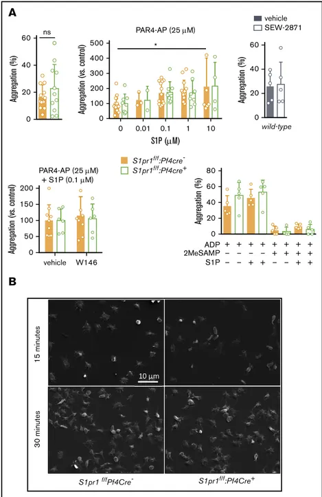
S1P1is dispensable for activation and spreading of mouse platelets
These results also question a reported role for S1P1in platelet aggregation and thrombosis in mice.7 S1P alone did not trigger aggregation of washed murine platelets (not shown), although it slightly enhanced PAR4 activating peptide (PAR4-AP)-induced aggregation (Figure 3A). This effect of S1P persisted in the absence of S1pr1, and pharmacologic S1P1modulators did not affect PAR4-AP-induced platelet aggregation (Figure 3A). S1P1 deficiency also did not affect ADP-induced platelet aggregation in the presence or absence of exogenous S1P at a concentration suggested to modulate platelet activation,7even when sensitizing the system by inhibiting P2Y12-mediated Gai activation with
2MeSAMP. Platelet adhesion and spreading on fibrinogen were also unaffected by S1pr1 deletion, S1P addition, or S1P1antagonism (Figure 3B; supplemental Figure 7). Thus, aggregation, spreading, and thrombosis phenotypes reported in Sphk-deficient platelets6,7 and mice7likely reflect on activation of a different receptor, receptor-independent effects, or thrombocytopenia,22rather than on a role for S1P1in platelet activation.7
Deregulated S1P production by Mx1Cre-sensitive hematopoietic cells suppresses thrombopoiesis in Sphk22/2mice
Increased platelet counts in Sphk1f/2:2f/2:Mx1Cre1mice (Figure 1B) not only argue against a necessary role for the S1P gradient20but also
A
0 20 40 60 Aggregation (%) ns 0 20 40 60 Aggregation (%) vehicle SEW-2871 wild-type S1pr1f/f :Pf4cre-S1pr1f/f:Pf4cre+ 0 0 0.01 0.1 1 10 100 200 300 400 500 S1P (M) Aggregation (vs. control) * PAR4-AP (25 M) vehicle 0 50 100 150 200 Aggregation (vs. control) PAR4-AP (25 M) + S1P (0.1 M) W146 0 + – – + – – + – + + – + + + – + + – + + + + + + 20 40 60 80 Aggregation (%) 2MeSAMP S1P ADPB
S1pr1 f/fPf4Cre- S1pr1f/f:Pf4Cre+ 3 0 minutes 15 minutesFigure 3.Platelet S1P1is dispensable for platelet aggrega-tion and spreading in mice.Platelets from mice in which S1pr1 was deleted in MKs (S1pr1f/f
:Pf4Cre1; green), littermate controls (orange), or wild-type mice (gray) were isolated, washed, and tested for their capacity to aggregate (A) and spread (B). (A, upper) Platelet aggregation in response to submaximal concen-trations (25mM) of PAR4-AP (thrombin receptor agonist) in the absence (left) or presence of exogenous S1P (0.1-10mM; middle) or of S1P1agonist SEW-2871 (0.5mM; right). (A, lower) Platelet aggregation in response to submaximal PAR4-AP in the presence of S1P (0.1mM) in the presence or absence of S1P1antagonist W146 (10mM; left) or in response to the weak platelet agonist ADP (2mM; right), with and without exogenous S1P (10 mM) or P2Y12 antagonism (2MeSAMP, 40mM) to address potential redundancy with P2Y12, which, similar to S1P1, is Gai coupled. (B) Representative scanning electron microscopy images (upper) showing the extent of platelet spreading 15 and 30 minutes after plating on fibrinogen in the presence of S1P (0.5mM; quantifica-tion in supplemental Figure 7). Note that although S1P did not trigger aggregation (not shown), it slightly increased PAR4-AP induced aggregation. However, neither aggregation nor spreading was influenced by selective S1P1modulation or S1pr1 deficiency. S1P also could not compensate for the absence of functional P2Y12 by alternative engagement of Gai. Statistical analyses by 2-way analysis of variance or the Mann-Whitney U test, as appropriate. Mean6 standard deviation is shown, symbols represent the number of mice.
against a necessary intracellular, receptor-independent role for S1P in platelet production.22The latter was deduced from the observation that mice globally deficient in Sphk2 display relative thrombocyto-penia and defective PP fragmentation.22 We confirmed a 25% reduction in circulating platelets and a slight increase in mean platelet volume (MPV) in Sphk22/2 mice; an intermediate phenotype in Sphk21/2littermates suggested a dose-dependent effect of Sphk2 deficiency (Figure 4A). As reported, the phenotype was present, although less profound, when the line was inbred to C57BL/6J background, and Sphk1 deficiency did not influence platelet counts despite being associated with a ;50% reduction in plasma S1P levels (supplemental Figure 8A).22Bone density, BM progenitors, platelet life span, and spleen size were unaltered, and genotype-dependent differences in platelet counts persisted after splenec-tomy (supplemental Figure 8B-F). Despite thrombocytopenia, MK numbers were higher in spleen and BM, consistent with a defect in thrombopoiesis (supplemental Figure 8G-H).22 Although PPs appeared larger, as reported by Zhang et al,22we did not observe a decrease in the capacity of fetal liver-derived MKs to produce PPs and shed platelets (supplemental Figure 8I). Concordantly, isolat-ed deletion of Sphk2 in MKs had no effect on platelet counts (Figure 4B). Consistent with a paracrine effect of Sphk2 deficiency on MK function, Sphk2 deletion with Lyve1Cre, active in lymphatic endothelial cells, some blood endothelial cells, macrophages, and other CD451 cells,48did reduce platelet counts (Figure 4B). This presented the possibility that the apparent increase in platelet counts in plasma S1Pless mice (Sphk1f/2:Sphk2f/2:Mx1Cre1; Figure 1B) represented a rescue of Sphk21/2-induced thrombocytopenia (Figure 4A) with pan-hematopoietic Sphk1 deficiency. This would imply that the Sphk22/2phenotype results from a redistribution of sphingosine toward Sphk1 rather than from loss of Sphk2-derived S1P (Figure 4C).16 Consistent with this notion, sphingo-sine did not build up in BM cells, as observed in plasma and RBCs of Sphk22/2 mice; Sphk1 expression remained unaltered (Figure 4D).9 To test this possibility more directly, we deleted Sphk1 in Mx1Cre-sensitive cells in a background globally deficient in Sphk2 (Sphk1f/f:22/2:Mx1Cre1). Consistent with our hypoth-esis, this returned platelet counts to wild-type levels (Figure 4E). The rescue was conferred by bone marrow transplantation (Figure 4F), but was not reproduced with selective Sphk1 deletion in MKs in a Sphk22/2background (Figure 4G). Compound deletion of the 2 kinases did not significantly affect MKP numbers or platelet life span, and therefore did not appear to provide rescue by an independent mechanism (Figure 4H). Thus, instead of reflecting on a critical intracellular signaling role for Sphk2-derived S1P in MKs, thrombocytopenia in Sphk2-deficient mice appears to arise from paracrine effects of S1P generated on redistribution of hematopoietic cell sphingosine to Sphk1.
Deregulated S1P production suppresses thrombopoiesis via S1P2
The above results suggest that Sphk2 deficiency induces thrombo-cytopenia by a receptor-dependent mechanism, and we further show that S1P1can suppress megakaryopoiesis. However, S1P1 antagonism did not ameliorate thrombocytopenia in Sphk22/2mice, whereas antagonism of S1P2, which is expressed on murine MKs (Figure 2C),20,23did (Figure 5A). S1P2deficiency did not by itself affect platelet production (Figure 5B), but when S1pr21/2 inter-crosses were performed in an Sphk22/2 background, S1P2 deficiency rescued Sphk22/2-induced thrombocytopenia (Figure 5C).
Conversely, when Sphk21/2 intercrosses were performed in an S1pr22/2background, Sphk2 deficiency no longer induced thrombocytopenia (Figure 5D vs Figure 4A). MPVs were also normalized by S1P2deficiency (Figure 5C-D). Transmission electron microscopy revealed a high density of MKs in Sphk22/2 BM (Figure 5E). Among these, we observed peri-sinusoidal MKs with scarce demarcation membrane systems (DMS), poorly resolved DMS regions sometimes without granules, and low-contrast MK “ghosts” that appeared to be undergoing necrosis. This contrasted with Sphk21/1:S1pr21/1 and Sphk22/2:S1pr22/2 BM, in which most mature MKs were large with well-defined DMS. This suggests that aberrant S1P2activation impairs MK maturation. S1P2is known to repel B cells when they encounter high S1P concentrations at the germinal center perimeter, a process that depends on Rho kinase, which also negatively regulates platelet formation by suppressing the actions of Rac1 and Cdc42 on cytoskeletal reorganization and microtubule assembly.17,49-51 Consistent with a role for the Rho pathway, the Rho kinase inhibitor Y-27632 significantly increased platelet counts in Sphk2-deficient mice with no effect on littermate controls (Figure 5F). Collectively, these observations suggest that Sphk1-derived S1P suppresses MK maturation in Sphk2-deficient mice by aberrant activation of S1P2and Rho kinase downstream. Although compound deficiencies of Sphk1 and S1P2reversed Sphk2 deficiency-induced thrombocytopenia, it is noteworthy that neither fully normalized the MK phenotype in Sphk22/2mice. Neither compound deficiency eliminated the higher density of MKs in Sphk22/2spleens (supplemental Figure 9), compound Sphk1 deficiency did not normalize MPV in Sphk22/2mice (Figure 4E), Sphk22/2PP extensions appear thicker also ex vivo (supplemen-tal Figure 8I22), and Sphk-deficient platelets display defective activation and spreading ex vivo.6,22These persistent phenotypes may reflect on imbalanced membrane lipids within Sphk22/2MKs and platelets, although not a simple buildup of upstream metab-olites, as we observed a paradoxical decrease in sphingosine levels in Sphk-deficient platelets (Figure 5G).6,22
Discussion
We here address roles for S1P in platelet production and function, using genetic and pharmacologic approaches in mice. Contrasting recent literature, our observations support neither a necessary role for the S1P gradient or MK/platelet S1P1in platelet production or aggregation nor a necessary intracellular signaling role for S1P in platelet production. They instead reveal that S1P1 signaling continuously restrains megakaryopoiesis and that S1P2 signaling can further suppress platelet production when sphingosine metab-olism is disturbed in the hematopoietic niche.
Three recent reports have positioned S1P as a critical facilitator of platelet biogenesis and signaling by S1P1-dependent attraction of PPs into BM sinusoids and subsequent shedding during plate-let budding from MKs,20receptor-independent regulation of plate-let shedding by Sphk2-derived S1P,22and amplification of platelet aggregation by an autocrine S1P1activation loop acting downstream of conventional platelet agonists.7 Our observations do not fully support either model, and argue that S1P plays a limited role in platelet biology bar major disturbances in S1P metabolism or signaling. First, we do not observe a necessary role for S1P1-mediated blood sensing in platelet formation. Genetic impairment of S1P provision to plasma or S1P breakdown in tissue, both with profound effects
Sphk2
A
0 200 400 600 800 1000 1200 0.0 0.5 4.0 4.5 5.0 * Platelets (1 000 /ul blood) MPV (fL) Sphk2f/f (C57BL6) Lyve1-Cre- (n=14) Lyve1-Cre+ (n=16) nsB
0 3 6 9 12 Sphingosine (pM /1 0^6 B M C) Sphk2+/+ (n=3) Sphk2-/- (n=3)D
sphingosine Sphk1 S1P S1PR SFK? S1P Mfsd2b Spns2C
E
Mx1Cre - (n=7) Mx1Cre + (n=8) Mx1Cre - (n=7) Mx1Cre + (n=8) 0 200 400 600 800 1000 1200 1400 ** Platelets (K/ L) Sphk1 f/f:Sphk2 -/- B M -> W ild-type ns W ild-type B M -> Sphk1 f/f:Sphk2-/-F
1 0 2 3 4 0 25 50 75 100 Time (days)Biotinylated platelets (%) Mx1Cre
- (n=4) Mx1Cre+ (n=4) Sphk1f/f:Sphk2 -/-0.0 0.2 0.4 0.6 0.8 ns M KP (% of B M C) Sphk1f/f:Sphk2 -/-Mx1Cre+ (n=5) Mx1Cre- (n=5)
H
G
S1pr1 S1pr2 S1pr4 Sphk1 Sphk2 0.00 0.02 0.04 0.06 0.08 0.10 0.12 Sphk2+/+ (n=4) Sphk2-/- (n=4) mR N A (relative to ) Gapdh 0 200 400 600 800 1000 1200 0.0 0.5 4.0 4.5 5.0 ** **** Platelets (K/ L) M PV (fL) Sphk2+/+ (n=54) Sphk2+/- (n=56) Sphk2-/- (n=46) ** 0 200 400 600 800 1000 1200 0.0 0.5 4.0 4.5 5.0 Platelets (K/ L) ns * Sphk2f/f (C57BL6) Pf4Cre- (n=18) Pf4Cre+ (n=29) M PV (fL) 0 200 400 600 800 1000 0.0 0.5 4.0 4.5 5.0 **** Platelets (K/ L) M PV (fL) * Sphk1f/f:Sphk2 -/-Mx1Cre-(n=116) Mx1Cre+ (n=83) 0 200 400 600 800 1000 0.0 0.5 4.0 4.5 5.0 ns Platelets (K/ L) ns M PV (fL) Sphk1f/f:Sphk2 -/-Pf4Cre- (n=41) Pf4Cre+ (n=31)Figure 4.Sphk2 deficiency induces thrombocytopenia by redirection of sphingosine to Sphk1.(A-B) Platelet counts and MPV from Sphk2 heterozygous intercrosses in C57BL/6J:129SVJ mixed background. (B) Effect of MK (Pf4Cre)- and lymphatic endothelium/CD451 (Lyve1Cre)–selective Sphk2 deletion on platelet counts. (C) Thrombocytopenia in Sphk22/2mice could be explained by redistribution of sphingosine to Sphk1 rather than by loss of Sphk2-derived S1P. This, in turn, could impair MK maturation by a receptor-dependent mechanism after S1P export by Spns2 or Mfsd2b, depending on cell type. (D) Impact of Sphk2 deficiency on the expression of Sphks and S1PRs and levels of sphingosine in total bone marrow cells (S1P was below the detection threshold). (E) Effect of deletion of Sphk1 in Mx1Cre-sensitive cells on Sphk2 deficiency-induced thrombocytopenia and MPV. (F) Effect of transplantation of BM cells from mice lacking Sphk1&2 in Mx1Cre-sensitive cells to lethally irradiated wild-type recipients and vice versa on platelet counts in the host. Note that the rescue conferred by Sphk1 deficiency is BM cell-derived, as the Sphk22/2phenotype itself.22(G) Effect of deletion of Sphk1 in MKs on Sphk2 deficiency-induced thrombocytopenia and MPV. (H) Effect of compound Sphk1 deficiency on MKP and platelet life span in Sphk22/2 mice. Statistical analyses by Mann-Whitney U test or 2-way analysis of variance.
on lymphocyte trafficking, did not reduce platelet counts, nor did complete or partial deletion of MK S1pr1 by 4 different transgenic approaches. Also at variance with Zhang et al,20 we did not observe effects of S1P1-selective pharmacological modulation on platelet counts when controlling for effects of vehicle and prior bleeding, nor did we confirm S1P1expression on murine MKs ex vivo or in situ. Our experiments were carried out in different strain backgrounds, littermate controlled, and sufficiently powered to reveal an important role for S1P gradient sensing. These observations are also in line with clinical experience, which has not revealed thrombocytopenia as an important adverse effect of S1P1-targeting drugs.52,53
In direct contrast, we observed elevated platelet counts with widespread neonatal deletion of either Sphk1&2 or S1pr1. Although modest, this effect was highly significant and replicated with constitutive S1pr1 deletion in hematopoietic cells, but not in MKs. Thrombocytosis was not conferred by transplantation of S1pr1-deficient bone marrow or induced with adult deletion or acute pharmacological S1P1modulation, suggesting developmental or delayed effects. A slight increase in MKs and a concomitant decrease in RBC counts suggested that S1P1may drive the differentiation of a common progenitor toward the erythroid lineage. Whether this reflects a direct role for S1P1 signaling in cell fate decisions or indirect effects on the hematopoietic niche or stem cell trafficking42,45,46remains to be determined.
A
B
C
D
0 200 400 600 800 1000 1200 1400 1600 Platelets (K/ul) Basal W146 ns ns Sphk2+/+ (n=7)Sphk2-/- (n=6) basal JTE-013 0 200 400 600 800 1000 1200 Platelets (K/ul) ns * Sphk2+/+ (n=12)Sphk2-/- (n=9) 0 200 400 600 800 1000 0.0 0.5 4.0 4.5 5.0 Platelets (K/ L) ns M PV (fL) Sphk2+/+ S1pr2+/+ (n=15) S1pr2 (n=34) S1pr2 (n=29) 0 200 400 600 800 1000 0.0 0.5 4.0 4.5 5.0 Platelets (K/ L) ** M PV (fL) *** S1pr2+/+ (n=37) S1pr2 (n=56) S1pr2 (n=26) Sphk2 -/-M PV (fL) 0 200 400 600 800 1000 0.0 0.5 4.0 4.5 5.0 Platelets (K/ L) S1pr2 -/-Sphk2+/+ (n=27) Sphk2 (n=29) Sphk2 (n=23)E
Sphk2+/+:S1pr2+/+ Sphk2-/-:S1pr2 -/-Sphk2-/-:S1pr2+/+G
0 1 2 3 4 5 Sphingosine (pmol/1 0^7 platelets) **** Sphk1 f/-:2 f/- Pf4Cr e- (n=5) Sphk1 f/-:2 f/- Pf4Cr e+ (n=5)F
0 200 400 600 800 1000 1200 Platelets (K/ul) ** ns Sphk 2+/+ (n=7) Sphk 2-/- (n=8) Basal Y27632Figure 5.Sphk2 deficiency induces thrombocytopenia by aberrant S1P2activation.(A) Effects of S1P1(W146, 10 mg/kg, left) or S1P2(JTE-013, 1.2 mg/kg) antagonism on Sphk2 deficiency-induced thrombocytopenia (24-hour platelet counts). (B-D) Platelet counts and MPV in litters from independent intercrosses of S1pr21/2in a wild-type background (B), S1pr21/2in a Sphk2-deficient background (C), and of Sphk21/2in a S1P2-deficient background (D). Note that although S1P2deficiency does not itself affect platelet counts, it rescues Sphk2 deficiency-induced thrombocytopenia. (E) Transmission electron micrographs of bone marrow from Sphk21/1, Sphk22/2, and Sphk22/2:S1pr22/2mice. Although the majority of MKs from Sphk21/1and Sphk22/2:S1pr22/2mice were singular and large, with a mature appearance and well-defined demarcation membrane systems (upper), MKs in Sphk22/2were highly heterogeneous, with clusters of immature MKs or mature MKs with limited DMS next to blood sinusoids (middle), low-contrast MK“ghosts” that appeared to be undergoing necrosis (bottom left, next to a normal MK) and platelet release within the bone marrow (bottom right). Representative images from n5 4 mice per genotype are shown. Scale bars, 2 mm. (F) Effect of a bolus injection of the Rho kinase inhibitor Y27632 (10mg/kg) on platelet counts in Sphk22/2and Sphk21/1controls. Note a significant increase in platelet counts only in the knockout. (G) Sphingosine content of Sphk deficient platelets. Statistical analyses by 2-way analysis of variance (A,F) or the Mann-Whitney U test.
Second, our results do not support a critical intracellular role for Sphk2-derived S1P in platelet production. As reported in a second Zhang et al study,22we observed mild thrombocytope-nia in Sphk22/2 mice. Yet whereas Zhang et al deduced an intracellular role for Sphk2-derived S1P from observations in mice globally deficient in Sphk2, we did not reproduce throm-bocytopenia with Pf4Cre-mediated selective deletion in MKs, even if this results in near complete loss of platelet S1P.6 Reversal of Sphk2 deficiency-induced thrombocytopenia with Sphk1 deletion in hematopoietic cells further suggested that the phenotype reflected on an increase rather than a decrease in S1P production, a hypothesis substantiated by similar reversal with S1P2deficiency. Although our interpretation of how Sphk2 deficiency induces thrombocytopenia differs, detailed charac-terization of the phenotype by Zhang et al remains consistent with our data. Elegant 2-photon microscopy showed abnormal extension of proplatelets into BM sinusoids of Sphk22/2mice, followed by retraction without efficient platelet shedding.22Our results suggest that the activation of S1P2 and Rho kinase downstream could contribute to impaired platelet shedding, and that disturbed membrane lipid composition could also contrib-ute to the gross aspects of the phenotype. S1P2 has been reported to mediate blood repulsion in osteoclast precursors19 and to confine B cells to germinal centers by preventing their exit to high S1P environment.17One could thus imagine high plasma S1P as observed in Sphk2-deficient animals to constitute a repulsive cue during platelet formation. Yet transfer of thrombo-cytopenia,22but not high S1P levels,9with transfer of Sphk22/2 bone marrow cells does not support this model, and Sphk22/2 PPs extended far into the bone marrow sinusoids before retracting.22 Impaired MK maturation was reminiscent of compound deficiency in Cdc42 and Rac1, and our observations would be consistent with Ga12/13-coupled S1P2 suppressing Cdc42 and Rac1 through RhoA, thus inhibiting terminal MK maturation or platelet shedding.20,49
Third, our results argue against an important role for S1P1 in amplifying platelet aggregation. Urtz et al reported protection from arterial thrombosis with global Sphk2 deficiency and reduced platelet aggregation in response to thrombin and other agonists in platelets derived from these mice.7This phenotype was attributed to a lack of activation of platelet S1P1by platelet-derived S1P, based mainly on studies performed in human platelets or whole blood.7Although we previously confirmed defective aggregation and spreading in Sphk-deficient platelets in the absence of exogenous S1P, we did not observe protection from thrombosis in mice with MK-selective Sphk deficiency.6Our current study shows no effect of S1pr1 deletion in assays in which we observed clear effects of Sphk deficiency,6 arguing against an autocrine platelet signaling loop involving S1P1. S1P1modulation was shown to affect human platelet activation in whole blood,7and it is possible that another S1P receptor takes on this function in mice, and that S1P1plays a more important role in humans. Yet as plasma S1P should already saturate S1P1,54,55it is unclear how platelets would sense further elevation of S1P levels after platelet activation by an S1P1-dependent mechanism. Whereas the current dogma would predict substantial effect of S1P1 modulation on platelet counts and thrombosis, our observations argue that these effects are likely to be minimal. Even if developmental deficiency of S1P1 in Mx1Cre-sensitive cells resulted in a modest increase in platelet counts, this was not
observed with adult deletion, and neither chronic treatment with oral fingolimod nor acute dosing with S1P1 modulators had a measurable effect on platelet counts. In contrast, even partial deficiency in Sphk2 reduced platelet counts by redistribution of sphingosine to Sphk1, a kinase that is frequently upregulated in cancer, inflammation, and other disease conditions. Increased Sphk1 activity in cells within the hematopoietic niche capable of S1P export could thus be predicted to suppress platelet production. This could be relevant to cancer, Gaucher disease, and other conditions in which there is evidence of both S1P pathway deregulation and thrombocytopenia.56
In conclusion, our observations argue that the S1P gradient and MK S1P1are both dispensable for platelet formation, and that S1P1is not critically involved in platelet activation or spreading in mice. Although it remains possible that S1P1is expressed and functional on human MKs and platelets, the current model, which predicts that S1P1modulation could be associated with a risk of bleeding and thrombosis, is based extensively on experiments performed in mice. The model therefore warrants revision, especially in light of clinical experience and with recent expansion of S1P1-directed therapies to pathologies associated with considerable bleeding risk.
Acknowledgments
The authors thank the Cellular and Molecular Imaging Platform, CRP2-UMS 3612 CNRS-US25 Inserm-IRD-Universit ´e Paris Descartes, Blandine Dizier, and Paris Cardiovascular Research Center (PARCC) platform staff for support, Camille Brunaud (PARCC) and Philippe Rameau (Gustave Roussy, Integrated Biology Core Facility, Villejuif, France) for cell sorting and flow cytometry analysis, and William Vainchenker and Alain Schmitt for helpful advice.
This work was funded by The Leducq Foundation (SphingoNet) (R.L.P. and E.C.), the French National Research Agency (ANR-10-MIDI-0003) (E.C.), Fondation de France (E.C.), the Intramural Research Program of the National Institutes of Health, the National Institute of Diabetes and Digestive and Kidney Diseases (R.L.P.), Higher Education Commission, Pakistan (H.N. and R.I.), the Marie Curie Prestige program (A.N.), the French Society of Arterial Hypertension (B.M.), and Promex Stiftung f ¨ur die Forschung (B.D.). H.N. is a graduate student at Paris Descartes University. This work is submitted in partial fulfillment of the requirement of the PhD.
Authorship
Contribution: H.N., N.Z, L.C., A.L., A.N., M.L.A., B.M., R.I., Y.A., P.H.B., S.L.G., S.P.-C., B.D., V.B., E.D.C., M.K., A.B., P.T., and E.C. designed and performed experiments and analyzed data; E.C. H.N., and A.N. wrote the manuscript; and M.L.A., M.K., P.G., P.-L.T., J.C., S.P., N.D., R.L.P., and C.B.-L. provided reagents and conceptual advice and critically reviewed the manuscript.
Conflict-of-interest disclosure: The authors declare no compet-ing financial interests.
ORCID profiles: A.N., 0003-0567-6790; E.D.C., 0003-0942-2819; M.K., 0003-2447-4350; P.G., 0002-9139-2147; P.-L.T., 0002-6062-5905; J.C., 0003-3964-0921; R.L.P., 0003-0456-1270; E.C., 0000-0002-6271-7125.
Correspondence: Eric Camerer, INSERM U970, Paris Cardio-vascular Research Center, 56 Rue Leblanc, 75015 Paris, France; e-mail: [email protected].
References
1. Proia RL, Hla T. Emerging biology of sphingosine-1-phosphate: its role in pathogenesis and therapy. J Clin Invest. 2015;125(4):1379-1387. 2. Obinata H, Hla T. Sphingosine 1-phosphate in coagulation and inflammation. Semin Immunopathol. 2012;34(1):73-91.
3. Mendelson K, Evans T, Hla T. Sphingosine 1-phosphate signalling. Development. 2014;141(1):5-9.
4. Olivera A, Mizugishi K, Tikhonova A, et al. The sphingosine kinase-sphingosine-1-phosphate axis is a determinant of mast cell function and anaphylaxis. Immunity. 2007;26(3):287-297.
5. Xiong Y, Yang P, Proia RL, Hla T. Erythrocyte-derived sphingosine 1-phosphate is essential for vascular development. J Clin Invest. 2014;124(11): 4823-4828.
6. Gazit SL, Mariko B, Th ´erond P, et al. Platelet and erythrocyte sources of S1P are redundant for vascular development and homeostasis, but both rendered essential after plasma S1P depletion in anaphylactic shock. Circ Res. 2016;119(8):e110-e126.
7. Urtz N, Gaertner F, von Bruehl ML, et al. Sphingosine 1-phosphate produced by sphingosine kinase 2 intrinsically controls platelet aggregation in vitro and in vivo. Circ Res. 2015;117(4):376-387.
8. Mizugishi K, Yamashita T, Olivera A, Miller GF, Spiegel S, Proia RL. Essential role for sphingosine kinases in neural and vascular development. Mol Cell Biol. 2005;25(24):11113-11121.
9. Sensken SC, Bode C, Nagarajan M, Peest U, Pabst O, Gr ¨aler MH. Redistribution of sphingosine 1-phosphate by sphingosine kinase 2 contributes to lymphopenia. J Immunol. 2010;184(8):4133-4142.
10. Cyster JG, Schwab SR. Sphingosine-1-phosphate and lymphocyte egress from lymphoid organs. Annu Rev Immunol. 2012;30:69-94.
11. Pappu R, Schwab SR, Cornelissen I, et al. Promotion of lymphocyte egress into blood and lymph by distinct sources of sphingosine-1-phosphate. Science. 2007;316(5822):295-298.
12. Venkataraman K, Lee YM, Michaud J, et al. Vascular endothelium as a contributor of plasma sphingosine 1-phosphate. Circ Res. 2008;102(6):669-676. 13. H ¨anel P, Andr ´eani P, Gr ¨aler MH. Erythrocytes store and release sphingosine 1-phosphate in blood. FASEB J. 2007;21(4):1202-1209.
14. Vu TM, Ishizu AN, Foo JC, et al. Mfsd2b is essential for the sphingosine-1-phosphate export in erythrocytes and platelets. Nature. 2017;550(7677): 524-528.
15. Mendoza A, Br ´eart B, Ramos-Perez WD, et al. The transporter Spns2 is required for secretion of lymph but not plasma sphingosine-1-phosphate. Cell Reports. 2012;2(5):1104-1110.
16. Yanagida K, Hla T. Vascular and immunobiology of the circulatory sphingosine 1-phosphate gradient. Annu Rev Physiol. 2017;79(1):67-91. 17. Green JA, Suzuki K, Cho B, et al. The sphingosine 1-phosphate receptor S1P2maintains the homeostasis of germinal center B cells and promotes niche
confinement. Nat Immunol. 2011;12(7):672-680.
18. Skoura A, Michaud J, Im DS, et al. Sphingosine-1-phosphate receptor-2 function in myeloid cells regulates vascular inflammation and atherosclerosis. Arterioscler Thromb Vasc Biol. 2011;31(1):81-85.
19. Ishii M, Kikuta J, Shimazu Y, Meier-Schellersheim M, Germain RN. Chemorepulsion by blood S1P regulates osteoclast precursor mobilization and bone remodeling in vivo. J Exp Med. 2010;207(13):2793-2798.
20. Zhang L, Orban M, Lorenz M, et al. A novel role of sphingosine 1-phosphate receptor S1pr1 in mouse thrombopoiesis. J Exp Med. 2012;209(12): 2165-2181.
21. Hla T, Galvani S, Rafii S, Nachman R. S1P and the birth of platelets. J Exp Med. 2012;209(12):2137-2140.
22. Zhang L, Urtz N, Gaertner F, et al. Sphingosine kinase 2 (Sphk2) regulates platelet biogenesis by providing intracellular sphingosine 1-phosphate (S1P). Blood. 2013;122(5):791-802.
23. Golfier S, Kondo S, Schulze T, et al. Shaping of terminal megakaryocyte differentiation and proplatelet development by sphingosine-1-phosphate receptor S1P4. FASEB J. 2010;24(12):4701-4710.
24. Bigaud M, Guerini D, Billich A, Bassilana F, Brinkmann V. Second generation S1P pathway modulators: research strategies and clinical developments. Biochim Biophys Acta. 2014;1841(5):745-758.
25. Pyne S, Adams DR, Pyne NJ. Sphingosine kinases as druggable targets [published online ahead of print 20 February 2018]. Handb Exp Pharmacol. doi: 10.1007/164_2018_96.
26. Weske S, Vaidya M, Reese A, et al. Targeting sphingosine-1-phosphate lyase as an anabolic therapy for bone loss. Nat Med. 2018;24(5):667-678. 27. Zhu Z, Fu Y, Tian D, et al. Combination of the immune modulator fingolimod with alteplase in acute ischemic stroke: a pilot trial. Circulation. 2015;
132(12):1104-1112.
28. Fu Y, Hao J, Zhang N, et al. Fingolimod for the treatment of intracerebral hemorrhage: a 2-arm proof-of-concept study. JAMA Neurol. 2014;71(9): 1092-1101.
29. Allende ML, Tuymetova G, Lee BG, Bonifacino E, Wu YP, Proia RL. S1P1 receptor directs the release of immature B cells from bone marrow into blood. J Exp Med. 2010;207(5):1113-1124.
30. Ramos-Perez WD, Fang V, Escalante-Alcalde D, Cammer M, Schwab SR. A map of the distribution of sphingosine 1-phosphate in the spleen. Nat Immunol. 2015;16(12):1245-1252.
31. Choi JW, Gardell SE, Herr DR, et al. FTY720 (fingolimod) efficacy in an animal model of multiple sclerosis requires astrocyte sphingosine 1-phosphate receptor 1 (S1P1) modulation. Proc Natl Acad Sci USA. 2011;108(2):751-756.
32. Allende ML, Bektas M, Lee BG, et al. Sphingosine-1-phosphate lyase deficiency produces a pro-inflammatory response while impairing neutrophil trafficking. J Biol Chem. 2011;286(9):7348-7358.
33. Camerer E, Regard JB, Cornelissen I, et al. Sphingosine-1-phosphate in the plasma compartment regulates basal and inflammation-induced vascular leak in mice. J Clin Invest. 2009;119(7):1871-1879.
34. Kono M, Tucker AE, Tran J, Bergner JB, Turner EM, Proia RL. Sphingosine-1-phosphate receptor 1 reporter mice reveal receptor activation sites in vivo. J Clin Invest. 2014;124(5):2076-2086.
35. Tiedt R, Schomber T, Hao-Shen H, Skoda RC. Pf4-Cre transgenic mice allow the generation of lineage-restricted gene knockouts for studying megakaryocyte and platelet function in vivo. Blood. 2007;109(4):1503-1506.
36. Schmitt A, Drouin A, Mass ´e JM, Guichard J, Shagraoui H, Cramer EM. Polymorphonuclear neutrophil and megakaryocyte mutual involvement in myelofibrosis pathogenesis. Leuk Lymphoma. 2002;43(4):719-724.
37. Bektas M, Allende ML, Lee BG, et al. Sphingosine 1-phosphate lyase deficiency disrupts lipid homeostasis in liver. J Biol Chem. 2010;285(14): 10880-10889.
38. Claxton S, Kostourou V, Jadeja S, Chambon P, Hodivala-Dilke K, Fruttiger M. Efficient, inducible Cre-recombinase activation in vascular endothelium. Genesis. 2008;46(2):74-80.
39. Swendeman SL, Xiong Y, Cantalupo A, et al. An engineered S1P chaperone attenuates hypertension and ischemic injury. Sci Signal. 2017;10(492): eaal2722.
40. Sanna MG, Wang SK, Gonzalez-Cabrera PJ, et al. Enhancement of capillary leakage and restoration of lymphocyte egress by a chiral S1P1 antagonist in vivo. Nat Chem Biol. 2006;2(8):434-441.
41. Sanna MG, Liao J, Jo E, et al. Sphingosine 1-phosphate (S1P) receptor subtypes S1P1 and S1P3, respectively, regulate lymphocyte recirculation and heart rate. J Biol Chem. 2004;279(14):13839-13848.
42. Massberg S, Schaerli P, Knezevic-Maramica I, et al. Immunosurveillance by hematopoietic progenitor cells trafficking through blood, lymph, and peripheral tissues. Cell. 2007;131(5):994-1008.
43. Joseph C, Quach JM, Walkley CR, Lane SW, Lo Celso C, Purton LE. Deciphering hematopoietic stem cells in their niches: a critical appraisal of genetic models, lineage tracing, and imaging strategies. Cell Stem Cell. 2013;13(5):520-533.
44. Sartawi Z, Schipani E, Ryan KB, Waeber C. Sphingosine 1-phosphate (S1P) signalling: Role in bone biology and potential therapeutic target for bone repair. Pharmacol Res. 2017;125(Pt B):232-245.
45. Golan K, Vagima Y, Ludin A, et al. S1P promotes murine progenitor cell egress and mobilization via S1P1-mediated ROS signaling and SDF-1 release. Blood. 2012;119(11):2478-2488.
46. Ishii M, Egen JG, Klauschen F, et al. Sphingosine-1-phosphate mobilizes osteoclast precursors and regulates bone homeostasis [published correction appears in Nature. 2010;465(7300):966]. Nature. 2009;458(7237):524-528.
47. Rankin EB, Wu C, Khatri R, et al. The HIF signaling pathway in osteoblasts directly modulates erythropoiesis through the production of EPO. Cell. 2012; 149(1):63-74.
48. Pham TH, Baluk P, Xu Y, et al. Lymphatic endothelial cell sphingosine kinase activity is required for lymphocyte egress and lymphatic patterning. J Exp Med. 2010;207(1):17-27.
49. Pleines I, D ¨utting S, Cherpokova D, et al. Defective tubulin organization and proplatelet formation in murine megakaryocytes lacking Rac1 and Cdc42. Blood. 2013;122(18):3178-3187.
50. Chang Y, Aurad ´e F, Larbret F, et al. Proplatelet formation is regulated by the Rho/ROCK pathway. Blood. 2007;109(10):4229-4236.
51. Chen Z, Naveiras O, Balduini A, et al. The May-Hegglin anomaly gene MYH9 is a negative regulator of platelet biogenesis modulated by the Rho-ROCK pathway. Blood. 2007;110(1):171-179.
52. Kappos L, Radue EW, O’Connor P, et al; FREEDOMS Study Group. A placebo-controlled trial of oral fingolimod in relapsing multiple sclerosis. N Engl J Med. 2010;362(5):387-401.
53. Cohen JA, Barkhof F, Comi G, et al; TRANSFORMS Study Group. Oral fingolimod or intramuscular interferon for relapsing multiple sclerosis. N Engl J Med. 2010;362(5):402-415.
54. Lee MJ, Van Brocklyn JR, Thangada S, et al. Sphingosine-1-phosphate as a ligand for the G protein-coupled receptor EDG-1. Science. 1998;279(5356): 1552-1555.
55. Rosen H, Sanna MG, Cahalan SM, Gonzalez-Cabrera PJ. Tipping the gatekeeper: S1P regulation of endothelial barrier function. Trends Immunol. 2007; 28(3):102-107.