Alma Mater Studiorum – Università di Bologna
DOTTORATO DI RICERCA IN
SCIENZE MEDICHE VETERINARIE
Ciclo XXVI
Settore Concorsuale di afferenza: 07/H5 Settore Scientifico disciplinare: VET/09
TITOLO TESI
CLINICOPATHOLOGIC FINDINGS IN THOROUGHBRED
HORSES DURING A HIGH SPEED – SHORT TERM
COMPETITION
Presentata(da:( Dott.(Robert(Mihai(Lukacs(
Coordinatore Dottorato Relatore
Prof. Carlo Tamanini Prof. Alessandro Spadari
INDEX
INTRODUCTION...4
1 EXERCISE AND MUSCULAR DAMAGE...5
1.1 REVIEW OF HUMAN SPORT MEDICINE LITERATURE...5
1.2 REVIEW OF EQUINE SPORT MEDICINE LITERATURE...11
2 EXERCISE AND OXIDATIVE STRESS...…..16
2.1 REVIEW OF HUMAN SPORT MEDICINE LITERATURE...16
2.2 REVIEW OF EQUINE SPORT MEDICINE LITERATURE...19
3 EXERCISE-INDUCED INTRAVASCULAR HEMOLYSIS...23
3.1 REVIEW OF HUMAN SPORT MEDICINE LITERATURE...23
3.2 REVIEW OF EQUINE SPORT MEDICINE LITERATURE...25
4 EXERCISE AND COAGULATION-FIBRINOLYSIS AXIS...28
4.1 REVIEW OF HUMAN SPORT MEDICINE LITERATURE...28
4.2 REVIEW OF EQUINE SPORT MEDICINE LITERATURE...30
EXPERIMENTAL PART...33
5 OBJECTIVES...33
6 MATERIALS AND METHODS...33
6.1 ANIMALS...33
6.2 NIBALO HORSE RACE...34
6.2.1 ENVIRONMENTAL CONDITIONS...36
6.3 BLOOD SAMPLES...36
6.3.1BLOOD SAMPLING TECHNIQUES...36
6.3.2 SAMPLE PROCESSING...37
6.4 PARAMETERS...38
6.4.1 COMPLETE BLOOD COUNT...38
6.4.2 MAIN METHODS USED...39
6.4.2.1 HAPTOGLOBIN...39 6.4.2.2 URIC ACID...41 6.4.2.3 FIBRINOGEN...42 6.4.2.4 D-DIMERS...44 6.4.2.5 FERRITIN...46 6.4.2.6 TOTAL IRON...49 6.4.2.7 TIBC...50
6.4.2.8 UIBC...50 6.4.2.9 SATURATION...52 6.4.3 STATISTICAL ANALYSIS...52 7 RESULTS...53 8 DISCUSSION...59 REFERENCES...67
Introduction
Blood plays an essential role during exercise. Its components provide transport of oxygen, electrolytes, hormones, water and nutrients to the exercising muscle, and at the same time provide removal of carbon dioxide and metabolism waste products, which result from exercise. Moreover, blood components are dynamically involved in the active reestablishment of acid-base imbalances created by exercise.
Throughout the years, various tests were created to evaluate changes of blood constituents related to exercise. The aim of these tests was to assess athlete’s fitness condition, performance potential, and to investigate and correct poor performance. However, a wide range of changes in blood constituents are influenced by various factors like athlete’s training condition, type of exercise, and sampling method, therefore throughout the years many studies have focused on different test combinations or on standardization of new tests.
The aim of this thesis was the evaluation of the occurrence of myopathy, hemolysis and hypercoagulation that occur in equine athletes after a high speed-short term competition. This is a three-year study, and for this purpose Thoroughbred horses that attend the Niballo horse race within Faenza City’s medieval reenactment were included in the present study. Pre- and post-race blood samples were taken from each subject and assessed for clinicopathological data. The results have evidenced myopathy, oxidative stress, hemolysis and alteration of the coagulation-fibrinolysis axis, which occurred after the race in the cohort of the present study.
1. Exercise and muscular damage
Poor athletic performance is a common sign encountered in equine sport medicine. Evaluation and understanding of the causes that lead to this condition represented and still remains a current interest for veterinarians. Most of the studies in equine sport medicine regarding athletic performance were based on human sport medicine study designs. Two of the most studied topics in human sport medicine and equine sport medicine are represented by exercise related muscular damage and oxidative stress.
Many studies have been conducted on human and equine athletes to establish the cause and pathological mechanism of muscular damage related to exercise. These studies have considered various aspects, but the most relevant topics focused on anatomy and physiology of skeletal muscular during exercise, on the influence of various types of exercise on promoting skeletal muscle damage, and on metabolic changes during exercise that lead to myopathy.
1.1. Review of human sport medicine
literature
Numerous studies in human literature have attempted to evaluate and understand the structural changes that occur with exercise in the skeletal muscle. The studies on specific characteristic of myosin, especially heavy chain myosin (HCM), led to the identification of several isoforms (Brooke M.H. & Kaiser K.K., 1970). With the aid of special methods, histochemical staining for oxidative enzymes, like adenosine
triphosphatases (ATPases), immunohistochemistry for HCM (Brooke M.H. & Kaiser K.K., 1974), and electrophoresis (Billeter R. et al., 1981), further studies on isolated single muscle fibers have evidenced that muscle fibers contain a single or multiple HCM isoforms, naming them pure type muscle fibers and hybrid type muscle fibers, respectively. Based on major MCH isoform, the pure type fibers are further divided in types: slow type I fiber, fast type fiber IIA, fast type fiber IID (formerly known as IIB) and fast type fiber IIX (Schiaffino S. & Reggiani C., 1994; Pette D. & Staron R.S., 2000).
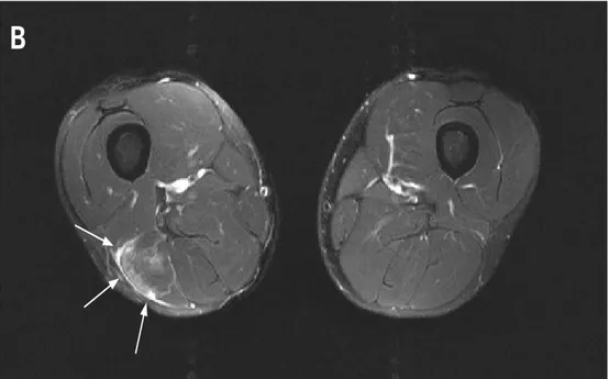
The human skeletal muscle is composed of a mixture of slow and fast muscular fibers, and the proportion of these fibers can vary within different parts of the same muscle (Lexell J. et al., 1983). All types of exercise, but in particular ones that involve eccentric contraction, promote damage within fast type fibers because of their lower oxidative capacity and their shorter length (Lieber R.L. & Fridén J., 1999; van Wessel T. et al., 2010). In another study, Guerrero and colleagues, using diagnostic imaging techniques (Fig. 1-3), brought more data reinforcing the concept of higher susceptibility of fast type fiber to exercise-induced damage in comparison with slow type fibers (Guerrero M. et al., 2008).
! 7!
Figure 1 (Guerrero M. et al., 2008). Magnetic resonance image from an axial view showing the presence of a lesion on the biceps femoris, upper part of posterior side of right thigh. The white arrow is showing a muscular edema. This type of lesions are mainly present in the fast type fibers.
Figure 2 (Guerrero M. et al., 2008). Magnetic resonance image from an axial view showing a lesion present in the medial-distal part of the posterior side of the left thigh. The presence of edema is noted along with a fibrillar defect lesion in the long head of the biceps femoris. This is a more pronounced lesion, indicated by white arrows, and involves both, type I and type II fibers.
Subjects
Thirty-six sportsmen aged 18–25 years (athletes, jockeys, tennis
players, football players, basketball players and pentathletes)
who had suffered some pain and/or injury were studied. The
control group comprised 51 non-sportsmen aged 18–55 years.
Muscle injuries were classified into three categories according to
clinical findings: grade I (delayed onset muscle soreness and
elongation, very small muscle tear); grade II (fibrillar disruption,
moderate muscle tear); grade III (fibre disruption, evident muscle
tear). Two ml of blood were obtained from the 51 controls and
from the athletes 48 h after they had suffered the muscle problem.
Serum was used for measurement of myosin levels.
Treatment of the sample
The blood samples were obtained in a Vacutainer tube and
centrifuged at 2000g at 4uC for 10 min. The serum could be kept
at 280uC without any loss of myosin for 15 days. Serum protein
was determined by the Bradford method.
15Myosin is present as a
blood marker at a very low concentration compared with other
serum proteins. It was therefore concentrated from the serum
sample by immunoprecipitation with A protein linked to
agarose-antibody. The pellet was resuspended with 15
ml loading buffer
(60 mM HCl-Tris, 10% glycerol, 2% SDS, 5% b-mercaptoethanol,
0.025% bromophenol blue, pH 6.8), centrifuged and warmed for
10 min to 70uC. The sample (about 25 ml) was prepared to be
carried out on Nupage Novex 3–8% Tris-acetate gels and
immunodetection. One or two fast myosin and slow myosin
standards were loaded in every gel. Electrophoresis was run in X
Cell SureLock Electrophoresis Cell (Invitrogen) at 150 V for 1 h at
room temperature. The gel was cut into strips of 7
61.2 cm. The
gel for immunoblot analyses was transferred to a PVDF
sequencing membrane (Immobilon PSQ Millipore) at 33 V for
70 min at room temperature.
The blots were treated with SuperBlocking buffer in PBS
(Pierce) at 0.01% to Tween 20 for 1.5 h at room temperature. One
gel was reacted with 1:90 000 anti-myosin fast monoclonal
antibody and the other with 1:150 000 anti-myosin slow
monoclonal antibody. Membranes were washed with 0.01%
Tween 20 at PBS six times for 5 min and incubated with
horseradish peroxidase conjugated rabbit anti-mouse IgG
(1:90 000) for 1 h at room temperature, followed by additional
washes (six times for 5 min in 0.01% Tween 20 at PBS). Each strip
was mixed with 80
ml of a mix (1:1) of Ultra Supersignal for
5 min. Proteins were visualised by enhanced chemiluminescence
(SuperSignal West Dura Trial Kit, Pierce, Rockford, Illinois, USA).
After drying, the strips were printed on the Hyperfilm TM ECL
for 10 min. This process was prepared with two samples, one for
detecting fast myosin and the other for slow myosin.
The films were scanned with a Hewlett Packard Scanjet
5200C and quantification was done with Quantity One 1-D
(BioRad, Hercules, California, USA).
Fast and slow myosin standards
The protein standards were prepared from fast and slow rabbit
muscle (tibialis anterior for fast fibres and soleus for slow
fibres). Purified myofibrils were obtained according to the
method described by Hasten et al.
16After carrying out this
process, myofibril suspensions of about 1 mg/ml of protein
were obtained. The suspension was denatured with the loading
buffer 1:1 v/v for 2 min at 100uC. 70 mg of protein was applied to
an acrylamide gel (7.5% T, 2.5% C) to separate the proteins. The
electrophoresis was run for 75 min at 150 V. The presence of
myosin was localised by staining a gel with Coomassie Blue G-250
R-250 (BioRad). The bands from an undyed gel were cut and
electroluted with a commercial electroluter (BioRad Model 422)
during 60 mA, 5 h with the electrolution buffer (25 mM HCl-tris,
192 mM glycine, 0.1% SDS, pH 6.8). We obtained 0.93 mg/ml fast
myosin and 1.55 mg/ml of slow myosin. The myosins were diluted
with 10 mM HCl-Tris, 300 mM NaCl, 2 mM EDTA at pH 6.8.
Standards were kept in fractions of 50
ml at 230uC.
Measurement of enzymatic activities
CK was determined by a Technicon DAX System autoanalyser
according to the method of Szasz et al.
17Imaging evaluation
Echography was carried out at the Centre d’Estudis d’Alt
Rendiment Esportiu (CEARE) and in the ultrasonographic
department of the FIATC Clinic using Toshiba Medical System
ultrasonography equipment with a multifrequency probe
(Just-Vision in CEARE, Power(Just-Vision in FIATC). Magnetic resonance
studies were done at the department of magnetic resonance of the
Coracha´n Clinic using a Siemens Symphony device (1.5 TESS).
Both types of soundings gave results which increased in direct
proportion with the grade of the lesion. In grade I lesions US
usually shows the lesion (haematic suffusion and defect of some
Figure 1 Magnetic resonance images of different grades of muscle
lesion: (A) grade I lesion; (B) grade II lesion; (C) grade III lesion.
Original article
582
Br J Sports Med 2008;42:581–584. doi:10.1136/bjsm.2007.037945
Subjects
Thirty-six sportsmen aged 18–25 years (athletes, jockeys, tennis
players, football players, basketball players and pentathletes)
who had suffered some pain and/or injury were studied. The
control group comprised 51 non-sportsmen aged 18–55 years.
Muscle injuries were classified into three categories according to
clinical findings: grade I (delayed onset muscle soreness and
elongation, very small muscle tear); grade II (fibrillar disruption,
moderate muscle tear); grade III (fibre disruption, evident muscle
tear). Two ml of blood were obtained from the 51 controls and
from the athletes 48 h after they had suffered the muscle problem.
Serum was used for measurement of myosin levels.
Treatment of the sample
The blood samples were obtained in a Vacutainer tube and
centrifuged at 2000g at 4
uC for 10 min. The serum could be kept
at 280
uC without any loss of myosin for 15 days. Serum protein
was determined by the Bradford method.
15Myosin is present as a
blood marker at a very low concentration compared with other
serum proteins. It was therefore concentrated from the serum
sample by immunoprecipitation with A protein linked to
agarose-antibody. The pellet was resuspended with 15
ml loading buffer
(60 mM HCl-Tris, 10% glycerol, 2% SDS, 5% b-mercaptoethanol,
0.025% bromophenol blue, pH 6.8), centrifuged and warmed for
10 min to 70uC. The sample (about 25 ml) was prepared to be
carried out on Nupage Novex 3–8% Tris-acetate gels and
immunodetection. One or two fast myosin and slow myosin
standards were loaded in every gel. Electrophoresis was run in X
Cell SureLock Electrophoresis Cell (Invitrogen) at 150 V for 1 h at
room temperature. The gel was cut into strips of 7
61.2 cm. The
gel for immunoblot analyses was transferred to a PVDF
sequencing membrane (Immobilon PSQ Millipore) at 33 V for
70 min at room temperature.
The blots were treated with SuperBlocking buffer in PBS
(Pierce) at 0.01% to Tween 20 for 1.5 h at room temperature. One
gel was reacted with 1:90 000 anti-myosin fast monoclonal
antibody and the other with 1:150 000 anti-myosin slow
monoclonal antibody. Membranes were washed with 0.01%
Tween 20 at PBS six times for 5 min and incubated with
horseradish peroxidase conjugated rabbit anti-mouse IgG
(1:90 000) for 1 h at room temperature, followed by additional
washes (six times for 5 min in 0.01% Tween 20 at PBS). Each strip
was mixed with 80
ml of a mix (1:1) of Ultra Supersignal for
5 min. Proteins were visualised by enhanced chemiluminescence
(SuperSignal West Dura Trial Kit, Pierce, Rockford, Illinois, USA).
After drying, the strips were printed on the Hyperfilm TM ECL
for 10 min. This process was prepared with two samples, one for
detecting fast myosin and the other for slow myosin.
The films were scanned with a Hewlett Packard Scanjet
5200C and quantification was done with Quantity One 1-D
(BioRad, Hercules, California, USA).
Fast and slow myosin standards
The protein standards were prepared from fast and slow rabbit
muscle (tibialis anterior for fast fibres and soleus for slow
fibres). Purified myofibrils were obtained according to the
method described by Hasten et al.
16After carrying out this
process, myofibril suspensions of about 1 mg/ml of protein
were obtained. The suspension was denatured with the loading
buffer 1:1 v/v for 2 min at 100
uC. 70 mg of protein was applied to
an acrylamide gel (7.5% T, 2.5% C) to separate the proteins. The
electrophoresis was run for 75 min at 150 V. The presence of
myosin was localised by staining a gel with Coomassie Blue G-250
R-250 (BioRad). The bands from an undyed gel were cut and
electroluted with a commercial electroluter (BioRad Model 422)
during 60 mA, 5 h with the electrolution buffer (25 mM HCl-tris,
192 mM glycine, 0.1% SDS, pH 6.8). We obtained 0.93 mg/ml fast
myosin and 1.55 mg/ml of slow myosin. The myosins were diluted
with 10 mM HCl-Tris, 300 mM NaCl, 2 mM EDTA at pH 6.8.
Standards were kept in fractions of 50
ml at 230uC.
Measurement of enzymatic activities
CK was determined by a Technicon DAX System autoanalyser
according to the method of Szasz et al.
17Imaging evaluation
Echography was carried out at the Centre d’Estudis d’Alt
Rendiment Esportiu (CEARE) and in the ultrasonographic
department of the FIATC Clinic using Toshiba Medical System
ultrasonography equipment with a multifrequency probe
(Just-Vision in CEARE, Power(Just-Vision in FIATC). Magnetic resonance
studies were done at the department of magnetic resonance of the
Coracha´n Clinic using a Siemens Symphony device (1.5 TESS).
Both types of soundings gave results which increased in direct
proportion with the grade of the lesion. In grade I lesions US
usually shows the lesion (haematic suffusion and defect of some
Figure 1 Magnetic resonance images of different grades of muscle
lesion: (A) grade I lesion; (B) grade II lesion; (C) grade III lesion.
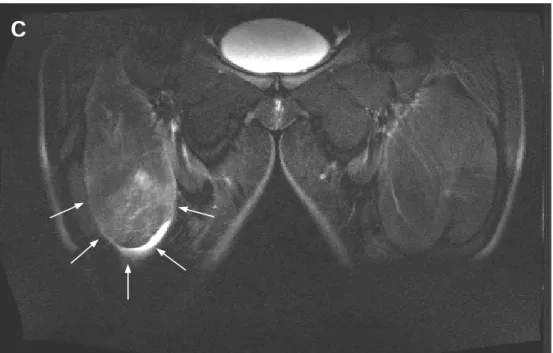
Figure 3 (Guerrero M. et al., 2008). Magnetic resonance image from a coronal view of the anterior side of the thigh. The arrows point out the presence of edema and a severe and extended fibrillar defect.
One of the first studies of sports-related muscular damage evidenced the occurrence of this phenomenon in athletes that are unaccustomed to exercise or in athletes that perform a sustained, intense workout (Hough T., 1900). Studies of human muscle mechanics have revealed that muscles can perform three types of contractions, which in the early 1900’s where termed as myometric, isometric and plyometric (Hubbard A.W. & Stetson R.H., 1938). These terms were formed using prefixes from Greek language and added to the noun “metric”, where “myo” means shorter, “iso” means the same, and “plyo” means larger. Later, in 2003, Faulkner explained how the scientific community and literature have changed the terminology, introducing new words for describing muscular contraction, “concentric” and “eccentric” (Faulkner J.A., 2003). Therefore, an eccentric muscular contraction presumes an action where the muscle lengthens under contraction, a concentric muscular contraction presumes an action where the muscle shortens, and an isometric muscular contraction presumes an action where the length of the muscle remains unchanged.
Subjects
Thirty-six sportsmen aged 18–25 years (athletes, jockeys, tennis players, football players, basketball players and pentathletes) who had suffered some pain and/or injury were studied. The control group comprised 51 non-sportsmen aged 18–55 years.
Muscle injuries were classified into three categories according to clinical findings: grade I (delayed onset muscle soreness and elongation, very small muscle tear); grade II (fibrillar disruption, moderate muscle tear); grade III (fibre disruption, evident muscle tear). Two ml of blood were obtained from the 51 controls and from the athletes 48 h after they had suffered the muscle problem. Serum was used for measurement of myosin levels.
Treatment of the sample
The blood samples were obtained in a Vacutainer tube and centrifuged at 2000g at 4uC for 10 min. The serum could be kept at 280uC without any loss of myosin for 15 days. Serum protein was determined by the Bradford method.15Myosin is present as a
blood marker at a very low concentration compared with other serum proteins. It was therefore concentrated from the serum sample by immunoprecipitation with A protein linked to agarose-antibody. The pellet was resuspended with 15 ml loading buffer (60 mM HCl-Tris, 10% glycerol, 2% SDS, 5% b-mercaptoethanol, 0.025% bromophenol blue, pH 6.8), centrifuged and warmed for 10 min to 70uC. The sample (about 25 ml) was prepared to be carried out on Nupage Novex 3–8% Tris-acetate gels and immunodetection. One or two fast myosin and slow myosin standards were loaded in every gel. Electrophoresis was run in X Cell SureLock Electrophoresis Cell (Invitrogen) at 150 V for 1 h at room temperature. The gel was cut into strips of 761.2 cm. The gel for immunoblot analyses was transferred to a PVDF sequencing membrane (Immobilon PSQ Millipore) at 33 V for 70 min at room temperature.
The blots were treated with SuperBlocking buffer in PBS (Pierce) at 0.01% to Tween 20 for 1.5 h at room temperature. One gel was reacted with 1:90 000 anti-myosin fast monoclonal antibody and the other with 1:150 000 anti-myosin slow monoclonal antibody. Membranes were washed with 0.01% Tween 20 at PBS six times for 5 min and incubated with horseradish peroxidase conjugated rabbit anti-mouse IgG (1:90 000) for 1 h at room temperature, followed by additional washes (six times for 5 min in 0.01% Tween 20 at PBS). Each strip was mixed with 80 ml of a mix (1:1) of Ultra Supersignal for 5 min. Proteins were visualised by enhanced chemiluminescence (SuperSignal West Dura Trial Kit, Pierce, Rockford, Illinois, USA). After drying, the strips were printed on the Hyperfilm TM ECL for 10 min. This process was prepared with two samples, one for detecting fast myosin and the other for slow myosin.
The films were scanned with a Hewlett Packard Scanjet 5200C and quantification was done with Quantity One 1-D (BioRad, Hercules, California, USA).
Fast and slow myosin standards
The protein standards were prepared from fast and slow rabbit muscle (tibialis anterior for fast fibres and soleus for slow fibres). Purified myofibrils were obtained according to the method described by Hasten et al.16
After carrying out this process, myofibril suspensions of about 1 mg/ml of protein were obtained. The suspension was denatured with the loading buffer 1:1 v/v for 2 min at 100uC. 70 mg of protein was applied to an acrylamide gel (7.5% T, 2.5% C) to separate the proteins. The electrophoresis was run for 75 min at 150 V. The presence of myosin was localised by staining a gel with Coomassie Blue G-250 R-250 (BioRad). The bands from an undyed gel were cut and electroluted with a commercial electroluter (BioRad Model 422)
during 60 mA, 5 h with the electrolution buffer (25 mM HCl-tris, 192 mM glycine, 0.1% SDS, pH 6.8). We obtained 0.93 mg/ml fast myosin and 1.55 mg/ml of slow myosin. The myosins were diluted with 10 mM HCl-Tris, 300 mM NaCl, 2 mM EDTA at pH 6.8. Standards were kept in fractions of 50 ml at 230uC.
Measurement of enzymatic activities
CK was determined by a Technicon DAX System autoanalyser according to the method of Szasz et al.17
Imaging evaluation
Echography was carried out at the Centre d’Estudis d’Alt Rendiment Esportiu (CEARE) and in the ultrasonographic department of the FIATC Clinic using Toshiba Medical System ultrasonography equipment with a multifrequency probe (Just-Vision in CEARE, Power(Just-Vision in FIATC). Magnetic resonance studies were done at the department of magnetic resonance of the Coracha´n Clinic using a Siemens Symphony device (1.5 TESS).
Both types of soundings gave results which increased in direct proportion with the grade of the lesion. In grade I lesions US usually shows the lesion (haematic suffusion and defect of some
Figure 1 Magnetic resonance images of different grades of muscle lesion: (A) grade I lesion; (B) grade II lesion; (C) grade III lesion.
Original article
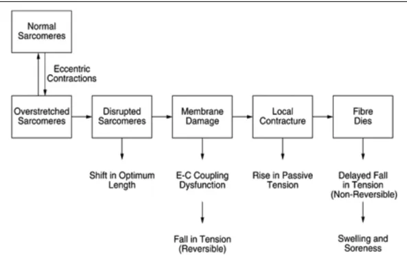
Numerous studies and reviews sustain that exercises that involve eccentric contractions have the most damaging effects on muscles. In a study from 2001, Proske and Morgan try to formulate and explain the events (Fig. 4) that lead to muscular damage following eccentric contractions, and promote the hypothesis of “popping sarcomere”. These authors sustain that a sudden change in the contracting force or a high intensity contraction lead to a non-uniform contraction of sarcomeres, and some of these sarcomeres are stretched at the point that they are damaged (Proske U. & Morgan D.L., 2001). !
Figure 4 (Proske U. & Morgan D.L., 2001). Diagram of events that lead to muscular damage following eccentric contractions.
There are other studies that promote another hypothesis. After eccentric contraction that induces a structural change follows an impairment of calcium (Ca2+) homeostasis (Proske U. & Morgan D.L., 2001; Tee J.C. et al., 2007). Taking inspiration from studies on mice (Duncan C.J. & Jackson M.J., 1987) and frogs (Duncan C.J., 1987), in human sport medicine it was hypothesized that the same mechanisms contribute and
exercise to the sarcoplasmic reticulum alters its function in Ca2+
homeostasis, which leads to accumulation of Ca2+ in the intracellular
space and subsequent activation of Ca2+ overload phase. The defense mechanisms against Ca2+ are overwhelmed, and Ca2+-dependent proteolytic and phospholipolytic enzymes are activated. These enzymes will damage myofibre structures, and contribute to muscular damage. Numerous studies are focused on finding the ideal markers of muscular damage. One of the most used markers is serum creatine kinase (CK) because of its magnitude of increase compared to other proteins and the relative low cost of the assay. From the studies conducted until now it has been noted that there are many variables that influence the relationship between muscular damage and release of CK from muscles, and between CK and other markers of muscular damage (Clarkson P.M. & Hubal M.J., 2002). Other markers used to evaluate muscular damage are lactate dehydrogenase (LDH), aspartate transaminase (AST), carbonic anhydrase isoenzyme II, heart fatty acid binding protein, troponin (Sorichter S. et al., 1999), myosin heavy chains (Sorichter S. et al., 1999; Guerrero M. et al., 2008) and myosin light chains (Guerrero M. et al., 2008).
Other phenomenon that contributes in amplifying muscular damage is the inflammatory response secondary to the initial trauma to the muscular fibers (Fielding R.A. et al., 1993; Clarkson P.M. & Hubal M.J., 2002). The inflammatory cells promote the formation of free radicals, which lead to oxidative damage of the muscular cells, which will be further discussed in the oxidative stress chapter. From the numerous studies available in human literature it has been seen that the extent of inflammatory response secondary to exercise-induced muscular damage is influenced by various factors, like type of exercise (Kokot K. et al., 1988; Peeling P. et al., 2009) and group of muscles (Hirose L. et al., 2004).
Despite the abundant literature the exact mechanisms of damage, reparation and adaptation have not been fully understood yet (Howatson G. & van Someren K.A., 2008; Nikolaidis M.G. et al., 2008).
1.2 Review of equine sport medicine
literature
In agreement with human sport medicine literature, in equine sport medicine there is an extensive specialized literature focused on exercise an its impact on skeletal muscle. A high percentage of these studies were concluded on Standardbred, Thoroughbred and endurance horses, and only a minor percentage on horses from other disciplines (Rivero J.-L. L., 2007).
As with human skeletal muscle, many studies were concluded to best identify the anatomy and physiology of the equine skeletal muscle. Immunohistochemistry, electrophoresis, staining for ATPase, were used to identify and classify MHC composition of the equine skeletal muscle fibers. Horses, like other mammals, have two main pure types of fibers, type I (slow oxidative) and type II (fast oxidative) (Fig. 5). Among the II type fibers, the horse has fast oxidative type IIA fibers and fast glycolytic type IIB fibers (Rivero J.-L. L.1 et al., 1996). The type IIB fiber can be found named as type IIX in more recent studies. In another study, Rivero and colleagues, with the ATPase method, have evidenced the presence of a forth type of fiber IIAB (IIAX) (Rivero J.-L. L.2 et al, 1996). Other authors confirm the presence of the fourth type of hybrid fiber, type IIA/IIX. Moreover, these authors have demonstrated by succinic dehydrogenase (SDH) analysis of each type of fibers that type IIA/IIX
fibers have a more fast contraction ability and higher resistance to fatigue (Yamano S. et al., 2002).
Figure 5. (Valberg S.J., 200) Metabolism in slow and fast twitch myofibers.
Numerous studies were performed to assess the influence of exercise and training protocols upon the transition and recruitment of muscular fibers. These studies have underlined that an increase in exercise duration and intensity will lead to the progressive transition from type I → type IIA → type IIB (IIX) (Hodgson D.R. et al., 1985; Valberg S., 1986; Essén-Gustavsson B. et al., 1995; Schuback K. & Essén-Essén-Gustavsson B., 1998). With the discovery of the fourth type of fiber, IIA/IIX, later studies have evidenced that higher intensity exercises leads to the recruitment of this type of fibers (Yamano S. et al., 2002; Rivero J.-L. L. et al., 2007).
Many articles in the equine sport medicine literature have studied different training schedules in order to prepare and improve the athletic condition of horses, and to avoid muscular damage (Rivero J.-L. L., 2007). The topic of exercise-induced muscular damage in horses is assessed with a wider focus on the markers of muscular injury. Terms like eccentric and concentric muscular contractions are not used in equine sport medicine. If in human literature exercises that presume eccentric contractions are considered high intensity exercises, in equine sport medicine the endurance race is considered as a high intensity exercise that requires superior athletic ability (Hinchcliff K.W. & Geor R.J., 2004).
In equine sport medicine literature it was hypothesized that imbalanced Ca2+ homeostasis might be involved in muscular damage related to exercise (Hodgson D.R., 1993). Ultrastructural alterations found in muscles from horses with rhabdomyolysis can lead to an imbalanced permeability of sarcoplasmic reticulum (Lindholm A. et al., 1974), which determines intracellular accumulation of Ca2+ (Byrd S.K. et al., 1989). This excessive accumulation of intracellular Ca2+ will determine the activation of enzymes that will contribute in amplifying muscular damage as seen in people (Proske U. & Morgan D.L., 2001). The Ca2+ hypothesis can be sustained by the studies on agents that interfere with the function of Ca2+ channels. Beech J. and collaborators have used phenytoin in horses and have managed to reduce the incidence of myopathy secondary to exercise, supposing that this agent could influence the permeability of sarcoplasmic reticulum (Beech J. et al., 1988).
The most frequently used makers used to detect muscular damage related to exercise are CK, LDH and AST. In one study, Anderson M.G. sustains that AST is released from cells due to a change in membrane permeability and not necessary due cellular breakdown. Moreover, the author sustains that CK is the most specific enzyme for muscular damage and it is parameter of choice in this type of studies (Anderson M.G., 1975). Later,
Volfinger L. and colleagues have studied the kinetics of CK in the horse and concluded that there are some differences between human and equine athletes, like the relationship between CK and athletic preparation encountered in human athletes, and bioavailability of CK in horses (Volfinger L. et al., 1994). Chiaradia L. and colleagues have studied exercise-induced muscular damage assessing CK and LDH, and in particular the isoenzymes specific of skeletal muscles. The authors have concluded that the intensity of exercise in their work led to lipid peroxidation, which was not intense enough to cause damage to muscular cells. In the same study the significant increase in LDH isoenzymes specific to myocardium lead to authors to hypothesize that beside skeletal muscles, myocardial muscles could be prone to exercise-induced damage (Chiaradia L. et al., 1998). In other study, Hargreaves B.J. and colleagues found AST and CK increased in association with decreased antioxidant status, suggesting leakage from damaged muscular cells following exercise (Hargreaves B.J. et al., 2002). Kim J. and colleagues in their study concluded that CK alone was not sensitive enough when compared to their findings on light and electron microscopy (Kim J. et al., 2005). Other authors studying carnosine have found it to be more selective than CK and AST plasma values in identifying muscular damage (Dunnett M. et al., 2002). Further studies are required to improve the evaluation of muscular damage in horses, perhaps by expanding the panel of markers for muscular damage.
Knowledge of exercise induced inflammatory reaction is rather limited in equine sport medicine. There are several studies that have demonstrated that exercise alter the immune response. In this study, besides demonstrating the negative effect of exercise on the immune response, the authors have revealed that post exercise samples showed an increase in leukocytes and neutrophils, as well as an increase in the production of reactive oxygen products (ROS) in non-stimulated leukocytes (Donovan
D.C. et al., 2007). There are studies that have documented the increase in myeloperoxidase (MPO) and elastase (ELT), enzymes contained mainly in the granules of neutrophils, that promote the formation of free radicals, which represents an efficient but non-selective mechanism of defense of white blood cells (Art T. et al., 2006; Lejeune J.P. et al., 2010; Serteyn D. et al., 2010). In the study of Serteyn D. and colleagues is has been also noted a correlation between MPO and ELT enzymes and CK, indicating that these enzymes can contribute in expanding the exercise-induced muscular damage (Serteyn D. et al., 2010).
2. Exercise and oxidative stress
The continuous research, in understanding the muscular damage related to exercise, led to the second most studied topic of sport medicine; exercise-induced oxidative stress. Exercise can induce an alteration to the oxidant/antioxidant balance causing oxidative stress, which has been suggested to be implicated in the pathogenesis of muscular damage, as seen previously, and in the pathogenesis of other exercise-related alterations, which will be further discussed.
2.1
Review of human sport medicine
literature
The first study to investigate the oxidative stress induced by exercise in human athletes it was the study of Dillard C.J. and colleagues in 1978. They have measured in the expired air a marker of lipid peroxidation, pentane, and saw that administering vitamin E, as antioxidant, reduced the quantity of this biomarker in the expired air. It was evidenced that exercise induced lipid peroxidation, but the source of this peroxidation was not identified (Dillard C.J. et al., 1978). Since then, numerous studies and reviews were conducted on exercise-induced oxidative damage (Powers S.K. & Jackson M.J., 2007; Nikolaidis M.G. et al., 2008). Human literature on this topic assessed on human patients is rather limited, and most of these studies were conducted on rats because of the similarity between human and rat muscle, which may share the same pathways in oxidants production.
Free radicals that are generated by the cells are superoxide (O2 .-) and
oxygen species (ROS), which have been more extensively studied, and reactive nitrogen species (RNS). Continuous research has evidenced that these oxidants can be produced in many tissues, including muscles (Powers S.K. et al., 2011).
In the muscular cell ROS can be formed in various sites: mitochondrial site (Fernström M. et al., 2007; Sahlin K. et al., 2010), sarcoplasmic reticulum site (Xia R. et al., 2003), transverses tubules site (Espinosa A. et al., 2006), and membrane site (Pattwell D.M. et al., 2004).
There are several mechanisms that contribute in producing these oxidants and vary with the endogenous sites in the skeletal muscle. Within the mitochondria complex I (Lustgarten M.S. et al., 2012) and complex III (Muller F.L. et al., 2004) have been identified as sources of superoxide production within the electron transport chain. As previously seen, several muscular components can participate at the production of ROS through the nicotinamide adenine dinucleotide phosphate (NADPH) oxidase, which transfers electrons to molecular oxygen (Xia R. et al., 2003; Pattwell D.M. et al., 2004; Espinosa A. et al., 2006). Other pathway that generates ROS is represented by the phospholipase-A2 (PLA2). This
enzyme cleaves mem
brane phospholipids, producing arachidonic acid, which is used as substrate by lipoxygenases to produce ROS (Zuo L. et al., 2004). Moreover, studies have evidenced that two types of PLA2 exist, Ca2+
-dependent and -in-dependent, and it was hypothesized that during exercise the Ca2+-dependent is the one that is more actively involved in ROS
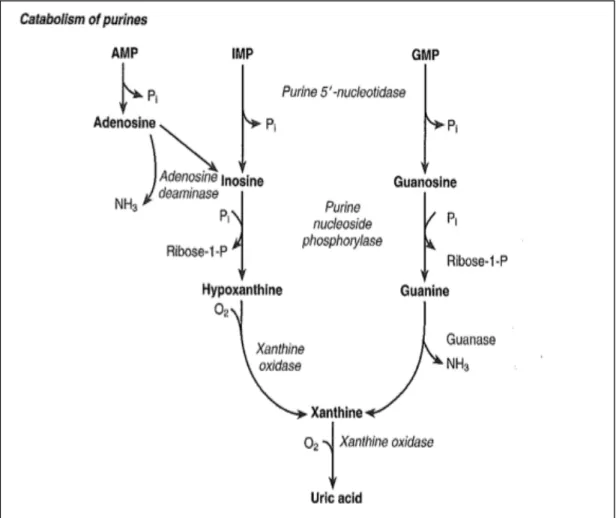
production (Gong M.G. et al., 2006). Xanthine oxidase pathway represents another mechanism promotes ROS formation through oxidizing hypoxanthine to xanthine (Fig. 6) that will generate superoxide radicals (Sjödin B. et al., 1990; Hellsten-Westing Y., 1993; Hellstan Y. et al., 1997).
Figure 6. (Lamb E.J. & Price C.P., 2008) Catabolism of purines and formation of xanthine.
At muscular level the production of these oxidants can be further increased by several conditions encountered during exercise like, increased muscular temperature, decreased cellular pH and increased carbon dioxide (CO2) tension (Reeder B.J. & Wilson M.T., 2001;
Arbogast S. & Reid M.B., 2004).
Following eccentric exercise and muscular damage there can be an inflammatory response, as seen previously, which can promote further extension of the oxidative stress (Smolen J.E. et al., 1980; Kokot K. et al., 1988). The damage process-induced by exercise to muscles is extended to the capillaries of endothelium (Crenshaw A.G. et al., 1993), which were found to be rich in xanthine oxidase (Hellstan Y. et al., 1997). Similarly to a reperfusion injury process, the damaged endothelium will increase the
availability of xanthine oxidase, which will interact with present inflammatory cells, and increase ROS production (Hellstan Y. et al., 1997).
2.2
Review of equine sport medicine
literature
In equine sport medicine literature in a 2008 review, Kirschvink and colleagues affirmed that there are three endogenous pathways related to exercise that lead to oxidative stress: electron transfer chain in the mitochondria, endogenous oxidant enzymes and oxidants that result from oxidative burst of inflammatory cells from the exercise-induced inflammatory process (Krischvink N. et al., 2008).
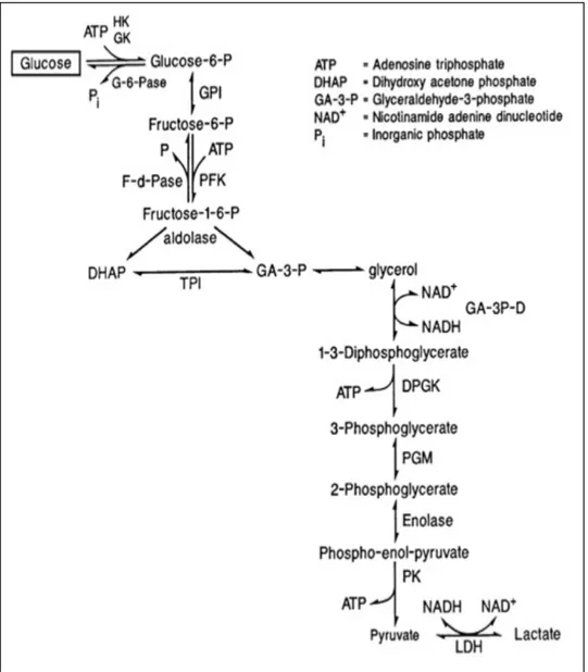
The most numerous studies regarding oxidative stress in equine sport medicine were focused on endogenous oxidant enzymes, and in particularly xanthine oxidase. As seen previously during exercise, as the intensity increases, the transition between fiber types leads to the recruitment of fiber type IIB (IIX). These fibers have low content in adenosine triphosphate (ATP), and have fast glycolytic properties (Valberg S. & Essén-Gustavsson B., 1987). Intense exercise increases the demand for ATP exceeding the production ability of the organism. Subsequently there is a shift from the aerobic production to anaerobic production, which is more efficient. The anaerobic production of ATP can occur through anaerobic glycolysis (Fig. 7) and high-energy phosphorylation (Fig. 8). The result of these processes is accumulation of lactic acid and adenosine monophosphate (AMP). Excessive AMP accumulation and reduced re-phosphorylation ability of the organism, will initiate purine catabolism with production of ammonia and inosine
monophosphate (IMP). The anaerobic environment will lead to a conversion of xanthine dehydrogenase in xanthine oxidase. The later enzyme not only will breakdown hypoxanthine to uric acid, which represents a substrate for ROS production, but will continue this process even when metabolism is restored to aerobic pathway increasing the release of superoxide (Harris R.C. et al, 1991). In humans the breakdown end product of IMP is uric acid, but in horses the process ends further with the formation of allantoin (Räsänen L.A. et al., 1993).
Figure 8. (Valberg S.J., 2008) Scheme of ATP production pathways.
Numerous studies were performed to evaluate the effect of variables, like training condition, type and intensity of exercise, on skeletal muscle metabolic response (Essén-Gustavsson B. & Valberg S., 1987; Valberg S. et al., 1989; Räsänen L.A. et al., 1995; Ronéus N. et al., 1995; Essén-Gustvasson B. et al., 1997; Ronéus N. et al., 1999; Evans D.L. et al., 2002; Viu J. et al., 2010). Schuback and Essén-Gustvasson have evidenced in their study that there is a positive correlation between increasing exercise intensity and increased depletion of ATP (Schuback K. & Essén-Gustavsson B., 1998). Harris R.C. and colleagues have suggested that ammonia (NH3) could represent a better marker of anaerobic metabolism due to the fact that peaks more rapidly than uric acid (Harris R.C. et al., 1999). In a study from 2008 Kirschvink and
colleagues evidenced that the balance between oxidant and antioxidant status varies between breed, gender and age (Kirschvink N. et al., 2008). Several studies focused on the damaging effect of exercise-induced oxidative stress. Chiaradia L. and colleagues have evaluated the exercise-induced oxidative stress and its damaging effects on muscles. Through the assessment of malondialdehyde, a product of lipid peroxidation, they have evidenced that exercise determined oxidative stress, but the extent of the oxidative stress was not great enough to damage skeletal muscle cells, as reflected by CK and LDH isoenzymes assessed in the study. They found a significant increase in LDH isoenzymes specific to myocardium hypothesizing the exercise-induced oxidative stress on myocardial muscle (Chiaradia L. et al., 1998). Other studies have demonstrated the exercise-induced oxidative damage by evidencing a positive correlation between decreased values of antioxidants and increased values of markers of muscle damage (White A. et al., 2001; Heargreaves B.J. et al., 2002).
3. Exercise-induced intravascular hemolysis
Intravascular hemolysis related to strenuous exercise represents another pathophysiological condition encountered and studied in human and equine athletes. In human sport medicine it has been postulated as a cause for sport anemia. In equine sport medicine this topic has been recently introduced thanks to the assessment of special markers, acute phase proteins (APPs).
3
.
1 Review of human sport medicine
literature
Human sport medicine literature contains a large amount of studies and reviews, which are evidencing that intravascular hemolysis, can be related to exercise as well. Several causes have been incriminated for exercise-induced intravascular hemolysis, and among these the first cause that has been investigated, it is represented by mechanical damage to erythrocytes during exercise. This mechanism was firstly studied in runner athletes, and the crucial moment for erythrocyte destruction is represented by the moment when the athlete strikes his foot on the ground. In this phase of the exercise several variables are involved like, hardness of the running ground, distance, intensity of the stride, type of athletic foot equipment and foot temperature (Davidson R.J.L., 1964). For these reasons this mechanism was termed as “foot strike”. Later studies have identified mechanical destruction of erythrocytes in other sports that do not involve running, like swimming (Selby G.B. & Eichner E.R., 1986), and weight lifting (Schobersberger W. et al., 1990). Telford and colleagues sustain
that foot strike during running represents the major mechanical cause for exercise-induced hemolysis (Telford R.D. et al., 2003).
Beside mechanical damage, intravascular hemolysis can be caused by oxidative stress and hemorheological changes that occur with exercise. Erythrocytes are subject to oxidative damage due to their rich content in iron and increased contact with oxygen, especially during exercise. These two aspects represent essential elements in free radicals generation and subsequent oxidative damage. Moreover, free radicals generated by other mechanisms during exercise, as seen previously, can sum and amplify oxidative damage to erythrocytes. The oxidative stress is exerted especially on erythrocyte’s membrane leading to a decreased deformability and consequently increased fragility (Huang Y. et al., 2000; Berzosa C. et al., 2011).
During exercise a series of hemorheological changes occur that lead to increased hematocrit (Hct) value. Plasma volume contraction, fluid shift, catecholamine-induced splenic contraction and subsequent release of stored erythrocytes and water loss with sweat, are conditions that increase blood viscosity and influence erythrocyte’s resistance (Connes P. et al., 2013). Decreased deformability and hemorheological changes are mechanisms that affect mainly senescent erythrocytes (Robinson Y. et al., 2006).
Increased blood lactate from exercise may be responsible for increased erythrocyte fragility (Smith J.A. et al., 1997). More recently, Simonds and colleagues sustain that lactate does not interfere with erythrocytes deformability (Simonds M.J. et al., 2013). More studies are needed to fully elucidate the exact pathophysiological mechanisms of exercise-induced erythrocyte damage.
Other studies have evaluated the presence of exercise-induced intravascular hemolysis through the assessment of hemoglobin scavengers and acute phase reactants (Peeling P. et al., 2008; Peeling P. et al., 2009).
3.2 Review of equine sport medicine
literature
Exercise-related intravascular hemolysis has been studied in equine sport medicine literature as well. In a recent study, Cywinska and colleagues have evidenced that in equine athletes, differently from what has been reported in human sport literature, male gender is more prone to intravascular hemolysis following exercise than female gender (Cywinska A. et al., 2011). In the middle of the 90’s, Schott and colleagues describe for the first time the presence of hematuria and pigmenturia following exercise. The authors have noted that urinary changes increased in parallel with exercise intensity. The origin of pigmenturia was not specified, but it was hypothesized to be a consequence of muscular damage or hemolysis (Schott II H.C. et al., 1995).
In horses, the mechanical damage responsible for intravascular hemolysis was evaluated through studies conducted erythrocytes released by splenic contraction induced by exercise. Hanzawa and colleagues in series of studies on normal and splenectomised horses have evidenced that continuous accumulation and release of erythrocytes from the spleen makes them susceptible for mechanical and chemical stress. The mechanical stress is represented by the passage of erythrocytes through the splenic vessels during splenic contraction (Hanzawa K. et al., 1995; Hanzawa K. et al., 1999a).
Several factors contribute to the osmotic stress of erythrocytes. Boucher proposed that splenic storage of erythrocytes represents the major cause for increased osmotic fragility. Within the spleen erythrocytes are kept in a hypoxic environment, which determines erythrocyte’s ATP depletion and subsequent increased osmotic fragility. These storage conditions are reflected by the release of “spiculated” erythrocytes (Boucher J.H. et al.,
1981). Later studies show contradictory results evidencing the lack of echinocytes in circulation following exercise, and that exercise-induced ATP depletion was similar for all erythrocytes, from the splenic pool and from outside the spleen (Smith J.E. et al., 1989; Snow D.H. & Martin V., 1990). Hanzawa and colleagues confirm the initial hypothesize that erythrocytes from splenic pool are more susceptible to lysis by evaluating the effect of different types of exercise on the erythrocytes from splenectomised and non-splenectomised horses, and by direct evaluation of hemolysis rate using a 0,56% hypotonic salt solution (Hanzawa K. et al 1999a; Hanzawa K. et al 1999b). Exercise-induced lactate accumulation, as well as changes in pH and temperature contribute and further extend the susceptibility of red blood cells to osmotic damage (Hanzawa K. et al., 1998; Hanzawa K. et al., 1999).
Exercise-induced oxidative stress, as seen in people, promotes intravascular hemolysis in horses by the same mechanisms. Matsuki and colleagues evidenced the effect of oxidative stress on erythrocytes related to exercise by assessing peroxidized phosphatidylethanolamine and MDA resulted from erythrocyte membrane lipid peroxidation (Matsuki N. et al., 1991). Avellini and colleagues demonstrated enhanced resistance of erythrocytes to oxidative stress in vitro by lower levels of MDA following antioxidant supplementation (Avellini L. et al, 1999).
Other studies have evaluated intravascular hemolysis-related to exercise by assessing acute phase proteins. Some of these proteins are positive acute phase reactants, but at the same time these proteins are hemoglobin scavengers as well. Due to this considerations, regardless of the inflammatory stimulus produced by intense exercise, a low value of these proteins reflect the occurrence of intravascular hemolysis following exercise. Among these reactants the most extensively studied acute phase protein is haptoglobin (Hp) (Willet K. & Blakmore D.J., 1979; Hanzawa
K. et al., 2002; Pellegrini Masini A. et al., 2003; Inoue Y. et al., 2005; Cywinska A. et al., 2011).
If in human medicine intravascular hemolysis has been postulated as a cause of sports anemia, in the horse, Inoue and colleagues have demonstrated by expanding the panel of acute phase proteins that exercise-induced intravascular hemolysis does not induce anemia (Inoue Y. et al., 2005).
4. Exercise and coagulation-fibrinolysis axis
It has been hypothesized that exercise has positive effect in maintaining health and prevents diseases such as coronary heart disease. Over the last two decades several studies has been conducted to understand the influence of exercise at various levels of coagulation and fibrinolysis processes, and how exercise can be of benefit from this point of view.
4.1 Review of human sport medicine
literature
Hemostatic abnormalities related to exercise that have been extensively studied in human sport medicine are platelet activation, activation of the coagulation cascade and activation of fibrinolysis (Colwell J.A, 1986; Smith J.E., 2003).
Following exercise it was reported that platelet number increased due to mobilization from organs like spleen, bone marrow, and lungs (Dawson A. & Ogston D., 1969; Prisco D. et al., 1994). Tissue damage induced by exercise leads to exposure of collagen, which will determine platelet adherence and activation. The more intense the exercise is, the more extended the platelet activation is (Hanke A.A. et al., 2010). Thromboxane A2, adenosine diphosphate (ADP), β-thromboglobulin and
platelet factor 4, are mediators subsequently released to adherence and activation of platelets at the site of exposed collagen and will extend platelet activation process. Parts of these mediators are derived from other exercise-induced processes, like oxidative damage to phospholipids (Prisco D. et al., 1994; Smith J.E., 2003).
Coagulation cascade is activated by exercise probably by similar mechanisms. Several studies and reviews have evidenced that exercise induces a rise in coagulation factors, like factor VIII (FVIII) and von Willebrand factor (vWF) due to catecholamine stimulation and endothelial activation (Prisco D. et al., 1994; Rock G. et al., 1996; Smith J.E., 2003; Ribeiro J. et al., 2007). Fibrinogen represents another parameter that has been assessed in sport medicine. The conflicting results obtained by the studies so far are evidencing that exercise-induced activation of coagulation cascade has not been thoroughly understood yet (Mandalaki T. et al., 1977; Prisco D. et al., 1998; El-Sayed M. et al., 1999; Smith J.E., 2003).
Exercise influences fibrinolysis cascade as well. The activation of this process was documented in human sport literature with the assessment of fibrinolysis activators (tissue-type plasminogen activator), fibrinolytic inhibitors (plasminogen activator inhibitor), fibrinolytic agents (antithrombin III-ATIII), fibrin degradation products (d-dimers - D-D, fibrin degradation products) and fibrinogen degradation products (fibrinopeptide A) (Marsh N.A. & Gaffney P.J., 1982; Colwell J.A., 1986; Herren T. et al., 1992; Prisco D. et al., 1994; Prisco D. et al., 1998; Hilberg T. et al., 2003; Smith J.E., 2003). The fibrinolytic pathway is activated through multiple mechanisms like exercise-induced catecholamine stimulus, endothelial activation by oxidative damage, exercise-induced metabolic changes. It has been evidenced that the accumulation of lactate following exercise promotes the activation of fibrinolysis, and the intensity of fibrinolysis is correlated to lactate concentration and intensity of exercise (Womack C.J. et al., 2006).
Even though human sports literature is abundant regarding studies on exercise and its effect on hemostasis, the contradictory results shown by these studies suggest that further studies are need to elucidate the effect of
exercise on the activation of coagulation and fibrinolysis cascades and the temporal relationship between these processes during exercise.
4
.2 Review of equine sport medicine
literature
Hemostasis topic in equine sport medicine has not been extensively studied as in human athletes. Nevertheless, several studies performed on horses in training and on horses performing different types of exercises, have evidenced that coagulation-fibrinolysis axis is influenced by exercise in equine athletes as well as in people (Ferguson E.W. et al., 1980; McKeever et al., 1990; Weiss D.J. et al., 1990; Monreal L. et al., 1995; Smith J.M. et al., 1997; Weiss D.J. et al., 1998; Kingston J.K. et al., 2002; Piccione G. et al., 2004a; Piccione G. et al., 2004b; Paltrinieri S. et al., 2008; Piccione G. et al., 2010; Assenza A. et al., 2013).
Some studies have evidenced that exercise can influence platelet function in horses. The mechanisms proposed for exercise-induced platelet aggregation are spontaneous platelet aggregation, shear stress-induced platelet activation, neutrophil-derived platelet activating factor, ADP released from erythrocytes (Weiss D.J. et al., 1997). The presence of neutrophil-platelet aggregates following exercise were identified in a few studies, but all of these studies failed to evidence of exercise-induced activated platelets (Weiss D.J. et al., 1998; Piccione G. et al., 2010). Kingston and colleagues have obtained the same results and failed to identify exercise-induced activated platelets using techniques described in human literature (Kingston J.K. et al., 2002). These neutrophil-platelet aggregates indicate exercise-induced platelet activation, but a failure to
identify activated platelets following exercise, demonstrates that further studies are required in the equine athlete to fully understand the influence of exercise on platelet function and its correlation with the coagulation-fibrinolysis axis during exercise.
Exercise-induced activation of coagulation and fibrinolysis cascades were studied and identified in equine athletes. Ferguson and colleagues identified increased fibrinolytic activity through the evaluation of euglobulin lysis time, plasma clot lysis time and the assessment of fibrin monomers (Ferguson E.W. et al., 1980). McKeever and colleagues have demonstrated an activation of the coagulation cascade induced by exercise by a reduced clotting time with no significant alterations in prothrombin time (PT) and activated partial thromboplastin time (aPTT), and activation of fibrinolysis through increased plasmin/plasminogen complex without a significant change in ATIII. These findings, previously seen in several studies in human literature, lead to the hypothesis that exercise might inhibit fibrinolytic factors (McKeever et al., 1990). In a later study Monreal and colleagues have evidenced concomitant activation of the coagulation and fibrinolysis cascades, expanding the panel of markers of hemostasis. Moreover, contrary to Mckeever’s study results, they have seen an increase in ATIII, which has been reported in human literature. Smith and colleagues have evidenced an exercise-induced activation of the coagulation cascade trough the assessment of vWF. Despite the long half-life of this parameter, their results allowed them to hypothesize that exercise might induce microvascular trauma, which enhances coagulation and rapid consumption of vWF (Smith J.M. et al., 1997). Paltrinieri and colleagues in a study in which have validated a new method for evaluating hemostasis, have evidenced through thromboelastometry an increase in fibrinolysis following exercise (Paltrinieri S. et al., 2008).
Hemostatic alterations in horses following exercise are mainly attributed to endothelial activation of the factors involved coagulation-fibrinolysis
cascades. This activation process can be further extended by leukocytes, especially neutrophils by direct mechanisms, like neutrophil-platelet aggregates, and indirectly through neutrophil mediated ROS production. Continuous studies inspired by human literature allow to enlarge the knowledge of exercise-induced alteration of hemostasis in equine athletes, but the results obtained until now suggest that the equine athlete is different from the human athlete, and that more studies are needed in equine sport medicine.
Experimental part
5. Objectives
The purpose of the present study is the evaluation of the influence of an short term –high speed competition on equine athletes, and in particularly focusing on the assessment of metabolic alterations related to muscular damage, oxidative stress, intravascular hemolysis and hemostatic abnormalities, as seen in human sport medicine and more recently in equine sport medicine. These alterations were highlighted using more recent chemistry and coagulation markers (uric acid-UA, D-D, Fibrinogen, Hp, and iron profile parameters comprehensive of serum iron, Total iron binding capacity-TIBC, TIBC saturation and Ferritin).
6. Materials and Methods
6.1 Animals
Thirty Thoroughbred horses were included in the present study (15 geldings and 15 mares) of age between 4 and 12 years (mean 7 years). This study has been conducted during consecutive three-year manifestations. For each year 10 horses were included in the study, 5 of which have participated at the race of first manifestation and 5 at the race of the second manifestation. All the horses competed in a real high speed – short term competition, Palio di Faenza “Niballo” horse race. Prior to the race, all horses follow an athletic preparation. The horses start their training program three months before the competition. The training
schedule presumes long walks and canter, for the first two months, followed by twice a week competition simulations during the last month.
6.2 Niballo horse race
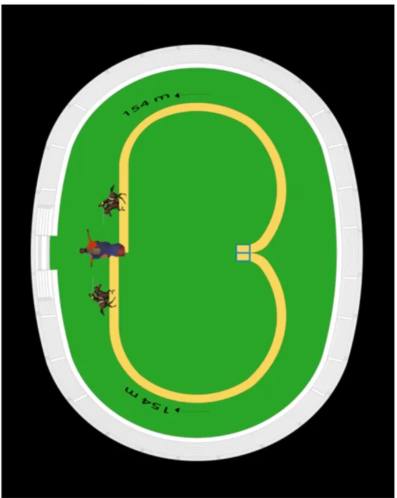
Niballo horse race is a manifestation within Faenza City’s medieval historical reenactment. This manifestation is composed of two races that are held on the second Saturday of June and on the fourth Sunday of June. This competition presumes that two challengers start galloping at the same time and aim to touch with a spare for first a computerized dummy. The racetrack has an oval elliptical shape, and challengers gallop in opposite directions following an arch of a circle trajectory that closes the track at the point where the dummy is placed, at the same distance from the start point for both of the participants (Fig 9). Each horse has to run over across a distance of approximately 154 meters in 12 seconds for a total of eight times in about 1 hour. The 5 horses attending the competition challenge each other following a race schedule where each participant challenges the other four. Each challenge has the duration of roughly 2 minutes and it takes up to 15 minutes for one horse to challenge the other four participants. The horses attending this race start their athletic preparation on February with a canter training session for two months. After two months the training program presumes race simulations.
Figure 9.(! http://www.paliodifaenza.it) The design of Faenza City’s Niballo horse racetrack. The starting point is indicated by the blue square in the figure, and the end of the racetrack is at the point where the dummy is placed in the figure
6.2.1 Environmental conditions
Environmental conditions were recorded during each day of the manifestations in order to verify their influence on the obtained results. These data were provided by ARPA Emilia-Romagna:
1. First year of study
First race: humidity 81%; temperature 19,4°C Second race: humidity 47%; temperature 29,1°C 2. Second year of study:
First race: humidity 82%; temperature 21°C Second race: humidity 38%; temperature 28,9°C 3. Third year of study:
First race: humidity 98%; temperature 17,6°C Second race: humidity 47%; temperature 27,3°C
6.3 Blood samples
6.3.1 Blood sampling technique
Blood samples were obtained by venipuncture from the jugular vein with a “closed system” using vials with negative pressure S-Monovette® SARSTEDT. If the sampling method was considered to be unsatisfactory, a second sample was withdrawn using the same system from the opposite jugular vein, in order to prevent interferences with the analysis of parameters. For each individual a first sample was taken 24 hours before the race and the second sample within 30 minutes by the end of the race. These samples were taken at the same time with anti doping samples. For
each sample were used: 1 vial containing K3EDTA anticoagulant, 1 vial
containing sodium citrate 1:9 anticoagulant, and 1 plain vial. All the vials were immediately refrigerated at +4ºC and processed within 3 hours after withdraw at SEPAC VET Laboratory of Department of Veterinary Medical Science of Alma Mater Studiorum - University of Bologna.
6.3.2 Sample processing
Blood samples with K3EDTA anticoagulant were collected using
S-Monovette® Sarstedt EDTA KE/2.6 mL vials and delivered refrigerated at +4ºC. At SEPAC VET Laboratory after vials were brought at room temperature a complete cell blood count (CBC) was performed using an automated hematology analyzer within 3 hours after withdraw.
Blood samples with sodium citrate anticoagulant were collected using S-Monovette® Sarstedt Coagulation 9 NC/5 mL vials (proportion blood and sodium citrate 1:9; sodium citrate solution 0.11 mol/L) and delivered refrigerated at +4ºC. At SEPAC VET Laboratory these vials were
centrifuged at 3000xG for 10 minutes. Plasma citrate supernatant was aspired with single use pipette avoiding the layer above the buffy coat, in order to obtain platelet poor plasma. The obtained plasma citrate was stored frozen at -20ºC until analysis. All these operations were concluded within 3 hours from withdraw.
Blood samples without anticoagulant were collected using S-Monovette® Sarstedt Serum Gel S/7.5 mL vials with cloth activator delivered refrigerated at +4ºC. At SEPAC VET Laboratory serum was obtained from centrifugation of vials at 3000xG for 10 minutes. Serum was divided in 2 aliquots and stored frozen at -20ºC until analysis. All
6.4 Parameters
The following hematological, chemistry and coagulation variables were evaluated in the present study: on K3EDTA samples a CBC
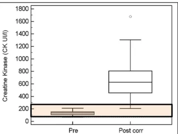
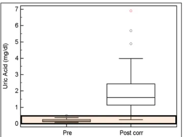
comprehensive of hematocrit percentage (Hct; %), mean cell hemoglobin concentration (MCHC; gr%); on serum samples a chemistry profile comprehensive of Creatinine (Crea; mg/dL), Urea (mg/dL), Uric Acid (UA; mg/dL), Albumin (Alb; g/dL), Total Protein (TP; g/dL), Calcium (Ca; mg/dL), Phosphate (P; mg/dL), Sodium (Na; mEq/L), Potassium (K; mEq/L), Chloride (Cl; mEq/L), Gamma-glutamyl Transferase (GGT; U/L), Bile Acids, Bilirubin (Bil; mg/dL), plasma muscular enzymes (PMEs) - Creatine Kinase (CK; U/L), Aspartate Aminotransferase (AST; U/L), Lactate Dehydrogenase (LDH; IU/L), Iron profile parameters - Total Iron (Fe++; µg/dL), Total Iron Binding Capacity (TIBC; µg/dL), TIBC Saturation (Sat; %), Acute Phase Proteins - Haptoglobin (HP; mg/dL), Ferritin (Ferr; mg/dL); on plasma citrate samples a coagulation profile comprehensive of Fibrinogen (Fib; g/L) and D-Dimers (D-D; µg/mL). All chemical and coagulation variables were assessed using previously validated methods and carried out on an automated analyzer Olympus AU 400.
6.4.1 Complete blood count
The CBC was performed using the Abbott Cell Dyn 3500 R automated hematology analyzer. Prior analysis, all samples were homogenized for 1 minute using Reamix 2789 Vortex instrument. For each sample a blood smear was made, which was used to evaluate platelet count microscopically.
6.4.2 Main methods used
6.4.2.1 Haptoglobin
Haptoglobin was assessed using an immunoturbidimetric method previously validated for horses in our laboratory (HAPTOGLOBIN OSR 6165, Olympus System Reagent).
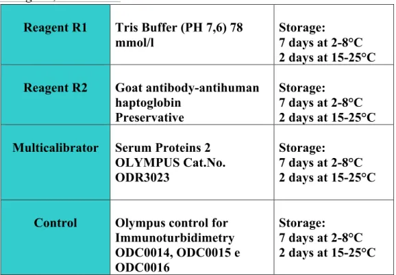
Reagents, Calibration:
Table 1. Reagents, Quality Control and Calibrators for the assessment of Haptoglobin. !!Method principle: when the sample is mixed with the buffer solution R1 and antiserum solution R2, haptoglobin will react specifically with anti-haptoglobin antibodies to yield insoluble aggregates. The absorbance of these aggregates is proportional to the haptoglobin concentration in the sample.
Linearity: the linearity of the method is comprised between 30-400 Reagent R1 Tris Buffer (PH 7,6) 78
mmol/l
Storage:
7 days at 2-8°C 2 days at 15-25°C Reagent R2 Goat antibody-antihuman
haptoglobin Preservative
Storage:
7 days at 2-8°C 2 days at 15-25°C Multicalibrator Serum Proteins 2
OLYMPUS Cat.No. ODR3023
Storage:
7 days at 2-8°C 2 days at 15-25°C Control Olympus control for
Immunoturbidimetry ODC0014, ODC0015 e ODC0016 Storage: 7 days at 2-8°C 2 days at 15-25°C
On-board stability: reagents (R1 and R2) are ready to use and can be directly placed on the instrument. Reagents are stable, if unopened and stored between +2 - +8ºC until expiration date. Reagents, once they are open and placed on the instrument, remain stable for 60 days.
Interferences:
Bilirubin: no significant interference until 40 mg/dL of added bilirubin
Hemolysis: added hemoglobin of 500 mg/dL lead to significant interference of 10%
Lipids: added lipid concentrations of 1000 mg/dL lead to significant interference of 20%