129 ACTA oTorhinolAryngologiCA iTAliCA 2015;35:129-131
129 Case seires and reports
Simultaneous nasopharyngeal and parotid gland
Warthin’s tumour: a case report
Un caso raro di tumore di Warthin sincrono della ghiandola parotide
e del rinofaringe
S. Pelucchi1, c. Bianchini1, a. ciorBa1, F. Stomeo1, a. Ferron2, a. PaStore1
1 otorhinolaryngology Department, and 2 Section of anatomy histology and Pathological cytology, university
hospital of Ferrara, italy SummAry
herein, a rare case of synchronous cystoadenolymphoma (Warthin’s tumour) of the right parotid gland and the nasopharyngeal space is described. Although Warthin’s tumour (WT) of the parotid gland is a common benign pathology, the occurrence of extra-parotid cystoad-enolymphoma is rare. Extra-parotid WT have been mainly localised in the submandibular gland, periparotid region and occasionally in other sites, such as the oral cavity, hard palate and nasopharynx. The simultaneous occurrence of an intra-parotid and extra-parotid WT localisation, as in the case presented, is extremely uncommon.
KEy WordS: Warthin’s tumour • Parotid Gland • Nasopharynx
riASSunTo
Obiettivo del presente lavoro è descrivere un raro caso di cistoadenolinfoma (tumore di Warthin) sincrono della ghiandola parotide destra e del rinofaringe. Il tumore di Warthin della ghiandola parotide è una neoplasia benigna relativamente comune; più raro è il suo riscontro in ambito extra-parotideo (per lo più, a livello della ghiandola sottomandibolare, del cavo orale o del rinofaringe). Molto più raro è poi il riscontro di due localizzazioni simultanee, una intra-parotidea ed una extra-parotidea, come nel caso qui descritto.
Parole chiave: Tumore di Warthin • Ghiandola Parotide • Rinofaringe
Acta Otorhinolaryngol Ital 2015;35:129-131
Introduction
Warthin’s tumour (WT) is a benign salivary gland tu-mour, almost exclusively located in the parotid gland
and periparotid region 1. From 12-19% of patients
devel-op more than one WT, frequently with a bilateral parotid
involvement (incidence: 5-14%) 1. WT has been rarely
reported to occur at multiple sites and at the same time; to the best of our knowledge, in the literature there is only another case report of a simultaneous intra-parotid
and extra-parotid WT 2.
Case report
A 63-year-old man was referred to the EnT department of the university hospital of Ferrara for the assessment of a right, non-painful, parotid mass. The lesion had been present for 4-6 months, but was increasing in size very slowly. he was also complaining the onset of snoring since 6 months with occasional nasal discharge.
Apart from a 40-year habit of smoking 20 cigarettes a
day and the onset of a non-insulin dependent diabetes mellitus two years ago, his medical history was unre-markable.
EnT examination revealed a mobile 3.5 × 2.0 cm mass just behind the angle of the jaw (right side). Facial nerve function was normal. nasopharyngeal endoscopy re-vealed an oval mass (1.0 cm) with a smooth surface in the left side of the nasopharynx. There were no other notable findings on physical examination.
Fine needle aspiration of the right parotid mass was per-formed. histological examination revealed mixed lym-phoid cells (lymphocytes and macrophages). The exact anatomical location of both lesions was confirmed by mri (Fig. 1A-B).
under general anaesthesia, the parotid mass was excised, via a superficial parotidectomy; at the same time, the na-sopharyngeal mass was removed under FESS guidance. At histological examination, a synchronous parotid and nasopharyngeal WT was diagnosed (Figs. 2, 3). There have been no signs of tumour recurrence at 16 months postoperatively.
S. Pelucchi et al.
130
Fig. 1. A. T1 weighted MRI scan, coronal section: a 3.5 × 2.0 cm mass is located in the superficial lobe of the right parotid gland. B. T1 weighted MRI scan, axial section: an oval mass of about 1.0 cm in diameter is located in the left side of the nasopharynx.
A
B
Fig. 2. Parotid lesion showing the classic histological pattern of Warthin’s tumour: bilayered oncoctic epithelium and lymphoid stromal tissue. Haema-toxylin-eosin, magnification 20×.
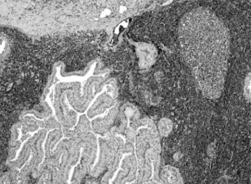
Fig. 3. The nasopharyngeal lesion shows the same histological pattern of Warthin’s tumour. A. Haematoxylin-eosin, magnification 10×. B. Haematoxy-lin-eosin, magnification 20×.
Discussion
Warthin’s tumour (adenolymphoma, papillary cystadeno-ma lymphocystadeno-matosum, cystoadenolymphocystadeno-ma) is one of the most common benign salivary gland tumours, generally
involving the parotid gland 1-3. Even if benign tumours of
the salivary glands are more common in women, WT, on the other hand, is found more frequently in men between the ages of 55 and 70 years. An association with cigarette
smoking has been described 3-5.
Although some cases have been reported in extra-parotid locations such as the cervical lymph nodes, submandibu-lar gland, lip, cheek, tongue and hard palate, WT of the
nasopharynx are extremely rare 2-10. only few isolated
cases of primitive nasopharyngeal involvement have been described so far, and we could find (through a PubMed database search performed in January 2011) only one published case of a nasopharyngeal WT with a
simulta-neous associated parotid tumour 2. A synchronous
intra-parotid and extra-intra-parotid WT, as in our case, can therefore be considered exceptional.
A
A case of simultaneous cystoadenolymphoma of nasopharynx and parotid
131
histologically, WT is an adenoma in which bilayered co-lumnar and basaloid oncocytic epithelium forms multiple cysts with multiple papillae and accompanied by a
prolif-eration of lymphoid tissue 1. Sometimes, oncocytic cells
can also form in nodal tissue. our case was judged to meet these criteria and the lesion was diagnosed as WT. it is difficult to explain the occurrence of a synchronous intra-parotid and extra-parotid WT. To date, there is no
evidence in either the previously reported case 2 or our
patient that one or more systemic or local factors might have influenced or induced the development of WT in two different sites: the hypothesis that we can make is of a multiple and simultaneous origin.
The origin of WT is still controversial and has been much debated. The most accepted hypothesis suggests that parotid WT could arise from salivary duct epithelium inclusions in the parotid gland lymph nodes, during
on-togeny 2 11. nonetheless, other authors propose that the
lymphocytic component is the result of an immunological reaction to the epithelial component, or could arise as an
inflammatory response 2 3.
At the same time, the pathogenesis of nasopharyngeal WT also remains unclear. By using monoclonal antibodies, Fantozzi et al. found the ratio of T to B cells in extrapa-rotideal WT to be similar to that of a normal lymph node, thus strengthening the theory of salivary organogenesis rather than that of reactive proliferation or
hypersensitivi-ty 12. Extraparotideal WT may then arise from components
of the minor salivary glands that are engaged in a pre-existing lymphoid stroma, and chronic inflammation in the nasopharynx could induce the formation of oncocytic
metaplasia of glandular tissues in the stroma 2 9.
The question also arises as to whether cigarette smoking, a chronic inflammatory stimulus, as well as a reported risk
factor for the onset of WT 3-5, could have acted as a trigger
for the simultaneous occurrence of WT in this case. in conclusion, WT is a predominantly benign lesion almost exclusively found in the parotid, which can exceptionally appear simultaneously in other areas, such as the naso-pharynx. head and neck surgeons should always be aware of extra parotideal WT and consider performing mri in highly suspicious cases during initial workup. Available
data 2 6 support a surgical, conservative, approach to the
management of WT, even in case of synchronous lesions, as in the patient presented.
it is likely that once the details of the pathogenesis of WT are better clarified, it will be possible to understand the occurrence of synchronous and/or multifocal lesions.
References
1 ellis Gl, auclair Pl. Tumors of the salivary glands. in: Atlas of tumor pathology. in: ellis Gl, auclair Pl. Tumors of the
sali-vary glands. Washington, Dc: american registry of Pathology and armed Forces institute of Pathology; 2008. pp. 259-68.
2 low WK, ng SB. Synchronous parotid and nasopharyngeal Warthin’s tumors: first report of a case. Ear nose Throat J 2002;81:839-41.
3 Teymoortash A, Krasnewicz y, Werner J. Clinical features of cystadenolymphoma (Wartins tumour) of the parotid gland: a retrospective comparative study of 96 cases. oral oncol 2006;42:569-73.
4 Klussmann JP, Wittekindt c, Preuss SF, et al. High risk for bilateral warthin tumour in heavy smokers-review of 185 cases. Acta otolaryngol 2006;126:1213-7.
5 de ru Ja, Plantinga rF, Majoor Mh, et al. Warthin’s tumour and smoking. B-EnT 2005;1:63-6.
6 hilton JM, Phillips JS, hellquist hB, et al. Multifocal multi-site Warthin tumour. Eur Arch otorhinolaryngol 2008;265:1573-5.
7 Kristensen S, Tveteras K, Friedmann i, et al. Nasopharyn-geal Warthin’s tumour: a metaplastic lesion. J laryngol otol 1989;103:616-9.
8 rydzewski B, Glowczewska M, Paprzycki W. Warth-in’s tumors with atypical location. otolaryngol Pol 1998;52:495-8.
9 yeh yA, Baker ll, Wang WJ, et al. Nasopharyngeal Warth-in’s tumor. otolaryngol head neck Surg 1999;120:942-4.
10 Griffiths aP, Dekker P. Oncocytic metaplasia of the na-sopharynx or extra-parotid Warthin’s tumour? J clin Pathol 1991;44:1030-2.
11 Kawakami m, ito K, Tanaka h, et al. Warthin’s tumor of the nasopharynx: a case report. Auris nasus larynx 2004;31:293-8.
12 Fantozzi rd, Bone rC, Fox r. Extraglandular Warthin’s tu-mors. laryngoscope 1985;95:682-8.
Address for correspondence: Chiara Bianchini, otorhinolaryn-gology department, university hospital of Ferrara, corso gio-vecca 203, 44100 Ferrara, italy. Tel. +39 0532 236383. Fax +39 0532 247709. E-mail: [email protected]
![[8] http: / / www.boatdesign.net](data:image/gif;base64,R0lGODlhAQABAIAAAP///wAAACH5BAEAAAAALAAAAAABAAEAAAICRAEAOw==)