R E S E A R C H
Open Access
Chitotriosidase: a biomarker of activity and
severity in patients with sarcoidosis
David Bennett
1*, Paolo Cameli
1,2, Nicola Lanzarone
1,2, Loredana Carobene
1,2, Nicola Bianchi
1, Annalisa Fui
1,2,
Luigi Rizzi
3, Laura Bergantini
1,2, Giuseppe Cillis
1, D
’ Alessandro Miriana
1,2, Maria Antonietta Mazzei
2,4,
Rosa Metella Refini
1,2, Piersante Sestini
1,2, Elena Bargagli
1,2and Paola Rottoli
2Abstract
Background: Serum chitotriosidase is a promising biomarker that has shown high specificity and sensitivity in patients with sarcoidosis. The aim of this study was to investigate correlations between serum chitotriosidase, clinical phenotypes, disease localizations and different radiological lung involvement and to identify clinical features associated with over-expression of chitotriosidase in a large cohort of sarcoidosis patients.
Methods: Chitotriosidase activity was evaluated in a population of 694 consecutive patients (males 39%, age 55.8 ± 12.8 years). Clinical and respiratory functional characteristics, Clinical Outcome Scale (COS) classification, clinical phenotypes proposed by the GenPhenResA project, and radiological assessment, including CT scan, were collected. Serum sampling and clinical and functional assessments at follow-up were also included.
Results: Significantly higher chitotriosidase activity was observed in sarcoidosis patients than in healthy controls (p < 0.0001). Evidence of lung fibrosis with reticular abnormalities and traction bronchiectasis at High resolution CT, presence of multiple extrapulmonary sarcoid localizations and increased 24-h urinary excretion of calcium were associated with significantly higher chitotriosidase activity (p < 0.005). Patients with remitted or minimal disease had lower values of chitotriosidase than patients with persistent disease. At follow-up, patients who required an increase in steroid dose showed an increase in its activity.
Conclusions: Chitotriosidase is a reliable biomarker of sarcoidosis. It is increased in patients with sarcoidosis correlating with disease activity, severity and multiorgan dissemination. Steroid therapy tended to reduce chitotriosidase expression, however it responded in cases of disease relapse.
Keywords: Chitotriosidase, Sarcoidosis, Biomarkers, Clinical evaluation Background
Sarcoidosis is a multi-organ granulomatous disease asso-ciated with abnormal T-lymphocyte and alveolar macro-phage activation and migration into affected organs [1]. Since clinical course ranges from spontaneous recovery to severe deterioration [2], specific biomarkers would be useful to predict clinical outcome and guide therapeutic management [3].
Chitotriosidase is a chitinase involved in defense against chitin-containing pathogens [4]. Recent studies showed that the immunomodulatory effects of chitotriosidase go beyond innate immunity to involve macrophage maturation and differentiation, non-chitin antigen presentation and direct stimulation of many cytokines, such as IL-8 and TGF-β [5–
9]. The enzyme has been found elevated in serum and
bronchoalveolar lavage (BAL) of patients with sarcoidosis than in patients with other interstitial lung diseases, pul-monary tuberculosis and healthy controls [10, 11]. In sar-coidosis patients, chitotriosidase showed higher sensitivity and specificity than other biomarkers, including angiotensin converting enzyme (ACE), lysozyme and soluble IL-2 receptor [11,12]. It has been found increased in active
sar-coidosis patients [11] and showed to predict clinical
© The Author(s). 2020 Open Access This article is distributed under the terms of the Creative Commons Attribution 4.0 International License (http://creativecommons.org/licenses/by/4.0/), which permits unrestricted use, distribution, and reproduction in any medium, provided you give appropriate credit to the original author(s) and the source, provide a link to the Creative Commons license, and indicate if changes were made. The Creative Commons Public Domain Dedication waiver (http://creativecommons.org/publicdomain/zero/1.0/) applies to the data made available in this article, unless otherwise stated.
* Correspondence:[email protected]
1Respiratory Diseases and Lung Transplantation Unit, Azienda Ospedaliera Universitaria Senese, Siena, Italy
course, steroid responsiveness and relapse of the dis-ease [13].
The aim of the present study was to determine serum chitotriosidase activity in a large cohort of sarcoidosis patients in order to test its utility for identifying clinical phenotypes, different radiological lung involvement, extrapulmonary organ involvement, and evaluating its reliability in patients on steroid therapy.
Materials and methods
Study population and study design Population
Between September 2015 and October 2017, serum chito-triosidase activity was assayed in 694 consecutive sarcoid-osis patients (males 39%, age 55.8 ± 12.8 years) monitored at the Regional Referral Centre for Sarcoidosis and other Interstitial Lung Diseases, Siena, Italy. No quantifiable chitotriosidase activity (< 10 nmol/ml/h) was detected in 25 patients (3.6%), these patients were suspected with CHIT1 polymorphism, but they were included into ana-lysis since no confirmatory genetic anaana-lysis was available. Healthy volunteers control group (n = 101, 34 males, mean age 52.2 ± 17.2 years; 73 non-smokers, 8 current smokers) without history of respiratory diseases and not receiv-ing any therapy was also enrolled.
Diagnosis and measurements
Sarcoidosis was diagnosed according to international ATS/ ERS/WASOG criteria [1]. All patients underwent complete physical evaluation and pulmonary function tests (PFT) in-cluding diffusing capacity of the lung for carbon monoxide (DLCO) and determination of serum concentrations of ACE and lysozyme. Combined physiological index (CPI) was cal-culated according to Walsh et al. [14] Extra-pulmonary sar-coid involvement (liver, spleen, chest and abdominal lymph nodes, skin, joints, heart, central nervous system) was assessed by specific diagnostic methods. Calcium metabol-ism was checked by 24-h urinary calcium excretion the same day as serum sampling.
Radiological evaluation
Chest x-ray alterations were classified according Scadding [15]. A high resolution CT (HRCT) scan of the chest was available for 228 patients. On the basis of the CT findings, patients were classified as having: 1) no lung or lymph node involvement; 2) parenchymal micronodules with or without lymph node enlargement; 3) parenchymal micro- and macro-nodules associated or otherwise with consolidation; 4) lung fibrosis, characterized by reticular abnormalities with or without traction bronchiectasis.
Patient stratification
According to therapy, patients were divided into two groups: steroid-free (n = 362) and steroid (n = 307). The
latter included patients on steroids or other specific medi-cations for sarcoidosis. Sarcoid patients were also classi-fied according to the clinical phenotypes proposed by GenPhenResA [16] and, those with a follow-up of at least 5 years, with the Clinical Outcome Scale (COS) [17].
Follow-up
Clinical assessment and chitotriosidase assay was re-peated at follow-up in a subgroup of patients (n = 416).
Patients were classified at follow-up as “improved”,
“stable” or “worse” according the physicians’ decisions on therapy (patients whose therapy was reduced or sus-pended were considered to have improved, patients whose therapy was maintained unchanged were consid-ered stable, and patients whose therapy was increased (steroid dose or new drug added) were considered to be worse). All data was collected retrospectively.
Chitotriosidase assay
Chitotriosidase activity was determined by a fluorimetric
method using 22 μM 4-methylumbelliferryl β
D-NNN-triacetylchitotriosidase (Sigma Chemical Co.) in citrate-phosphate buffer, pH 5.2; 100μl substrate was incubated for 1 h at 37°C and the reaction was stopped with 1.4 ml 0.1 M glycine-NaOH buffer, pH 10.8, as previously de-scribed [10,11]. Fluorescence was read at 450 nm with a Perkin Elmer LS40 fluorimeter (excitation wavelength 365 nm). Chitotriosidase activity in serum was expressed in nmol/ml/h.
Statistical analysis
Data was expressed as mean ± standard deviation (M ± SD). Comparisons between groups were performed by t-test and one-way ANOVA with significance set at p ≤ 0.05. The Pearson test was used for correlation analysis. Contingency tables were analysed by Fisher’s exact test and Chi-square test. Outcome analysis was performed using the Mantel Cox test. Statistical analysis and graphic representations of the data were obtained using GraphPad Prism Version 5.0 software for Windows, while ROC curves were plotted using SPSS Statistics 20.
Results
Clinical, radiological and functional parameters
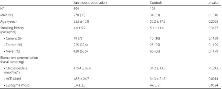
Demographic data, baseline pulmonary function test values and serum chitotriosidase, ACE and lysozyme ac-tivities of patients with sarcoidosis (n=694) and controls (n=101) are reported in Table 1. No significant differ-ences in sex, age and smoking status were found be-tween sarcoidosis patients and controls. On average, no significant impairment of PFT parameters, including
DLCO, was found in our cohort of patients. Table 2
radiological staging and extra-pulmonary localizations of our sarcoidosis cohort.
Chitotriosidase assay
Chitotriosidase activity was significantly higher in sarcoid-osis patients than in healthy controls (t=5.490, p<0.0001). It was significantly higher in both steroid-free and treated groups of patients than in healthy controls (180.1 ± 99.2 and 168.2 ± 118.2 nmol/ml/h vs. 34.2 ± 13.8 nmol/ml/h, t= 5.588 and t=5.492, respectively; p<0.0001 for both) (Fig.1). The same was true for ACE levels (49.3 ± 24.2 U/l and 48.5 ± 24.8 U/l vs. 34.3 ± 21.8 U/l, t=3.344, p=0.0004 and t= 2.883 p=0.004, respectively), whereas lysozyme levels were only significantly higher in steroid-free patients than healthy controls (5.8 ± 1.8 and 5.4 ± 2.4 mg/l vs. 4.6 ± 2.1;, t=1.998, p=0.046 and t=1.808 p=0.0721, respectively).
In sarcoidosis patients, chitotriosidase activity and ACE concentrations were directly correlated (r = 0.34, p<0.0001) (Fig.2). No significant differences were found in chitotriosidase activity according to sex, smoking sta-tus, age and comorbidities. Regarding specific symptoms, exertional dyspnea (t=2.714; p=0.0068) and cough were associated with higher chitotriosidase activity, however the latter was only statistically significant in steroid-free patients (t=2.287; p=0.0281).
Pulmonary function parameters did not significantly correlate with chitotriodase activity; however, stratifying patients according to specific patterns of alterations at PFTs (obstructive, restrictive or mixed respiratory de-fect), we observed that restrictive patients and those with reduced DLCO (TLC below the 5th percentile of the predicted value or DLCO < 75% of predicted) showed higher chitotriosidase activity (204 ± 305 vs 157 ± 242 nmol/ml/h, t=2.180, p=0.0296 and 310.8 ± 188 vs 162 ±
174 nmol/ml/h, t=2.682, p=0.0076, respectively).
Chitotriosidase showed a significant correlation with CPI index as well (r=0.09434, p=0.0199).
Second line therapy approach included the use of methotrexate in 42 patients as steroid-sparing agent (27 patients) or as steroid resistant second-line treatment (15 patients): no differences of chitrotriosidase expres-sion were found among these subgroups.
Chitotriosidase and radiological features
Chitotriosidase activity was higher in patients with chest X-ray stages 1, 2, 3 and 4 than in those with stage 0 (p< 0.0001).
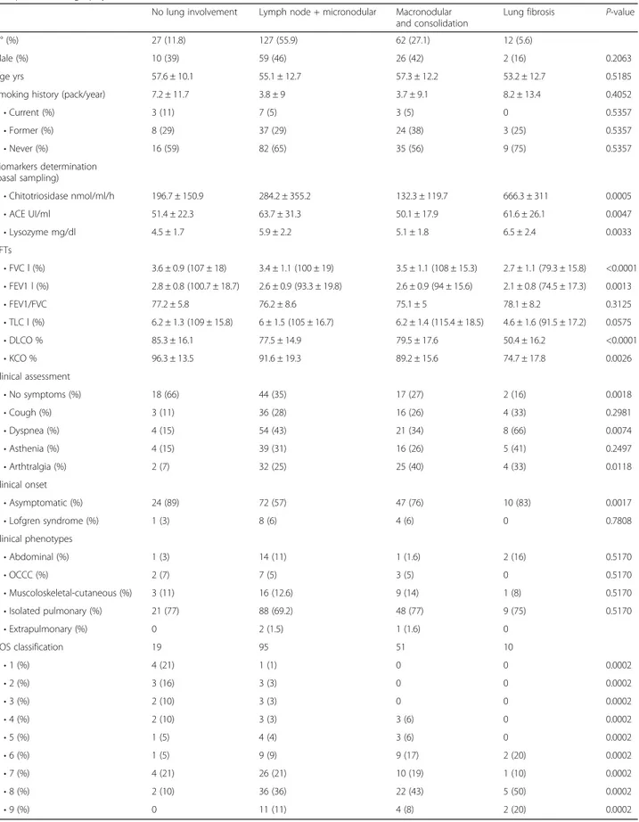
High resolution CT evidence of lung fibrosis with re-ticular abnormalities and traction bronchiectasis (group 4) (n=12) was associated with significantly higher chitotriosi-dase activity than in patients with no evidence of lung involvement (q=4.654; p<0.001) or with only parenchymal micronodules and lymph node enlargement (q=4.286; p< 0.01) or with parenchymal micro- and macro-nodules as-sociated with consolidation (q=5.828; p<0.0001). Patients with HRCT evidence of lung fibrosis showed significant impairment of FVC, FEV1, DLCO and KCO compared to other patients and more frequently reported dyspnea; whereas arthralgia was more common in group 3 (paren-chymal micro- and macro-nodules with consolidation) and group 4. Patients with lymph node enlargement and parenchymal micronodules (group 2) more frequently ex-perienced an acute or subacute clinical onset of the dis-ease (Table3).
Chitotriosidase activity in relation to extrapulmonary localizations and GenPhenResA phenotypes
The number of patients with at least one extrapulmonary localization of sarcoidosis was 267 out of 694 (38.4%). They showed significantly higher chitotriosidase activity than
Table 1 Demographic features, smoking status and biomarker assessment in the sarcoidosis cohort and healthy controls
Sarcoidosis population Controls p-value
N° 694 101 Male (%) 270 (39) 34 (33) 0.1310 Age (years) 55.8 ± 12.8 52.2 ± 17.2 0.2065 Smoking history (pack/year) 4.4 ± 9.7 5.1 ± 11.6 0.3457 • Current (%) 49 (7) 10 (10) 0.1199 • Former (%) 225 (32.4) 25 (25) 0.1199 • Never (%) 420 (60.5) 66 (66) 0.1199 Biomarkers determination (basal sampling) • Chitotriosidase nmol/ml/h 175.4 ± 89.4 34.2 ± 13.8 < 0.0001 • ACE UI/ml 48.5 ± 26.7 34.3 ± 21.8 0.0014 • Lysozyme mg/dl 5.4 ± 2.3 4.6 ± 2.1 0.0524
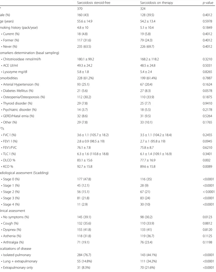
Table 2 Demographic data, smoking, comorbidities and functional, radiological and clinical parameters of the sarcoidosis cohort, divided into steroid-free and treated
Sarcoidosis steroid-free Sarcoidosis on therapy p-value
N° 370 324
Male (%) 160 (43) 128 (39.5) 0.4012
Age (years) 55.6 ± 14.9 54.2 ± 13.4 0.5978
Smoking history (pack/year) 4.8 ± 10 5.1 ± 10.4 0.1849
• Current (%) 18 (4.8) 19 (5.8) 0.4012
• Former (%) 117 (31.6) 79 (24.3) 0.4012
• Never (%) 235 (63.5) 226 (69.7) 0.4012
Biomarkers determination (basal sampling)
• Chitotriosidase nmol/ml/h 180.1 ± 99.2 168.2 ± 118.2 0.3210 • ACE UI/ml 49.3 ± 24.2 48.5 ± 24.8 0.5031 • Lysozyme mg/dl 5.8 ± 1.8 5.4 ± 2.4 0.8265 Comorbidities 228 (61.2%) 199 (61.4%) 0.7887 • Arterial Hypertension (%) 93 (25.1) 67 (20.4) 0.1624 • Diabetes Mellitus (%) 21 (5.6) 27 (8.3) 0.0578 • Osteopenia/Osteoporosis (%) 112 (30.2) 110 (33.9) 0.1875 • Thyroid disorder (%) 29 (7.8) 25 (7.7) 0.9410 • Psychiatric disorder (%) 14 (3.7) 18 (5.5) 0.2178 • GERD/Hiatal ernia (%) 32 (8.6) 31 (9.5) 0.5264 • Other (%) 29 (7.8) 33 (10.1) 0.1765 PFTs • FVC l (%) 3.6 ± 1.1 (105.7 ± 18.2) 3.5 ± 1.1 (104.2 ± 18.4) 0.2455 • FEV1 l (%) 2.8 ± 0.9 (98.5 ± 19) 2.7 ± 1 (95.8 ± 19) 0.0945 • FEV1/FVC 76.1 ± 7.8 75.8 ± 8.7 0.6210 • TLC l (%) 6.3 ± 1.6 (110.8 ± 18.8) 6.1 ± 1.4 (109.1 ± 16.9) 0.3401 • DLCO % 83.1 ± 15.6 77.7 ± 16.9 0.002 • KCO % 92.7 ± 15.8 89.6 ± 15.8 0.0089
Radiological assessment (Scadding)
• Stage 0 (%) 177 (47.8) 116 (35) <0.0001 • Stage 1 (%) 45 (12.1) 28 (9) <0.0001 • Stage 2 (%) 56 (15.1) 67 (21) < 0.0001 • Stage 3 (%) 81 (21.8) 83 (24) <0.0001 • Stage 4 (%) 11 (2.9) 30 (10) <0.0001 Clinical assessment • No symptoms (%) 145 (39.1) 98 (30.2) 0.0123 • Cough (%) 132 (35.6) 110 (33.9) 0.8812 • Dyspnea (%) 155 (41.8) 133 (41) 0.8120 • Asthenia (%) 118 (31.8) 119 (36.7) 0.1125 • Arthtralgia (%) 71 (19.1) 76 (23.4) 0.1198 Localizations of disease • Isolated pulmonary 284 (76.7) 143 (44.1%) <0.0001 • Lung + extrapulmonary 55 (14.8%) 111 (34.2%) <0.0001 • Extrapulmonary only 31 (8.3%) 70 (21.6%) <0.0001
patients with isolated pulmonary disease (t=5.257, p< 0.0001). Chitotriosidase activity tended to be progressively higher in patients with multiple organ involvement. In par-ticular, patients with three or more disease localizations re-ported significantly higher chitotriosidase activity than those with one or two localizations (q=10.79, p<0.0001 and q=5.05, p<0.001, respectively) (Fig.3). Extrapulmonary dis-ease was associated with significantly incrdis-eased chitotriosi-dase activity, regardless pulmonary involvement (F=10.51;
p<0.0001). Chitotriosidase in patients with lung
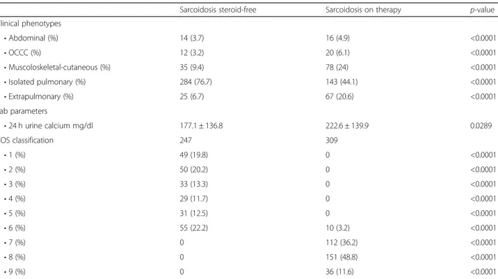
Table 2 Demographic data, smoking, comorbidities and functional, radiological and clinical parameters of the sarcoidosis cohort, divided into steroid-free and treated (Continued)
Sarcoidosis steroid-free Sarcoidosis on therapy p-value
Clinical phenotypes • Abdominal (%) 14 (3.7) 16 (4.9) <0.0001 • OCCC (%) 12 (3.2) 20 (6.1) <0.0001 • Muscoloskeletal-cutaneous (%) 35 (9.4) 78 (24) <0.0001 • Isolated pulmonary (%) 284 (76.7) 143 (44.1) <0.0001 • Extrapulmonary (%) 25 (6.7) 67 (20.6) <0.0001 Lab parameters • 24 h urine calcium mg/dl 177.1 ± 136.8 222.6 ± 139.9 0.0289 COS classification 247 309 • 1 (%) 49 (19.8) 0 <0.0001 • 2 (%) 50 (20.2) 0 <0.0001 • 3 (%) 33 (13.3) 0 <0.0001 • 4 (%) 29 (11.7) 0 <0.0001 • 5 (%) 31 (12.5) 0 <0.0001 • 6 (%) 55 (22.2) 10 (3.2) <0.0001 • 7 (%) 0 112 (36.2) <0.0001 • 8 (%) 0 151 (48.8) <0.0001 • 9 (%) 0 36 (11.6) <0.0001
PFT pulmonary function test, COS clinical outcome status and biomarker assessment in steroid-free and treated sarcoidosis patients, GERD gastro-esophageal reflux disease
Fig. 1 Comparison of chitotriosidase activity in steroid-free and treated sarcoidosis patients and healthy controls. Data expressed as mean ± standard deviation. ***: p < 0.0001. One-way ANOVA
Fig. 2 Correlation between chitotriosidase activity and ACE levels in the sarcoidosis cohort (n = 669) (R = 0.3513, p < 0.0001). Pearson’s correlation coefficient
Table 3 Demographic, clinical and functional data and biomarker assessment of sarcoidosis patients in relation to High resolution computed tomography (HRCT) evidence of involvement
No lung involvement Lymph node + micronodular Macronodular and consolidation
Lung fibrosis P-value
N° (%) 27 (11.8) 127 (55.9) 62 (27.1) 12 (5.6)
Male (%) 10 (39) 59 (46) 26 (42) 2 (16) 0.2063
Age yrs 57.6 ± 10.1 55.1 ± 12.7 57.3 ± 12.2 53.2 ± 12.7 0.5185
Smoking history (pack/year) 7.2 ± 11.7 3.8 ± 9 3.7 ± 9.1 8.2 ± 13.4 0.4052
• Current (%) 3 (11) 7 (5) 3 (5) 0 0.5357 • Former (%) 8 (29) 37 (29) 24 (38) 3 (25) 0.5357 • Never (%) 16 (59) 82 (65) 35 (56) 9 (75) 0.5357 Biomarkers determination (basal sampling) • Chitotriosidase nmol/ml/h 196.7 ± 150.9 284.2 ± 355.2 132.3 ± 119.7 666.3 ± 311 0.0005 • ACE UI/ml 51.4 ± 22.3 63.7 ± 31.3 50.1 ± 17.9 61.6 ± 26.1 0.0047 • Lysozyme mg/dl 4.5 ± 1.7 5.9 ± 2.2 5.1 ± 1.8 6.5 ± 2.4 0.0033 PFTs • FVC l (%) 3.6 ± 0.9 (107 ± 18) 3.4 ± 1.1 (100 ± 19) 3.5 ± 1.1 (108 ± 15.3) 2.7 ± 1.1 (79.3 ± 15.8) <0.0001 • FEV1 l (%) 2.8 ± 0.8 (100.7 ± 18.7) 2.6 ± 0.9 (93.3 ± 19.8) 2.6 ± 0.9 (94 ± 15.6) 2.1 ± 0.8 (74.5 ± 17.3) 0.0013 • FEV1/FVC 77.2 ± 5.8 76.2 ± 8.6 75.1 ± 5 78.1 ± 8.2 0.3125 • TLC l (%) 6.2 ± 1.3 (109 ± 15.8) 6 ± 1.5 (105 ± 16.7) 6.2 ± 1.4 (115.4 ± 18.5) 4.6 ± 1.6 (91.5 ± 17.2) 0.0575 • DLCO % 85.3 ± 16.1 77.5 ± 14.9 79.5 ± 17.6 50.4 ± 16.2 <0.0001 • KCO % 96.3 ± 13.5 91.6 ± 19.3 89.2 ± 15.6 74.7 ± 17.8 0.0026 Clinical assessment • No symptoms (%) 18 (66) 44 (35) 17 (27) 2 (16) 0.0018 • Cough (%) 3 (11) 36 (28) 16 (26) 4 (33) 0.2981 • Dyspnea (%) 4 (15) 54 (43) 21 (34) 8 (66) 0.0074 • Asthenia (%) 4 (15) 39 (31) 16 (26) 5 (41) 0.2497 • Arthtralgia (%) 2 (7) 32 (25) 25 (40) 4 (33) 0.0118 Clinical onset • Asymptomatic (%) 24 (89) 72 (57) 47 (76) 10 (83) 0.0017 • Lofgren syndrome (%) 1 (3) 8 (6) 4 (6) 0 0.7808 Clinical phenotypes • Abdominal (%) 1 (3) 14 (11) 1 (1.6) 2 (16) 0.5170 • OCCC (%) 2 (7) 7 (5) 3 (5) 0 0.5170 • Muscoloskeletal-cutaneous (%) 3 (11) 16 (12.6) 9 (14) 1 (8) 0.5170 • Isolated pulmonary (%) 21 (77) 88 (69.2) 48 (77) 9 (75) 0.5170 • Extrapulmonary (%) 0 2 (1.5) 1 (1.6) 0 COS classification 19 95 51 10 • 1 (%) 4 (21) 1 (1) 0 0 0.0002 • 2 (%) 3 (16) 3 (3) 0 0 0.0002 • 3 (%) 2 (10) 3 (3) 0 0 0.0002 • 4 (%) 2 (10) 3 (3) 3 (6) 0 0.0002 • 5 (%) 1 (5) 4 (4) 3 (6) 0 0.0002 • 6 (%) 1 (5) 9 (9) 9 (17) 2 (20) 0.0002 • 7 (%) 4 (21) 26 (21) 10 (19) 1 (10) 0.0002 • 8 (%) 2 (10) 36 (36) 22 (43) 5 (50) 0.0002 • 9 (%) 0 11 (11) 4 (8) 2 (20) 0.0002
involvement only was 144.1 ± 188.2 nmol/ml/h; in patients with lung and extrapulmonary was 239.4 ± 398.6 nmol/ml/ h; in patients with extrapulmonary only was 249.5 ± 393.1 nmol/ml/h.
A correlation between chitotriosidase and 24h-urinary cal-cium excretion was observed in both steroid-free and steroid subgroups (r=0.4201, p<0.0001; r=0.3528; p=0.0004).
According GenPhenResA phenotypes [16], patients
with abdominal organ involvement had higher chitotrio-sidase activity than other groups (q=8.155, p<0.0001 vs ocular-cardiac-cutaneous-central-nervous-system; q=11.32, p<0.0001 vs musculoskeletal-cutaneous; q=14.97, p<0.0001 vs isolated pulmonary disease; q=11.49, p<0.0001 vs extra-pulmonary disease), while ocular–cardiac–cutaneous–cen-tral-nervous-system (OCCC) localization showed higher chitotriosidase activity than isolated pulmonary involvement (q=4.494; p<0.05) (Fig.4).
Chitotriosidase activity in relation to COS classification
The number of patients that could be classified by COS [17] was 556: 99 were in remission (COS 1-2), 62 reported minimal disease (COS 3-4) and 395 showed persistent dis-ease (COS 5 to 9). Statistically significant differences in chitotriosidase activity were found between COS-1 and COS-5-6-8-9 (p<0.001), COS-2 and COS-5-9 (p<0.01), COS-4 and COS-5-6-8-9 (p<0.01, p<0.05, p<0.05 and p< 0.01, respectively) and COS-7 and COS-9 (p<0.05). Clinic-ally persistent disease was associated with significantly higher chitotriosidase activity than minimal or remitted disease (t=3.824, p=0.0001) (Fig.5).
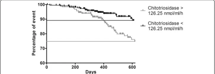
Patients with chitotriosidase activity exceeding 126.25 nmol/ml/h more frequently showed persistent disease as defined by COS classification (sensitivity 57%, specificity
72%) and more frequently needed a higher daily dose of steroid (48 vs 26 events, prevalence: 24.8% and 11%, re-spectively) (Fig.6).
Chitotriosidase activity at follow-up
Clinical assessment and chitotriosidase determination was available at follow-up in 416 patients (175 males,
Fig. 3 Comparison of Chitotriosidase activity in Sarcoidosis patients, classified on the basis of the number of extrapulmonary
localizations. Data expressed as mean ± standard deviation. **: p <
0.01; ***: p < 0.0001. One-way ANOVA Fig. 4 Comparison of chitotriosidase activity in differentGenPhenResA phenotypes. 1: abdominal; 2: ocular-cardiac-cutaneous-central nervous system; 3: muscoloskeletal-cutaneous; 4: pulmonary; 5: extrapulmonary. Data expressed as mean ± standard deviation. ***: p < 0.0001; *: p < 0.05. One-way ANOVA
Fig. 5 Comparison of chitotriosidase activity in relation to COS classification. Significant differences emerged between COS 1 and COS 5–6–8-9 patients (p < 0.0001 in all cases), COS 2 and COS 5–6-9 patients (p < 0.05, p < 0.05 and p < 0.0001, respectively), COS 4 and COS 5–6–8-9 patients (p < 0.001, p < 0.001, p < 0.05 and p < 0.0001, respectively), COS 5 and COS 7 patients (p < 0.05) and COS 7 and COS 9 patients (p < 0.001). Data expressed as mean ± standard deviation. One-way ANOVA
age 56 ± 13.8 years) (baseline to follow-up interval: 414 ± 265 days).
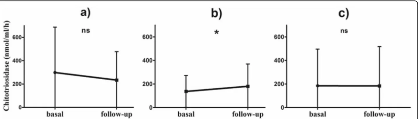
When physicians decided to change steroid dose or otherwise modify therapy, a significant increase in chito-triosidase activity was observed in "worse" patients (n= 215) (Table 4), whereas "improved" (n=47) and "stable" (n=164) patients did not show any significant change (Fig.7).
Discussion
The present study offers new insights into the clinical utility of chitotriosidase as a biomarker in patients with sarcoidosis. Sarcoidosis is a complex, pleiotropic disease with many phenotypes and many clinical classifications
have been proposed [18]. Numerous biomarkers have
been proposed over time in sarcoidosis, but none of these have been universally recognized in clinical prac-tice [19–21]. Ideal characteristics of candidate bio-markers should be highly specific and sensitive for the diagnosis, but more importantly, in sarcoidosis patients, from a prognostic point of view, to discriminate among different phenotypes and states of the diseases predicting remission or progression [22]. In the present study we aimed to test chitotriosidase utility for identifying clin-ical phenotypes, different radiologclin-ical lung involvement,
extrapulmonary organ involvement, and for evaluating its reliability in a large cohort of sarcoidosis patients with and without steroid therapy.
Chitotriosidase is a chitinase involved in defense against chitin-containing pathogens [4]. This enzyme, implicated in innate immunity, macrophage maturation and differen-tiation, has been found elevated in serum and bronchoal-veolar lavage (BAL) of patients with sarcoidosis and has been proposed some years ago as prognostic biomarker by our group of research [11]. It showed higher sensitivity and specificity than other biomarkers [23–26], including angiotensin converting enzyme (ACE), lysozyme and
soluble IL-2 receptor [11, 12] and it has been found
increased in active sarcoidosis patients [11] showing to predict clinical course, steroid responsiveness and relapse of the disease [13].
In the present large case series (n = 694), chitotriosi-dase activity proved to be a reliable biomarker of sar-coidosis; it was significantly increased in patients with sarcoidosis than in healthy controls and it correlated with respiratory symptoms (exertional dyspnea and cough) and with serum ACE concentration. It also proved to correlate with disease severity, expressed as severe progressive pulmonary and extrapulmonary in-volvement, to be able to detect disease relapse and to identify patients requiring escalation of therapy.
Chitotriosidase correlated with radiological findings in our cohort: patients with chest X-ray stages different from 0 showed significantly higher concentrations. In the present study, we firstly investigated correlations between chitotriosidase activity and different CT patterns of pres-entation. Severe pulmonary involvement, indicated by HRCT by evidence of lung fibrosis with reticular abnor-malities and traction bronchiectasis, was associated with a significant increase in chitotriosidase activity. In sarcoid-osis patients, fibrsarcoid-osis is a result of persistent and
Fig. 6 Log-rank test comparing outcome (increase in daily dose of steroid) in sarcoidosis patients with basal chitotriosidase above or below the cut-off of 126 nmol/ml/h. (Chi-square = 10.396; p = 0.001). Long rank test
Table 4 Changes in chitotriosidase activity between basal and follow-up sampling, in relation to subjective symptoms and therapy adjustments during the observation period
Δ Chitotriosidase (nmol/ml/h) p-value Therapy modification
• Improved (n = 47) −64.5 ± 118.1 0.2045
• Stable (n = 164) −1.3 ± 214.4 0.9389
uncontrolled disease [27] and chitotriosidase may reflect persistent granuloma activity. We also explored Chitotrio-sidase activity in relation the composite physiological index (CPI), proposed by the Brompton group of research, that showed to identify patients at high clinical risk correl-ating with radiological extent of fibrosis [14] and we found a slightly significant correlation with chitotriosidase activ-ity. In our cohort strength of such correlation is rather weak, even if significant (r = 0.09434, p = 0.0199); prob-ably it is underestimated by the overwhelming percentage of patients with normal PFTs. Further studies are needed to better understand values of CPI in combination with chitotriosidase in sarcoidosis, but indirectly suggests the association of chitotriosidase expression with fi-brosis development. Overexpression of chitotiosidase has been previously associated with abnormal produc-tion of TGF-beta, leading to initiaproduc-tion and perpetuaproduc-tion of interstitial lung disease in systemic sclerosis [9] and its ex-pression, and of other chitinases (i.e. YKL-40), has been associated to airway remodelling in severe asthma [28,
29]. Moreover, in BAL from patients with sarcoidosis,
chitotriosidase has been found increased in progressive than stable patients [11, 24]. These results suggest that this enzyme could be involved in fibrogenesis in diffuse lung diseases, expression of high activation of certain macrophage pathways responsible for remodeling and fi-brosis development.
In our study, population was divided according to Gen-PhenReSa phenotypes classification [16], that have been proposed to make homogenous cohorts in biomedical stud-ies, in five subgroups: 1) abdominal organ involvement; 2) ocular–cardiac–cutaneous–central; nervous system disease involvement; 3) musculoskeletal–cutaneous involvement; 4) pulmonary and intrathoracic lymph node involvement; 5)
extrapulmonary involvement [15]. We did observe that
patients with multiorgan sarcoid involvement (39% of our population), particularly with abdominal involvement, had higher chitotriosidase activity: patients with three or more extrapulmonary localizations showed higher chitotriosidase.
In 2016, Popevic et al. failed to demonstrate higher chito-triosidase levels in patients with extrapulmonary sarcoidosis detected by PET-CT scan [23]. In the present study, the definition of extrapulmonary involvement was based on specific clinical, radiological and pathological investigations, including PET-CT scan in some cases. This approach, probably more complete and sensitive than PET-CT scan alone, might explain our findings. Patients with active and/ or persistent disease showed increased activity of chitotrio-sidase. Definition of sarcoidosis activity is difficult and there is still no universal consensus. In the present study, we used the COS classification scale proposed some years ago by WASOG in which patients are classified in relation to need for therapy, disease persistence, relapse of symptoms and modification of therapy at follow-up [17]. It has been previ-ously reported by us and others [10,23] that chitotriosidase activity is expressed differently among COS stages. Even if we could not apply COS to all our population, but only to patients with 5 or more years of follow-up at time of chito-triosidase determination (n = 553), in the present study we confirmed this association: patients in remission or with minimal disease (COS stages 1 to 4) had lower chitotriosi-dase activities than patients with persistent disease (COS stages 5 to 9) and, among the latter, patients whose condi-tions deteriorated showed higher activities than stable patients.
Chitotriosidase expression showed to increase in pa-tients with multiple organ involvement suggesting that its level correspond to the amount of active organ in-volvement reflecting the number of active granulomas harbored by a patient at a given time. In a previous paper from our group [24], we reported a significant dis-crepancy between serum and BAL expression of
chito-triosidase suggesting the potential extrapulmonary
production of this biomarker, however in that paper population was not such well characterized as in this present work. Anyway, extrapulmonary production of chitotriosidase is supported by other studies describing
Fig. 7 Line plots of Chitotriosidase activity at basal and follow-up sampling: a therapy modification, improved; b therapy modification, worsened; c therapy modification, stable. *: 0.0012. Paired t test
chitotriosidase as a reliable marker of macrophage acti-vation in different organs and apparatus [30–32].
Chitotriosidase is mainly secreted by macrophages and is involved in maturation of monocytes into both M1 and M2 macrophages subtypes, even without the pres-ence of chitin. How chitotriosidase is implicated in pathogenic mechanisms of sarcoidosis is unknown. The pathogenesis of sarcoidosis is complex and not yet com-pletely understood; environmental, genetic and immuno-logical factors demonstrated several interactions that lead to macrophage activation and granuloma formation. The innate immune system has also been shown to play a role in the triggering and maintenance of
granuloma-tous inflammatory phenomena [33]. However, the events
preventing dissolution of granulomas and maintaining inflammation, evolving toward fibrosis are not yet fully characterized [33] and specific studies to better under-stand chitotriosidase role in sarcoidosis mechanisms are lacking.
The chitotriosidase activity cut-off of 48.8 nmol/h/ml was proposed by our research group to distinguish sar-coid patients from healthy subjects with high sensitivity and specificity (89 and 93%, respectively) [10]. Chitotiro-sidase has been found overexpressed in sarcoidosis than patients with idiopathic pulmonary fibrosis, interstitial lung disease associated to systemic sclerosis and tuber-colosis [24,25], however increased levels have been also reported in COPD, asbestosis and lung cancer patients [26]. Diagnostic accuracy of chitotriosidase for sarcoid-osis is still to be addressed, prospective studies evaluat-ing its levels in patients with differential diagnosis with sarcoidosis are strongly needed.
In the present study, we identified a second cut-off to predict disease persistence among sarcoidosis: patients with chitotriosidase activity higher than 126 nmol/h/ml proved to have active disease requiring an increase in therapy. These observations confirm the prognostic value of this biomarker, which can identify patients most likely to have a persistent disease and therefore requiring particular attention. In fact, chitotriosidase analysis at follow-up showed that patients requiring an increase in steroid dosage showed higher chitotriosidase activity. In line with this observation, Harlander et al. reported a significant increase in chitotriosidase activity during re-lapse of sarcoidosis [13]. Unfortunately, due to retro-spective nature of this study, we could not explore chitotriosidase in relation with quality of life as stan-dardized specific questionnaires were not available for the majority of patients included in the study.
The main limitations of the present study are its retro-spective and monocentric nature and the absence of a validation cohort. Moreover, the low number of patients with Löfgren syndrome prevented us to explore chitotrio-sidase in this particular subgroup of patients. However,
this is the biggest study ever conducted on chitotriosidase in sarcoidosis and the results clearly show its potential utility as a clinical biomarker correlating with many clin-ical and radiologclin-ical parameters. Prospective studies to evaluate its role in clinical decision-making are needed to definitely establish its role in sarcoidosis patients.
Conclusion
The present study demonstrated that chitotriosidase activity is a reliable biomarker of sarcoidosis. Our results showed that it is correlated with disease activity, severity and multiorgan dissemination. Taken together, our find-ings support an intriguing hypothesis that chitotriosidase
production, mainly by sarcoid macrophages [34], could
reflects the number of active granulomas harbored by a patient at a given time. In favor of this speculation, we observed chitotriosidase activity differently expressed in patients with severe pulmonary involvement (presence of lung fibrosis at CT scan), with multi vs. single organ involvement (in particularly in patients with abdominal involvement and with three or more extrapulmonary lo-calizations), with active vs. non-active disease and with remitted/minimal vs. persistent disease and in patients at high clinical risk (expressed by CPI index). Moreover, chitotriosidase expression reduces with steroid therapy and it proved able to detect disease relapse and to iden-tify patients requiring escalation of therapy. Together with these positive clinical findings, chitotriosidase la-boratory determination has been shown feasible, quick and accurate with reasonable costs [35].
Combined with clinical, radiological and physiological findings, chitotriosidase activity proved to be an excellent non-invasive prognostic biomarker for management of pa-tient with sarcoidosis, with a cost-benefit ratio highly posi-tive, that should be part of the regular follow-up of sarcoidosis patients in the daily clinical practice.
Abbreviations
ACE:Angiotensin converting enzyme; BAL: Bronchoalveolar lavage; COS: Clinical Outcome Scale; FEV1: Forced expiratory volume in 1 s; FVC: Forced vital capacity; HRCT: High resolution computed tomography; KCO: Diffusing capacity of the lung for carbon monoxide/alveolar volume; PFT: Pulmonary function tests; DLCO: Diffusing capacity of the lung for carbon monoxide
Acknowledgements Not applicable Authors’ contributions
DB: study concept and design; analysis and interpretation of data; drafting of the manuscript; critical revision of the manuscript for important intellectual content; statistical analysis; study supervision. PC: acquisition of data; analysis and interpretation of data; statistical analysis; critical revision of the manuscript for important intellectual content. NL, LC, AF, LR: acquisition of data. NB: analysis and interpretation of data; laboratory analysis; statistical analysis. LB and GC: laboratory analysis. MAM: radiological interpretation; analysis and interpretation of data. RMR: acquisition of data; analysis and interpretation of data. EB: analysis and interpretation of data, critical revision of the manuscript for important intellectual content. PR: critical revision of
the manuscript for important intellectual content; study supervision. All authors read and approved the final manuscript.
Funding
This study did not receive any specific grant from funding agencies in the public, commercial or non-profit sectors.
Availability of data and materials
databases and all relevant raw data can freely available on request. Ethics approval and consent to participate
The research was approved by the local ethical committee (Azienda Ospedaliera Universitaria Senese, protocol OSS_REOS n° 12908). All subjects gave their written informed consent to the study.
Consent for publication Not applicable Competing interests
The authors declare that they have no competing interests. Author details
1Respiratory Diseases and Lung Transplantation Unit, Azienda Ospedaliera Universitaria Senese, Siena, Italy.2Department of Medical and Surgical Sciences & Neurosciences, University of Siena, Siena, Italy.3Internal Medicine Unit“C. Frugoni”, Centre for Rare Diseases, University Hospital of Bari, Bari, Italy.4Diagnostic Imaging Unit, Azienda Ospedaliera Universitaria Senese, Siena, Italy.
Received: 5 June 2019 Accepted: 18 December 2019 References
1. Hunninghake GW, Costabel U, Ando M, Baughman R, Cordier JF, du Bois R, Eklund A, Kitaichi M, Lynch J, Rizzato G, Rose C, Selroos O, Semenzato G, Sharma OP. ATS/ERS/WASOG statement on sarcoidosis. American Thoracic Society/European Respiratory Society/world Association of Sarcoidosis and other granulomatous disorders. Sarcoidosis Vasc Diffuse Lung Dis. 1999; 16(2):149–73.
2. Spagnolo P, Rossi G, Trisolini R, Sverzellati N, Baughman RP, Wells AU. Pulmonary sarcoidosis. Lancet Respir Med. 2018;6(5):389–402.
3. Carleo A, Bennett D, Rottoli P. Biomarkers in sarcoidosis: the contribution of system biology. Curr Opin Pulm Med. 2016;22(5):509–14.
4. Funkhouser JD, Aronson NN Jr. Chitinase family GH18: evolutionary insights from the genomic history of a diverse protein family. BMC Evol Biol. 2007;7:96.
5. Di Rosa M, Tibullo D, Cambria D, Distefano G, Saccone S, Di Raimondo F, Malaguarnera L. Chitotriosidase expression during monocyte-derived dendritic cells differentiation and maturation. Inflammation. 2015;38(6):2082–91.
6. Di Rosa M, Malaguarnera G, De Gregorio C, Drago F, Malaguarnera L. Evaluation of CHI3L-1 and CHIT-1 expression in differentiated and polarized macrophages. Inflammation. 2013;36(2):482–92.
7. van Eijk M, van Roomen CP, Renkema GH, Bussink AP, Andrews L, Blommaart EF, Sugar A, Verhoeven AJ, Boot RG, Aerts JM. Characterization of human phagocyte-derived chitotriosidase, a component of innate immunity. Int Immunol. 2005;17(11):1505–12.
8. Di Rosa M, Malaguarnera G, De Gregorio C, D'Amico F, Mazzarino MC, Malaguarnera L. Modulation of chitotriosidase during macrophage differentiation. Cell Biochem Biophys. 2013;66(2):239–47.
9. Lee CG, Herzog EL, Ahangari F, Zhou Y, Gulati M, Lee CM, Peng X, Feghali-Bostwick C, Jimenez SA, Varga J, Elias JA. Chitinase 1 is a biomarker for and therapeutic target in scleroderma-associated interstitial lung disease that augments TGF-β1 signaling. J Immunol. 2012;189(5):2635–44.
10. Bargagli E, Bennett D, Maggiorelli C, Di Sipio P, Margollicci M, Bianchi N, Rottoli P. Human chitotriosidase: a sensitive biomarker of sarcoidosis. J Clin Immunol. 2013;33(1):264–70.
11. Bargagli E, Margollicci M, Perrone A, Luddi A, Perari MG, Bianchi N, Refini RM, Grosso S, Volterrani L, Rottoli P. Chitotriosidase analysis in
bronchoalveolar lavage of patients with sarcoidosis. Sarcoidosis Vasc Diffuse Lung Dis. 2007;24(1):59–64.
12. Bargagli E, Maggiorelli C, Rottoli P. Human chitotriosidase: a potential new marker of sarcoidosis severity. Respiration. 2008;76(2):234–8.
13. Harlander M, Salobir B, Zupančič M, Dolenšek M, Bavčar Vodovnik T, Terčelj M. Serial chitotriosidase measurements in sarcoidosis--two to five year follow-up study. Espir Med. 2014;108(5):775–82.
14. Walsh SL, Wells AU, Sverzellati N, Keir GJ, Calandriello L, Antoniou KM, Copley SJ, Devaraj A, Maher TM, Renzoni E, Nicholson AG, Hansell DM. An integrated clinicoradiological staging system for pulmonary sarcoidosis: a case-cohort study. Lancet Respir Med. 2014;2(2):123–30.
15. Scadding JG. Prognosis of intrathoracic sarcoidosis in England. A review of 136 cases after five years' observation. Br Med J. 1961; 2(5261):1165–72.
16. Schupp JC, Freitag-Wolf S, Bargagli E, Mihailović-Vučinić V, Rottoli P, Grubanovic A, Müller A, Jochens A, Tittmann L, Schnerch J, Olivieri C, Fischer A, Jovanovic D, Filipovic S, Videnovic-Ivanovic J, Bresser P, Jonkers R, O'Reilly K, Ho LP, Gaede KI, Zabel P, Dubaniewicz A, Marshall B, Kieszko R, Milanowski J, Günther A, Weihrich A, Petrek M, Kolek V, Keane MP, O'Beirne S, Donnelly S, Haraldsdottir SO, Jorundsdottir KB, Costabel U, Bonella F, Wallaert B, Grah C, Peroš-Golubičić T, Luisetti M, Kadija Z, Pabst S, Grohé C, Strausz J, Vašáková M, Sterclova M, Millar A, Homolka J, Slováková A, Kendrick Y, Crawshaw A, Wuyts W, Spencer L, Pfeifer M, Valeyre D, Poletti V, Wirtz H, Prasse A, Schreiber S, Krawczak M, Müller-Quernheim J. Phenotypes of organ involvement in sarcoidosis. Eur Respir J. 2018;51(1).
17. Baughman RP, Nagai S, Balter M, Costabel U, Drent M, du Bois R, Grutters JC, Judson MA, Lambiri I, Lower EE, Muller-Quernheim J, Prasse A, Rizzato G, Rottoli P, Spagnolo P, Teirstein A. Defining the clinical outcome status (COS) in sarcoidosis: results of WASOG task force. Sarcoidosis Vasc Diffuse Lung Dis. 2011;28(1):56–64.
18. Bargagli E, Prasse A. Sarcoidosis: a review for the internist. Intern Emerg Med. 2018;13(3):325–31.
19. Ungprasert P, Carmona EM, Crowson CS, Matteson EL. Diagnostic utility of angiotensin-converting enzyme in Sarcoidosis: a population-based study. Lung. 2016;194(1):91–5.
20. Uysal P, Durmus S, Sozer V, Gelisgen R, Seyhan EC, Erdenen F, Simsek G, Uzun H. YKL-40, Soluble IL-2 Receptor, Angiotensin Converting Enzyme C-Reactive Protein: Comparison of Markers of Sarcoidosis Activity. Biomolecules. 2018;8(3).
21. Ramos-Casals M, Retamozo S, Sisó-Almirall A, Pérez-Alvarez R, Pallarés L, Brito-Zerón P. Clinically-useful serum biomarkers for diagnosis and prognosis of sarcoidosis. Expert Rev Clin Immunol. 2019;15(4):391–405. 22. Drucker E, Krapfenbauer K. Pitfalls and limitations in translation from
biomarker discovery to clinical utility in predictive and personalised medicine. EPMA J. 2013;4(1):7.
23. Popević S, Šumarac Z, Jovanović D, Babić D, Stjepanović M, Jovičić S, Šobić-Šaranović D, Filipović S, Gvozdenović B, Omčikus M, Milovanović A, Videnović-Ivanov J, Radović A, Žugić V, Mihailović-Vučinić V. Verifying Sarcoidosis activity: Chitotriosidase versus ACE in Sarcoidosis - a case-control study. J Med Biochem. 2016;35(4):390–400.
24. Bargagli E, Margollicci M, Luddi A, Nikiforakis N, Perari MG, Grosso S, Perrone A, Rottoli P. Chitotriosidase activity in patients with interstitial lung diseases. Respir Med. 2007;101(10):2176–81.
25. Bargagli E, Margollicci M, Nikiforakis N, Luddi A, Perrone A, Grosso S, Rottoli P. Chitotriosidase activity in the serum of patients with sarcoidosis and pulmonary tuberculosis. Respiration. 2007;74(5):548–52.
26. Tercelj M, Salobir B, Simcic S, Wraber B, Zupancic M, Rylander R. Chitotriosidase activity in sarcoidosis and some other pulmonary diseases. Scand J Clin Lab Invest. 2009;69(5):575–8.
27. Bonham CA, Strek ME, Patterson KC. From granuloma to fibrosis: sarcoidosis associated pulmonary fibrosis. Curr Opin Pulm Med. 2016;22(5):484–91. 28. James AJ, Reinius LE, Verhoek M, Gomes A, Kupczyk M, Hammar U, Ono J,
Ohta S, Izuhara K, Bel E, Kere J, Söderhäll C, Dahlén B, Boot RG, Dahlén SE. BIOAIR (longitudinal assessment of clinical course and biomarkers in severe chronic airway disease) consortium. Increased YKL-40 and Chitotriosidase in asthma and chronic obstructive pulmonary disease. Am J Respir Crit Care Med. 2016;193(2):131–42.
29. Lai T, Wu D, Chen M, Cao C, Jing Z, Huang L, Lv Y, Zhao X, Lv Q, Wang Y, Li D, Wu B, Shen H. YKL-40 expression in chronic obstructive pulmonary disease: relation to acute exacerbations and airway remodeling. Respir Res. 2016;17:31.
30. Musumeci M, Palermo A, D'Onofrio L, Vadalà G, Greto V, Di Stasio E, Maddaloni E, Di Rosa M, Tibullo D, Angeletti S, Napoli N, Denaro V, Manfrini
S. Serum chitotriosidase in postmenopausal women with severe osteoporosis. Osteoporos Int. 2016;27(2):711–6.
31. Malaguarnera L, Di Rosa M, Zambito AM, dell’Ombra N, Nicoletti F, Malaguarnera M. Chitotriosidase gene expression in Kupffer cells from patients with non-alcoholic fatty liver disease. Gut. 2006;55(9):1313–20. 32. Malaguarnera L, Di Rosa M, Zambito AM, dell’Ombra N, Di Marco R,
Malaguarnera M. Potential role of chitotriosidase gene in nonalcoholic fatty liver disease evolution. Am J Gastroenterol. 2006;101(9):2060–9 Epub 2006 Jul 18. Erratum in: Am J Gastroenterol. 2012 Dec;107(12):1932.
33. Bennett D, Bargagli E, Refini RM, Rottoli P. New concepts in the pathogenesis of sarcoidosis. Expert Rev Respir Med. 2019;13(10):981–91. 34. Boot RG, Hollak CE, Verhoek M, Alberts C, Jonkers RE, Aerts JM. Plasma
chitotriosidase and CCL18 as surrogate markers for granulomatous macrophages in sarcoidosis. Clin Chim Acta. 2010;411(1–2):31–6.
35. Amick AK, Liu Q, Gautam S, Chupp G, Dela Cruz CS, Sharma L. Measurement of Chitinase Activity in Biological Samples. J Vis Exp. 2019;(150).
Publisher’s Note
Springer Nature remains neutral with regard to jurisdictional claims in published maps and institutional affiliations.