Study on microbial
communities in domestic
kitchen sponges: Evidence
of Cronobacter sakazakii
and Extended Spectrum Beta
Lactamase (ESBL) producing
bacteria
Stefania Maria Marotta, Filippo Giarratana, Anastasia Calvagna, Graziella Ziino, Alessandro Giuffrida, Antonio Panebianco
Department of Veterinary Sciences, University of Messina, Italy
Abstract
Domestic environment, in particular, kitchen setting is a well-established source of microbial contamination. Kitchen sponges represent an important vehicle of microbial transmission and maintenance of spoilage bacteria and pathogenic strains responsible for food borne diseases. The aim of this study was to evaluate the micro-bial communities of 100 ‘in-use’ kitchen sponges, improving the knowledge on their role in cross-contamination in domestic environment and transmission of ESBL-producing strains. Sponges were processed for: aerobic mesophilic bacteria (AMB), Enterobacteriaceae (EB), yeasts and molds (YM), coagulase-positive staphylococci (CPS), micrococci (MCC), anaerobic sulfite reducing bacteria (ASR), and for the detec-tion of Listeria monocytogenes, Salmonella spp. and Yersinia enterocolitica. A total of 309 enterobacteria strains were identified and then processed for ESBL (Extended Spectrum Beta Lactamase) phenotypical expression. A high contamination level of kitchen sponges was observed (mean value AMB 8.25±1.1; EB 5.89±1.2; YM 5.57±1.1; MCC 4.82±0.1 log CFU/g). Identified enterobacteria strains revealed several opportunistic and pathogenic agents such as Enterobacter cloacae (28%), Citrobacter freundii (23.3%), Cronobacter sakazakii (14.6%) and other strains in lower percentage. Listeria monocytogenes was found in only one sponge (1%). A total of 69 (22.3%) enterobacteria resulted ESBL+, with the following prevalence: P. rettgeri (50%), L. adenocarboxilata (30%), K. pneumoniae (25%), K. oxytoca (25%), C. sakazakii (20%), E. cloacae (20.7%), C. freundii (20.1%). Results confirm the
potential role of kitchen sponges as vehicle for food-borne pathogens such as, C. sakazakii for the first time, infectious agents and spoilage microorganisms. The observed high contamination level and the presence of several ESBLs opportunistic pathogens, stresses the necessity to improve a proper education of the consumers on the effective treatment to reduce their microbial loads.
Introduction
In the last few years, numerous studies indicate that several food-borne diseases are related to domestic infection sources (EFSA, 2015). Improper food handling and un-hygienic practices are considered the major factors in foodborne illness episodes. Cross-contamination in household kitchens represent another important domestic source of infection (Azevedo et al., 2014). However, consumers risk perception of food borne illness in home environment is reported as very low (European Commission, 2016). Kitchens are included among the most contaminated domestic environments, even more of some bath’s areas (toilette seat, door knob, light switch, toilet handle) or commonly used objects such as pen, keys, cellular phone, key-boards, etc. (Donofrio et al., 2012). Furthermore, 37.3% of food borne out-breaks in EU in 2014, founded their infec-tion sources in home environments (EFSA, 2015). This evidence is confirmed by the frequent isolation of food-borne pathogens in tools, cloths, towels, sponges and kitchen surfaces (Mattick et al., 2003). Among these, dish sponges were the most contami-nated item in the household, and deserve great attention, for their potential role as vehicle for foodborne pathogens (Donofrio et al., 2012). Their role as microbiological hot spots in domestic settings was recently well established (Cardinale et al., 2017). They are, indeed, frequently used not only to clean dishes and cookware, but also, dif-ferent surfaces or even refrigerators shelves, increasing the risk of cross-contamination (Catellani et al., 2014). Thanks to their large surface/volume ratio, their constant humid-ity and the nutrients for bacterial growth contained, sponges are, indeed, an ideal habitat for microorganisms (Cardinale et al., 2017). Several authors have investigat-ed the microbiological quality of sponges used in domestic kitchens, reporting a high-level contamination and the frequent isola-tion of pathogens, such as Salmonella spp., Staphylococcus aureus, Campylobacter spp. and Listeria monocytogenes (Hilton
and Austin, 2000).
These findings induce much more con-cern, considering that common dishwashing soaps or chemical compounds do not reduce significantly microbial load in kitchen sponges (Sharma et al., 2009). Moreover, multi-drug resistance bacteria were com-mon in household’s environment, and no significant differences were noted between biocide users and non-users; as well as the frequency of pathogen recovery (Marshall et al., 2012). Among this, ESBLs (Extended Spectrum Beta Lactamase) are a group of evolving enzymes that are able to hydrolyze extended spectrum cephalosporin. ESBL producing enterobacteria are a wide range of resistant strains that have gained much more importance in public health in the recent years. Infections due to ESBL pro-ducers range from uncomplicated urinary tract infections to life-threatening sepsis representing a serious challenge for clinical treatments. It is well established the role of animals and derived products as a source of diffusion of resistant strains (Beninati et al., 2015; EFSA, 2011) For all these reasons, the aim of the present study was to improve the knowledge on the microorganisms fre-quently involved “in-use” kitchen sponges’ colonization, and their role in maintaining and diffuse ESBL-producing organisms in domestic environment.
Correspondence: Graziella Ziino, Department of Veterinary Sciences, University of Messina, Polo Universitario della Annunziata, 98168 Messina, Italy.
Tel.: +39.090.3503761. E-mail: [email protected]
Key words: kitchen sponges; Cronobacter sakazakii; enterobacteria; microbiology; ESBL producing bacteria.
Contributions: the authors contributed equally. Conflict of interest: the authors declare no potential conflict of interest.
Funding: none
Received for publication: 4 July 2018. Revision received: 12 November 2018. Accepted for publication: 12 November 2018. This work is licensed under a Creative Commons Attribution-NonCommercial 4.0 International License (CC BY-NC 4.0).
©Copyright S.M. Marotta et al., 2018 Licensee PAGEPress, Italy
Italian Journal of Food Safety 2018; 7:7672 doi:10.4081/ijfs.2018.7672
Materials and Methods
Samples collection and macroscopic
evaluation
A total number of 100 sponges from domestic kitchens was collected within 24 hours from the last use and transported to our laboratory in sterile sampling bags. All processed sponges were characterized by a rough thin layer (scrub pad) covering a spongy synthetic material and by the fol-lowing dimension 120 ± 20 mm (h) × 80 ± 10 mm (w) × 20 ± 10 mm (d). Samples were also macroscopically evaluated by 3 mem-bers of the staff of Laboratory of Animal Origin (Department of Veterinary Sciences, Messina, Italy) and classified by an increas-ing scorincreas-ing from 1 to 3 for the followincreas-ing parameters: i) consumption (1: like new; 2: normal consumption; 3: excessive con-sumption); ii) dirt (1: like new; 2: slight color change; 3: intensive blackening) and iii) presence of food debris/extraneous par-ticles (1: from 0 to 2; 2: from 3 to 5; 3: more than 5). For each sample, the days of actual domestic use were recorded.
Microbiological analysis
For quantitative determinations, a total amount of 3±0.5 g from each sponge was collected and transferred to a stomacher bag and buffered peptone water (Biolife, Milan, Italy) was added with a ratio of 1:9 (w/v). The suspension was then homogenized for 60 s at 230 rpm in a peristaltic homogenizer (Stomacher 400 Circulator, Seward, UK) and 1:10 serial dilutions of the homogenate in BPW were prepared. Each sample was then processed for the following determina-tions: i) aerobic mesophilic bacteria count (AMB) according to UNI EN ISO 4833-1: 2013; ii) Enterobacteriaceae count (EB) according to UNI EN ISO 21528-2: 2017; iii) yeasts and molds count (YM) according to UNI EN ISO 21527-2: 2008; iv) Coagulase-positive staphylococci (CPS) and Micrococci (MCC) count according to UNI EN ISO 6888-1:2004; v) Salmonella spp. detection according to UNI EN ISO 6579-1: 2017; vi) Listeria monocytogenes detection according to UNI EN ISO 11290-1: 2017; vii) detection of presumptive path-ogenic Yersinia enterocolitica according to UNI EN ISO 10273:2003; viii) anaerobic sulfite reducing bacteria count (ASR) on sulfite-polymyxin-sulfadiazine (SPS) agar (Biolife, Milano, Italy) incubated under anaerobic conditions at 37°C for 24h.
Enterobacteria strains identification
A total of 309 confirmed enterobacteria colonies, collected from the required five colonies from the highest dilution for each sample according to ISO UNI EN ISO
21528-2: 2017, were sub cultured on Tryptic Soy Agar (TSA) plates with 5% sheep blood (Biolife, Milano, Italy), then incubated at 37°C for 24h. Isolated colonies were then identified by Matrix-assisted laser desorption ionization–time of flight mass spectrometry (MALDI-TOF MS). Single cells were selected and directly transferred as a thin film on the 48-well sample plate and overlaid with 1 µL of matrix solution (saturated solution of alfa-cyano-4-hydroxycinnamic acid in 50% ace-tonitrile, and 2.5% tri-fluoracetic acid). E. coli ATCC 8739 was used as standard and loaded in specific control wells. After the crystallization of the matrix and microbial material, the metal plate was introduced in the mass spectrometer Vitek MS, (bioMérieux, Firenze, Italy) and was bom-barded with brief laser pulses. MALDI-TOF generates unique MS signatures (spec-tra) for microorganisms, that were trans-ferred into the AgnosTec-SARA-MIS soft-ware (Spectral Archive and Microbial Identification System) (bioMérieux, Firenze, Italy) were they were compared to the database containing the reference spec-tra of common bacteria.
ESBL phenotypic expression
Screening test
To evaluate the presence of presumptive ESBL producing bacteria, all 309 strains were tested with a chromogenic media, con-taining a mixture of antimicrobic and chro-mogenic substances that allow the growth of ESBL producing strains with a specific color (ChromaticTM ESBL, Liofilchem, France). Considering that only few species (E. coli, Klebsiella spp., Enterobacter spp., Serratia spp., Proteus spp.) have been checked by the producer for the specific coloration expressed we confirmed for ESBL also the strains that showed a not col-ored growth. Each sample was spread on the surface of a dried media plate and incu-bated for 24h at 37°C.
Confirmation test: combination disk assay
To confirm phenotypic expression of ESBL producing strains, the combination disk assay was carried out. As proposed by Luzzaro et al. (2007), the combination
assay consists in testing a β-lattamic alone and in the presence of an inhibitor of the β-lactamase, in order to evaluate the recovery activity of the antibiotic in the presence of the inhibitor. At this purpose, each sample that resulted positive for the screening test, was sub cultured in Brain Heart Infusion Broth (BHIB, Biolife, Italy) and incubated at 37°C for 24h. The overnight cultures were spread on the surface of Mueller-Hinton agar plates (MH, Biolife, Italy). Then antibiotic disk (6 mm Ø) of Cefotaxime (CTX) 30 µg, Cefotaxime + Clavulanic acid (CTL) 40 µg, Ceftazidime (CAZ) 30 µg and Ceftazidime + Clavulanic acid (CAL) 40 µg (Liofilchem, France) were placed on the inoculated medium. After incubation at 37°C for 24 h, the inhi-bition diameters were calculated. For each tested antibiotic, the ESBL production was considered positive if the inhibitory diame-ter was ≥5 mm, compared to the β-lactam tested alone according to CLSI method (2016).
Data analysis
Least squares linear and multiple regression analysis were performed to esti-mate the influence of microbiological parameters loads on AMB count and coeffi-cients of determination (R2and adjusted R2)
were calculated to estimate the strength of the relationship between our models and the observed data. Spearman’s rank correlation coefficient (rs)was estimated to verify rela-tionships between macroscopic scoring and microbiological parameters. F-test was per-formed in order to estimate significative associations. Significance level was assumed as P<0.05. Microbial loads were converted in log CFU/g to facilitate the expression of results.
Results
Macroscopic evaluation scoring
Results are showed in Table 1. Considering consumption level, 40.0% of samples revealed the maximum score 3 conditions, showing in some cases the lack of the superficial scrub pad and/or the spongy tissue. For dirt scoring, 34.0% of Table 1. Sample distribution on macroscopic observations scoring.
Parameter Score, % 1 2 3
Consumption 17.0 43.0 40.0 Dirt 14.0 52.0 34.0 Food debris/Extraneous materials 55.0 30.0 15.0
Consumption: (1) like new; (2) normal consumption; (3) excessive consumption. Dirt: (1) like new; (2) slight color change; (3) intensive blackening. Food debris/extraneous particles: (1) from 0 to 2 units; (2) from 3 to 5 units; (3) more than 5 units.
processed sponges achieved the higher score 3. Those samples resulted, indeed, very soiled and/or with an intensive black-ening of the spongy material. Some samples presented an overall low level of dirt/con-sumption but a consistent presence or for-eign particles, which were minimally repre-sented by papers fragments or organic mate-rial and largely by human or pets’ hairs. Spearman’s Rank correlation coefficient rs=0.77 attests a strong and significative (P<0.05) relationships between consump-tion and dirt. Nevertheless, only 15.0% of samples showed more than five extraneous particles and/or food debris. No significa-tive relationship (P>0.05) was reported between this parameter and the previous macroscopic observation. Considering the period of actual utilization, 30% of sponges were used from 5 to 15 days, another 30% from 15 to 30 days and the last 40% for a period greater than 30 days. It is interesting to highlight that no significative relation-ships (P>0.05) were observed between the period of utilization and the other macro-scopic observations.
Microbiological results
Table 2 summarized microbiological results. The 15.0% of samples presented AMB loads ranging from 5.0 to <7.0 log CFU/g, while the remaining 85.0% reported values from 7.0 to <10.0 log CFU/g (Figure 1). The poor hygienic condition was also assessed by the detection of EB in all the samples. EB count showed a wide distribu-tion range (Figure 1) from 3.0 to <6.0 log CFU/gin 50% of samples and the remaining 50% from 6.0 to <9.0 log CFU/g. MCC and YM were isolated in all the samples with loads ranging from 2.0 to <8.0 log CFU/g and from 3.0 to <8.0 log CFU/g respective-ly (Figure 1). CPS and ASR bacteria were occasionally isolated in 11% samples with
mean value of 3.29±0.4 and 1.68±0.8 CFU/g respectively. Multiple regression analysis revealed a significative (P<0.05) influence of EB, MCC and YM on AMB loads. Linear regression test revealed that each of these parameters was significative (P<0.05) related to AMB. No significative (P>0.05) association was observed among CPS vs AMB and ASR vs AMB.
The association between AMB and EB, MCC and YM have been expressed in sta-tistical models by fitting a linear equation to observed data, in which AMB and the other parameters were considered as dependent and independent variable respectively. According to our statistical models, EB have a considerable influence on AMB (45%) (Figure 2A). MCC loads influenced Table 2. Microbiological loads and prevalence in kitchen sponges.
Parameter log CFU/g Prevalence,%
Aerobic mesophilic bacteria count 8.25±1.1 100
Enterobacteriaceae count 5.89±1.2 100
Micrococci 4.82±1.1 100
Yeasts and Molds count 5.57±1.2 100
Coagulase-positive staphylococci 3.29±0.4 11.0 Anaerobic sulfite reducing bacteria count 1.68±0.8 11.0 Listeria monocytogenes + 1.0 Salmonella spp. - 0
Yersinia enterocolitica - 0
+ detected; - not detected.
Figure 1. Distribution of Aerobic Mesophilic Bacteria (AMB), Enterobacteriaceae (EB), Micrococci (MCC), Yeast and Mold (YM) loads in 100 kitchen sponges.
Figure 2. Linear regression scatter plots, trend lines and R2 values, relating: A) Enterobacteriaceae count (EB) to Aerobic Mesophilic Bacteria count (AMB); B) Micrococci count (MCC) to Aerobic Mesophilic Bacteria count (AMB); C) Yeast and Mold count (YM) to Aerobic Mesophilic Bacteria count (AMB).
AMB variance for 44.0% (Figure 2B), while YM gave a less contribution (28.0%) (Figure 2C), but were constantly detected and can be considered as characterizing microbiota within the already mentioned parameters. No significative (P>0.05) rela-tionships were detected between microbio-logical loads (AMB and EB) and macro-scopic scoring including the period of usage.
Listeria monocytogenes was detected only in one sample characterized by: AMB 9.7 log CFU/g; EB 7.1 log CFU/g; MCC 5.5 log CFU/g; YM 6.7 log CFU/g; ASR 3.6 log CFU/g. Moreover, the sample with Listeria monocytogenes was classified as score 3 for dirt and 2 both for consumption and pres-ence of particles. No Salmonella spp. and Yersinia enterocolitica were found.
Enterobacteria identification
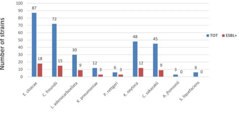
The identification of enterobacteria, which, as reported, are the predominant flora in kitchen sponges, showed a limited variability of strains identified. On the 309 strains, in fact, 87 were identified as Enterobacter cloacae (28.2%), 72 as Citrobacter freundii (23.3%), 48 as Klebsiella oxytoca (15.5%), 45 as Cronobacter sakazakii (14.6%), 30 as Leclercia adecarboxylata (9.7%), 12 as Klebsiella pneumoniae (3.9%), 6 as Providencia rettgeri (1.9%), 6 as Serratia liquefaciens (1.9%) and finally 3 as Acinetobacter johsonii (1.0 %) (Figure 3).
ESBL phenotypical expression
A total of 69 (22.3%) among 309 tested strains resulted positive for ESBL pheno-typical test. The chromogenic media employed revealed a high rate (78%) of false-positive ESBL strains, probably due to an over-production of cephalosporinases or chromosomal penicillases, by some Enterobacteriaceae. Moreover, strains belonging to genus Citrobacter, Leclercia, Acinetobacter and Providencia showed growth with variable colorations (green, blue, pink) for the same strain, whether or not they were confirmed as ESBL produc-ers. The confirmation test reported the fol-lowing prevalence of resistance: P. rettgeri (50%); L. adenocarboxilata (30%); K. pneumoniae (25%); K. oxytoca (25%); E. cloacae (20.7%); C. freundii (20.1%); C. sakazakii (20%) (Figure 3). No ESBLs were reported among S. liquefaciens and A. johsonii.
Discussion
The macroscopic results reported in our
sponges confirm the low consumers risk perception of food borne illness related to cross-contamination in home environment (European Commission, 2006). The pres-ence of foreign particles such as human or pets’ hairs, in fact, underlines the promiscu-ous use of sponges in real-life domestic conditions, like the cleaning of pet’s bowls or other tools and surfaces non-related to kitchen environment. The microbiological data of this study confirms a great level of sponge contamination and poor hygienic conditions, as observed by other authors (Azevedo et al., 2014; Mattick et al., 2003; Cardinale et al., 2017). The role of kitchen cleaning tools in bacterial transfer to sur-faces was, already, confirmed by Hilton and Austin (2000) on cloths contamination. In that study, no significant differences between the microbial load of wet cloths, dry cloths, those used for short and pro-longed periods and cloths used for different activities were observed. Our results con-firm, as just previously reported, that in kitchen sponges’ bacterial colonization and subsequent replication occur rapidly and high loads were maintained all over their period of use (Cardinale et al., 2017).
The detection of Listeria monocyto-genes, also if only in one sample, confirms
the potential role of kitchen sponges as food-borne pathogen vehicle, considering its ability in adhesion and surviving on dif-ferent surfaces (Kilonzo-Nthenge et al., 2012; Mattick et al., 2003). Among food-borne pathogen bacteria deserve particular attention the presence in 16 sponges (16%) of Cronobacter sakazakii (previously Enterobacter sakazakii). This occurrence confirms, as reported by Kilonzo-Nthenge et al. (2012), that it can colonize several kitchen districts. To date and based on the recently paper of Cardinale et al. (2017) on the microbiome analysis on used kitchen
sponges this is the first evidence of Cronobacter sakazakii presences among the bacteria involved in kitchen sponges’ colo-nization. These bacteria, frequently isolated from environment and humans and animals’ intestinal tract, can cause necrotizing ente-rocolitis, bacteremia, and meningitis in chil-dren and infants, with a 40-80% mortality rate. It can also cause diarrhea and urinary tract infections in people of all ages, espe-cially in YOPI subjects (young, old, preg-nant, immunosuppressed) (Healy et al., 2010). C. sakazakii has been found in sever-al foods, but mostly powdered infant formu-la has been linked to disease outbreaks (Adekunte et al., 2010). Its presence in this food is related to contamination in raw ingredients, during the manufacturing process or during the preparation/reconsti-tution process. Contamination also may occur through blenders, feeding bottles and utensils used to cleanse feeding bottles (Adekunte et al., 2010). This occurrence deserves particular attention considering that kitchen sponges are commonly used for baby tools cleaning (baby bottles, children’s cutlery, bowls ecc.). This study confirms the high stress, environmental resistance of C. sakazakii and also reports the diffusion of ESBL+ strains in household settings (Abdel-Galil et al., 2015). In regards of other Enterobacteriaceae identified in our kitchen sponges, the higher prevalence reported for Enterobacter cloacae, is proba-bly related to its high diffusion and resist-ance. Moreover, these bacteria, with Citrobacter freundii, Klebsiella oxytoca and Klebsiella pneumoniae have been reported as important opportunistic and multiresis-tant bacterial pathogens for humans during the last three decades in hospital wards (Mezzatesta et al., 2012; Munoz-Price et al., 2013; Whalen et al., 2007). L. adecar-boxylata, often found in water
environ-Figure 3. Prevalence of ESBL (Extended Spectrum Beta Lactamase) strains among 309 identified Enterobacteriaceae.
ments, is reported as occasionally pathogen-ic agent, even in healthy subjects (Keren et al., 2014). The pathogenic potential of Providencia rettgeri is well known, as well as the diffusion of extended spectrum beta lactamase (ESBL) and metallo-beta lacta-mase (NDM-1) strains (Tada et al., 2014). Finally, S. liquefaciens is widely isolated in nature, including river water, mineral water, domestic sewage, fish, minced meat and pasteurized milk or cream (Muscolino et al., 2014; Ziino et al., 2010). In humans, it has been, rarely, reported as a cause of nosocomial infections, including urinary tract infection, pneumonia, neonatal menin-gitis, endocarditis and septicemia (Mossad, 2000). As reported, in our study the highest prevalence of ESBLs was assigned to P. rettgeri and L. adenocarboxilata whose beta-lactamase resistance was already reported for nosocomial infections (Sheng et al., 2013). Resistance patterns for Klebsiella genus, are well-known and sev-eral strains represent the most prevalent clinical isolates in complicated infections (Barrios et al., 2017). The situation becomes even more worrying, considering that Klebsiella is one of the most wide-spread opportunistic pathogens in nosoco-mial infections with fatal prognosis (Tuon et al., 2011). Klebsiella ESBL-producing strains, often, carry resistance determinants against fluoroquinolones, cotrimoxazole and aminoglycosides. These microorgan-isms are rapidly evolving in response to the selective pressure created by antibiotics abuse, resulting in a serious clinical and epi-demiological problem (Tuon et al., 2011). E. cloacae and C. freundii ESBL strains are more and more frequently implied in multi-ple resistance patterns against insidious infections (Lagha et al., 2016).
The results of this study (Figures 1 and 2) obtained with traditional microbiology methodology on a relevant number of sam-ples (n. 100) reveled difference on Enterobacteriaceae abundance respect to the those of Cardinale et al. (2017) on the total of 223,741 raw sequences obtained by 454-pyrosequencing of 16S rRNA gene amplicon libraries from 28 sponge samples. In our samples Enterobacteriaceae have a considerable influence (45%) on the AMB, while for Cardinale et al. (2017) have a cumulative relative abundance of 1.18% only after Bacteroidaceae, Bdellovibrionaceae, Brevibacteriaceae,
Caulobaceriaceae, and
Comamobacteriaceae family. Except for Escherichia, were in accordance the report-ed genera Enterobacter, Citrobacter and Leclercia with our results among those reported in the Enterobacteriaceae family (Cardinale et al., 2017).
Conclusions
In conclusion, the high contamination level observed in our samples must be assumed as unacceptable for a kitchen tool, considering the potential role of sponges in cross contamination events. Sponges create a setting for colonization and subsequent microbial replication thanks to favorable factors such as high-level humidity, pres-ence of organic residuals and promiscuous use. Moreover, the presence of ESBL-pro-ducing strains in kitchen sponges confirms how the domestic settings are potential transmission pathways, explaining the spread of ESBL Enterobacteriaceae from the food chain to humans (Tschudin-Sutter et al., 2014).
Microorganisms, in fact, in kitchen cloths and sponges are protected by soil aggregates, surviving thermal or chemical stress of washing (Park et al., 2006). For all these reasons in order to reduce microbial loads, it would be necessary change, fre-quently, kitchen sponges and make, period-ically, efficient sanification treatments. Microwave and dishwasher, reaching high temperatures, represent the most effective treatments to reduce, significantly, micro-bial loads (Park et al., 2006; Sharma et al., 2009). High temperature in combination with washing is more effective in reducing bacteria in kitchen sponges than using heat alone (Tate, 2006). As reported by Erdoğrul and Erbilir (2005), the regular dish washing liquid was not effective in reduction of bac-teria in the house hold in use sponges, as the presence of food residues strongly reduces the product’s efficacy, while in the laborato-ry tests, the regular dish washing liquid was demonstrated to be effective in reduction of bacteria. The necessity of efficient sanifica-tion treatments is supported by the detecsanifica-tion of pathogenic and ESBL producing strains, confirming kitchen sponges as vehicle of their transmission and permanence in domestic settings.
References
Abdel-Galil FY, Abdel-Latif HK, Ammar AM, Serry FM, 2015. Studies on preva-lence, antimicrobial resistance and sur-vival of Cronobacter sakazakii. Zagazig J Pharm Sci 23:95-106.
Adekunte A, Valdramidis VP, Tiwari BK, Slone N, Cullen PJ, Donnell CPO, Scannell A, 2010. Resistance of Cronobacter sakazakii in reconstituted powdered infant formula during ultra-sound at controlled temperatures: a quantitative approach on microbial
responses. Intl J Food Microbiol 142:53-9.
Azevedo I, Albano H, Silva J, Teixeira P, 2014. Food safety in the domestic envi-ronment. Food Control 37:272-76. Barrios H, Garza-Ramos U, Mejia-Miranda
I, Reyna-Flores F, Sánchez-Pérez A, Mosqueda-García D, Silva-Sanchez J, 2017. ESBL-producing Escherichia coli and Klebsiella pneumoniae: The most prevalent clinical isolates obtained between 2005 and 2012 in Mexico. J Globl Antimicrob Resist 10:243-6. Beninati C, Reich F, Muscolino D,
Giarratana F, Panebianco A, Klein G, Atanassova V, 2015. ESBL and MRSA isolated from poultry and turkey prod-ucts imported from Italy. Czech J Food Sci 33:97-102.
Cardinale M, Kaiser D, Lueders T, Schnell S, Egert M, 2017. Microbiome analysis and confocal microscopy of used kitchen sponges reveal massive colo-nization by Acinetobacter, Moraxella and Chryseobacterium species. Sci Rep 7:5791.
Catellani P, Scapin RM, Alberghini L, Radu IL, Giaccone V, 2014. Levels of micro-bial contamination of domestic refriger-ators in Italy. Food Control 42:257-62. CLSI, 2016. Clinical and Laboratory
Standards Institute. Performance Standards for Antimicrobial Susceptibility Testing: Twenty-first Informational Supplement M100-S26, 2016. Wayne, PA, USACLSI.
Donofrio R, Bechanko R, Hitt N, O’Malley K, Charnauski T, Bestervelt LL, Saha R, Saha N, 2012. Are we aware of micro-bial hotspots in our household? J Environ Health 75:12-9.
EFSA, 2011. Scientific Opinion on the pub-lic health risks of bacterial strains pro-ducing extended-spectrum β-lactamases and/or AmpC β-lactamases in food and food-producing animals. EFSA Journal 9:2322.
EFSA, 2015. The European Union summa-ry report on trends and sources of zoonoses, zoonotic agents and food-borne outbreaks in 2014. EFSA Journal 13:4329.
Erdoğrul Ö, Erbilir F. 2005.Microorganisms in kitchen sponges. Int J Food Safety 6:17-22.
European Commission. 2006. “Risk issues 2006 Special Eurobarometer”. Available from: http://ec.europa.eu/ commfrontoffice/publicopinion/archive s/ebs/ebs_238_en.pdf
Healy B, Cooney S, O’Brien S, Iversen C, Whyte P, Nally J, Callanana JJ, Fanning S, 2010. Cronobacter (Enterobacter sakazakii): an opportunistic foodborne
pathogen. Foodborne Pathog Dis 7:339-50.
Hilton AC, Austin E, 2000. The kitchen dishcloth as a source of and vehicle for foodborne pathogens in a domestic set-ting. Int J Environ Health Res 7:18-22. ISO 10273:2003. Microbiology of food and animal feeding stuffs. Horizontal method for the detection of presumptive pathogenic Yersinia enterocolitica. International Standardization Organization Geneva, Switzerland. ISO 11290-1:1996. Microbiology of food
and animal feeding stuffs. Horizontal method for the detection and enumera-tion of Listeria monocytogenes. Part 1: Detection method. International Standardization Organization Geneva, Switzerland.
ISO 21527-1:2008. Microbiology of food and animal feeding stuffs. Horizontal method for the enumeration of yeasts and moulds. Part 1: Colony count tech-nique in products with water activity greater than 0,95. International Standardization Organization Geneva, Switzerland.
ISO 21528-2:2017. Microbiology of the food chain - Horizontal methods for the detection and enumeration of Enterobacteriaceae Part 2: Colony-count method. International Standardization Organization Geneva, Switzerland.
ISO 4833-1:2013. Microbiology of the food chain .Horizontal method for the enu-meration of microorganisms. Part 1: Colony count at 30 degrees C by the pour plate technique. International Standardization Organization Geneva, Switzerland.
ISO 6579-1:2017. Microbiology of the food chain - Horizontal method for the detec-tion, enumeration and serotyping of Salmonella - Part 1: Detection of Salmonella spp. International Standardization Organization Geneva, Switzerland.
ISO 6888-1:2004. Microbiology of food and animal feeding stuffs. Horizontal method for the enumeration of coagu-lase-positive staphylococci (Staphylococcus aureus and other species). Part 1: Technique using Baird-Parker agar medium. International Standardization Organization Geneva, Switzerland.
Keren Y, Keshet D, Eidelman M, Geffen Y, Raz-Pasteur A, Hussein K, 2014. Is Leclercia adecarboxylata a New and Unfamiliar Marine Pathogen? J Clin Microbiol 52:1775-6.
Kilonzo-Nthenge A, Rotich E, Godwin S, Nahashon S, Chen F, 2012. Prevalence and antimicrobial resistance of Cronobacter sakazakii isolated from domestic kitchens in middle Tennessee, United States. J Food Protect 75:1512-7.
Lagha N, Hassaine H, Robin FE, Bonnet R, Abdelouahid DE, 2016. Prevalence and molecular typing of extended-spec-trum-lactamases in Escherichia coli, Enterobacter cloacae and Citrobacter freundii isolates from Laghouat Hospital, Algeria. Afr J Microbiol Res 10:1430-8.
Luzzaro F, Gesu G, Pagani L, Rossolini, GM, 2007. Diagnostica delle ß-lattama-si a spettro esteso (ESBL) nelle Enterobacteriaceae: problemi e racco-mandazioni nella realtà epidemiologica italiana. Microbiola Med 22:281-90. Marshall BM, Robleto E, Dumont T, Levy
SB, 2012. The frequency of antibiotic-resistant bacteria in homes differing in their use of surface antibacterial agents. Curr Microbiol 65:407-15.
Mattick K, Durhman K, Dominigue G, Jorgensen F, Sen M, Schaffner DW, Humphrey T, 2003. The survival of foodborne pathogens during domestic washing up and subsequent transfer onto washing-up sponges, kitchen sur-faces, and food. Int J Food Microbiol 85:213-26.
Mezzatesta ML, Gona F, Stefani S, 2012. Enterobacter cloacae complex: clinical impact and emerging antibiotic resist-ance. Future Microbiol 7:887-902. Mossad SB, 2000. The world’s first case of
Serratia liquefaciens intravascular catheter-related suppurative throm-bophlebitis and native valve endocardi-tis. Clin Microbiol Infect 6:559-60. Munoz-Price LS, Poirel L, Bonomo RA,
Schwaber MJ, Daikos GL, Cormican M, Cornaglia G, Garau J, Gniadkowski M, Hayden MK, Kumarasamy K, Livermore DM, Maya JJ, Nordmann P, Patel JB, Paterson DL, Pitout J, Villegas MV, Wang H, Woodford N, Quinn JP, 2013. Clinical epidemiology of the global expansion of Klebsiella
pneumo-niae carbapenemases. Lancet Infect Dis 13:785-96
Muscolino D, Giarratana F, Beninati C, Tornambene A, Panebianco A, Ziino G, 2014. Hygienic-sanitary evaluation of sushi and sashimi sold in Messina and Catania, Italy. Ital J Food Safety 3:134-6.
Park DK, Bitton G, Melker R, 2006. Microbial inactivation by microwave radiation in the home environment. J Environ Health 69:17.
Sharma M, Eastridge J, Mudd C, 2009. Effective household disinfection meth-ods of kitchen sponges. Food Control 20:310-3.
Sheng WH, Badal RE, Hseuh PR, 2013. Distribution of Extended-spectrum β-lactamases (ESBLs), AmpC β-lacta-mases, and carbapenemases among Enterobacteriaceae isolates causing intra-abdominal infections in Asia-Pacific: the Study for Monitoring Antimicrobial Resistance Trends (SMART). Antimicrob Agents Ch AAC-00971.
Tada T, Miyoshi-Akiyama T, Dahal RK, Sah MK, Ohara H, Kirikae T, Pokhrel BM, 2014. NDM-1 Metallo-β-Lactamase and ArmA 16S rRNA methylase producing Providencia rettgeri clinical isolates in Nepal. BMC Infect Dis 3:5.
Tate NJ, 2006. Bacteria in Household Sponges: A study testing which physical methods are most effective in deconta-minating kitchen sponges. Saint Martin’s University Biol J 1:65-74. Tschudin-Sutter S, Frei R, Stephan R,
Hächler H, Nogarth D, Widmer AF, 2014. Extended-Spectrum β-Lactamase (ESBL)–Producing Enterobacteriaceae: A Threat from the Kitchen. Infect Control Hosp Epidemiol.35:581-4. Tuon FF, Kruger M, Terreri M,
Penteado-Filho SR, Gortz L, 2011. Klebsiella ESBL bacteremia-mortality and risk factors. Braz J Infect Dis 15:594-8. Whalen JG, Mully TW, English JC. 2007.
Spontaneous Citrobacter freundii infec-tion in an immunocompetent patient. Arch Dermatol 143:115-26.
Ziino G, Giuffrida A, Giufrè N, Greco V, Panebianco A. 2010. Survey on Serratia marcescens isolated from cooked refrig-erated pork meat. Ind Aliment-Italy 49:15-23.