University of Catania
Ph.D. IN NEUROBIOLOGY
25
thCYCLE
EXPERIMENTAL THESIS
Dementia in Parkinson’s Disease: relationship between
clinical and neurobiological aspects
Ph.D. Coordinator: Prof. Roberto Avola
Dr. Maria Elena Di Battista
University of Rome “Sapienza”
Tutor: Prof. Giuseppe Meco
Background
Parkinson’s disease (PD) is the one of the most common neurodegenerative disorders.
The clinical motor syndrome of PD have been classically associated with neuronal loss in
the substantia nigra and inclusions containing the synaptic protein α-synuclein (α-syn) in
the cell bodies and processes of surviving neurons (known as Lewy bodies and Lewy
neurites, respectively) in this region.
However, PD is now recognized as being a more complex clinicopathological entity: a
“proteiform” multiorgan disease resulting from synergistically convergent multi factorial
biomolecular substrates.
Consistent with this expanded view of the malady, Dementia and Cognitive Impairment in
Parkinson’s Disease are now going to be depicted as the resultant of complex
con-nections between variable neurobiological “aetiologies” rather that the expression of a
linear pathological process.
Clinically, fairly subtle cognitive disturbances may be characteristically present at the initial
more consistent cognitive impairment or, in some cases, worsen to the clinical picture of
dementia.
At earliest stages, symptoms of cognitive impairment are mainly related to frontal–striatal
dysfunctions and involve executive defects in planning, initiation, monitoring of
goal-directed behaviors and working-memory (Pagonabarraga J, 2012). Other initial symptoms,
which era not necessarily related to a frank dementia, may also include visuospatial deficit
as expression of posterior cortical malfunctioning. However, major differences in the
overall rate of cognitive decline among Parkinson patients, supporting the existence
different pattern of progression. At least two differentiating patterns of decline have been
described: a relatively slow decline of fronto-striatal deficits and a more rapid decline of
posterior–cortical deficits (Williams-Gray, 2009). However, Parkinson dementia likely
presents even a more remarkably variability, probably due to the interaction of
pathophysiological, neuropathological and genetic substrates.
Understanding the basis of this heterogeneity and the correlation with the overall clinical
picture of the disease is the purpose of the study. Starting from the “nosological” question
contributing not only to cognitive impairment, but also impact the motor phenotype of the
disease.
Dementia in Parkinson’s disease is a crucial determinant of reduced life expectancy in
patients with this movement disorder.
Studies from the prelevodopa era include Lewy, who in 1923 reported that 54 of 70 (77%)
developed dementia and Monroe, who reported that approximately one third of patients
develop dementia.
A recent review accounted for a prevalence of PDD of 40% among 4336 patients included
from 27 studies. However, applying different methodological criteria, other authors found a
prevalence of PDD of 31.3% (Aarsland, 2005).
How common dementia is in PD still remain a matter of debate.
Two longitudinal studies, are now available: the Sydney study in 149 denovo PD patients, the prevalence of dementia was of 28% after 5 year, 48% at 15 years and 83% after 20 years (Hely, 2008). In the Stavanger study the prevalence of dementia increased up to 78% after 17 years of observation (Aarsland et al., 2003).
Clinical characteristics
Cognitive impairment in PD is present since the earliest stages and can be detected even
in untreated ‘de novo’ patients although is not generally apparent to the patient or clinician.
Muslimovic at al. (Neurology 2005) assessed the neuropsychological profile in a cohort of
patients with newly diagnosed PD, the group of 129 patients was characterized with an
extensive neuropsychological battery and well matched with a group of healthy control.
The authors found that 23.5% of PD patients had cognitive deficits: PD group displayed
alterations on variables of psychomotor speed, language, attention and executive
functions, memory, and visuospatial abilities. Impairment was observed in the domain of
language was relatively rare (22%), whereas deficit in the domain of attention/executive
functions was found in the entire subgroup of patients with cognitive impairment (100%).
The same authors found that older age at onset is an independent predictor of cognitive
impairment in patients with PD. This finding is in accordance with a number of study
indicating that the time of onset of the disease, rather than the disease duration, promote
Interestingly, a study carried out by Marini P. el al (Neurol Sci, 2003) on a small de-novo
and levodopa naïve group of PD patients found that, at least a specific component of
executive dysfunctions, reverse after administration of levodopa.
A recent study (Pfeiffer HC, Acta Neurol Scand. 2013) investigated the association
between cognitive impairment and motor symptoms in a cohort of patients without a
diagnosis of dementia demonstrated that cognitive dysfunction in non demented PD
patients robustly correlate with Levodopa ‘non-responding’ symptoms, such as impairment
of gait, posture abnormalities and dysarthria, underlying the contribution of
non-dopaminergic systems.
Initial deficits, in non-demented PD patients include:
- Alternating and phonemic verbal fluency.
- Speech fluency: “tip-of-the-tongue phenomenon”.
- Sustained attention.
- Set-shifting, set-acquisition, conceptualization: Trail-Making Test,
- Wisconsin Card Sorting Test, Tower of London, WAIS similarities.
- Cognitive processing speed: Digit Symbol Test.
- Free-recall verbal and visual memory.
- Unprompted clock or figure drawing.
- Visuospatial skills: mental figure rotation, line orientation.
With the progression of the disease can be observed, in conjunction with a worsening of
the symptoms above mentioned, the occurrence of deficiency regarding the following
domains:
- Semantic verbal fluency.
- Confrontation naming.
- Recognition (cued-recall) memory.
Cognitive impairment remain however highly heterogeneous, regarding both temporal
progression as well dysfunctional profile.
Importantly, early deficits on frontostriatally based tasks are not necessarily related to
subsequent development of dementia (Williams-Gray 2009).
Neuropathology of Parkinson Dementia
a-Synucleinopathy
The presence of neuronal intracytoplasmic inclusions known as LBs and inclusions
confined to neuronal processes known as Lewy neurites (LN) is the classical
neuropathological feature of Parkinson’s Disease. The involvement of the substantia nigra
with the destruction of dopaminergic neurons in the pars compacta is unanimously
considered the PD “hallmark”. A study undertaken to examine the neuropathological
substrates of cognitive dysfunction and dementia in Parkinson disease (Irwin DJ, Ann
“synucleinopathy”, is the most significant correlate of dementia in PD. This results are in
line with the Braak hypothesis of alpha-synuclein progression.
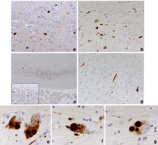
Figure from Kalaitzakis ME (Acta Neuropathol 2009). Pathology in cortical and subcortical regions. a Cortical LB pathology in the cingulate gyrus of a demented PD case. b LB and LN pathology in the NBM of a demented PD case. c Selective vulnerability of the CA2 sector of the hippocampus to LN-type pathology. Inset higher magnification showing the web
of LNs. d Neurofibrillary pathology in the CA2 sector of the hippocampus of a demented PD case. e Multiple ‘classical’ LB in a pigmented neuron of the medial substantia nigra. A single LB in a pigmented neuron is also evident. A single (f) and multiple (g) LB in a neuron of the NBM magnification: a and b 920, c 94 and for inset 910 and d 920, e, f and g 9100. PD Parkinson’s disease, LB Lewy bodies, LN Lewy neurites, NBM nucleus basalis of Meynert, CA Cornu Ammoni.
AD Pathology
Although several studies indicate cortical AD pathology as an important determinant for
questionable (Kalaitzakis ME, Acta Neuropathol 2009), and there is no consistent
correlation between cortical Ab burden and various measures of clinical deficit in patient
with Parkinson Dementia.
A recent study (DJ Irwin, Ann Neurol 2012) based on analysis of a large cohort of PD
patients from shows that the most robust correlate of dementia in PD is the severity of
CLBs/LNs (in association with APOE4 genotype). However, in the same study, the authors
observed that subanalysis of the PDD group designed to examine variables predictive of a
comorbid AD diagnosis and demographics of PDD patients suggests that plaque and
tangle pathology may influence cognitive status and the course of disease progression in a
subset of PDD patients.
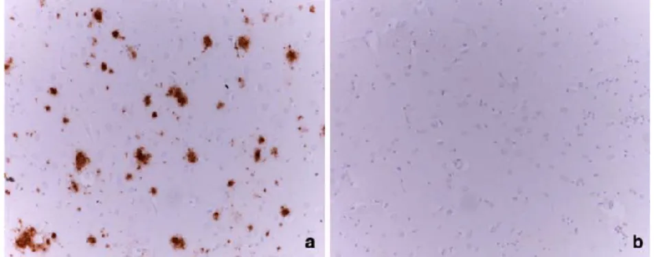
Figure from Kalaitzakis ME (Acta Neuropathol 2009) Immunostaining for b-amyloid deposits in the
caudate nucleus. a A large number of Ab deposits in the caudate nucleus of a PD case with dementia. b Caudate nucleus without Ab deposits from a non-demented PD case. Magnification 920. PD Parkinson’s disease, Ab amyloid b peptide.
White Matter Lesions
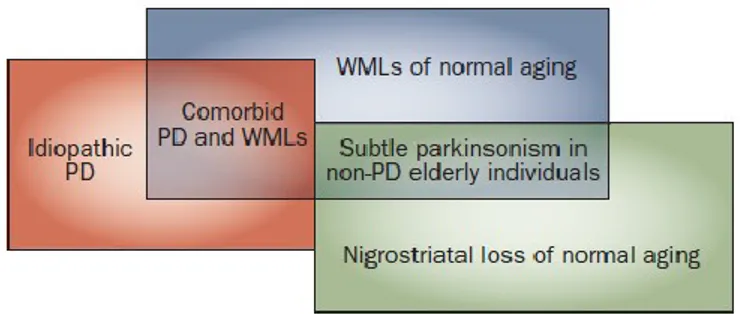
White matter lesion are classically associated with motor disability and cognitive
symptoms in non-demented and non-PD elderly individuals, therefore, comorbid
vasculopathy is expected to contribute to clinical symptoms in PD, included cognitive
deficits. An important review by Bohnen NI (Nat Rev Neurol 2011) highlights this concept.
Figure from Bohnen NI, Nat Rev Neurol, 2011.
However, the impact of vascular pathology on cognitive dysfunctions in Parkinson’s
disease has not been jet commonly accepted, a recent study by Gonzalez-Redondo R.
(Eu J Neurol 2012) analyzed WMHs in cerebral magnetic resonance images (MRI) from a large cohort of patients with PD using a cross-sectional and longitudinal design, to assess the possible relationship between silent vascular lesions and dementia or MCI in this
population. The author found that white matter hyperintensities do not influence the cognitive status of patients with PD. A specific pattern of Frontal WMHs have a negative impact on a single cognitive domain, specifically semantic fluency, but no effect of global cognitive functioning. Nevertheless , the mean follow-up period of the study was 30
months, given that vascular burden may have an effect on cognitive impairment in patients with PD as WMHs increase overtime there is need further studies with larger series of patient with relevant follow-up period.
At the state of the art, the elective brain changes yielding to dementia in PD have not been
yet adequately characterized to permit a “consensus definition” from a neuropathological
point of view. Although some studies indicate LB-type pathology in cortical and limbic
structures as the main histological substrate of PDD, it is now evident that the mere
spreading of alpha-Synuclein pathology cannot reliably account for the presence of
dementia and the co occurrence of concomitant AD-type changes and/or vascular
Genetics
Carriers of mutations in the alpha-synuclein gene (SNCA) tend to develop dementia at an early stage of the disease, although these are rare conditions (Multiplication of the SNCA locus and the three known missense mutations: A30P, A53T, and E46K). SNCA dosage has been found to influence disease progression directly including development of dementia [56,57]. Functional studies on brain tissue revealed that SNCA genomic copy number and gene expression are related, as increasing number of SNCA copies lead to increase in alphasynuclein expression as well as Lewy body formation.
The apolipoprotien (APOE) ε4 allele is associated with a higher risk and earlier disease onset of Alzheimer's disease. Several studies have explored the role of APOE in PD, with inconsistent results (Aarsland D., 2010).
Recent findings from Tsuang D. (JAMA Neurol, 2013) indicate that The APOE ε4 allele is a strong risk factor across the LBD spectrum, specifically in Dementia with Lewy Body (DLB) and Parkinson Dementia (PDD). Interestingly, the high ε4 frequency in the PD dementia patients groups, id not correlate with overall brain neuritic plaque burden, indicating a contribute of apoE on neurodegeneration through mechanisms unrelated to amyloid
processing. At this regard, an association of the ε4 allele with the severity of Lewy body pathology has been reported.
In a recent prospective study based on an incidence cohort, an association between the H1 haplotype and increased risk for dementia was reported (Williams-Gray, 2009).
An association between COMT genotype and performance on a measure of attention has been recently reported (Morley JF, Mov Dis 2012), as has been described by others (Williams-Gray, 2009), and thought to reflect modulation of a frontostriatal network. On the other hand, the same authors found that H1 MAPT haplotype is related to the progression of deficits with respect to measures of the domains of memory and attention, over the entire follow-up period, but not with the overall rate of cognitive decline and the progression to the condition of Parkinson Dementia.
Biomarkers of Parkinson Dementia
Biological markers
While the failure of ancient studies (Molina 1997, Jensen 1998) on CSF biomarkers, subsequent publication have given rise to a wide research . In 2006 Mollenhauer reported
a specific biomarker profile on patients with PD, especially those patients with cognitive deficits and worse motor performances, present CSF τ higher and CSF Aβ lower. Alves G (2010) demonstrated that CSF Aβ levels are altered in a subset of patients with early PD and relate to memory impairment. This results study suggests that alterations in Aβ protein metabolism may contribute to the heterogeneity in pattern and course of cognitive decline associated with PD. T-tau increases in CSF in AD and other degenerative and destructive diseases of brain and is widely thought to signify damage to neurons. The same group (Montine TJ, 2011) found that CSF T-tau levels trended to lower values in the PD-D group, these were not significant and thereby concordant with the results of some14 but not others who observed an increase in average CSF T-tau in patients with PD-D.13 In contrast, we observed that average P181-tau concentrations in PD and PD-CIND groups were significantly 20% lower than age-matched controls, and this result differs from others who have reported no difference or increased average CSF P181-tau in these g groups. CSF P181-tau is more difficult to interpret than T-tau as its levels presumably reflect at least two potentially related mechanisms, cellular processes that lead to phosphorylation and release from damaged neurons. The reasons for these discrepant results among
studies are not clear. However, one interpretation of our results is that patients with PD, PD-CIND, and PD-D may have less neuron damage than patients with aMCI.
Neuroimaging
STRUCTURAL MAGNETIC RESONANCE IMAGING: Structural magnetic resonance imaging (MRI)
is a validated biomarker for Alzheimer’s disease (AD); it is capable of correlating the
topographic pattern of cerebral atrophy with clinical progression identifying changes
associated with MCI, and stratifying those patients with MCI at highest risk for AD. Recent
research demonstrates that the topographic patterns of neurodegeneration underlying
cognitive impairment in PD are also detectable with MRI. On the other hand, the
heterogeneity and non linear progression of cognitive deficits in PD, as opposed to the
progression of Mild Cognitive Impairment (MCI) to Alzheimer disease, is a significant
confounding factor. A wide variations in cognitive profiles, disease duration, and age (that
reflect the complex phenomenology of cognitive impairment in PD) give inconsistent and
difficulty comparable results.
Cerebral Atrophy Associated with PD In established PDD, cortical atrophy may present a
(Burton, 2004; Beyer 2007). Burton et al. (2004), using Voxel Based Morphometry (VBM,
automated and unbiased MRI technique), observed a relative gray matter (GM) atrophy
in right superior, middle, and inferior frontal lobe in PD patients compared with control
group; while a GM loss was reported in temporal hippocampus and parahippocampal
gyrus, in occipital lobes; frontal lobes and left parietal lobe, atrophy of right caudate tail
and of putamen and thalamus bilaterally in PDD (vs control group). These data have been
substantially replicated in other subsequent PDD studies using VBM (Duncan 2013).
Although not a universal finding and less severe than in AD, using various MRI
methodological approach (visual rating scales, ROI analyses and VBM) hippocampal
atrophy is frequently reported in PDD (for rev. Duncan 2013).
In a recent study, Melzer et al. (2012), using VBM, showed that PD-MCI present a limited
GM atrophy in the temporal, parietal e frontal cortex as well as the bilateral caudal
hippocampus, amygdala and right putamen; interestingly specific cognitive impairments
have been observed in relation to the relevant neuroanatomic structure in this study.
Author concluded that some gray matter atrophy may precedes the development of
dementia but may be accelerated once frank dementia begins.
Regions that display areas of significant grey matter atrophy in patients who had (A) Parkinson’s disease (PD) with normal cognition (PD-N), (B) PD with mild cognitive impairment (PD-MCI), and (C) PD with dementia (PD-D), relative to controls, are displayed on conventional, study-specific average grey matter neurological images (the left in each image is the left brain). (Melzer TR. J Neurol Neurosurg Psychiatry 2012) .
In contrast with these findings, other authors failed to detect significant correlations
between cognitive domain test performance and GM loss in PD-MCI (Dalaker TO 2010).
Substantia innominata, another structure potentially involved in cognitive decline of PD,
has been investigated using MRI protocols. A volume reduction of substantia innominata
has been reported across all stage of PD, and degree of atrophy has been correlated with
White Matter Imaging White Matter Hyperintensities (WMH), also referred to as
leukoaraiosis, are commonly observed on brain imaging studies in older adults. WMH
have been linked with general cognitive decline, in particular with impairment of attention,
executive functions and processing speed; these cognitive dysfunction are frequently
reported among PD patients. Studies of WMH severity and association with cognitive
decline or motor impairment in PD yielded inconsistent results (Lee, 2010; Dalaker
TO,2009; Shin J, 2012; Bohnen NI,2011; Herman T,2013). Different WM analysis
methods, study population and concomitant vascular burden are some confounding
factors, that contribute to these conflicting results.
Diffusion Tensor Imaging Diffusion Tensor Imaging (DTI) represents a post-processing
evaluation of MRI imaging, used to evaluate white matter tract integrity. In a recent paper,
Hattori T (2012) identified decreased Fractional Anisotropy (FA, directionality of water
molecules movement and then index of structure integrity) in many major tracts in PD-MCI
and PDD patients, but not in PD without cognitive impairment, compared with control subject (the superior longitudinal fasciculus, inferior longitudinal fasciculus, inferior
fronto-occipital fasciculus, uncinate fasciculus, cingulum, and corpus callosum). Interestingly, FA values in bilateral parietal white matter were significantly correlated with MMSE scores in
PD patients. These data are partially consistent with a previous study of Matsui (2007), that observed a reduction of FA in posterior cingulated bundles, frontal, temporal, and occipital white matter in PDD patients compared with PD control group.
PET-STUDIES Several PET studies have suggest that striatal dopaminergic depletion
presents a positive correlation with cognitive dysfunction. Jokinen P (2009) observed an impairment of cognitive performance (assessing visual memory and verbal memory) in PD patients with a more pronounced reduction of [18F]-Fluorodopa uptake in the caudate
nucleus. In another study PET study, Cropley VL (2008) illustrated that striatal dopamine denervation contributes to frontostriatal cognitive impairment in PD. Right caudate, ventral striatum and the anterior cingulate cortex are other structures potentially implicated in cognitive impairment in PD, as emerged in a study of Ito (2002) who compared PD
patients with and without dementia. PET studies of brain metabolism with FDG have shown decreased resting metabolic activity in various cortical regions: PDD showed a an impairment of glucose metabolism in frontal, in parietal and in occipital cortices and in posterior and anterior cingulate cortex (Jokinen P,2010). PET studies have also provided evidence that cognitive decline in PD is associated with significant degeneration of the
cholinergic system: cortical cholinergic denervation (assessed using [11C]-PMP-AChE
PET) were associated with decreased performance on test of executive functioning but not motor symptoms (Bohnen NI, 2006). In another study, PD patients with dementia had significantly lower parietal [11C]-MP4A uptake (used to assess cortical AChE activity) than PD patients without cognitive impairment (Hilker R, 2005). In a multitracer studies, demonstrating the interdependence of imaging biomarkers, Klein et al verified that cholinergic degeneration in PDD patients was spatially congruent with the metabolic changes measured with [18F]FDG.
Image adapted from Klein et al. (2010).44 (A) Brain areas with significant reduction of [11C]MP4A uptake in PDD versus PD. (B) Brain areas with significant reduction of [18F]FDG uptake PDD versus controls. CMRglc, cerebral metabolic rate of glucose.(Ray and Stafella, Mov Dis, 2012).
The b-amyloid imaging, using [11C]-PIB, has been used to assess the amyloid load
associated in Lewy-bodies disease spectrum. The Authors (Edison P,2008) observed that,
whereas the majority of DLB patients showed an increased agent binding, only a small
number of PDD patients (and none of the PD patients investigated) had PET images
Progression of cognitive deficit in PD
The existence of a cognitive continuum that goes to from normal aging to frank dementia
(Alzheimer Disease) and passing for a phase of mild impairment was postulated in 1995
by Petersen. The “transposition” of the MCI notion to the dyscognition area of PD was
made in the last years (Caviness JN, 2007), with aprioristic assumptions and applying the
same methodological criteria used for Alzheimer Disease. A ‘critical review’ of the concept
of Mild Cognitive Impairment in PD has been published on 2011 by the Movement
Disorders Society (MDS). This condition emerges as a frequent (27% prevalent) and
heterogeneous in terms of number and type of cognitive domains involved, and is
considered to be a risk factor for the development of dementia. However, this criteria will
The construct of Mild Cognitive Impairment in Parkinson has received increased attention
from clinicians and the term is now widely accepted, however, prospective longitudinal
studies using formal MCI criteria are few and often inconsistent.
A recent study (Pedersen KN, Ann Neurol 2013, The Norwegian ParkWest Study), a
prospective longitudinal cohort study designed to examine the course of MCI and its
progression to dementia in an incident PD cohort, found that Mild cognitive impairment at
PD diagnosis predicts a highly increased risk for early dementia. The authors also found
that impairment in verbal memory and attention was associated with conversion from MCI
to PDD. This is in line with previous studies that have shown frontal/executive and verbal
memory deficits to be associated with the development of PDD although others have
found that visuospatial based dysfunction were more prone to predict dementia in PD.
These inconsistencies deserve further investigations but may reflect different definitions of
cognitive impairment and PDD, differences in sample characteristics, or biological
heterogeneity.
Research from the group of Williams-Gray C., conducted on an incident cohort of PD
to the development of Dementia in PD patients rather than deficit in executive dysfunction
driven by fronto-striatal dopamine related circuitry.
The authors demonstrated that early deficits on frontostriatally based tasks are not related
to subsequent dementia risk. The domain of “verbal fluency” is paradigmatic of the results
and the subsequent discussion worked out by the authors. The dissociation between
semantic verbal fluency (frontally driven) and phonemic verbal fluency (a posterior
temporal lobe task) in terms of predicting dementia indicate that it is the semantic,
temporal lobe component of the fluency task which is predictive of cognitive decline rather
than the frontally based strategic retrieval common to both fluency tasks. Williams-Gray et
Clinical Characterization of PD Dementia in our “Parkinson Centre”.
The emerging importance of Parkinson Dementia, the lack of specific clinical diagnostic criteria as well necessity of a reliable work-up for diagnosis, follow-up and treatment led to start a comprehensive job of defining the epidemiological characteristics, clinical and neurobiological features of this condition, involving hundreds referred to our tertiary PD Centre.
First, the epidemiology and the characteristics associated to progression to Dementia were assessed in our population in a recent retrospective study. Patient were diagnosed in accordance with the criteria defined by the United Kingdom Parkinson’s Disease Society (UKPDS) Brain Bank (Gibb e Lees, 1988). The diagnosis of dementia was made from clinical notes reported or when a Cholinesterase-inhibitor was prescribed. The incidence of Dementia was calculated from the entire population of patient referring to our Parkinson Outpatients Ambulatory, patients were included in the analysis of Dementia incidence if they underwent at least two visits or three months of clinical observation.
Beside the epidemiological tool of prevalence, that indicate the proportion of subject suffering of a given condition, it is possible to assess the incidence of pathologic condition, which gives a dynamic assessment of the phenomenon inside the general population. In particular, density of incidence allows the calculation the frequency of the phenomenon inside the cohort of interest that are free from the disease at the baseline.
We calculated the incidence index for 1000 years person, a CI of 95% was fixed according to the following formula:
We analyzed 480 clinical notes of the study period (January 2004-May 2008), patients were classified as: Casi afferenti n = 480 Altre diagnosi n = 95 Casi con MP n = 385 Parkinsonismi n = 29 Tremore essenziale n = 32 Demenze n = 8 Miscellanea n = 26 vascolare (11) iatrogeno (8) PSP (4) MSA (3) CBD (1) DBL (3) Huntington (2) Alzheimer (2) FTD (2) Demenza nda (1) Senza diagnosi (14) Lieve sind.extrapr (10) Sindrome ansioso-dep (2) Periodo 1.1.2004-31.05.08
Clinical characteristic of patients with PD [M= 200, (53,5%) ; F = 174, (46,5%)]
Variables N Mean
Standard
deviation Min Max Age at onset 353 61,80 10,16 29 84 Age at diagnosis 153 62,45 9,68 34 88 Age at first visit 374 65,90 9,61 33 90 UPDRS at first visit 237 16,95 9,78 2 50 Age at last visit 373 70,94 9,14 44 92 UPDRS at last visit 199 19,58 11,39 1 55 Levodopa equivalent dose 344 803,60 444,13 100 2370 Year-person 374 5,07 4,01 0 21
Estimated incidence. For our incidence value we considered 374 patients with a total amount of follow up of 1896 year-person (mean period of follow-up 5,07 ± 4,01). In the study period 35 new cases of Dementia occurred with an estimated incidence of 18.4 cases for 1000 year-person (IC95% 12,4-24,4) and 17 cases of cognitive impairment, with an estimated incidence of 9 cases for 1000 year-person (IC95% 4,8-13,2).
In order to ascertain possible predictive features for Dementia we compared clinical characteristic, at first and last evaluation, of our two groups of patients: patients with dementia (n=35) and patients without dementia (n=339).
Clinical features associated with Dementia. Were compared to the clinical characteristics of the first and the last visit of the two groups of patients with (n = 35) and without dementia (n = 339) in order to envy possible predictive variables of the phenomenon under study and to estimate the progression of the disease during the period of observation of the patients in the study. The following table describes the demographic and clinical characteristics of patients with Parkinson's disease with and without dementia. The analysis of data documenting an age of onset of PD on average four years more advanced for patients who later developed dementia. In addition, it is noted that the male patients with Parkinson's disease have a probability of about 3 times higher than those of female to meet to dementia within five years. Patients who develop dementia have the same profile of schooling than those who did not develop.
Variables PD-D (n=35) PD-ND OR IC95% P Age at onse 65,5±8,7 61,4±10,2 0.023 Age at diagnosis 70,4±5,2 61,8±9,7 0,006 Gender (M/F) 26/9 165/159 2,78 1,26-6,13 0,009 Formal education none 0 2 (1,1%) n.s. 5 yrs 7(41,2%) 71 (40,8%) 8 yrs 4(23,5%) 43(24,7%) 13 yrs 5 (29,4%) 43 (24,7% ) College degree 1 (5,9%) 15 (8,6%)
This other table shows the clinical characteristics performed at the first visit at the outpatient clinic of the Department of Neurology. The analysis documents an older age of about 5 and a half years, statistically significant at the first visit for patients who develop dementia. Variables PD-D (n=35) PD-ND (n=339) OR IC95% P Age at I visit 70,8±8,1 65,4±9,6 0,001 Familiarity for dementia 1/30 10/276 0,92 0,11-7,44 n.s.
Vascular risk factors (Y/N) 10/12 107/95 0,74 0,31-1,79 n.s. Clinica subtype at disease onset Tremo 13 (46,4%) 157 (52,0%) n.s. Akinetic-rigid 11 (39,3%) 97 (32,1%) Mixed 4(14,3%) 48 (15,9%)
Gait disordets (Y/N) 5/25 28/276 1,97 0,70-5,55 n.s. Postural instability (Y/N) 5/24 30/273 1,90 0,67-5,33 n.s. Hallucinations (S/N) 3/26 4/289 8,34 1,77-39,3 0,002 Hoehn-Yahr Stage I 7 (28,0%) 129 (44,9%) n.s. II 15 (60,0%) 135 (47,0%) III 3 (12,0%) 22 (7,7%) IV 0 1 (0,3%) UPDRS at I visit 22,11±10,36 16,57±9,62 0,021
The following summarizes the clinical features at the last visit in the clinic of the two groups of patients. Statistical analysis shows a significant difference in the frequency of disturbance of gait and postural instability (about two times greater for patients suffering from dementia), a clinical stage recorded at Hoehn-Yahr more advanced for people with dementia, a higher prevalence of hallucination (about 12 times more) and a greater motor deficit as measured in the UPDRS. Finally, the average amount of levodopa-equivalent expressed in mg is statistically reduced in patients with dementia compared to those without dementia. Variables PD-D (n=35) PD-ND (n=339) OR IC95% P
Age at last visit 76,7±6,20 70,42±9,15 0,001
Gait disorders (Y/N) 12/16 65/228 2,63 1,18-5,84 0,014 Postural instability (Y/N) 12/15 65/224 2,76 1,23-6,18 0,011 Hallucination (Y/N) 16/10 34/258 12,14 5,1-28,9 0,001 Hoehn-Yahr Stage I 1 (5,9%) 51 (24,9%) 0.001 II 2( 11,8%) 111 (54,1%) III 11 (64,7%) 27 (13,2%) IV 2 (11,8% ) 15( 7,3%) V 1( 5,9%) 1( 0,5%)
UPDRS at last visit 31,83±13,60 18,81±10,59 0,001 Levodopa
equivalent dose (last visit)
625,81±300,96 825,85±453,7 0,017
The histogram below shows the trend of Parkinson's disease observed in its motor component for the group of patients with dementia than those who did not report such occurrence. It is known that patients with dementia did not differ in the definition of the clinical stage of Hoehn-Yahr than other patients at the first visit, while the last visit reveals
a statically significant difference. For an assessment of the mean differences observed in the two patient groups in scores on the UPDRS is noted that in the period of the study there was a decrease of about 44% for the group with dementia while the other group is evidence of worsening of motor 13.5%.
0% 10% 20% 30% 40% 50% 60% 70% 1 2 3 4 MP con demenza MP I I I 0% 10% 20% 30% 40% 50% 60% 70% 1 2 3 4 5 MP con demenza MP p = n.s. p = 0.001
ANDAMENTO DI MALATTIA NEL CORSO DI CINQUE ANNI PRIMA VISITA (STADIO HY) ULTIMA VISITA (STADIO HY)
MP 16,57±9,62 MP con demenza 22,11±10,36 UPDRS MP 18,81±10,59 MP con demenza 31,83±13,60
44% di aumento medio del punteggio UPDRS (MP con dem) 13,5% di aumento medio del punteggio UPDRS (MP)
The bivariate analyzes shown previously documented the identification of four predictor variables in the onset of dementia, such as age of onset age, the greater motor impairment as measured by the UPDRS, male sex, and the presence of hallucinations at the first visit in the clinic . The logistic regression model showed the greatest role of hallucinations and more advanced age of onset of parkinsonian symptoms in the onset of dementia than the other two variables identified in the bivariate analysis (male and a motor deficit with a higher score 15).
A logistic regression analysis for the identification of identification of the predictor variables for teh occurrence of Dementia.
Variables B E.S. Sig. OR IC95,0%
Inferior Superior Age at onset 1,336 0,601 0,026 3,804 1,171 12,358 UPDRS 0,736 0,514 0,152 2,088 0,762 5,721 Hallucinations 2,411 0,850 0,005 11,150 2,106 59,039 Gender -0,379 0,503 0,451 0,684 0,255 1,834
Dipendet variable: dementia Indipendent variables: - Age at onset
- UPDRS score
- Allucinations at first visit (Y/N) - Gender (M/F)
Experimental procedures. Clinical Setting-Part 1.
Validation study of clinical criteria for PDD diagnosis
Introduction
The nosological classification of dementia in PD patients has been based on the DSM IV criteria (American Psychiatric Association 2000). These criteria define Parkinson’s disease dementia as a clinical condition associated with memory loss and at least one of the following disorders: apraxia, agnosia, aphasia or executive dysfunctions; the cognitive deficits must cause a significant impairment in global functioning. The DSM criteria, which are only descriptive, have certain limitations: first, they are not specific to PD-D; second, memory or cortically based disturbances may not be prominent in PD even in the very late stages; lastly, it may be difficult to determine the specific effects of motor and cognitive impairment on the patients overall functioning. A PD-D Task Force was created to draw up specific diagnostic criteria for dementia in PD and to design tools that can be used to assess the cognitive state in parkinsonian patients. This group of specialists, named the Movement Disorders Society Task Force (MDSTF), proposed specific ‘‘clinical criteria’’ for the diagnosis of dementia in PD in a paper published in 2007 (Emre et al. 2007), in which
the authors described the core clinical features of dementia in PD and the behavioral features that closely correlate with PD-D and help to make a diagnosis. These criteria were not, however, validated. In a subsequent publication, the same authors provided a framework of the neuropsychological tests that could be used to assess cognitive deficits and to make a diagnosis, providing the ‘‘diagnostic criteria’’ (Dubois et al. 2007). Two levels of analysis were proposed for the diagnostic procedures: step I and the step II. Step I is based on simple tests that analyze various cognitive domains, such as: attention, executive functions, visuo-constructive abilities and memory, and rules out major depression and other possible cause of cognitive impairment (Vascular dementia, vitamin deficits, hypothyroidism). Once step I has been accomplished, the neurologist should be able to establish whether the patient has ‘‘possible’’ or ‘‘probable’’ Parkinson’s disease with dementia (Dubois et al. 2007). Step II is based on extensive neuropsychological tests designed to thoroughly explore different cognitive domains, thereby allowing the pattern and severity of the dementia in PD patients to be accurately defined. The MDSTF recommended that the second level of tests be used for pharmacological trials and research, though the authors of one paper suggested that step II should be applied even in cases in which the step I results are borderline or somewhat uncertain (Goetz et al. 2008).
Step I of the MDSTF only lasts a few minutes and has been proposed to clinicians as a tool for the diagnosis of PD-D that obviates the need for a formal neuropsychological assessment. The aim of our study was to validate step I proposed by the MDSTF and assess its accuracy as a screening instrument for Parkinson’s disease dementia. For this purpose, we decided to measure the diagnostic sensitivity and specificity of step I when compared with the diagnosis made on the basis of an extensive neuropsychological examination. We rigorously applied the DSM IV criteria for Parkinson’s disease dementia which are, despite certain limitations, validated and represent the current gold standard for the diagnosis of this pathology.
Methods
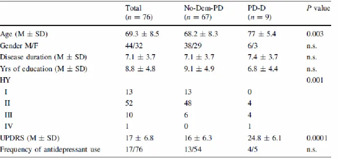
Patients and recruitment. Patients were randomly recruited from June 2009 to April 2010 from the outpatient service of the Parkinson’s disease and Extrapyramidal disorders Unit at the Department of Neurology and Psychiatry of the ‘‘Sapienza’’ University of Rome. The study population consisted of 76 patients. Table 1 summarizes the demographic and clinical characteristic of the study population. Informed consent was obtained from all the patients or their relatives. The local ethics committee approved the study.
Inclusion and exclusion criteria Inclusion and exclusion criteria. The inclusion criteria were: idiopathic Parkinson’s disease according to the United Kingdom Parkinson’s Disease Society Brain Bank criteria. No other inclusion criteria, such as age, disease duration or motor subtypes, were applied in order to minimize a possible selection bias.
Table 1 Demographic and clinical characteristics of the study population
The exclusion criteria were:
• Definite vascular dementia according to the NINDS AIREN criteria;
Patients taking antidepressants for moderate depression were not excluded from the study. Fifteen of the patients enrolled were on antidepressant treatment during the study: eight patients with Duloxetine, three patients with Venlafaxine, two patients with Sertraline, one patient with Escitalopram and one patient with Paroxetine.
Study design. The patients were evaluated by a first neurologist, as recommended by the MDSTF, who administered the tests and applied the recommended cut-off values (Dubois et al. 2007). At the end of step I, the first neurologist attributed a score to each patient according to the 8-point rule. Thereafter, the patients underwent the second part of the examination, i.e. step II, which yielded a neuropsychological report. Lastly, the patients underwent a neurological examination and a second neurologist, who was blind to the scores obtained in step I, made the diagnosis of PD-D according to the current gold standard DSM IV criteria.
The patients underwent the following neuropsychological tests:
- Global efficiency: Mattis DRS
- Working memory: Digit span, Spatial span (CANTAB), Digit ordering test - Conceptualization: Similarities (WAIS-III), Wisconsin CST
- Set activation: Verbal fluency - Set shifting: TMT
- Memory: RAVLT
- Language: Boston naming test
- Visuo-constructive: Copy of the clock - Visuo-spatial Benton: line orientation test - Visuo-perceptive: Benton face recognition test
Neuropsychiatric complains were assessed with the following tests:
- Apathy: Apathy scale
- Depression: Beck depression inventory - Psychosis: NPI
The second neurologist used the neuropsychological reports (in step II) to appraise the patient’s cognitive status and make a diagnosis of PD-D. We assessed the sensitivity, specificity, positive predictive value (PPV), negative predictive value (NPV) and accuracy of step I. Moreover, we calculated the kappa index of step I versus that of step II. A kappa index of 0.70 was considered satisfactory.
Results
Applying the DSM IV criteria after a full battery of neuropsychological tests, a standard neurological examination and an interview with the caregivers, we diagnosed nine patients with PD-D (12% of our cohort). In our setting, step I had a sensitivity of 78% and a specificity of 95.5%, while its positive predictive value was 70%, and its negative predictive value was 97%; moreover, step I displayed an accuracy of 93.4% (Table 2).
Table 2 A contingency table showing the diagnosis of PD-D according to the DSM IV following a neuropsychological examination (column 1) and the diagnosis of PD-D by means of step I (line 1)
The kappa index stood at 70%. Two patients with PD-D according to the DSM IV criteria were not demented according to the step I diagnostic scheme (Table 2). In one false negative case (MMSE = 26, normal at serial-7 and 3-word recall), step I did not capture
substantial memory and attention deficits that did not escape detection by the psychometric tools; moreover, this patient had but slight, though detectable, cortically based deficits such apraxia and anomia (below cut-off in the Boston naming test). A diagnosis of dementia was, however, made in this patient in view of his high educational level and his performances in the premorbid condition. The other false negative case also yielded normal results in tests regarding attention and memory, while his MMSE value was just above the cut-off. Worthy of note is the fact that both false negative patients displayed abnormalities in the copy of pentagons and design of the clock tests. Ten patients were diagnosed as having PD-D according to the step I procedure. Three of these ten patients did not fulfil the criteria of PD-D for the DSM IV (Table 2).
Memory deficits were not detected in the neuropsychological tests of the three false positive cases; also they were not a subjective impression or reported by the caregiver; these three patients proved to have a considerable dysexecutive syndrome though no cortical disturbances.
In accordance with the current literature, we found that PD-D patients were not only older than those without dementia (No-Dem-PD) but even had a more severe motor form of parkinsonism (Table 1). We observed a statistically significant difference in age, UPDRS
and HY. There was a difference between the two groups in the disease duration values, though this difference did not reach statistical significance. None of the patients with PD-D according to both the DSM IV criteria and step I checklist had significant neuroimaging or laboratory abnormalities.
Discussion
We assessed the screening properties of step I of the MDSTF for the diagnosis of PD-D. Our results indicate an average match between the cases diagnosed by means of step I and those diagnosed by means of a neuropsychological examination.
Dujardin and colleagues recently assessed the validity of step I for the diagnosis of PD-D in a group of 188 PD patients (Dujardin et al. 2010). The estimated clinical parameters reported in their paper were: specificity of 95%, sensitivity of 66%, PPV of 77% and NPV of 91%.
Step I is burdened, at least in our setting, by defects in specificity (1) and sensitivity (2): 1. A PPV of 70% indicates that 30% of the patients may be misdiagnosed as PD-D. We found three false positive cases in our cohort; these three patients had a long disease duration, moderate-to-severe motor symptoms and serious executive dysfunctions,
resulting in performances below the cut-off in memory, attention and global efficiency tests at the screening level, not accompanied, however, by cortical disturbances or memory deficits. The analysis conducted by Dujardin
and colleagues yielded a PPV value (77%) that is comparable to ours (70%).
2. Sensitivity was 78% in our study and 66% in the study conducted by Dujardin and colleagues. In keeping with the results of their paper, our findings raise doubts regarding the sensitivity of the MMSE cut-off, particularly as regards highly educated patients. In (2008) O’Bryant et al. also reported that the discriminative power of MMSE was lower for college graduates.
Since tests designed to assess memory and attention as well as visuo-constructive abilities all adopt the MMSE cut-off values, the use of such tests in highly educated individuals should be assessed in further studies.
The pill questionnaire appears to be a consistent indicator of the global functioning and organization of daily living in PD patients. In our cohort, PD-D had an impact on the activities of daily living in all the patients according to the pill questionnaire; however, numerous No-Dem-PD patients, including the three false positive cases, were unable to describe their therapies. Nonetheless, the DSM IV criteria specify that the decline in
activities of daily living must represent a decline from a previously higher level of functioning, as the authors themselves point out. Indeed, this highlights the need for formal validation of the pill questionnaire. Dujardin and colleagues believe that with an easy-to use, short battery of tests that are commonly used in routine clinical practice, it is possible to diagnose PDD in accordance with reference criteria and with the same sensitivity and specificity as in a more extensive evaluation (Dujardin et al. 2010). We affirm the contrary that while diagnosing dementia and cognitive impairment has become part of the clinical practice for neurologists, the use of step I as only diagnostic tool for PD-D may lead to a significant proportion of misdiagnoses as a consequence of both type I (false positive) and type II (false negative) diagnostic errors.
However, step I, which has a NPV of 97%, may be considered an otherwise valid screening test. Indeed, future fine-tuning of the cut-off values combined with more sensitive domain-related psychometric tests will maximize the overall screening properties of step I. Once step I has been optimized, it may be effectively be used as a screening test, while reserving step II for step I-positive individuals and borderline cases. Further studies on larger series of patients are warranted to assess the validity of step I and to draw up consensus criteria for the definition of PD-D and other forms of cognitive
impairment associated with PD. In the meantime, we suggest that the diagnosis of PD-D should still be based on a formal neuropsychological examination and longitudinal follow-up.
Experimental procedures. Clinical Setting-Part 2.
Follow-up analysis for cognitive decline in our cohort
Background
Cognitive impairment is one of the most common and important aspects in Parkinson Disease (PD), and greatly affects functioning and quality-of-life. The spectrum of cognitive dysfunction in PD has been lately coded in terms of mild cognitive impairment (PD-MCI) and dementia (PDD) with clinical and diagnostic criteria provided by specialists of the Movement Disorders Society (MDS). This nosological effort has been structured in order to support the identification of cognitive decline trajectories in PD population. Indeed, the term of MCI, initially conceptualized as a prodrome of Alzeheimer disease and subsequently extended to others degenerative disorders including PD, is generally codified as a transitory stage prior to the development of a full-blown dementia. However,
identify this intermediate stage of cognitive impairment presents a relative clinical relevance given that a large proportion of PD presents cognitive deficits (classified as MCI) even in earliest disease stage and the rate of cognitive decline remain largely unpredictable. Considering that cognitive disturbances are integral to the course of PD and this nosological characterization do not always demonstrate a reliable prognostic value, we evaluated the phenomenon of “cognitive conversion” at five years, focusing on clinical and neuropsychological features that characterized patients who worsen their cognitive abilities with respect to cognitively steady-staying patients, independently from baseline cognitive asset.
Methods
Patients. Patients were randomly recruited from June 2009 to November 2009 from the outpatient service of the Parkinson’s disease and Extrapyramidal disorders Unit at the Department of Neurology and Psychiatry of the “Sapienza” University of Rome; 54 patients of 67 of the initial cohort enrolled for a previous published study were revaluated about five years later. The inclusion criteria were: idiopathic Parkinson’s disease according to the United Kingdom Parkinson’s Disease Society Brain Bank criteria. No other inclusion criteria, such as age, disease duration or motor subtypes, were applied in order to
minimize a possible selection bias. The exclusion criteria were 1) PDD according to the MDS – Task force (MDS-TF) 2) Definite vascular dementia according to the NINDS-AIREN criteria 3) Major depression according to the DSM IV-TR criteria.
Clinical and neuropsychological evaluation. All patients underwent a detailed neuropsychological evaluation including all tests of level II diagnostic criteria for PD-D of the Movement Disorders Society. Motor examination included: H&Y, UPDRS part III and axial score. Clinical characteristics regarding “milestones “ of PD progression were also reported, including: presence of levodopa induced dyskinesia, and occurrence of falls; presence of motor fluctuation at baseline were also recorded. Patients were classified in PD-normal cognition and PD-MCI at baseline (the diagnosis of MCI was made in accordance with MDS-TF criteria on the base of neuropsychological evaluation performed in 2009).
At follow-up patients were classified as:
a) Non-converter patients group (PD-NC): patients with normal cognition or MCI either at baseline or follow-up
b) Converter patients group (PD-C): patients normal cognition at baseline who worsened their cognitive performances developing a MCI and PD-MCI at baseline that developed a PDD at follow-up.
Statistical Analysis. We used the t-test for continuous variables and chi-square test for categorical variables. We performed a logistic regression analysis to assess the association between cognitive conversion adjusted for variables statistically relevant to univariate analysis . P values < 0.05 were considered as statistically significant. All statistical analyses were performed using SPSS software (version 20.0, SPSS inc. Chicago, IL, USA).
Results
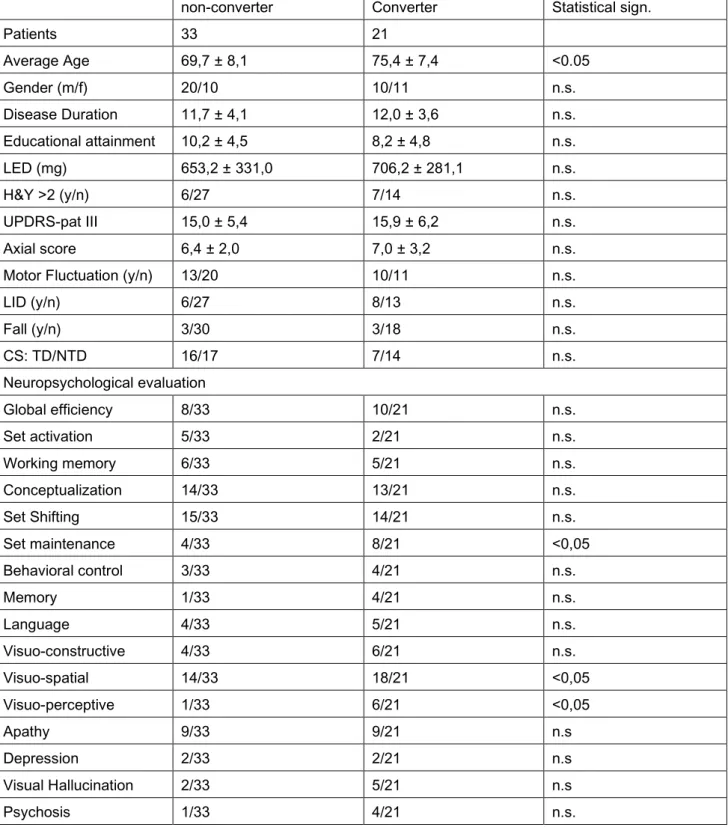
At follow-up visit, 33 of 54 patients (61%, 19 with normal cognition and 14 with MCI) presented a stable cognitive assessment and were classified as non-converter, while 21 patients (11 normal cognition at baseline who developed a MCI, 8 MCI and 2 normal cognition at baseline who developed incident dementia meeting the criteria of probable PDD) worsened their cognitive performance and were classified as converter.
non-converter Converter Statistical sign. Patients 33 21 Average Age 69,7 ± 8,1 75,4 ± 7,4 <0.05 Gender (m/f) 20/10 10/11 n.s. Disease Duration 11,7 ± 4,1 12,0 ± 3,6 n.s. Educational attainment 10,2 ± 4,5 8,2 ± 4,8 n.s. LED (mg) 653,2 ± 331,0 706,2 ± 281,1 n.s. H&Y >2 (y/n) 6/27 7/14 n.s. UPDRS-pat III 15,0 ± 5,4 15,9 ± 6,2 n.s. Axial score 6,4 ± 2,0 7,0 ± 3,2 n.s.
Motor Fluctuation (y/n) 13/20 10/11 n.s.
LID (y/n) 6/27 8/13 n.s. Fall (y/n) 3/30 3/18 n.s. CS: TD/NTD 16/17 7/14 n.s. Neuropsychological evaluation Global efficiency 8/33 10/21 n.s. Set activation 5/33 2/21 n.s. Working memory 6/33 5/21 n.s. Conceptualization 14/33 13/21 n.s. Set Shifting 15/33 14/21 n.s. Set maintenance 4/33 8/21 <0,05 Behavioral control 3/33 4/21 n.s. Memory 1/33 4/21 n.s. Language 4/33 5/21 n.s. Visuo-constructive 4/33 6/21 n.s. Visuo-spatial 14/33 18/21 <0,05 Visuo-perceptive 1/33 6/21 <0,05 Apathy 9/33 9/21 n.s Depression 2/33 2/21 n.s Visual Hallucination 2/33 5/21 n.s Psychosis 1/33 4/21 n.s.
Patients that converted cognitive status were older at baseline whoever, disease duration “per se” was not associated with a significant risk of cognitive conversion. No other differ significantly in demographic feature emerged. Non statistically difference were observed in motor performance at baseline, included clinical motor symptoms subtype at onset. There were no significant differences in the use of dopaminergic therapy (table 1).
At univariate analysis patients that converted cognitive status showed statistically significant deficits on measures of set maintenance, visual-spatial and visual-perceptive function; moreover a trend of significance were observed in test that explore memory function. Not other differ in neuropsychological variables emerged. Concerning neuropsychiatric complains, in converters group visual hallucinations and psychosis were more frequent without reaching, however, a statistical significance (table 1).
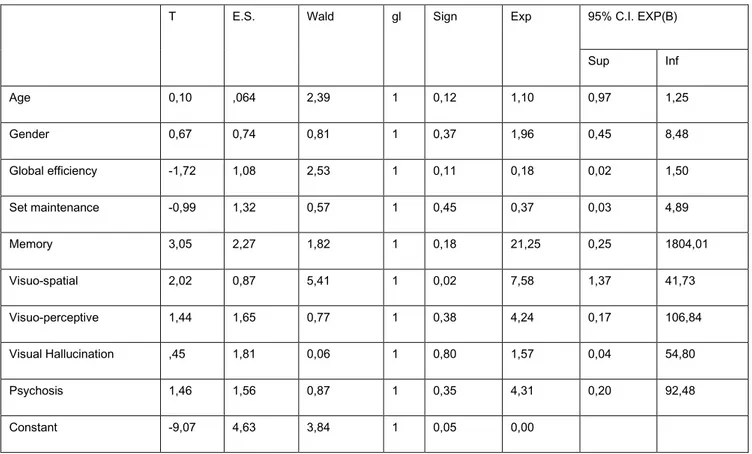
After logistic regression, test assessing visuospatial functions was the only variable that survived; a trend of significance was observed for the variables age and test assessing memory functions (Table 2).
T E.S. Wald gl Sign Exp 95% C.I. EXP(B) Sup Inf Age 0,10 ,064 2,39 1 0,12 1,10 0,97 1,25 Gender 0,67 0,74 0,81 1 0,37 1,96 0,45 8,48 Global efficiency -1,72 1,08 2,53 1 0,11 0,18 0,02 1,50 Set maintenance -0,99 1,32 0,57 1 0,45 0,37 0,03 4,89 Memory 3,05 2,27 1,82 1 0,18 21,25 0,25 1804,01 Visuo-spatial 2,02 0,87 5,41 1 0,02 7,58 1,37 41,73 Visuo-perceptive 1,44 1,65 0,77 1 0,38 4,24 0,17 106,84 Visual Hallucination ,45 1,81 0,06 1 0,80 1,57 0,04 54,80 Psychosis 1,46 1,56 0,87 1 0,35 4,31 0,20 92,48 Constant -9,07 4,63 3,84 1 0,05 0,00
Table 2. Logistic regression analysis.
Discussion
The aim of our study was to intercept a possible disease profile, including motor, neuropsychological and neuropsychiatric complains, that is associated with cognitive involution.
Regardless of their nosological characterization at baseline, namely if they are MCI or normal cognition, a proportion of 61% of PD patients remained overall cognitive stable over the observational period of five years, while 39% of patients consistently reduced their cognitive reserve during the follow-up period. Focusing on demographic
characteristics, age was the only variable significantly associated with cognitive conversion; these data are in line with the view of the ageing effects as the overall weakening of metabolic reserve, increasing the susceptibility to biochemical insults that are proper of the disease process. In our setting, clinical motor characteristics at baseline, including motor phenotype at onset (Tremor dominant vs akinetic-rigid), the presence of milestone of PD progression as well as LED assumption were not associated with cognitive conversion; this data may suggest that cognitive decline do not necessarily share the exactly alike pathophysiologic mechanism observed for motor disability progression.
With regard to neuropsychological profile we found that tests assessing the domains of visuo-spatial functions, visuo-perceptive functions, attention and, to a lesser extent, memory deficits and neuropsychiatric complains, were associated with cognitive conversion at univariate analysis. However, after logistic regression, only visuo-spatial deficits survived at a level of statistical significance and a trend of significance was observed for memory deficit.
Our findings may suggest that patients with “posterior” dysfunctions (visuo-spatial deficits and likely memory) are more prone to consistently worsen their cognitive abilities (whether they are cognitively preserved or MCI). Nevertheless, we have to underline that
visuospatial dysfunctions were presented in a consistent proportion of non converter, thus resulting in poor specificity. On the other hand, the measures of set maintenance, although associated with cognitive conversion at univariate analysis, showed a poor relevance after logistic regression; this may indicate that, although frequently represented in converter group, set maintenance as discrete variable has a limited predictive value for cognitive decline. Taken together these data are consistent with previous line of evidence, that hypothesized at least two pattern of discognitive progression. Indeed, other authors found a relative benign progression of fronto-striatal cognitive deficit when compared with posterior cortical dysfunction, that predispose to the development of dementia. Therefore, the seminal concept of different progression for cognitive patterns is replicable in a study design that assess the phenomenon of cognitive involution per se, across groups with different cognitive performance at baseline (normal-cognition or MCI). Moreover, although neuropsychiatric symptoms tended to be more present in converter group, no statistical difference emerged. Taking in to account the combined analysis of demographic, motor, neuropsychiatric and neurosychological profiles we can speculate that, according to our results, the presence of a MCI (dysexecutive-type) associated with motor complication, along with depressive symptoms and a long disease duration is not necessarily associated
to the collapse of cognitive function. On the other hand, older patients with cortical posterior dysfunction and memory impairment are more likely prone to cognitively deteriorate, in spite of the overall motor impairment.
The study present some limits: first, our approach is merely interpolative since our evaluation at baseline was performed in patients with a mean disease duration of about ten years and not at disease onset; second, the limited numerosity of our cohort do not allow generalization and translation in clinical practice; third: the categorization in the two groups (converter and non-converter), although original, is arbitrary and include patients consistently difference with regard to cognitive asset. Even though these limits, the notion of PD cognitive-conversion was not conceptualized to provide diagnostic criteria or clinical guidelines: the construct of cognitive-conversion represent an observational analysis focused on the phenomenon of cognitive breakdown in a cohort of patient followed for five years and assessed with a comprehensive neuropsychological battery. In this terms, the observation of an overall cognitive stability in a consistent proportion of patients, even when tagged as MCI, raises the question of the clinical utility and relevance (including therapeutic strategies) of the diagnosis of PD-MCI.
In conclusion, tracking with confident accuracy the profile of patients with risk of cognitive conversion remains difficult to obtain, partially ascribable to the complexity of the phenomena; however the role of visuospatial and memory deficit deserves attention and further analysis.
Experimental Procedures. Laboratory Setting-Part 1
The impact of MAPT haplotype on PD motor phenotype
Introduction
Parkinson’s disease (PD) is a multiorgan disease resulting from degeneration in different populations of neurons. Although the extent and progression of the degenerative process may to some extent be predictable (Braak et al 2003), PD patients present a wide range of clinical phenotypes, including symptoms in the pre-motor phase, motor signs and disabilities and, most importantly, a varying severity and rate of progression of motor and non-motor features. The clinical heterogeneity of PD is likely to reflect the contribution of different environmental and genetic factors, which in turn lead to different neuropathological substrates (Selikhova et al. 2009). Neither environmental factors nor individual genetic traits are currently recognized as determinant of clinical or pathological subtype of PD; for example, it has been observed that for LRRK2 patients (the most common autosomal dominant gene for PD) the extent of LB cortical involvement differs across patients and, interestingly, also inclusion of tau protein were present in a variable distribution and severity of brain autopsies (Poulopoulos et al. 2012). The
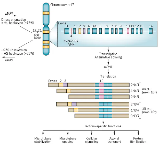
microtubule-associated protein tau (MAPT or tau) is being widely investigated in the field of neurodegeneration as there is a well-established genetic link between the MAPT gene locus and neurodegenerative diseases. There are two major haplotypes: H1 and H2, resulting from a common genomic inversion of approximately 900 kb in
chromosome17q21of the MAPT gene. Recent studies have revealed haplotype-specific
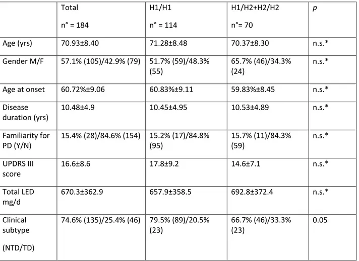
differences in expression and alternative splicing of MAPT transcripts in neurodegenerative diseases. One of the issues believed to underlie the heterogeneity of PD motor features is the interaction of pathogenic proteins involved in neurodegeneration, and tau protein is likely involved in this process. The association between MAPT haplotypes and motor features of PD has, however, not yet been clearly characterised. To evaluate the role of the MAPT gene in motor phenotype in PD, we performed a phenotype-genotype association study to assess the possible influence of H1 and H2 haplotypes.
The human MAPT locus lies on chromosome 17q21, and six isoforms of the encoded tau protein are expressed in the adult human CNS. The isoforms are generated via alternative splicing of exons 2, 3 and 10. Alternative splicing of exon 10 results in a protein containing either three tandem repeats (3R; exon 10–) or four tandem repeats (4R; exon 10+) of a microtubule-binding motif. Alternative splicing of exons 2 and 3 generates a protein with no
N-terminal inserts or with one or two N-terminal inserts (Wade-Martin, Nat Rev Neurolo 2012).
Progress and distribution pattern of PD-related neuronal pathology (ab, accessory basal nucleus of amygdala; ac, accessory cortical nucleus of amygdala; ad, anterodorsal nucleus of amygdala; am,
anteromedial nucleus of thalamus; an, abducens motor nucleus; ba, basal nucleus of amygdala; bn, basal nucleus of Meynert; ca1, first Ammon’s horn sector; ca2, second Ammon’s horn sector; ca, caudate nucleus; cc, corpus callosum; ce, central nuclei of amygdala; cg, central gray of mesencephalon; cl, claustrum; co, cortical nuclei of amygdala; cr, central nucleus of raphe; db, nucleus of the diagonal band; dm, dorsomedial hypothalamic nucleus; dr, dorsal nucleus of raphe; ds, decussation of superior cerebellar peduncles; dv, dorsal nuclear complex of vagal nerve; en, entorhinal region; fn, facial motor nucleus; fo, fornix; gi, gigantocellular reticular nucleus; gr, granular nucleus of amygdala; hn, hypoglossal motor nucleus; in, infundibular nucleus; ir, intermediate reticular zone; lc, locus coeruleus; ld, laterodorsal nucleus of the thalamus; lg, lateral geniculate body; li, nucleus limitans thalami; lt, lateral nuclei of the thalamus; md, mediodorsal nuclei of thalamus; me, medial nuclei of amygdala; mf, medial longitudinal fasciculus; mg, medial geniculate body; ml, medial lemniscus; mm, medial mamillary nucleus; ms, medial septal nucleus; mt, mamillothalamic tract, mv, dorsal motor nucleus of vagal nerve; oi, oliva inferior; os, oliva superior; ot,optic tract; pe, external pallidum; pf, parafascicular nucleus; ph, posterior hypothalamic nucleus; pi, internal pallidum; po, pontine gray; pr, praepositus nucleus; pu, putamen; pv, paraventricular nucleus; re, reticular nucleus of the thalamus; rm, nucleus raphes magnus; ru, nucleus ruber; sb, subiculum; sc, superior cerebellar peduncle; sf, solitary fascicle; so, supraoptic nucleus; sn, substantia nigra; sp, subpeduncular nucleus; st, nucleus of the stria terminalis; su, subthalamic nucleus; te, transentorhinal region; tl, lateral