University of Catania
Research Doctorate in Biotechnology XXIII Cycle
October 31th, 2007 - October 31th, 2011
Coordinator of Doctorate: Dear Professor Federico Cicirata
Cooperation between potassium
channels and gap junctions:
interaction between Kv1.1 channel
and Pannexin 1
Author: Corsaro Veronica Carmen
Supervisor: Dear Professor Federico Cicirata
Academic year
2010-2011
The thesis work has been developed at the Institute of Molecular and Cell Biology, Miguel Hern´andez University, Elche (Spain).
”There is a motive force greater than steam, electricity and atomic energy : the willpower.”
Abstract
The beta 3 subunit of voltage gated potassium channels has been recently identified as a modulatory macromolecule of pannexin 1. Our interest has focused on the possible interaction between the alpha subunit of Kv1.1 chan-nel and pannexin 1. Through voltage clamp studies we have analyzed the electrical activity of the single channels and their behavior when they were coexpressed. With our results we have demonstrated that pannexin 1 was less susceptible to its inhibitors, like probenecid and DTT, when it was co-expressed with Kv1.1 channel, on the contrary pannexin 1 did not seem to influence the activity of Kv1,1 channel. Through immunocytochemistry on HEK-hBK1 cells expressing in stable way Kv1.1 channel we have observed the colocalization of the two channels but through coimmunoprecipitation we proved the lack of a physical interaction between these proteins, therefore the interaction should be functional. Moreover previous studies has reported an involvement of pannexin 1 in apoptosis at elevated concentrations of ex-tracellular potassium. So we wanted to estimate the cell death in presence and absence of pannexin 1 inhibitors, using like control SH-SY5Y cells, being a cell line that does not express Kv1.1 channel. With our results we have ob-served a decrease in the cell death when HEK-hBK1 cells were treated with 1 mM probenecid in presence of 140 mM KCl, suggesting that this behavior was the consequence of pannexin 1 inhibition, therefore in these conditions Kv1.1 channel did not influence in some way its activity; on the contrary the treatment with 10 mM DTT did not produce any beneficial effects in HEK-hBK1 cells. These findings confirmed that Kv1.1 channel influenced the sensibility of pannexin 1 to its inhibitors only when redox potential was altered and then in presence of reducing agents, or when a depolarization of the membrane was induced like in oocytes, but this phenomenon didn’t occur in other conditions. Probably this indirect interaction is mediated by other proteins, such as calmodulin and kinase proteins or by lipids of the
membrane such as PIP2; it could represent a regulatory mechanism that re-places or enhances that exercised by beta 3 subunit on pannexin 1, in order to control the ‘potassium buffering’ and then the cellular excitability and survival, both in pathological and physiological conditions.
Contents
1 Introduction 10
1.1 Electrical Cell communication:
the synapse . . . 11 1.2 Gap junctions . . . 14 1.3 Pannexins, a new family of gap junction proteins . . . 20 1.4 Mechanisms of regulation in gap junctions: pannexins and
connexins . . . 23 1.5 Action Potential . . . 28 1.6 Potassium channels . . . 34 1.7 Mechanisms of inactivation and destabilization in potassium
channels . . . 37 1.8 Eukaryotic voltage gated potassium channels . . . 43 1.9 Potassium Channelopathies . . . 47 1.10 Potassium channels and gap junctions involved in the same
physiological and pathological events . . . 56 1.11 Comparison of aminoacid sequence between beta 3 auxiliary
subunit and Kv1.1 channel of mouse and human . . . 62 1.12 Analysis of electrical currents in ion channels . . . 67
2 Objectives and methods 72
2.1 Objectives and methods . . . 72 2.2 Midiprep of pcDNA3-1 and PCI-neo plasmids containing the
DNA inserts of pannexin 1 and Kv1.1 channel . . . 74 2.3 Linearization of plasmids . . . 75
2.4 Purification of digested DNA . . . 77
2.5 Retrotrascription of purified DNA to RNA . . . 78
2.6 Oocyte extraction from Xenopus Laevis frog . . . 80
2.7 Injection of RNA in oocytes . . . 80
2.8 Two microelectrods voltage clamp (TEVC) . . . 82
2.9 Transient expression of mouse pannexin 1 in HEK-hBK-1 cell line . . . 86
2.10 Midiprep of pBMN-I-GFP plasmid containing the DNA insert of pannexin 1 . . . 86
2.11 Maintenance of cell lines . . . 87
2.12 Cell Transfection . . . 87
2.13 Immunocytochemistry . . . 88
2.14 Valuation of cell death . . . 89
2.15 Preparation of protein extracts . . . 90
2.16 Quantification of proteins . . . 91
2.17 Western Blot . . . 92
2.18 Coimmunoprecipitation . . . 93
3 Results 95 3.1 Expression of pannexin 1 in frog oocytes and electrical activity in presence of potassium chloride . . . 95
3.2 Expression of human Kv1.1 channel in frog oocytes and elec-trical activity in presence of potassium chloride . . . 96
3.3 Coexpression of Kv1.1 channels and pannexin1 in frog oocytes and electrical activity in presence of potassium chloride . . . . 103
3.4 Analysis of transient expression of mouse pannexin 1 in HEK-hBk1 cells by immunocytochemistry . . . 110
3.5 Valuation of cell death during treatment with potassium chlo-ride and inhibitors of Kv1.1 channel 1 and pannexin 1 . . . 121
3.6 Transient expression analysis of mouse pannexin 1 in HEK-hBk1 cells by western blot . . . 125
4 Discussion and Conclusions 131 4.1 Functional interaction between pannexin 1 and Kv1.1 channel 131 4.2 Mechanisms of potassium’s cellular redistribution . . . 132 4.3 A new perspective to define channelopathies . . . 134 4.4 Kv1.1 channel’s influence on pannexin 1 in HEK-hBk1 cell
death mediated by high extracellular potassium chloride . . . 136 4.5 Conclusion and future perspectives . . . 139
Glossary
Br− bromide ion Ca2+ calcium ion Cl− chloride ion Cs+ cesium ion H+ proton K+ potassium ion Li+ litium ion M gCl2 magnesium chloride N O3− nitrateN aHCO3 sodium carbonate
N a+ sodium ion
Rb+ rubidium ion
∆E equilibrum potential for ions
µA microamper µF microfaraday µM micromolar µg microgram µl microlitre Ala Alanine Arg arginine Asn asparagine
Asp aspartic acid
b.p base pairs
BSA bovin serum albumin
C capacity
CaCl2 calcium chloride
CaMKII calcium calmodulin-dependent protein kinase cAMP cyclic adenosine monophosphate
CaNO3 calcium nitrate
cDNA complementary DNA
cGMP cyclic guanosine monophosphate CK1 casein protein kinase
Cx connexin
DAPI 4’,6-diamidino-2-phenylindole DEPC diethyl pyrocarbonate
dH2O distilled water
DMEM modified eagle medium DNA deoxyribonucleic acid
DTT dithiothreitol
Eag ether-`a-go-go potassium channel EDTA ethylenediaminetetraacetic acid EGTA ethylene glycol tetraacetic acid
ER endoplasmic reticulus
FBS fetal bovine serum
FITC fluorescein isothiocyanate
g gram
GΩ gigaohm
GFP fluorescent green protein
Gln glutamine
Glu glutamic acid
HEK-293 normal human embryonic kidney cell line HEK-hBk1 normal human embryonic kidney cell line
sta-bly expressing Kv1.1 channel Hepes
4-(2-hydroxyethyl)-1-piperazineethanesulfonic acid
HERG human channel ether-`a-go-go-related
His histidine
Ic current intensity
IgG immunoglobulin
KATP ATP-dependent potassium channel
Kb kilobase)
KchAP K channel-associated protein
KCl potassium chloride
KCNQ M-type potassium channel
KcsA potassium channel from streptomycens livi-dans
kDa kilodalton (1000 dalton; 1 dalton=1,650 × 10-27)
kHz kiloherz
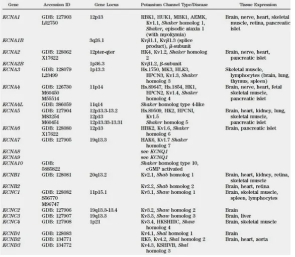
Kv1.1 voltage-gated potassium channel, subfamily A, member 1
Kv1.2 voltage-gated potassium channel, subfamily A, member 2
Kv1.3 voltage-gated potassium channel subfamily A, member 3
Kv1.5 voltage-gated potassium channel subfamily A, member 5
Kv1.6 voltage-gated potassium channel subfamily A , member 6
Kv3.4 voltage-gated potassium channel subfamily C, member 4
L.B standard medium used to grow bacteria LTP long-term potentiations
Lys lysine
M molar
MΩ megaohm
M-type currents voltage-dependent potassium current that persists at slightly depolarized membrane po-tentials
MAPK mitogen-activated protein kinase MEM minimum essential medium
mg milligram
MgSO4 magnesium sulphate
mM millimolar
mm millimetre
mRNA messenger RNA
ms millisecond
nA nanoamper
NaCl sodium chloride
ng nanogram
nl nanolitre
nm nanometre
p.m molecular weight
P2X7 purinoreceptor for ATP
Panx pannexin
PBS phosphate buffered saline PCR polymerase chain reaction
PDZ domain that binds to a short region of the C-terminus of other specific proteins
pF picofaraday
PIP2 Phosphatidylinositol 4,5-bisphosphate
PK kinase protein
PMSF phenylmethylsulfonyl fluorid
Q electric charge
R resistence
RNA ribonucleic acid
Rpm revolutions per minute SDS sodium dodecyl sulphate
SEM standard error
Ser serine
SH-SY5Y human derived neuroblastoma cell line SNC central nervous system
SNP peripheral nervous system
Src tyrosine kinase
STP short-term potentiations
TBST tween phosphate buffered saline
TEA tetraethylammonium
Tes
2-[(2-Hydroxy-1,1-bis(hydroxymethyl)ethyl)amino]ethanesulfonic acid
TM transmembrane
TRICT tetramethyl rhodamine isothiocyanate
Trp tryptophan
Tyr tyrosine
U.V ultraviolet radiation
Chapter 1
Introduction
Gap junctions and ion channels play an important role in the cellular home-ostasis and in the synchronization of electrical signals. A functional interac-tion between potassium channels and gap juncinterac-tions has been already docu-mented (GJ Christ, Drug News Perspective, 13 (1): 28-36, 2000; Kotsias et al, J Biol States, 203 (3 ): 143 -50, 2005), but more recent researchs have shown that also a direct interaction between ion channels and gap junctions occurs (Chanson et al, Prog Biophys Mol Biol, 94 (1-2) : 233-244, 2007). In particular i want to focus on a recent study that described the existence of a physical interaction between the mouse β3 subunit of voltage-dependent potassium channel and the C-terminal region of mouse pannexin 1 (Bunse et al ., FEBS Journal, 276, 6258-6270, 2009) , presenting the beta 3 subunit as new modulator of pannexin 1. In fact this investigation reported that the electrical activity’s inhibition of pannexin 1 by reducing agents and block-ers (like probenecid) is compromised when it is coexpressed with the Kvβ3 subunit, confirming the idea that the latter controls the pannexin activity. This is the first example of functional modulation and physical interaction between pannexin and potassium channels. The study of this cooperation has a great importance because it could clarify the physiological pathways that underlie the electrical synapse and explain the functions of this interac-tion. Before discussing in detail the work i’ve done i’m going to do a brief introduction about the electrical communication, the gap junctions and the
potassium channels, in order to better understand their relationship.
1.1
Electrical Cell communication:
the synapse
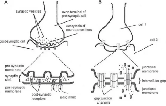
Today it is demonstrated that there are basically two different intercellu-lar communication mechanisms, one that involves the release of secreted molecules such as hormones and neurotransmitters that interact with re-ceptors on neighboring cells and the other is based on the formation of channels that permit a direct contact between the cells with the passage of ions, metabolites and second messengers, allowing the electrical coupling. Synapses are structures through which two neurons communicate (one is called ‘presynaptic’ and the other that receives the signal is called ‘postsy-naptic’). Interneuronal communication has been discussed for years, some neuroscientist was in favor of electrical transmission, in which the action po-tential in the presynaptic cell induces a current flow to the post-synaptic cell, therefore there is no an actual anatomical separation between the two cells but, small molecules and small ions pass directly from the cytoplasm of one cell to another through gap junctions (G.Js) and ion channels, the passage of the potential is direct and faster; while other scientists were in favor of chemical transmission, in which between the presynaptic membrane and the post-synaptic membrane there is an inter-synaptic space, then can’t exist a direct electrical communication and transmission of signals occurs because a chemical substance released by the presynaptic neuron interacts with the re-ceptor channel in the post-synaptic membrane activating a series of intracel-lular responses (release of cytosolic calcium, phosphorylation and activation of PK, CaMKII etc), allowing the propagation of the nerve impulse. Exam-ples of neurotransmitters are acetylcholine, GABA, monoamines, dopamine, that bind their corresponding receptors causing the opening and the entry of ions. These events cause a depolarization with the further opening of addi-tional voltage-gated channels. Anyway has been proved the existence of both chemical and electrical transmission and it is assumed that in vertebrates one
could be complementary to the other (see fig. 1.1). For several years it has been believed that the chemical synapses were only present in the brain of vertebrates. The presence of electrical synapses has been shown for the first time in the axon of the crayfish motor nerve, in these synapses the contact between two neurons is by G.Js (Furshpan&Potter, Nature 180:342-43, 1957; Watanabe, Jpn. J. Physiol. 8:305-18 , 1958; B. Litch et al. Journal of Neurocytology, 18 (6), 749-761; WJHeitler et al. Journal of Neurocytolo-gy, Vol 20, No 2, 109-123, 1991). Electrical synapses function as ’low pass filter’, that transmit low frequency stimuli; the transmission power is more rapid than in chemical synapse, moreover the electrical synapse can be two-directional. This type of communication for many years was considered a feature of the structures of invertebrates, but in reality it plays a key role also in the development of the vertebrate CNS, when chemical synapses are still immature and their number is limited. It has been observed that the electrical synapses is linked to certain events during the development of the CNS, such as cell differentiation, migration and the formation of neuronal circuits. This coupling decreases with the differentiation proceeding. The electrical synapses are abundantly present in the brains of mammals, Cx36 is widely expressed in central nervous system where it plays a key role in coupling neuronal electrical, and probably other proteins are involved like connexin 43 and pannexins. In the CNS electrical synapses seem to occur in most regions of the brain, as in the lower olivary nucleus, in the interneurons of the cerebellum, in the the reticular nucleus of the thalamus, in the retina, in the hippocampus, in the neocortex, in the olfactory bulb and also between the motoneurons synapses. This type of synapse could also allow the passage of small signaling molecules between cells, previous experiments of ’dye cou-pling’ support this idea (Hatton GI., 1998, Cell Biol. Int 22:765-80; Roerig B, Feller MB, 2000, Brain Res Rev 32:86 -114, BW Connors et al. Annu. Rev. Neurosci. 2004, 27:393-418). Several studies have also demonstrated the ex-istence of mixed synapses (chemical and electrical), in which gap junctions play an important role (for example in motoneurons). Mixed synapses are considered a single functional unit (Michelson and Wong, J. Physiol. 477, 35-45, 1994; Bernard, J. Neurophysiol. 77, 3134-3144,1997, Gibson et al. Nature
402:75-79, 1999; Mann-Metzer and Yarom, J Neurosci.19(9):3298-306, 1999; Fukuda and Kosaka, Neurosci. 20(4):1519-28, 2000). Several studies have suggested that mixed synapses are usefuls to synchronize the ‘inflammation’ of neurons (Bernardo, J. Neurophysiol. 77, 3134-3144, 1997; Galarreta and Hestrin, Nature 402, 72-75, 1999, Tamas et al., Nat Neurosci 3:366–37, 2000) and myocytes in the myocardium, or that they represent a combination to facilitate the excitation of the post-synaptic fiber; it is like electrical trans-mission prepares the chemical nerve excitation, (Galarreta and Hestrin, Nat. Rev. Neuro. 2, 425-433, 2001). In addition, mixed synapses are implicated in neuronal ritmogenesis (Galarreta and Hestrin, Nat. Rev. Neuro. 2, 425-433, 2001), providing a mechanism of correction and adjustment of the excitation. Important studies have been performed on the pyloric neurons, in which the two types of communication presented opposite effects in the transmission of the signal from lateral pyloric neuron to the constrictor neuron , in fact when the lateral pyloric neuron (PL) explodes, the electrical transmission promotes the activity of the pyloric constrictor (PY) in which the chemical component acts by killing it (through dopamine) (A. Mamiya et al., The Journal of Neuroscience, October 22, 2003 23 (29):9557-9564). In motoneurons about 3-5% of the axo-somatic and axo-dendritic synapses are mixed and about 30-100% are excitatory synapses.So it is clear that the mixed synapse have a significant influence on the activity of post-synaptic neurons (in the CNS and PNS) and that the chemical and electrical transmissions don’t work al-ternatively, but rather they cooperate. In vitro experiments have shown that the conditions of the medium in which cells growth can modulate the forma-tion of synapses. In fact, when the nerve-cells were cultured and juxtaposed in vitro they formed inappropriate electrical synapses, presumably because of the contact conditions between cells, instead when they were grown sepa-rately they formed chemical synapses. The G. Js in the CNS have been found also in oligodendrocytes and astrocytes (Massa and Mugnaini, Neuroscience, 7 : 523-538, 1982; 1982; Giaume et al. Neuron 6, 133-143, 1991; Altevogt et al., Neurosci 22:6458-6470, 2002) and play an important role both during embryogenesis and in the adult brain, in fact transgenic studies have shown the presence of electrical synapses in the rodent adult brain like in
motoneu-rons of the rat (Jerash et al. Neurobiology, Vol 93, pp. 4235-4239, April 1996; Galarreta and Hestrin, Nature 402, 72-75, 1999; Beierlein et al. Nat. Neurosci. 3, 904-910, 2000; Blatow et al., Neuron, Vol. 38, 79-88, 2003), these data confirms again that these two types of communication are com-plementary. In conclusion the mixed synapses are widely present in most regions of the CNS of mammals, the abundance of these synapses suggests that they play an important role in the transmission of electrical signals but the mechanism by which they function are still unclear.
1.2
Gap junctions
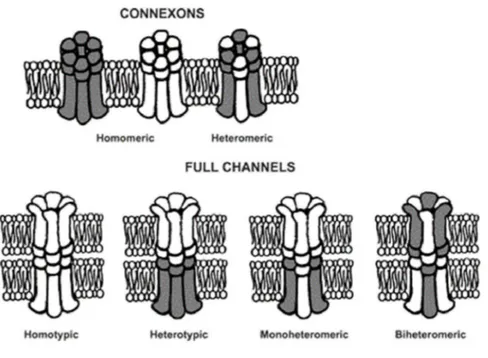
The junctions between cells are of various types, generally consist of: oc-cluding junctions (tight junctions), their main components are the occludins and claudins that seal the gaps between adjacent epithelial cells; adherens junctions, that are formed by cadherins and catenins and create adhesion between cells; the desmosomes, that serve to connect the cells through a fil-ament structure and also connect the cytoskeleton filfil-aments of two adjacent cells; finally the gap junctions, they constitute transmembrane channels that permit the communication between the cytoplasm in adjacent cells through the passage of small ions, small metabolites (< 1kDa) and second messen-gers, (WR Loewenstein, Phy-siol. Rev 61, 829-913, 1981) . The term ’gap’ means ’regular separation’. In fact in two adjacent cells their cytoplasmatic membrane is separated by a regular space of 2 or 3 nm. Gap junctions are multicellular channels large about 16-20 A ➦.These cylinders have been identified using staining with lanthanum hydroxide (Revel and Karnovsky, J.Cell Biol. 33: C7-C12, 1967). Each cell contributes to the adjacent chan-nel with a hemichanchan-nel that consists of six protein subunits, that are called connexins (Beyer et al., J.Membr.Biol. 116, 187-194, 1990, Willecke et al., Eur.J.Cell.Biol. 56, 1-7, 1991; Bennett et al., Neuron 6, 305-320, 1991, Bruz-zone et al., BioEssay 18, 709-718, 1996). The hemichannels are bound with the help of other proteins, the cadherins; when they consist of identical con-nexins are called ‘homomeric’ , if are formed by different concon-nexins are called ‘heteromeric’. Moreover the gap junction is called homotypic when consists
of two identical hemichannels, it is called heterotypic when consists of dif-ferent hemichannels. So we can have four possibilities: G. J homotypic / homomeric, G. J heterotypic / heteromeric, G. J homotypic/heteromeric, G. J heterotypic / homomeric (see fig. 1.2). The closing and opening of these channels is influenced by change in voltage, pH and intra-extracellular ion concentration, by the interaction with macromolecules, by phosphoryla-tion reacphosphoryla-tions (because the conformaphosphoryla-tion of the protein changes), moreover their expression and permeability is controlled by hormons like estrogens in the uterus. The mechanism by which hormones control the permeability of these channels is still unclear, probably this modulation occurs through cyclic AMP. Neurotransmitters may also modulate the permeability of GJS, such as dopamine with an inhibitory action, and acetylcholine, that also reduces the permeability of gap junctions, but how this happens is not yet clear. In both cases the inhibition is reversible. The modulation of the gap junction conductance is of two types: short-term and long-term. The short term mod-ulation is due to changes in permeability in the range of time between seconds and minutes, is too fast that can’t cause changes in the turnover of these pro-teins, it is possible that it is due to alterations of the basic properties of the channels , like variations in pH, in intracellular calcium, in neurotransmitter signaling. Surely this fast modulation permits the cell to respond rapidly to specific stimuli, this phenomenon is called ’functional plasticity’. The long term modulation occur during a larger time, is due to changes in properties of gap junctions or changes in their number. It can be caused by interac-tion with extracellular messengers, in this case the turnover of these proteins changes. For example, this type of modulation occurs during development (Neyton et al., J. exp. Biol. 124, 93-114 (1986)). Communication mediated by gap junctions is required for many biological processes, including cell growth and differentiation, embryonic morphogenesis, metabolic homeosta-sis, muscle contraction and secretion, synchronization of electrical activity. Their pore size is about 1.5-2 nm and permit the passage of ions and small molecules of 1-1.5 kDa (metabolites, second messengers, etc.), but gap junc-tions are not selective pore, cajunc-tions and anions can cross the channel, the passage is free also for dyes such as LY and DAPI (Cao et al, Journal of Cell
Science 111, 31-43 (1998)). The channel formed by Cx43 and Cx40 is perme-able to various ions such as Cs+, Rb+, K+, N a+, Li+, H+, Br−
, Cl−
, N O3−
, acetate, glutamate, but with different affinity for these ions, this affinity de-pends on the composition and the internal charge. The positive ions cross the pore that is partially hydrated and anions move in the opposite direction to the flow and can be complexed with cations (Wang et al, J.Gen.Physiol, 109, 491-507, 1997; Beblo et al, J . Gen.Physiol, 109, 509-522, 1997). The channel is also permeable to divalent cation, such as M g++, Ba++, (Fireck
Ludwick et al, J Mol Cell Cardiol, 27, 1633-1643, 1995, Matsuda et al, Prog Biophys Mol Biol. 2010 Sep; 103 ( 1):102-1) and Ca++. The closure is
of-ten triggered by phosphorylation of the subunits constituting the channel, closure can be observed also by increasing the concentration of intracellular calcium. On cell membrane gap junctions are often present in the form of plaques or agglomerations of many channels (Wang et al, Journal of Cell Sci-ence 108, 3501-3508, 1995). The junctions are easily detectable with the use of dyes such fluorescein. Connexins form a family of membrane proteins that are different for their molecular weight and aminoacid sequence; according to the nomenclature they are indicated with the abbreviation Cx followed by a number indicating the molecular weight. All connexins have four transmem-brane regions (TM1-4) in alpha helix structure, two extracellular loops (E 1-2), one intracellular loop (L), and two cytoplasmic terminal parts, the one shorter is placed before TM1 region and is the amino terminal part, the one longer is placed after TM4 region and is the carboxyl terminal part. Con-nexins are divided into three groups α, β and γ, according to the structure of their gene. The two extracellular loops E1 and E2 and the transmembrane regions are the portions most conserved, for this reason show high degree of homology. For example all connexins have the repetition of three cysteine residues in the extracellular loops. Instead the intracellular loop L and the C-terminal part are not conserved regions, this confer specificity to the pro-tein, in fact these portions are needed for the recognition between two cellular connexons. Furthermore, by varying the length of these regions varies also the molecular weight. The loop E1 moreover should be the voltage sensitive region.Twenty different connexins have been identified in the genome of mice
and rats and three pannexins (Bruzzone et al., Eur J Biochem 238(1):1-27, 1996, Condorelli et al., J. Neurosci. 10, 1202-1208, 1998, Sohl et al., FEBS Lett 428:27-31, 1998). Connexins have been found both in vertebrates and in chordates.
Figure 1.1: chemical (A) and electrical (B) (Zygon , Volume 44, Issue 4, pages 807–824, December 2009)
The specificity of connexins to certain tissues is related to their function, the same connexins can be expressed in different tissues and a specific tissue may coexpress different connexins, allowing the phenomenon of redundancy or compensatory expression that could be helpful in pathological conditions and is important to maintain cellular homeostasis. It is possible that this redundancy exist also between pannexins and / or between pannexins and connexins. The anomalous variation in the number of gap junctions results in pathological states, but this variation can also have a physiological signif-icance. In mammals the G .Js in the uterus are highly expressed in pregnant females, this phenomenon contributes to improve the synchronization of con-tractions during the labour. The increase in the number is possibly caused
Figure 1.2: homomeric and heteromeric gap junctions (Cardiovasc Res (2004) 62 (2):276-286).
by hormonal stimulation. After delivery, the gap junction number decreases again, because these proteins are removed by endocytosis and degraded as all proteins in the proteasome. The compatibility code of connexins allows only selective interactions between connexons. In some cases, the connex-ins can form only homotypic channels , in other cases can form heterotypic channels. For example, Cx46 may form channels with at least 5-6 connexins (Elfgang et al., J Cell Biol. May;129(3):805-17, 1995; White et al., Kidney Int. , Oct;48(4):1148-57, 1995), instead Cx31 can only produce homotypic combinations. In addition, the connexin composition of gap junctions in-fluence the permeability of the channel. For example, the heteromeric gap junction formed by Cx32 and Cx26 is not permeable to cGMP and cAMP, instead the homomeric channel formed by Cx32 is permeable to both signal molecules. The compatibility is strongly linked to the second extracellular domain E2 (White et al., J. Cell Biol. 125: 879-892, 1994). For example Cx43 functionally interacts with Cx46 but not with the Cx50, but swapping the domain E2 of Cx50 with that of Cx46 the capacity to form channels with
Figure 1.3: model of a connexin with cellular partners
Cx43 is moved to the new chimeric macromolecule. This ability of connexins to discriminate between one and the other is very important because it may represent a mechanism to limit rather than promote cellular communication, this is necessary in certain physiological situations. It represent infact a basic mechanism for the ‘compartmentalization’ within the same tissue, so that the cells communicate in the area of the same group but not between different groups. This event mainly occurs during embryonic development. The gap junctions, however, have further partners of interaction, infact connexins can bind calmodulin, kinase proteins and the zonula occludens (ZO-1), a mem-brane protein especially present in tight junctions of epithelial and endothe-lial cells; this protein plays a role in the connexin targeting to the plasma membrane, it mediates also their interaction with the cytoskeleton and per-haps also influences the interaction between different connexins. The GJS can bind also other components of tight junctions, Cx26 for example directly interacts with the occludin (Nusrat et al., Biol Chem. 22;275(38):29816-22, 2000) and has binding sites for calmodulin. These interactions must be still
explored to understand the function and effects on gap junctions.
1.3
Pannexins, a new family of gap junction
proteins
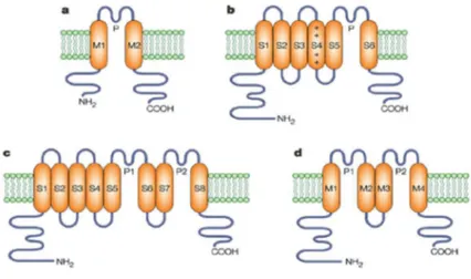
Pannexins (from the greek ”pan,” neuter of the adjective ”pas,” which means ”all,” ”whole,” ”entire” to reflect their broad expression in both protostomes and deuterostomes) have recently been discovered, they form a second family of proteins belonging to the gap junctions. Like connexins they are trans-membrane porins and consist of two hexameric hemichannels. They are structurally similar to connexins, they have four transmembrane portions, two extracellular loops, an intracellular loop and the N-and C- terminal por-tions facing on the cytoplasm (see fig. 1.4). But pannexins are genetically different, they derive from innexins, a group of proteins that form intercellu-lar channels in invertebrates (Panchin, Journal of Experimental Biology 208, 1415-1419, 2005; Barbe et al, Physiology 21: 103 -114, 2006.). There is in-fact some sequence similarity between pannexins and innexins from shellfish, viruses and insects (see fig. 1.4 A). Also in poly DNA virus they have been recently identified and are called vinnexins. Pannexins don’t have aminoacid sequence similarity with connexins but contain two conserved residues of cys-teine in the extracellular loop. Today we know only three pannexins : pan-nexin 1, panpan-nexin 2 and panpan-nexin 3 (panx1,panx2,panx3), their functions are still unclear. Pannexin 1 is more expressed in the central nervous system but is also present in other tissues, pannexin 2 is expressed only in the CNS, are both localized mainly in the interneurons of the hippocampus and in Purkinje cells, pannexin 1 alone or in combination with pannexin 2 induces formation of active intercellular channels, instead homomeric hemichannel of pannexin 2 is not active (plausibly the formation of this channel is in-volved in the compartmentalization process); the expression of pannexin 3 has been identified in the brain but mainly in the skin, in osteoblasts and in fibroblasts with pannexin 1 (Bruzzone et al., PNAS, 2003 November 11, 100 (23): 13644-13649; Baranova et al., Genomics 83, 706-716, 2004, Bench et al.
Curr. Biol. 10, R473-R474, 2000; Bench, The Journal of Experimental Bi-ology 208, 1415 - 1419, 2005). Pannexin 1 forms mechano-sensitive channels in fact, electrophysiological studies have shown that pannexon is active after ’stretching’ (see fig. 1.4 C); it is probably also voltage-sensitive like channels formed by connexins whose opening and closing is dependent on changes in electrical potential (Bao et al. FEBS Letters 572 (2004) 65-68; Yang Qu et al. PNAS, January 22, 2002, vol. 99, no. 2, 697-702). Pannexin 1 molecular weight changes from 37 kDa to 48 kDa, according to the degree of glycosyla-tion. Pannexins infact can exist in different isoforms due to post-translational modifications such as phosphorylation and glycosylation that occur during their synthesis and are important for their formation (Boasso et al, The Journal of Biological Chemistry, 282 (43), 31733 -31743, 2007, Jiang et al, Investigative Ophthalmology & Visual Science, Vol 34, No. 13, 3558-3565.., 1993;. Shearer et al, Investigative Ophthalmology Visual Science &, April 2008, Vol 49, No..4,1553-1562). The post-translational modifications (PMT) can affect the folding of proteins and are important for their function, it is in-teresting that some proteins with different structure and sequence, but with similar PMT show similar function. Pannexins unlike connexins are highly glycosylated (from mannose), but Pannexin 1 exist also in a non-glycosylated form (Pe˜nuela et al, Journal of Science 120, 3772-3783, 2007; Pe˜nuela et al, MBoC, Vol 20, Issue 20, 4313 -4,323, 2009); glycosylation occurs in predicted aminoacid sites as in the arginine of extracellular loop in position 254, that is important to address the protein on the cytoplasmic membrane (Boasso et al, The Journal of Biological Chemistry, 282 (43) , 31733-31743, 2007), but the reaction may also occur in the C-terminal part (Pe˜nuela et al, Journal of Science 120, 3772-3783, 2007; Pe˜nuela et al, Molecular Cell Biology, Vol.20, 4313-4323 , 2009). Glycosylation is important for the cellular localization of pannexins and also for their interaction and assembly. In fact when these pro-teins exist in non-glycosylated form they have intracellular distribution be-cause fail to reach the membrane. N-glycosylation occurs on arginine residues and O-glycosylation occurs on serine or threonine residues present mainly in the cytoplasmic portions. In pannexins happens mainly N-glycosylation (see fig. 1.4 B). Panx1 moreover facilitates the location of panx 2 on the cell
surface and also colocalizes with panx3, so panx1 should be important for the functionality of the other two pannexins. The function of pannexins is not yet clear. They could have an important role in the synchronous activity in the brain, for example pannexins are also expressed in certain types of interneurons, it is interesting to note that the residual gamma activity in the knockout animal can be completely wiped out with carbenoxolone, which is a potent pannexin blocker (Pais I et al., J Neurophysiol 89: 2046–2054, 2003). A relationship between schizophrenic symptoms and disruption of neural synchrony has been suggested by a recent paper that reported a lower frequency of gamma-band oscillations in schizophrenic patient (Ray A et al., Eur J Neurosci 21: 3277–3290). Moreover it has been demonstrated that the function of the nucleus accumbens may be disturbed in schizophrenia; pan-nexin 1 is distributed also in this nucleus (O’Donnell et al., Ann NY Acad Sci 877: 157–175, 1999) .Thus it is probable that all neuronal gap-junction pro-teins are candidate genes in the familiar forms of schizophrenia. Pannexins can work like hemichannels (Huang et al.Neuroscience 104 (15):6436-6441, 2007), because past studies through oocyte voltage clamp have reported that, first injection of synthetic RNA for rat Panx1 results in the development of nonselective, voltage-activated currents in the nonjunctional plasma mem-brane, indicating that this pannexin can assemble homomeric hemichannels; second, Panx1 hemichannels are permeable to small molecules; third, Panx1 shows the ability to form functionally competent intercellular channels that can be closed by commonly used gap-junction blockers (Bruzzone et al., J Neurochem 92: 1033–1043, 2005; Bruzzone et al., Proc Natl Acad Sci USA 100: 13644–13649, 2003; Locovei et al., FEBS Lett 572: 65–68, 2004). More-over previous studies have shown that pannexins have a role in the ATP release and in the calcium homeostasis; for example the hemichannel activ-ity is important in erythrocytes where in response to low oxygen it releases ATP, and in astrocytes where hemichannels are formed only by pannexin 1 and not by connexins (Bao et al. FEBS Lett, 572, 65 - 68, 2004; Locovei et al, PNAS, 2006 May 16, 103 (20): 7655-7659; Iglesias et al., The Journal of Neuroscience, May 27, 2009, 29 (21):7092-7097). Pannexon activity may be required in many cellular pathways that are still unclear or unknown, an
example is the involvement in ILβ1 release and in caspase-1 cascade, that are mechanisms induced by activation of ATP-gated P2X7 receptor expressed on macrophages; recent studies have shown that pannexin 1 is associated with this receptor, moreover that its blocking inhibits the release of ILβ1 and that when it is overexpressed it works like a non-selective hemichannel stimulat-ing the cascade of caspases (Pellegrin et al. The EMBO Journal (2006) 25, 5071-5082). Panx 1 is also involved in the calcium wave or indirectly by ATP releasing and cooperation with P2Y receptor and through IP3 diffusion, or directly with calcium diffusion between cells (Barbe et al, Physiology, 21:. 103 -114, 2006;. Abeele V. et al, The Journal of Cell Biology, Vol 174, No. 4, 535-546, 2006). Anyway further studies are required to clarify the cellular pathways in which pannexins are involved.
1.4
Mechanisms of regulation in gap
junc-tions: pannexins and connexins
The opening of gap junctions is certainly regulated but further analysis are necessary to clarify the mechanism of this regulation. For example intra-cellular calcium appears to play a role in the modulation of gap junctions (as in Cx32) through calmodulin, that interacts with the C-terminal part of connexins (Torok et al, Biochem J. 326, 479-483, 1997.) and possibly with the C-terminal region of pannexins. Also the extracellular ATP might have a regulatory action, infact previous studies reported that ATP has an allosteric effect on pannexin 1, inhibiting its permeability (Qiu et al, Cell Physiol, 296: 250-255, 2009). Another factor that regulates the activity of gap junctions is the temperature. A recent research documented that hemichannels formed by Cx26 increases its conductivity after heating, this temperature dependence may have a physiological significance in thermoreg-ulation, in fact, Cx26 is expressed in the skin (Steffens et al. BBA, Vol 1778, 1206 -1212, 2008). It is interesting to note that Panx1 is also expressed in the skin. Many studies have also shown that cytoplasmic acidification affects the activity of gap junctions and that can cause alterations on phosphorylation
sites in the C-terminal part with a consequent conformational change and closing of the channel (Alan F. Lau, Sci STKE, 2005 (291). In particular, studies performed on Cx43 show that acidification leads to protonation of the histidine residues in Cx43L2 sequence (in the intracellular loop) facilitating the interaction with Cx43CT (the C-terminal part), moreover this acidifica-tion increases the formaacidifica-tion of alpha-helices, in particular in 119-144 domain that is important for dimerization between Cx43CT and Cx43CL (Alan F Lau Sci STKE;. 2005 (291), J. Hirst-Jensen et al, The Journal of Biolog-ical Chemistry Vol 282, No. 8, pp 5801-5813, 2007; Sorgen et al, Biophys J, Vol 87, 574-581, 2004). This is a hypothetical model of ’ball-and-chain’ inactivation (Duffy et al, J Biol Chem, Vol 277, No. 39, 36706-36714, 2002, Duffy et al, Circ Res 94, 215-222, 2004) where the low pH facilitates the process of dimerization that is also regulated by phosphorylation (Sorgen et al, Biophys J, Vol 87, 574-581, 2004; Hirst-Jensen et al, The Journal of Bi-ological Chemistry Vol 282, No. 8, pp 5801-5813, 2007). Pannexins could have a similar mechanism of dimerization. Acidification inhibits activity of gap junctions also because reduces their coupling (Rorig et al., January 1, 1996 The Journal of Physiology, 490, 31-49). In addition, pure lipids can play a role in the modulation of pannexins, in particular in their conformation, in fact, cholesterol is involved in the remodeling of gap junctions (Biswas et al, Molecular Vision 2009, 15:1492-1508) and can stimulate or inhibit their assembly and their permeability depending on the concentration of choles-terol (Lars Bastians et al, Cardiovasc Res 33, 272-283, 1997, Meyer et al, J Cell Sci, 96:231-238, 1990). Lipids may be also involved in the formation of plaques, where gap junctions shows an increased activity (Ghosh et al , Bio-electrochemistry, 68, 150-157, 2006); studies on the clustering of pannexins must still be done.
The reversible phosphorylation by kinases is another mechanism of mod-ulation of a large number of proteins, including Panx1. For example, the permeability of Cx43 is affected by phosphorylation in Ser 368, resulting in a conformational change in the C-terminal part (Bao et al., J Biol Chem, 279 (19), 20058-20066, 2004). NetPhosK program identifies many sites of phos-phorylation in the sequence of Panx1 and particularly in the C-terminus,
for example, it reports that Ser 328 could be phosphorylated by PKA and DNAPK, Ser 343 by PKC, Thr 383 by PKC, Ser 405 by CKII etc. C-terminal part is the portion that mainly interact with other macromolecules such as zonula occludens ZO-1, calmodulin and kinase proteins, the phosphorylation itself can modulate these interactions (The Journal of Biological Che- mis-try, Sorgen et al, 279 (52), 54695-54701, 2004;. The Journal of Biological Chemistry, Singh et al, 280 (34), 30416-30421, 2005, Toyofuku et al., J Biol Chem, 276 ( 3), 1780-1788, 2001). Modulation mechanisms of pannexins are little known, so further studies on their phosphorylation, glycosylation and possible interactions with calmodulin, ZO and other macromolecules should be performed. Finally, it is also interesting to consider the pharmacologi-cal modulation of pannexins, in particular carbenoxolone (anti-ulcer medica-tion), FFA (NSAIDs) and the probenecid (drug used for gout) have already been identified as blockers of pannexin 1, also reducing agents like DTT (1,4-dithio-D-threitol) and TCEP (Tris(2-carboxyethyl)phosphine) cause its inhi-bition, as shown by the analysis of electrical activity (Bruzzone et al, Journal of Neurochemistry, 2005, 92, 1033-1043; Silverman et al, Am J Physiol Cell Physiol 295:761-767, 2008; But Weihong, The Journal of Pharmacology and Experimental Therapeutics, Vol 328, No. 2, 2008; Bunse et al., FEBS Jour-nal 276 (2009) 6258). Previous patch and voltage clamp studies in oocytes show that the channels costituted of pannexins are activated by extracellu-lar potassium and have a extracellu-large conductance of up to 500 ps when perfused with 150 mM KCl (Li Bao et al, FEBS Letters 572 (2004 ) 65-68). How potassium chloride actives pannexon in vivo is still unclear, the hypothesis is that the ion binds the extracellular domain directly or through an aux-iliary molecule, causing the activation, moreover if the pannexin activation by potassium chloride occurs also in vivo is not yet known (Silverman et al, The Journal Of Biological Chemistry Vol 284, No. 27, pp. 18143-18151, July 3, 2009). Inhibitors such as carbenoxolone and probenecid significantly inhibit the electrical activity in a dose-dependent manner (Silverman et al, The Journal of Biological Chemistry, Vol.284, N 27 , 2009). Carbenoxolone abolishes the electrical activity of pannexins with much lower doses than those requests to inhibit connexins, probenecid seems to be enough specific
for pannexins rather than connexins. The mechanism by which probenecid abolishes the activity is not clear, may interact with the hydrophilic parts of pannexin or access to the channel through the lipid layer of membrane considering that it is lipophilic. Moreover it induces depolarization of the plasma membrane, interferes with mitochondrial oxidative phosphorylation and ATP production (Masereeuw British Journal of Pharmacology (2000) 131, 57 – 62). At a concentration of 1 mM there is almost total inhibition of electrical activity (Silverman et al, Am J Physiol Cell Physiol 295: C761-C767, 2008; But Weihong, The Journal of Pharmacology and Experimental Therapeutics, 328:409-418, , Vol 328, No. 2., 2009). Recent researchs have shown that the susceptibility of pannexin 1 to reducing agents and to their inhibitors is reduced when coexpressed with the Kvβ3 subunit (Bunse et al. FEBS Journal, 276, 6258-6270, 2009) and that a physical interaction occurs between both proteins. These data are interesting because the same phe-nomenon could occur during hypoxic conditions such as ischemia. However, pharmacological characterization of gap junctions and in particular of pan-nexins requires further evaluations, as well as their functional and structural characterization.
Figure 1.4: A: topography of pannexins and connexins (The Journal of Experimental Biology 208, 1415-141, 2005); B :glycosylation sites of pannexins (Journal of Cell Science 120, 3772-3783, 2007); C: mechanical activation of pannexin channels (FEBS Letters 572 (2004) 65–6).
1.5
Action Potential
Electricity plays an important role in biology, when solutes such as phos-phate, aminoacids and inorganic ions are transported across cell membranes the movement of their charges produces an electrical current that generates a membrane voltage difference. In general, electrical signals are mainly gener-ated by the flow of inorganic ions such as N a+, K+, Ca2+, and Cl−
that pass through large transmembrane proteins called ion channels. To generate an action potential in a neuron are required millions of ions in one millisecond, so to fill this request without using millions of proteins is necessary that each single channel presents high adaptability and high selectivity. A cell can be compared to a RC circuit (resistor-capacitor) based on the resistance and on the presence of a dynamic element, the condensator that stores and releases electrical charges (see fig. 1.5). The membrane is in fact able to conduct ionic current, and has both resistive and capacitive elements. The resistance depends on the cell ability to transport ions, it is inversely proportional to the amount of water present in the cell (for example the lipidic bilayer is a poor conductor, unlike water, and opposes a resistance to the passage of ions); seeing that the flow is given by the opening of ion channels, the higher the number of open channels the greater the conductance and less the resistance. The capacity of the membrane depends on its hydrophobic nature because the lipidic bilayer is a poor conductor. When the charges are separated by the lipidic bilayer generates a potential difference whose magnitude depends on the characteristics of the capacitor. The membrane capacity thus influ-ences the transition phase during which the action potential changes. At the opening of the ’circuit’ the current is predominantly capacitive, then when the potential reaches the stationary phase, the current becomes resistive. In fact, the flow of capacitive current occurs only at the beginning and at the end of the potential change, but, during the fixed phase of the potential, ionic current can be considered pure. Extracellular fluids have high con-centration of sodium and chloride instead intracellular fluid contains mostly potassium and organic anions, this difference of ionic distribution generates also a difference in electric charge between the membrane sides, the
inte-rior side will be negatively charged and the exteinte-rior side will be positively charged. The capacity of the membrane, as mentioned before, maintains this charge separation generating the potential difference. The amount of charge Q is proportional to the difference in potential and capacity:
Q = C × V
The movement of charges from one side to the other generates the elec-trical power that is proportional to the capacity and to the speed at which the voltage changes over time:
IC = C × dV dt
So we can express the variation of the voltage as a function of current and time during which the current is passed:
dV = Ic×dt
C
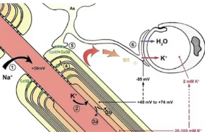
Seeing that the capacity increases with the size of the cell, to determine a change of membrane potential in a large neuron respect to a smaller neu-ron will be necessary more charges, and therefore more current, so cells of smaller dimensions are more excitable. Nervous and muscle tissues are for example excitable tissues, when cells do not conduct impulses they are at rest. Typical values of resistence for a cell move between 1 MΩ and 100 MΩ, current values move normally from fractions to hundreds nA. The rest membrane potential remains around -70mV, this value varies depending on the type of cells ( nerve, skeletal or smooth muscle cells), the resting poten-tial of neurons for example is - 65mV. During this resting phase anion and sodium channels are closed, but potassium channels remain open. The rest-ing potential is generated by an unequal distribution of ionic species between intracellular and extracellular fluid; sodium and chloride are in fact about 40 times more concentrated outside the cell and potassium 50 times more concentrated inside the cell. If the cell was permeable only to one ion it will reach the electro-chemical equilibrium.
Intracellular concentration Sodium : 12 mM Potassium: 150 mM chloride : 4 mM Extracellular concentration Sodium: 145 mM Potassium : 4 mM chloride: 118 mM
The Nernst equation allows to calculate the potential difference at the electro-chemical equilibrium of a single ion:
∆Ex = RTzF log[X][X]e
t
where:
❼ ∆Ex = equilibrium potential for ion X
❼ R= gas constant
❼ T = absolute temperature (Kelvin) ❼ z = valence of the ion
❼ F = Faraday constant (96,500 coulombs / gram equivalent of charge) ❼ [X]i = intracellular concentration of the ion X
❼ [X]E = extracellular concentration of the ion X
The Goldman equation instead gives a quantitative description of mem-brane potential, it can be considered as an approximation of Nernst equation but takes in consideration the permeability to different ions:
V m = RT zF log
[N a+]eP N a+[K+]eP k+[Cl−]iP Cl
[N a+]eP N a+[K+]eP K+[Cl−]eP Cl
With the help of these formulas we can obtain the contribution to the potential of ionic diffusion when the cell is at rest phase. An action poten-tial is a electrical fluctuation that travels along the surface of the cellular plasma membrane, it occurs when a cell is ”activated”, so when it carries out a pulse. The Hodgkin-Huxley model is a mathematical equation that de-scribes the process of depolarization in the neuron, is based on the analogy of the neuron with an electrical circuit. The findings of Hodgkin and Hux-ley scientists about cell depolarization, the membrane potential inversion and their dependency on external ion concentrations, led them to believe that there was an ionic species responsible for this behavior. Thus it born the idea of the ‘sodium current’, according this idea the membrane depo-larization causes first a rapid rise and then a slow descent of the membrane permeability to sodium ions (N a+) followed by a slow rise in permeability
to potassium ions (K+). The experiments carried out between 1938 and
1940 by Cole and Curtis, and then by Hodgkin and Katz, showed that the action potential of squid giant axon is due to an abrupt change in the N a+
flow crossing the membrane. The hypothesis of Hodgkin and Huxley was as follows: the membrane depolarization opens N a+ channels, allowing a massive entry of N a+ into the cell and thus producing the action potential
upswing, followed by the downturn of the action potential caused by the clo-sure of N a+ channels that reduces the influx of this ion, and the opening
of K+ channels that leads to greater release of K+ from the cell. To test
this hypothesis it was necessary to measure the changes of the conductances of N a+ and K+ channels when membrane potential E changes. Cole
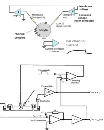
in-vented a method to measure these changes called ”at voltage block” (voltage clamp), which allowed Hodgkin and Huxley to perform their experiments. The block voltage circuit consists in connecting with two electrodes the ends of a battery to the fluids that contact the axon membrane sides. In this way the membrane potential is controlled by the experimenter, which may impose variations. The amperometer records the current that the battery must deliver to maintain a constant membrane potential, this current
repre-sent the current flowing through the membrane. Analyzing the conductance in correspondence to depolarizing pulses of different intensity and duration, Hodgkin and Huxley obtained the empirical equations that allow to predict with good accuracy the size and shape of action potential, refractory period and other characteristics of the nerve impulse (Hodgkin AL, Huxley, AF, andKatz, B., Measurement of current-voltage relations in the membrane of the giant axon of Loligo J. Physiol. 116: 424-448, 1952). The complexity of the model does not allow the mathematical resolution ( a significantly simplified model is that of Fitzhugh-Nagumo), however, it allowed to explore and understand the electrical circuit of a neuron. The model describes three types of channels: sodium channels, potassium channels and leak channels that are represented mainly by chloride channels.
The equations are as follows: CdV dt = gN am 3h(V − V N a) − gKm4(V − VK) − gL(V − VL) + Ia dm dt =αm(V )(1 − m)−βm(V )m dove:
❼ C is the capacity of the membrane ❼ Ia is the external electrical impulse
❼ gN a and gK are the conductance of sodium and potassium ions
❼ V is the equilibrium potentials
❼ gL is the leak conductance of linear channels
The phases of an action potential in general can be described like this:
❼ An adequate stimulus causes the opening of sodium channels. Sodium enters the cell and establishes a local depolarization
❼ If the local value of the depolarization reaches or exceeds a certain value (threshold potential, with an average of about - 50 mV) many voltage-gated sodium channels open. The potential is the minimum threshold value for the opening of these channels. If the threshold is not reached these voltage-gated sodium channels remain closed, without generating an action potential.
❼ The entry of sodium causes decrease in the cell membrane potential, which assumes positive values (on average up to +30+60 mV), so occurs an inversion of charge between the membrane sides.
❼ The voltage-gated channels remain open for about 1 millisecond, then close again.
❼ Once reached the action potential and after the closing of voltage-gated sodium channels, the cell begins the repolarization phase, the membrane potential returns to its resting negative value. The voltage-dependent potassium channels open only to values of around +30/+60mV. The potassium ions begin to go out from the cell for reasons of chemical and electrical gradient because there is an excess of positive charges in the intracellular side of the membrane; moreover during this phase the permeability to chloride increases and calcium channels open (plateau phase), then calcium gets in the cell and when the entry of calcium is equal to the exit of potassium occurs the plateau. Later also calcium channels close and potassium continues to get out from the cell. The flow of potassium towards the extracellular space restores the original excess of positive ions on the external surface. The potassium channels may remain open even when the cell has reached the resting potential: in this way a further amount of potassium comes out and for a short period of time the cell can be hyperpolarized reaching values more negative like -80/-90mV; at this stage it is impossible creating another depolarization.
❼ Finally the action of Na+/K+ pumps that bring out sodium ions and
The action potential works on ‘all or nothing law’, it means that it occurs maximally or not at all and only if the depolarization reachs the threshold value.The refractory period is the amount of time it takes for an excitable membrane to be ready for a second stimulus once it returns to its resting state following excitation. The absolute refractory period is the interval during which a second action potential absolutely cannot be initiated, no matter how large a stimulus is applied. The relative refractory period is the interval immediately following during which initiation of a second action potential is inhibited but not impossible.
Figure 1.5: RC circuit
1.6
Potassium channels
Ion channels are divided into: voltage-gated channels that open in response to changes in membrane potential, ligand-gated channels that open upon in-teraction with a chemical mediator, and mechanical channels that only open after mechanical movements of the membrane. The potassium channels are widely distributed, they transport potassium across the cellular membrane, they are classified in :
Calcium-dependent channels that open in the presence of calcium. Channels ’inwardly rectifying’ in which the ion current is directed to-ward the intracellular space.
❼ Channels ’tandem pore domain’, that are constitutively open or have high activity, maintaining the negative membrane potential; they are similar to leak channels represented mainly by chloride channels, that are independent of the voltage and are always open, participating in the natural permeability system of the cell.
❼ Voltage-gated channels that open and close in response to changes in membrane voltage.
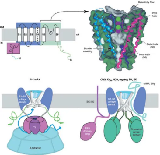
Potassium channels represent a large family of channels, including about 70 different genes, of which about 40 genes encode for voltage-gated potas-sium channels such as eukaryotic Shaker channels that are homologous to Shaker channels from Drosophila melanogaster. Structurally, Shaker potas-sium channel consists of 6 transmembrane alpha helices segments (S1-S2-S3-S5-S5-S6) and the N and C-terminal cytoplasmic portions (see fig. 1.6, 1.7 and 1.8). The C-terminal portion is the part that mainly interacts with the cytoplasmic macromolecules, for example in voltage-gated channels it contains the PDZ binding domain that binds ’like PSD-95’ protein, this in-teraction is important for the clustering. In addition, the C-terminal region belongs to the recent class of proteins called ’intrinsically disordered’, or rather proteins that lack a defined tertiary structure under physiological na-tive conditions. In fact, the C-terminal sequence has some unusual features for a folded protein ,it is rich in hydrophilic aminoacids, glutamine repeats and presents depletion of hydrophobic aminoacids. These features suggest that the primary sequence is unstructured and therefore belongs to the ’in-trinsically disordered protein’ family, thus with highly flexible structure that facilitates the different conformations required for the binding to enzymes or other proteins (Magidovich et al. Bioinformatics, Vol . 22 no. 13 2006, pages 1546-1550). The S1 and S4 segments are voltage sensitive, in particular S4 region contains residues of lysine and arginine that are positively charged and are involved in the voltage-sensitive function, infact their movement during the depolarization leads to the channel opening. The domain of TM S5-S6 P-core is the part involved in the selectivity, in the inactivation and in the tetramerization. All potassium channels are tetrameric with each monomer
placed around the central pore that presents structural differences between the various types of channels. The S4 segment contains cysteine residues, for example in Shaker channels has been studied the function of cysteine in position 361; these cysteine residues with the formation of disulfide bridges are important for the activation of the channel, infact the removal of these aminoacids leads to a mutant that is susceptible to oxidation, the latter oc-curs in absence of C-type inactivation, impeding the channel opening. So the formation of disulfide bonds is part of the activation process and contributes to the structural changes (Aziz et al., The Journal of Biological Chemistry, Vol 277, No. 45, Issue of November 8, pp. 42719-42725, 2002). Studies on ’voltage gating’ were performed in KvAP voltage-dependent channel (of Aeropyrum pernix). This channel is structurally different from Shaker chan-nels, however, presents some aminoacidic sequenc’s similarity; its 3D struc-ture is already known, it has been used to study the interactions between the subunit S1 and S4, and other regions of the channel like the pore, ob-taining various models of interaction (Shrivastava et al. Biophysical Journal Volume 87 October 2004 2255-2270). All potassium channels present in the pore region TM five aminoacids highly conserved , TVGYG in KcsA (bacte-rial channel), this domain is homologous to the eukaryotic domain (see fig. 1.12 and 1.14) and it is involved in the selectivity. Normally the potas-sium ion is solvated by water molecules, in particular, is surrounded by two groups of four oxygen atoms, the selective filter of the channel is designed precisely to mimic the structure of water around the potassium ion, in fact, the aminoacids involved in the selectivity expose oxygen groups for the ion solvation. The structure of the pore fails to adapt to the sodium ion, per-haps for the size, so the sodium ion can not cross it. In fact, the hypothesis is that the constriction of the channel is not sufficient to accommodate the dehydrated sodium ion that is too small and therefore the energy barrier to transfer the ion is too high.
1.7
Mechanisms of inactivation and
destabi-lization in potassium channels
Potassium channels must be able to open and close rapidly in response to biological signals, this process is called ’gating’ and is due to conformational changes of the channel protein. However, usually differences in the structure of the pore domain correspond to different gating mechanisms, for example the bacterial KcsA channel gating depends on the pH , the gating of KvAP (Aero-pyrum pernix) and eukaryotic Kv channels depends on voltage. A channel moves from closed state to open state in less than 10 microseconds, a channel never takes the shape of ’half open’. There are actually three states: closed, open and inactive. In the closed state the channel does not work but it can be activated after a stimulus, usually channels close when the membrane is at rest condition; instead in the inactive state the chan-nel can not be open from any pulse. The inactivation follows a period of prolonged activation, for example, is a phenomenon that occurs during the refractory period and it is reversible. One of the most important mechanisms of inactivation of these channels is the N-type Inactivation , also called A-type Inactivation or Fast Inactivation, in which a subunit of the N-terminal portion ( called ‘ball’ ) blocks physically the passage of ions. Alternatively, the inactivation may be mediated by the beta subunit, that is an auxiliary subunit of the channel, it blocks the channel with its N-terminal portion in a mechanism similar to N-type inactivation (see fig. 1.8 , 1.9 and 1.10). In mammals there are three beta subunits, the β1, β2 and β3. They are highly conserved. During the depolarization the channel opens transiently, then falls in the inactive state due to a conformational change and then opens again during repolarization. The model of ’ball-and-chain’ inactivation has been studied on potassium channels expressed in Drosophila melanogaster (encoded by the Shaker gene) and on channels expressed in oocytes of Xeno-pus frog. Experiments of deletions and insertions have shown that the first 83 aminoacids of the N-terminal portion are involved in the channel inac-tivation. In fact when some of the first 20 aminoacids were changed the channel lost the capacity of inactivation, but when deletions were induced
in the aminoacidic portion 20-83 the channel showed hyperinactivation, on the contrary insertions reduced it. Moreover intracellular trypsin removed the inactivation, suggesting that in the ’ball’ were present sites for trypsin cleavage (see fig. 1.11).
Figure 1.6: functional voltage-gated potassium channel domains (The Journal Of Biological Chemistry, Vol. 283, No. 37, pp. 25105–25109, 2009).
Figure 1.7: structure of potassium channel in various species: a: KcsA ; b: Shaker ; c: TOK1,motivi trovati nei funghi; d: Paramecium bursaria, (Nature Reviews Neuroscience 3, 115-121 , 2002).
Patch clamp studies have also shown that the ’ ball-and -chain’ inac-tivation is not voltage-dependent, infact only during acinac-tivation there is a movement of charges, but any electrical component seems to be associated to the inactivation. The latter is instead removed by proteolytic agents and the increase of extracellular potassium concentration causes a total recovery of activity ( Antz et al., News Physiol. Ski. Volume 13, August 1998), but
Figure 1.8: the tetrameric 6TM architecture of the K+ channel family (Nature 419, 35-42, 5
also an excess of extracellular potassium can inactivate the channel; more-over inactivation can be mimicked by blockers such as tetraethylammonium ( TEA) and 4-aminopyridine. Voltage gated potassium channels infact are very sensitive to TEA inhibition, this molecule binds the extracellular tyro-sine residue in position 449 in each subunit of the tetramer, the consequent inactivation is also temperature-dependent, suggesting that this interaction is not purely hydrophobic (Heginbotham L; Neuron. 1992 Mar;8(3):483-91).
Figure 1.9: ‘ball and chain’ inactivation (Bioinformatoc, Vol. 22 no. 13 2006, pages 1546–1550).
Figure 1.10: NH2-terminal ball (News in Physiological Sciences, Vol. 13, No. 4, 177-182, 1998)
In addition to the N -type inactivation and the inactivation mediated by beta subunit, there is also an additional blocking mechanism called C-type inactivation that is due to a transition of the selective filter and represent a slower inactivation. This type of inactivation has been studied in KcsA, the potassium channel from Streptomycens lividans. This channel is used
Figure 1.11: removal of trypsin inactivation (News in Physiological Sciences, Vol. 13, No. 4, 177-182, 1998)
as model to explore the molecular basis of selectivity and ion permeation, because it presents a simplified structure,it is a tetramer, each unit of the tetramer is composed of two transmembrane subunits M1 and M2 connected by an extracellular loop, that have sequence and structure homologies with S5 and S6 subunit of the eukaryotic channel (see fig. 1.7 and 1.14); it is permeable to potassium, rubidium, cesium, and is not very permeable to sodium and lithium. Moreover past studies have shown that mutations of some residues in TM domain of KcsA makes it similar to the Shaker channel, it gets high affinity binding to agitoxin ( AgTx2 ) that blocks Shaker channels, on the other hand chimeric channels obtained by the replacement of segments S5 -P- S6 of Shaker channels with M1-P- M2 segments of KcsA are functional channels, suggesting that the structure of the pore domains is similar in the two proteins. The structure of crystallized KcsA was the first to be identified and this has allowed to understand some of the workings mechanism of potassium channels.
In KcsA does not occur the N-type inactivation. The C-type inactivation is governed by the interaction between the residues W67, D80 and E71 (see 1.13), because previous studies have shown that the change of glutamate in position 71 with valine causes the lack of inactivation, the mutant infact has high conductance even in the presence of sodium (Cuello et al. Nature. 2010 July 8, 466 (7303): 203-208; Choi etc. al. Biophys J. 2004 April; 86 (4): 2137 - 2144.). Moreover this channel undergoes the destabilization in the presence of high concentrations of extracellular cations, not only in the presence of sodium ions to which the channel is not permeable, but also in the presence
Figure 1.12: KcsA structure and selectivity filter (Nature 414, 37-42(1), 2001)
of potassium, in fact the high extracellular concentration of this ion leads to the dissociation of the tetramer, to the affinity reduction for the ion and thus to the inactivation of the channel. Anyway similar process of destabilization could occur in the eukaryotic channels. As well as in KcsA, in eukaryotic voltage-gated channels the aminoacids that are involved in C-type inactiva-tion are adjiacent to the highly conserved region of the pore that is critical for the integrity of selectivity; this type of inactivation is voltage-dependent. The interaction between certain aminoacid sites present in the pore leads to a constriction of the channel inactivating it. The channel goes into a state of low opening probability that is sensitive to extracellular potassium con-centration, when the external potassium increases the channel passes in this reversible inactive state (Morales et al., Nature Structural Molecular Bio-logy &, Volume 13, Number 4 April 2006, Kiss et al. Biophysical Journal Volume 74 April 1998 1840-1849). Recent studies of patch clamp in giant liposomes showed that the ball peptide, that is the ‘ball’ domain of N-terminal portion of Shaker channels involved in the ball-and-chain mechanism, is able to give ’Fast Inactivation’ in KcsA channel. In fact, when the synthesized ball pep-tide is placed in the bath solution the electrical activity reduces (Molina et al., The Journal of Biological Chemestry Vol 283, No. 26, pp. 18076-18085, June 27, 2008).
Figure 1.13: aminoacidic interaction in KcsA C-type-inactivation (Nature Structural & Molec-ular Biology, Volume 13, Number 4, 2006)
1.8
Eukaryotic voltage gated potassium
chan-nels
Eukaryotic voltage-gated channels are formed by tetrameric transmembrane glycoproteins, called also ‘alpha subunits’. They form aqueous pores for the passage of potassium that open and close in response to changes in voltage. These channels are involved in the maintaining of the resting potential, in repolarization, and in the temporal control of action potential repetitions in neurons; they also participate in electrical activity of smooth muscle and myocardial cells (Koh, SDET al. delayed rectifier potassium currents of Con-tribution to the electrical activity of murine colonic smooth muscle, Ameri-can Journal of Physiology 515, 475-487, 1999) and in electrolyte homeosta-sis of epithelium. The first potassium channel was cloned from Drosophila Melanogaster fly (Papazian et al., Science (Wash DC) 237:749–753, 1987). Thereafter began the identification of this very large family of ion chan-nels,the voltage-dependent potassium channels. Initially, four genes of Kv were cloned in Drosophila: Shaker, Shab, Shal and Shaw. These genes had different isoforms by differential splicing. The first channels cloned into mam-mals were associated with these isoforms, for this reason the KV1 subfamily was called Shaker . The various isoforms are encoded by different genes al-lowing a differential expression in different cell types. Currently 11 human