Università degli Studi di Ferrara
DOTTORATO DI RICERCA IN
BIOCHIMICA, BIOLOGIA MOLECOLARE E BIOTECNOLOGIE
CICLO XXIICOORDINATORE Prof. FRANCESCO BERNARDI
HUMAN HERPESVIRUS 8 UPREGULATES ACTIVATING
TRANSCRIPTION FACTOR 4 (ATF4)
and
REAL TIME PCR TO ASSESS TOTAL BACTERIAL LOAD IN
CHRONIC WOUNDS
Settore Scientifico Disciplinare MED/07
Dottorando Tutore
Dott.ssa GENTILI VALENTINA Prof. DI LUCA DARIO
1
INDEX
ABSTRACT 4
RIASSUNTO 7
PART I: HUMAN HERPESVIRUS 8 UPREGULATES ACTIVATING
TRANSCRIPTION FACTOR 4 (ATF4) 10
INTRODUCTION 11
1. Herpesviruses: general features 11
1.1.Classification of herpesviruses 11
1.2.Structure of herpesviruses 13
1.3.Herpesviruses genome 14
1.4.Replication of herpesviruses 16
2. Human herpesvirus 8 18
2.1.Structure of human herpesvirus 8 19
2.2.HHV-8 genome structure 20
2.3.Replication cycle of HHV-8 23
2.4.Transactivator genes of HHV-8 26
2.5.Epidemiology and transmission of HHV-8 30
2.6.HHV-8 pathogenesis 31
2.7.HHV-8 and angiogenesis 35
3. The cellular activating transcription factor 4 36
AIM OF THE RESEARCH 40
MATERIALS AND METHODS 42
1. Cell cultures 42
2. Plasmids 42
3. Cell transfections 45
4. Virus purification and cell infection 46
5. DNA extraction 47
6. RNA extraction and retrotranscription 47
2
8. Real time PCR (qPCR) 50
9. Luciferase assay 51
10. Western blotting 52
RESULTS 53
1. HHV-8 infection increases ATF4 expression 53
2. ATF4 induces HHV-8 replication 56
3. The expression of ATF4 does not reactivate HHV-8 from latency 59
4. ATF4 does not activate HHV-8 promoters 61
5. ATF4 activates MCP-1 promoter 63
DISCUSSION 65
REFERENCES 68
PART II: REAL TIME PCR TO ASSESS TOTAL BACTERIAL LOAD IN
CHRONIC WOUNDS 75
INTRODUCTION 76
1. Microbial diversity in chronic wounds 76
2. Rapid molecular method for quantifying bacteria 80
3. Cutimed Sorbact 82
AIM OF THE RESEARCH 84
MATERIALS AND METHODS 85
1. Clinical study 85 2. Wound sampling 85 3. DNA extraction 86 4. Bacterial strains 86 5. Plasmid constructs 86 6. Real time PCR 89
7. Isolation of Staphylococcus and Pseudomonas spp by culture methods 91
8. Statistical analysis 91
RESULTS 92
1. Sensitivity of eubacterial real time PCR 92
3
3. Total bacterial load in chronic wounds 94
4. Real time PCR sensitivity of anaerobic bacteria 104
5. Quantification of anaerobes in chronic wounds 105
6. Classic microbiologic culture results 107
DISCUSSION 109
4
ABSTRACT
I. Human Herpesvirus 8 Upregulates Activator Transcription Factor 4
BACKGROUND: Human herpesvirus 8 (HHV-8) is the primary etiologic agent of Kaposi’s sarcoma, a highly vascularised neoplasm of endothelial origin characterized by inflammation, neoangiogenesis, and by the presence of characteristic spindle cells. HHV-8 angiogenic activity is due to the activation of NF-kB and the subsequent induction of MCP-1 synthesis. MCP-1 is a chemokine produced by macrophages and endothelial cells in response to different stimuli, and is a direct mediator of angiogenesis. HHV-8-induced angiogenesis is MCP-1 dependent. However, HHV-8 activation of MCP-1 is not completely dependent on NF-kB induction and another cellular factor is involved. A potential candidate is the cellular activating transcription factor 4 (ATF4), a stress responsive gene. ATF4 is upregulated in several condition, including ER-stress, viral infection (e.g. CMV) and in tumours.
AIM: The aim of the research is to study the interaction between HHV-8 and ATF4, and verify whether ATF4 is involved in angiogenesis characteristic of HHV-8 infection.
METHODS: To demonstrate the effect of HHV-8 on ATF4 expression, Jurkat cells were infected with a cell-free viral inoculum, obtained in our laboratory by stimulating reactivation of latent HHV-8 in chronically infected cells (PEL-derived). HHV-8 reactivation from latency was assessed by transfection of BC-3 and BCBL-1 cells (PEL-derived) with pCG-ATF4 recombinant plasmid. DNA or RNA were analysed by PCR, rtPCR or quantitative real time PCR. Promoters activation was assessed by luciferase assays.
RESULTS: HHV-8 upregulates the expression of ATF4 gene, and overexpression of ATF4 is able to increase replication and transcription of HHV-8, but not to reactivate HHV-8 from latency. Preliminary results show that ATF4 activates the MCP-1 promoter in absence of the NF-kB binding sites.
CONCLUSION: HHV-8 induces ATF4 expression during the productive infection, probably to obtain advantage for its replication and neoplastic development. ATF4 might be implicated in HHV-8 induced tumorigenesis, being involved in several aspects of viral
5
replication, and could represent a potential therapeutic target for HHV-8 induced transformation.
II. Real Time PCR To Assess Total Bacterial Load in Chronic Wounds
BACKGROUND: Wounds and wound healing are important issues in chronic patients (i.e. diabetes, pressure). Critical colonization and infection are strictly linked to a delay in wound healing. Analysis of pathogens present in chronic wound is an essential aspect for the wound care. Classic microbiologic methods have several limits to ensure the correct analysis of ulcer environment.
AIM: To analyse total bacterial load by a single quantitative real time PCR reaction in swabs and biopsies obtained from infected chronic wounds treated with an innovative hydrophobic dressing.
METHODS: Biopsies were collected at the beginning and after 4 weeks of treatment, and swabs were collected once a week for 4 weeks. Real time PCR was carried out on DNA extracted from biopsies and swabs amplifying a region of the 16s rRNA gene, highly conserved among bacteria. Moreover, DNA extracted from biopsies was also analysed for the detection of 2 anaerobic bacteria (B. Fragilis, F. necrophorum, frequently associated with delayed healing) by real time PCR amplifying specific unique regions of their genome. In parallel, classical culturing methods were performed on biopsies searching for Staphylococcus and Pseudomonas species.
RESULTS: We evaluated the correlation between the molecular data obtained by real time PCR and the clinical data, in particular considering the area of the wound. We observed a mean 253-fold decrease of the total bacterial load in 10/20 wounds those also showed an average 58% decrease of their area. This 10 wounds showed a positive correlation between clinical and molecular data. In 5/20 wound, we found a non significant 5,2-fold decrease of the total bacterial load, correlate with a 27% increase of the wound’s area. Thus, 75% of molecular results (15/20 wounds) were correlate to the clinical data. In contrast, classical culturing method did not correlate with the clinical data, confirming that classical methods have several limits and disadvantages. B. Fragilis was present in 10/20 wounds, and F. necrophorum in 2/20 wounds.
6
CONCLUSION: The molecular approach can be considered a reliable and rapid test to assess infection levels in chronic wounds, being more sensitive than the classic cultural techniques. The research of specific pathogens is not sufficient to assess the outcome of the wounds, whereas total bacterial load can give a prognostic value to the wound care.
7
RIASSUNTO
I. L’herpesvirus umano 8 (HHV-8) “upregola” il fattore d’attivazione trascrizionale ATF4.
INTRODUZIONE: HHV-8 è l’agente eziologico del Sarcoma di Kaposi, un tumore altamente vascolarizzato di origine endoteliale caratterizzato da infiammazione, neoangiogenesi, e dalla presenza di cellule dalla caratteristica forma spindle. L’attività angiogenica di HHV-8 è dovuta all’attivazione del fattore NF-kB e dalla conseguente induzione della sintesi di MCP-1, una chemochina prodotta da macrofagi e cellule endoteliali in risposta a diversi stimoli. MCP-1 è un mediatore diretto dell’angiogenesi. L’angiogenesi indotta da HHV-8 è MCP-1-dipendente, ed è in parte dovuta all’attivazione di NF-kB. Dato che l’attivazione di MCP-1 da parte di HHV-8 non è completamente dipendente da NF-kB, si è ipotizzato che fosse coinvolto un altro fattore cellulare. Un potenziale candidato è ATF4, un fattore cellulare di attivazione trascrizionale, ubiquitario, sovraespresso nei tumori e normalmente attivato in risposta a diversi stimoli di stress cellulare, come ad esempio lo stress del reticolo endoplasmatico, l’infezione virale.
SCOPO: Lo scopo della ricerca è stato quello di studiare le interazioni tra HHV-8 e ATF4, e di verificare se ATF4 fosse coinvolto nell’angiogenesi indotta da HHV-8.
METODI: Per dimostrare l’effetto di HHV-8 sull’espressione di ATF4, cellule Jurkat sono state infettate con un inoculo virale cell-free prodotto nel nostro laboratorio tramite la riattivazione di HHV-8 in cellule latentemente infettate dal virus (derivate da PEL). Per verificare l’azione di ATF4 sull’infezione virale, cellule derivate da PEL, BC-3 e BCBL-1, sono state transfettate con il plasmide ricombinante pCG-ATF4. I DNA e gli RNA estratti sono stati analizzati tramite la PCR, la rtPCR o la real time PCR quantitativa. Per analizzare l’attivazione su promotori genici è stato utilizzato il saggio della Luciferasi. RISULTATI: HHV-8 “upregola” l’espressione di ATF4, e la sovraespressione di ATF4 è in grado di aumentare la replicazione e la trascrizione di HHV-8. Nonostante ciò, ATF4 non è in grado di riattivare il virus dalla latenza, dal momento che non attiva i principali promotori di HHV-8. ATF4 è però in grado di attivare il promotore di MCP-1 in assenza dei siti di legame per NF-kB.
8
CONCLUSIONE: HHV-8 induce l’espressione di ATF4 durante l’infezione produttiva, probabilmente per trarne vantaggio per la replicazione e per lo sviluppo neoplastico. ATF4 potrebbe quindi essere coinvolto nella tumorigenesi indotta da HHV-8, essendo implicato in diversi aspetti della replicazione virale, e potrebbe rappresentare un potenziale target terapeutico contro la trasformazione indotta da HHV-8.
II. Utilizzo della tecnica della real time PCR per quantificare la carica batterica totale in ulcere croniche.
INTRODUZIONE: Le ulcere croniche più comuni includono le ulcere venose, diabetiche e da pressione. La condizione cronica indica la mancanza di guarigione per mesi o addirittura anni, gravando pesantemente a livello fisico e psicologico sul paziente. La colonizzazione e l’infezione di ferite croniche sono strettamente correlate alla mancata guarigione. L’analisi dei patogeni presenti è un aspetto fondamentale per il trattamento delle ulcere croniche. I metodi della microbiologia classica presentano diversi limiti per una corretta analisi del microambiente di un’ulcera, tra cui il maggior tempo impiegato per effettuare l’analisi, la minor sensibilità dei risultati, ma soprattutto l’impossibilità di coltivare tutti i patogeni presenti in una lesione. Per questi motivi, recentemente sono stati considerati con crescente interesse approcci molecolari per quantificare ed identificare i patogeni presenti nelle lesioni croniche.
SCOPO: Lo scopo della ricerca è stato quello di analizzare la carica batterica totale attraverso un’unica reazione di real time PCR in tamponi e biopsie di ulcere croniche infette trattate con una medicazione idrofobica innovativa.
METODI: Le biopsie sono state effettuate all’inizio e al termine di 4 settimane di trattamento e i tamponi una volta alla settimana per 4 settimane. Il DNA estratto da tali campioni è stato processato attraverso la real time PCR amplificando una regione del gene codificante per 16S rRNA, altamente conservato in tutti i batteri. Inoltre, il DNA estratto dalle biopsie è stato analizzato per la ricerca di 2 batteri anaerobi frequentemente associati alla mancanza di guarigione delle ulcere, B. fragilis e F. necrophorum. Parallelamente, le biopsie sono state analizzate tramite la microbiologia classica per verificare la presenza di Staphylococcus e Pseudomonas spp.
9
RISULTATI: Abbiamo valutato la correlazione tra i dati molecolari ottenuti mediante real time PCR e i risultati clinici riguardanti l’area dell’ulcera. E’ stata osservata una diminuzione di 253 volte della carica batterica in 10 ulcere su 20, le quali hanno mostrato anche una diminuzione media dell’area ulcerosa del 58%. In 5 ulcere su 20, non è stata osservata variazione tra la carica batterica all’inizio e al termine del trattamento, e anche il risultato clinico è stato negativo, con un aumento medio dell’area dell’ulcera del 27%. Quindi, il 75% dei risultati molecolari era correlato con i risultati clinici, mentre i dati ottenuti con la microbiologia classica non concordavano con l’andamento delle lesioni, confermando che i metodi culturali presentano diversi limiti e svantaggi. Per quanto riguarda gli anaerobi, il B. fragilis era presente in 10 ulcere su 20, ed il F. necrophorum in 2 su 20.
CONCLUSIONI: La tecnica molecolare può essere considerata un test più rapido, sensibile ed affidabile dei metodi microbiologici classici, per valutare i livelli d’infezione in ferite croniche e l’efficacia di trattamenti terapeutici. La ricerca di specifici patogeni all’interno di una lesione cutanea non è sufficiente per monitorare l’andamento delle ulcere, mentre sembra essere molto più rilevante la carca batterica totale, che può dare un valore prognostico all’esito della malattia.
10 PART I
HUMAN HERPESVIRUS 8 UPREGULATES ACTIVATING
TRANSCRIPTION FACTOR 4 (ATF4)
11
INTRODUCTION
1. Herpesviruses: general features
1.1. Classification of herpesviruses
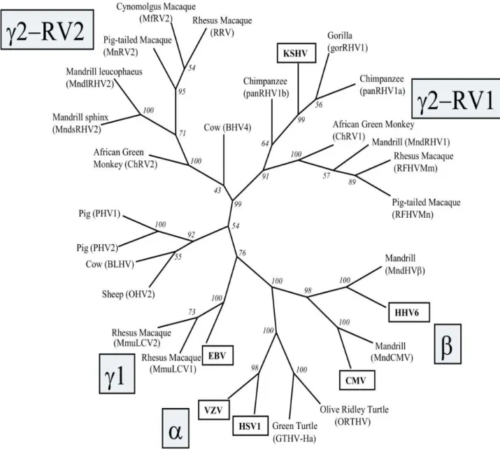
More than 100 herpesviruses have been discovered, all of them are double-stranded DNA viruses that can establish latent infections in their respective hosts. Eight herpesviruses infect humans. The Herpesvirinae family is subdivided into three subfamilies: the Alpha-, Beta-, or Gammaherpesvirinae1.
The Alphaherpesvirinae are defined by variable cellular host range, shorter viral reproductive cycle, rapid growth in culture, high cytotoxic effects, and the ability to establish latency in sensory ganglia. Human alpha-herpesviruses are herpes simplex viruses 1 and 2 (HSV-1 and HSV-2) and varicella zoster virus (VZV), and are officially designated human herpesviruses 1, 2, and 3.
The Betaherpesvirinae have a more restricted host range with a longer reproductive viral cycle and slower growth in culture. Infected cells show cytomegalia (enlargement of the infected cells). Latency is established in secretory glands, lymphoreticular cells, and in different tissues, such as the kidneys and others. In humans, these are human cytomegalovirus (HCMV or herpesvirus 5) and roseoloviruses (causing the disease roseola infantum in children) including human herpesviruses 6A and 6B (HHV-6A and -6B) and human herpesvirus 7 (HHV-7).
The Gammaherpesvirinae in vitro replication occurs in lymphoblastoid cells, but lytic infections may occur in epithelial and fibroblasts for some viral species in this subfamily. Gammaherpesviruses are specific for either B or T cells with latent virus found in lymphoid tissues. Only two human gammaherpesviruses are known, human herpesvirus 4, or Epstein-Barr virus (EBV), and human herpesvirus 8, referred to as HHV-8 or Kaposi's sarcoma-associated herpesvirus (KSHV). The gammaherpesviruses subfamily contains two genera that include both the gamma-1 or Lymphocryptovirus (LCV) and the gamma-2 or Rhadinovirus (RDV) virus genera. EBV is the only Lymphocryptovirus and HHV-8 is the only Rhadinovirus discovered in humans. LCV
12
are found only in primates but RDV can be found in both primates and subprimate mammals. RDV DNAs are more diverse across species and are found in a broader range of mammalian species.
HHV-8 has sequence homology and genetic structure similar to another RDV, Herpesvirus saimiri (HVS). The T-lymphotropic Herpesvirus saimiri establishes specific replicative and persistent infection in different primate host species, causes fulminant T-cell lymphoma in its primate host and can immortalize infected T-cells.
13
Human herpesvirus type
Sub
Family Target cell type Latency sites Pathologies
Herpes simplex-1
(HSV-1) Alpha Mucoepithelial cells Neuron
Oral and genital herpes (predominantly
orofacial)
Herpes simplex-2
(HSV-2) Alpha Mucoepithelial cells Neuron
Oral and genital herpes (predominantly
genital)
Varicella Zoster virus
(VSV) Alpha Mucoepithelial cells Neuron chickenpox
Epstein-Barr Virus
(EBV) Gamma
B lymphocyte,
epithelial cells B lymphocytes
Infectious mononucleosis, Burkitt’s lymphoma Cytomegalovirus (CMV) Beta Epithelial cells, monocytes, lymphocytes Monocytes, lymphocytes Mononucleosis-like syndrome
Human herpes virus-6
(HHV-6) Beta
T lymphocytes and
others T lymphocytes Roseola infantum
Human herpes virus-7
(HHV-7) Beta
T lymphocytes and
others T lymphocytes Roseola infantum
Human herpes virus-8 (HHV-8) Kaposi's sarcoma- associated herpes virus (KSHV) Gamma Endothelial cells B lymphocytes monocytes B lymphocytes and endothelial cells Kaposi’s sarcoma, Multicentric Castleman disease, Primary effusion lymphoma
Table 1: Human herpesviruses classification and characteristics.
1.2. Structure of herpesviruses
Herpesviruses have a central toroidal-shaped viral core containing a linear double stranded DNA. This DNA is embedded in a proteinaceous spindle2. The capsid is icosadeltahedral (16 surfaces) with 2-fold symmetry and a diameter of 100-120 nm that is partially dependent upon the thickness of the tegument. The capsid has 162 capsomeres.
14
The herpesvirus tegument, an amorphorous proteinaceous material that under EM lacks distinctive features, is found between the capsid and the envelope; it can have asymmetric distribution. Thickness of the tegument is variable depending on the location in the cell and varies among different herpesviruses3.
The herpesvirus envelope contains viral glycoprotein protrusions on the surface of the virus. As shown by EM there is a lipid trilaminar structure derived from the cellular membranes. Glycoproteins protrude from the envelope and are more numerous and shorter than those found on other viruses.
Figure 2: Representation of a herpesvirus virion.
1.3. Herpesviruses genome
Herpesvirus genome studied to date ranges in size from 130 to 235 kbp.
Herpesvirus DNA is characterized by two unique components, unique long (UL) and unique small (US) regions, each flanked by identical inverted repeat sequences. Herpesvirus genome also contains multiple repeated sequences
All known herpesviruses have capsid packaging signals at their termini4. The majority of herpes genes contain upstream promoter and regulatory sequences, an initiation site followed by a 5' nontranslated leader sequence, the open reading frame (ORF) itself, some 3' nontranslated sequences, and finally, a polyadenylation signal. Gene overlaps
15
are common, whereby the promoter sequences of antisense strand (3') genes are located in the coding region of sense strand (5') genes; ORFs can be antisense to one another.
Figure 3: Genomic organization of some herpesviruses. HSV, VZV and CMV have inverted repeated sequences.
Proteins can be embedded within larger coding sequences and yet have different functions. Most genes are not spliced and therefore are without introns and sequences for noncoding RNAs are present.
Herpesviruses code for genes that synthetize proteins involved in establishment of latency, production of DNA, and structural proteins for viral replication, nucleic acid packaging, viral entry, capsid envelopment, for blocking or modifying host immune defences, and for transition from latency to lytic growth. Although all herpesviruses
16
establish latency, some (e.g., HSV) do not necessarily require latent protein expression to remain latent, unlike others (e.g., EBV and HHV-8).
1.4. Replication of herpesviruses
Herpesviruses can establish lytic or latent infection. The lytic stage is divided in 6 phases:
1. Binding to the cell surface. As many other viruses, cell tropism is determined by the availability of the correct receptor on the surface of the cell
2. Envelope fusion with the plasma membrane
3. Uncoating. Degradation of the tegument. Capsid is carried to the nuclear membrane.
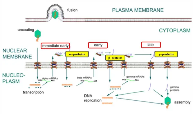
4. Transcription. The DNA genome then enters the nucleus. This is a very complex process, as expected considering the large size of viral genome. Viral DNA replicates by circularization followed by production of concatemers and cleavage of unit-length genome during packaging. The herpesvirus lytic replicative phase can be divided into four stages:
α or immediate early (IE), requiring no prior viral protein synthesis. The genes expressed in this stage are involved in transactivating transcription from other viral genes.
β or early genes (E), whose expression is independent of viral DNA synthesis. Following the E phase, γ1 or partial late genes are expressed in concert with the
beginning of viral DNA synthesis.
γ2 or late genes (L), where protein expression is totally dependent upon synthesis of viral DNA and expression of structural genes encoding for capsid proteins and envelope glycoproteins occurs.
Herpesvirus DNA is transcribed to RNA by cellular RNA polymerase I. The neo-formed viral mRNAs block cellular protein synthesis and activate the replication of viral DNA. Herpesviruses encode their own DNA-dependent DNA polymerase and other enzymes and proteins necessary to replication, such as ori-Lyt (replication start for the lytic phase), major DNA binding protein (MDBP) and origin DNA binding protein
17
(OBP). Herpesviruses can alter their environment by affecting host cell protein synthesis and host cell DNA replication, immortalizing the host cell, and altering the host's immune responses (e.g., blocking apoptosis, cell surface MHC I expression, modulation of the interferon pathway).
Figure 4: Representation of lytic phase of herpesviruses.
5. Assembly: capsids are assembled in the nucleus.
6. The viral particles bud through the inner lamella of the nuclear membrane which has been modified by the insertion of viral glycoproteins and leave the cell via the exocytosis pathway.
In the latent phase, the virus genome depends on the host replication machinery and replicates as closed circular episome. Latency typically involves the expression of only a few latency specific genes. Generally, most infected host cells harbour latent virus, as in the case of HHV-8: when KS tissue or HHV-8 infected cultured cells are analyzed, the virus is latent in majority of infected cells. Different signals such as inflammation and immunosuppression may cause the virus to enter into a new lytic phase.
18 Figure 5: Herpesviruses replication cycle.
2. Human herpesvirus 8
Human herpesvirus 8 (HHV-8), also known as Kaposi’s sarcoma associated herpesvirus (KSHV), is a member of the Rhadinovirus genus in the gamma-herpesvirus subfamily, first detected in 1994 in a patient affected by Kaposi sarcoma (KS)5, a neoplasm of endothelial origin. Since then, HHV-8 has been identified as the etiologic agent of all epidemiologic forms of KS, including classical, endemic African, iatrogenic and AIDS types. In addition, HHV-8 has been implicated in the pathogenesis of other neoplastic disorders affecting immunocompromised hosts: primary effusion lymphoma (PEL, a rare form of B-cell lymphoma)6, multicentric Castleman disease (MCD, a B-cell
19
lymphoproliferative disease)7, other lymphoproliferative disorders affecting patients infected with HIV8, and neoplastic complications in patients after transplantation9.
2.1. Structure of human herpesvirus 8
HHV-8 has the typical morphology of the herpesviruses.
The envelope contains proteins of cellular origin and virus-specific glycoproteins, such as gB, gM, gH and K8.1.
The tegument is an amorphous asymmetric proteinaceous layer between envelope and capsid and contains proteins encoded by ORFs 19, 63, 64, 67, 75.
Each capsid, 125 nm in diameter, contains 12 pentons and 150 hexons which are interconnected by 320 triplexes. These capsomers or structural components are arranged in a icosahedral lattice with 20 triangular faces. Each asymmetric unit (one-third of a triangular face) of the capsid contains one-fifth of a penton at the vertex. Several
proteins are involved in capsid assembling: the major capsid protein (MCP), three capsid proteins encoded by ORF62, ORF26, ORF65 and a protease encoded by ORF17. Hexons and pentons contain 5 or 6 MCPs, and triplexes contain ORF62 monomer and ORF26 dimer10.
20
Figure 6: Structural comparison of HHV-8 capsid and HSV-1 B capsid. The two capsid maps are radially coloured and are shown in a montage as viewed along the icosahedral threefold axis. One penton (5), three types of hexon (P, E, and C), and six types of triplexes (Ta to Tf) are labelled.
2.2. HHV-8 genome structure
In the viral capsid, HHV-8 DNA is linear and double stranded, but upon infection of the host cell and release from the viral capsid, it circularizes. Reports of the length of the HHV-8 genome have been complicated by its numerous, hard-to-sequence, terminal repeats. Renne et al.11 reported a length of 170 kilobases (Kb) but Moore et al.12 suggested a length of 270 Kb after analysis with clamped homogeneous electric field (CHEF) gel electrophoresis.
Base pair composition on average across the HHV-8 genome is 59% G/C; however, this content can vary in specific areas across the genome.
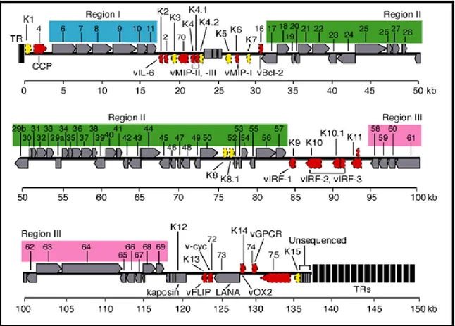
HHV-8 possesses a long unique region (LUR) of approximately 145 Kb, containing all the known ORFs (open reading frame), flanked by terminal repeats (TRs). Varying amounts of TR lengths have been observed in the different virus isolates. These repeats
21
are 801 base pairs in length with 85% G/C content, and have packaging and cleavage signals12. The LUR is similar to HVS and at least 66 ORFs have homology with the HSV genes. New genes are still being discovered through transcription experiments with alternative splicing. A "K" prefix denotes no genetic homology to any HVS genes (K1–K15).
Figure 7: HHV-8 genome. The genome consists of a long unique region (145 kb) encoding for over 80 ORFs, surrounded by terminal repeats regions.
HHV-8 possesses approximately 26 core genes, shared and highly conserved across the alpha-, beta-, and gammaherpesviruses. These genes are in seven basic gene blocks, but the order and orientation can differ between subfamilies. These genes include those for gene regulation, nucleotide metabolism, DNA replication (polymerase ORF9 and
22
thymidin kinase ORF21), and virion maturation and structure (envelope glycoproteins: ORF8, ORF22, ORF38).
HHV-8 encodes several ORFs homologous to cellular genes (at least 12), not shared by other human herpesviruses13. These genes seem to have been acquired from human cellular cDNA as evidenced by the lack of introns. Some retain host function, or have been modified to be constitutively active; an example of this is the viral cyclin-D gene14. Cellular homologs related to known oncogenes have been identified in HHV-8, including genes encoding viral Bcl-2 (ORF16), cyclin D (ORF72), interleukin-6 (K2), G-protein-coupled receptor (ORF74), and ribonucleotide reductase (ORF2).
23
Other genes have homologues in other members of the RDV genus, such as v-cyclin (ORF72), latency-associated-nuclear antigen (LANA, ORF73), viral G-protein coupled receptor (ORF74). A number of other genes encoding for capsid protein have been identified, including ORF25, ORF26, and ORF6513. In addition to virion structural proteins and genes involved in virus replication, HHV-8 has genes and regulatory components (e.g. ORFs K3, K4 and K5) that interact with the host immune system, presumably to counteract cellular host defenses15.
2.3. Replication cycle of HHV-8
Like other herpesviruses, HHV-8 genome structure and gene expression pattern varies depending on the replication state. The lytic phase consists of 6 steps:
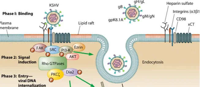
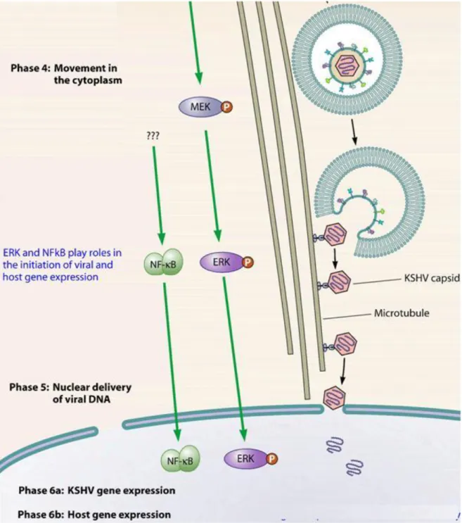
1. Binding to the cell surface mediated by glycoproteins B and K8.1, encoded by ORF8 and K8.116. HHV-8 can use multiple receptor for infection of target cells, and these receptors differ according to the cell type. HHV-8 utilizes the ubiquitous cell surface heparan sulphate (HS) proteoglycan to bind several target cells (e.g. B lymphocytes). Glycoprotein B also interacts with the host cell surface alpha3-beta1-integrin, a heterodimeric receptor containing transmembrane subunits. Another cellular receptor used is the dentritic cell specific intracellular adhesion molecule-3 (ICAM-3) for the binding to the myeloid dendritic cells and macrophages. Moreover, HHV-8 utilizes the transporter protein xCT for entry into cells (but not into the B cells); xCT molecule is a part of the membrane glycoprotein CD98 complex.
2. Fusion between envelope and plasma membrane. The binding between cellular receptors and HHV-8 glycoproteins leads to induction of the host signal cascades critical for maintenance of viral gene expression, such as protein kinase C (PKC), phosphatidylinositol 3-kinase (PI3K), and nuclear factor kB (NF-kB). In fact, HHV-8 reprogrammes the elements of host cell transcriptional machinery that are involved in regulating a variety of processes (apoptosis, cell cycle regulation, signalling, inflammatory response and angiogenesis).
3. Uncoating: capsid degradation by cellular enzymes and viral genome transfer to the cytoplasm.
24
Figure 9: Representation of the first three phases of early events of HHV-8 infection of target cells.
4. Transcription: viral genome migrates to the nucleus and regulator genes are transcribed by host RNA polymerase. mRNAs are translated in virus-specific proteins, able to block cellular synthesis and to start viral replication. Lytic gene expression begins with transcription of immediate-early (IE) genes that regulate the synthesis of other viral genes. Some of the genes transcribed in this step are: ORF6, coding the major DNA binding protein (MDBP), ORFs 9, 56, 59, encoding the DNA polymerase and ORFs 40, 41, 44 (helicase/primase complex). Expression of IE genes occurs independent of viral replication, and afterwards, early and late genes are expressed. The early genes (E) expression is activated by IE genes within 24 hours after infection or viral reactivation. Early genes encode proteins involved in viral DNA replication, nucleotides metabolism, virus assembly. Some early genes are: K2-5, T1.1, ORFs2, 41, 59, 70, 74.
Expression of late genes (L) begins after viral DNA replication. They encode structural proteins involved in virus assembly, such as glycoproteins B and H (ORF8 and ORF22), capsid proteins (ORFs 25, 26), the small viral capsid antigen (ORF65).
5. Assembly: transcription of genes coding structural proteins and production of viral particle. ORFs26 and 29 proteins are responsible for capsid assembly and viral DNA packaging.
25
6. The viral particles bud through the inner lamella of the nuclear membrane which has been modified by the insertion of viral glycoproteins and leave the cell via exocytosis.
26
After initial infection, HHV-8 may establish lifelong latency. Throughout latency, viral gene expression is tightly regulated and only a few viral genes are expressed. The latent HHV-8 genome is circularized by joining of GC rich terminal repeats (TRs) at the ends of the viral genome to form an extrachromosomal circular episome17. The latency associated nuclear antigen (LANA) regulates episome replication by host cell machinery18. LANA is a phosphoprotein expressed in latently infected cells and promotes the maintenance of latency by associating with the ORF50 promoter19 or binding cellular factors which normally interact with ORF50. HHV-8 infection can be reactivated from latency and the lytic gene expression may restart.
2.4. Transactivator genes of HHV-8
Two immediate-early genes play a key role in the reactivation from latent phase to lytic phase: ORF50 and ORF57.
ORF50
ORF50 is an immediate early gene whose product is the major transcriptional transactivator and his activity is required for viral reactivation by all known chemical inducer (e.g. tetradecanoyl phorbol acetate, TPA). The ORF50 gene is rapidly expressed, within 2 to 4 h after induction.
ORF50 belongs to the family of R transactivators, highly conserved among herpesviruses and is related to immediate-early transcriptional activator proteins of other gammaherpesviruses, such as ORF50a encoded by Herpesvirus saimiri and Rta encoded by the BRLF1 ORF of Epstein-Barr virus20.
During latency, ORF50 expression is repressed; however, ORF50 may be activated by physiological conditions, such as hypoxia, or by pharmaceutical agents and the activation triggers the start of the lytic replication cascade.
The genomic sequence of ORF50 is characterized by 5 exons and 4 introns and transcribes an mRNA of 3,6 Kb. The transcript initiates at position 71560, 23 nts downstream a potential TATA box; its first AUG is located at position 71596.
27 Figure 11: Schematic representation of ORF50 protein.
The ORF50 transcript encodes a 691 aa protein (110 kDa) located in the nucleus during the latent phase for the presence of two nuclear localization signals (NLSs). The N-terminus 272 amino acids of ORF50 binds independently to HHV-8 promoters and mediates sequence-specific DNA binding; N-terminal region is followed by a leucine zipper domain. The C-terminal domain contains multiple charged amino acids alternated with repeated bulky hydrophobic residues, a primary structure conserved in many eukaryotic transcriptional activation domains. The C-terminus is sufficient to activate transcription when targeted to promoters with a heterologous DNA binding domain21.
This region contains four overlapping domains termed activation domains (AD1, AD2, AD3, AD4), sharing significant homology to the R proteins encoded by other gamma-herpesviruses.
Analysis of the ORF50 amino acids sequence reveals multiple sites of phosphorilation, including a C-terminal region rich in serines and threonines, and 20 other consensus sites for phosphorilation by serine-threonine kinase and protein kinase C (PKC).
R response elements (RREs) have been identified within several lytic gene promoters. The response element contains a 12-bp palindrome with additional sequences flanking the palindrome which are also required for both DNA binding and activation by ORF50. The ORF50 protein binds directly to this palindromic sequence, and the N-terminal 272 aa is sufficient for binding in vitro. ORF50 can directly transactivate the early gene promoters22.
28
The ORF50-responsive promoters include the following: ORF 6 (single-stranded DNA binding protein), ORF21 (thymidine kinase [TK]), ORF57 (posttranscriptional activator), ORF59 (DNA polymerase associated processivity factor), K8 (K-bZip), K9 (viral interferon response factor), K12 (kaposin), and nut-1 or PAN or T1.1 (polyadenylated nuclear RNA)23.
Furthermore, recent studies suggest autoactivation of ORF50 by interaction with the cellular protein octamer-1 (oct-1) and an intact octamer element that is located approximately 200 bp upstream of the ORF50 transcription start site24.
Expression of ORF50 reactivates viral lytic cycle in cells containing the virus in a latent phase25, and is also able to activate heterologous viral promoters such as LTR HIV, synergizing with Tat26. This molecular transactivation increases cellular susceptibility to HIV infection and could have clinical consequences in patients co-infected with HHV-8 and HIV.
The constitutive expression of ORF50 in stable clones increases expression of several cellular transcription factors, including activating transcriptional factor-4 (ATF4) (Unpublished data).
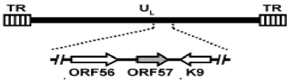
ORF57
ORF57 is a lytic gene expressed between 2 and 4 h after activation of the lytic phase, immediately following the appearance of ORF50 transcripts but prior to most early mRNAs21.
ORF57 is homologous to known posttranscriptional regulators in other herpesviruses. One of these, ICP27 of HSV is a regulator whose functions include downregulation of intron containing transcripts and upregulation of some late messages. ICP27 is essential for lytic viral replication, is required for inhibition of host cell splicing and shuttles from the nucleus to the cytoplasm to promote the export of intronless viral RNAs27. The other gammaherpesviruses, EBV and herpesvirus saimiri, also encode ICP27 homologs. ORF57 gene is positioned in a unique long region of the HHV-8 genome and is flanked by ORF56 (primase) and K9 (viral interferon regulatory factor, vIRF) genes. ORF56 and ORF57 have their own promoter to initiate transcription, but they share the same polyadenilation signal downstream of ORF57.
29
Figure 12: Schematic diagram of ORF57 gene in the context of HHV-8 genome.
ORF57 contains two coding exons and a single 108 bp intron. The exon 1 is relatively small, 114 nts, and has four ATG codons, clustered within a region of 33 nucleotides, in frame with each other and with the first exon and separated from it by a single stop codon. The intron is 109 nts in size and contains consensus splice donor and acceptor sites. The exon 2 is about 1,4 kb long. The transcriptional start site (TSS) is located at nt 8200327 and polyA signal starts at nt 83608. A TATA box 24 bp is identified upstream the TSS as well as several consensus transcription factor binding sites (NF-kB, AP-1, Oct-1), and at least four R responsive elements involved in the transcriptional activation by ORF50.
ORF57 expression is highly dependent on ORF50, and a RRE in the ORF57 promoter is responsible for ORF57 binding.
ORF57 is expressed predominantly in the nucleus and nuclear localization is driven by three independent nuclear localization signals (NLS) that form a cluster in the N-terminal.
ORF57 encodes a protein of 455 aa residues.
Analyses of amino acid sequence reveals several structural and functional motifs. The N-terminus contains a long stretch of arginine residues, two separate RGG-motifs, which are typical of RNA-binding proteins and four serine/arginine dipeptides, characteristic of SR proteins, the major cellular splicing factors. The three NLSs overlap the arginine rich region.
30
The C terminus of ORF57 is enriched in leucine residues and contains a leucine zipper motif, typical of cellular transcriprion factors. The C-terminus also contains the zinc-finger-like motif.
ORF57 promotes the expression of HHV-8 intronless genes, including several viral early and late genes, such as ORF59, T1.1 (PAN or nut-1), gB, MPC. ORF59 is an early gene encoding a viral DNA polymerase processivity factor involved in viral DNA replication. T1.1 is a non-coding RNA that accumulates at unusually high levels in the nucleus of lytically infected cells.
ORF50 and ORF57 have a synergic activity that is promoter specific: expression of some promoters that are upregulated by ORF50 can be synergistically enhanced by coexpression with ORF57. This synergy results from a post-translational enhancement of the transcriptional activity of ORF50. ORF57 transactivates specific viral promoters in synergy with ORF50, such as promoters of T1.1, ori-Lyt and Kaposin. ORF57 interacts with ORF50 via its N-terminal region and the central region of ORF50.
2.5. Epidemiology and transmission of HHV-8
The serologic prevalence of HHV-8 infection has been explored in most continents worldwide and in different populations with different levels of risk of HHV-8 infection. It should be noted that the comparisons of prevalence are limited by the fact that either antibodies to latent or lytic HHV-8 antigens were detected and by the test formats used. Several studies have confirmed that there is a low seroprevalence in central and northern Europe, North America and most of Asia, intermediate prevalence in the Middle East and Mediterranean, and high prevalence in southern Africa.
31
Figure 13: Worldwide geographic distribution of HHV-8 infection.
The virus, first thought to be transmitted only sexually, is now also considered transmissible through low risk or more casual behaviours. Important risk factors for transmission of the virus are a spouse's seropositivity and maternal seropositivity. Of all anatomic sites, HHV-8 DNA is found most frequently in saliva, which also has higher viral concentrations than other secretions28. For this reason, it has been hypothesized that saliva could be the route of casual transfer of infectious virus among family members.
Other possible transmissions are blood-borne and organ transplantation.
2.6. HHV-8 pathogenesis
Human herpesvirus 8 is associated with proliferative disorders including Kaposi’s sarcoma (KS), multicentric Castleman disease (MCD), primary effusion lymphoma (PEL), and other lymphoadenopathologies.
32
HHV-8 induces the formation of neoplasias in natural or experimental hosts, and reactivation from latency is essential for this activity.
Latent viral proteins, such as vFLIP and LANA, inactivate tumour suppressors and block apoptosis29.
However, lytic replication is also important for transmission of the virus in the population and in the pathogenesis of KS. HHV-8 vIL-6 is highly expressed during the lytic cycle, and promotes cellular growth and angiogenesis, while protecting against apoptosis30. Additional evidence for the importance of lytic replication includes the fact that inhibition of active HHV-8 replication by gancyclovir reduces the incidence of HHV-8 in HIV-infected individuals31.
Figure 14: Cellular transformation after HHV-8 infection.
Kaposi’s sarcoma
KS was first described by Moritz Kaposi in the 1870s32 and was described as an aggressive tumour affecting patients younger than those currently observed. For all epidemiological forms of KS, the tumour presents as an highly vascularised neoplasm that can be polyclonal, oligoclonal, or monoclonal. Its antigenic profile suggests either
33
endothelial, lymphoendothelial, or macrophage origins8. All forms of KS lesions contain a variety of cell types, including endothelium, extravasated erythrocytes, infiltrating inflammatory cells, and characteristic “spindle” cells of endothelial origin33. The spindle cells express both endothelial and macrophagic markers.
Extensive and aberrant neoangiogenesis in KS lesions is accompanied by elevated levels of many cytokines, including basic fibroblast growth factor (bFGF), interleukin-1 (IL-1), IL-6, IL-8, platelet-derived growth factor (PDGF), tumour necrosis factor (TNF), gamma interferon (IFN-), and vascular endothelial growth factor (VEGF). Many of these cytokines are secreted by spindle cells, are essential for spindle cell viability in culture, and are themselves proangiogenic.
HIV infection increases the risk for development of KS, and therefore, the incidence of KS has increased substantially during the HIV pandemic, particularly in younger HIV-infected patients34.
Figure 15: Cutaneous and visceral manifestations of Kaposi’s sarcoma.
Four forms of Kaposi’s sarcoma are known, differentiated on clinical parameters and epidemiology:
Classic KS: is an indolent tumour affecting the elderly population, preferentially men, in Mediterranean countries. The lesions tend to be found in the lower extremities and the disease, due to its non-aggressive course, usually does not kill those afflicted.
34
AIDS-KS: in the context of the acquired immunodeficiency syndrome (AIDS), KS is the most common malignancy and is an AIDS defining illness35. AIDS-KS is a more aggressive tumour than classic KS and can disseminate into the viscera with a greater likelihood of death. It presents more often multifocally and more frequently on the upper body and head regions.
Endemic KS: HHV-8 was prevalent in Africa prior to the HIV epidemic. Prior to HIV coinfections, endemic KS affected men with an average age of 35 and very young children36. HIV coinfection has raised the prevalence of KS significantly in Africa, where endemic KS is found more often in women and children than in other areas of the world37.
Iatrogenic KS: Immunosuppression, as that occurring in transplant recipients, is known to facilitate reactivation of herpesviruses and therefore transplant patients under immunosuppressive therapy can develop KS. Withdrawal of the therapy can cause the KS to regress38.
Multicentric Castleman disease
MCD is a rare polyclonal B-cell angiolymphoproliferative disorder. Most of the B-cells in the tumour are not infected with HHV-8, and the HHV-8 infected cells are primarily located in the mantel zone of the lymphatic follicle. It is thought that uninfected cells are recruited into the tumour through HHV-8 paracrine mechanisms, such as vIL-6, a known growth factor for the tumour. More than 90% of AIDS patients with MCD are HHV-8 positive, whereas MCD in the context of no HIV infection has a HHV-8 prevalence of approximately 40%39.
Primary effusion lymphoma
First identified as a subset of body-cavity-based lymphomas (BCBL), PELs contain HHV-8 DNA sequences6. These lymphomas are distinct from malignancies that cause other body cavity effusions. PEL cell lines have 50–150 copies of HHV-8 episomes per cell40.
35
2.7. HHV-8 and angiogenesis
The effects of acute HHV-8 infection on endothelial cell functions, induction of angiogenesis, and triggering of inflammatory processes are still largely unknown. In our laboratory, we demonstrated that HHV-8 selectively triggers the expression and secretion of high levels of monocyte chemoattractant protein 1 (MCP-1). We also found that this event is accompanied by virus-induced capillary-like structure formation at a very early stage of acute infection41.
Figure 16: HUVEC monolayer not infected or infected with HHV-8 41.
The MCP-1 expression is controlled by the nuclear factor kB (NF-kB), that induces the expression of chemokines promoting cell migration and angiogenesis, such as IL-8 and vascular endothelial growth factor (VEGF), several matrix metalloproteinases (MMPs) that promote tumour invasion of surrounding tissue. NF-kB is a critical regulator of the immediate early response to HHV-8, playing an important role in promoting inflammation, in the control of cell proliferation and survival, and in the regulation of virus replication. Several studies show that transfection of different HHV-8 genes results in NF-kB activation in different cell types, and in our laboratory we demonstrated that HHV-8 acute infection induces NF-kB activation in endothelial cells.
36
The human MCP-1 gene contains 2 NF-kB-binding sites in the enhancer region. The kB binding sites are required for TPA-induced expression. In HHV-8 infection we observed that the NF-kB pathway is involved in the enhancement of MCP-1 expression and is required for maximal production of the chemokine. However, mutations in both NF-kB sites in the enhancer region did not result in the complete loss of promoter induction in HHV-8 infected cells, and inhibitors of NF-kB do not prevent MCP-1 activation following HHV-8 infection suggesting that at least another signalling pathway may be involved in the control of MCP-1 expression in the course of acute HHV-8 infection.
3. The cellular activating transcription factor 4
ATF4 (also called cAMP responsive element binding 2, CREB2) belongs to the ATF/CREB family of transcription factors that represent a large group of basic region-leucine zipper (bZip) proteins. The basic region of the bZIP protein interacts with DNA, and they dimerize by their leucine zipper domains forming homodimers, heterodimers or both42.
CREB/ATF family members include ATF1 (also known as TREB36), CREB/CREM, CREB314 (also known as Aibzip or Atce1), CREB-H, ATF2 (also known as CRE-BP1), ATF3, ATF4, ATF6, ATF7, B-ATF and ATFX (also known as ATF5).
ATF4 gene is in chromosome 22 at the cytogenetic band 22q13.1, located at 38,241,069–38,243,191 bp, with a genomic size of 2122 and is constitutively expressed in many cells.
The structure of human ATF4 mRNA includes three short open reading frames (uORFs) in the 5’UTR that precede the functional coding sequence43
and are out of frame with the main protein-coding region. The organization of the 5’UTR uORFs in ATF4 is essential for the response of ATF4 to stress such as ER stress and hypoxia.
ATF4 protein consists of 351 amino acids. The protein is structured into several domains/motifs that are essential for ATF4 homo/heterodimerization and DNA binding. A transcriptional activation domain has been located at the N-terminus of ATF444.
37 Figure 17: Representation of ATF4.
The mammalian ATF4 can form a homodimer, and heterodimers with members of the AP-1 and C/EBP family of proteins, including Fos42 and Jun45, and several C/EBP proteins46. ATF4 has a very short half-life of about 30-60 minutes.
ATF4 has several interacting partners, which include p30047, RNA polymerase II subunit RPB348, ZIP kinase, a serine/threonine kinase, which mediates apoptosis49, HTLV1 transactivator Tax, which activates the expression of viral mRNA through a three 21 bp repeat enhancer located within the HTLV-1 LTR50. Tax transactivates the HTLV-1 promoter via the Tax responsive elements that contain the consensus ATF/CRE core sequence. ATF4 enhances the ability of Tax to transactivate the HTLV-1 promoter. The numerous dimerization and interaction partners determine the diverse functions of ATF4.
ATF4 can function as a transcriptional activator, as well as a repressor. It is a stress responsive gene, which is upregulated by several factors/stressors, including oxygen deprivation (hypoxia/anoxia), amino acid deprivation, endoplasmic reticulum stress (ER stress), oxidative stress, and by the growth factor heregulin51.
In mammalian cells, hypoxia/anoxia and perturbation of ER homeostasis induces a complex transcriptional program and triggers a reduction in protein translation (UPR: unfolded protein response). A central mediator of this translational response to anoxia is phosphorylation of the eukaryotic initiation factor 2 (eIF2) by PERK protein kinase. Although the phosphorylation of eIF2 results in global translational reduction, it specifically increases the translation of ATF4 mRNA43. The various stress signals
38
integrate in a common pathway of increased translation of ATF4, which subsequently ensures supply of amino acids for protein biosynthesis and protects cells against oxidative stress, by modulating a number of genes involved in mitochondrial function (e.g., Lon mitochondrial protease homologue), amino acid metabolism and transport (e.g., asparagine synthetase), as well as in redox chemistry (e.g., NADH cytochrome B5 reductase homolog)52. As a result of metabolic and ER stress that activate the PERK pathway of translational inhibition, ATF4 initiates a feedback regulatory loop to ensure the transient nature of protein synthesis inhibition. ATF4 induces GADD34 transcription, a component of the phosphatase complex that dephosphorylates eIF-2alpha.
Some of the genes that are induced by ATF4 include receptor activator of nuclear factor-kappa B (RANK) ligand (RANKL), osteocalcin, E-selectin, VEGF, Gadd153, gadd34, asparagine-synthesase, TRB3, and several genes involved in mitochondrial function, amino acid metabolism and redox chemistry52.
Figure 18: Three proximal sensors IRE1, PERK and ATF6 regulate the UPR through their respective signalling cascades.
39
One important stress factor relevant to cancer progression is hypoxia and more extremely, anoxia. Tumour hypoxia/anoxia is associated with a more aggressive clinical phenotype, and ATF4 protein has been observed to be in much greater levels in primary human tumours compared to normal tissues53.
ATF4 induces VEGF and E-selectin which may be associated with increased metastasis. Since ATF4 protein has shown to be present at greater levels in cancer compared to normal tissue, and it is upregulated by signals of the tumour microenvironment such as hypoxia/anoxia, oxidative stress, and ER stress, it could potentially serve as a specific target in cancer therapy. As a target ATF4 is attractive because it is also potentially involved in angiogenesis and adaptation of cancer cells to hypoxia/anoxia, which are major problems in cancer progression. The induction of VEGF (vascular endothelial growth factor) has a key role in angiogenesis, and preliminary study in our laboratory demonstrate that transfection of ATF4 induces capillary-like structure formation in vitro.
Many viruses have been shown to induce ER stress and activate the UPR: these include three members of the flavivirus, C hepatitis and HCMV. Viral infection induces the cell response to stress, which should lead to an attenuation of viral replication. However, some aspects of the UPR can be regulated and limited by the virus. For example, infection with HCMV (betaherpesvirus) induces the UPR by regulating specifically three signalling pathways: PERK, ATF6 (activating transcription factor 6) and Ire-154. Cells infected with this betaherpesvirus show an increase in ATF4 protein levels, leading to the activation of genes involved in metabolism and redox reactions and helping the virus to maintain a cellular environment permissive to infection.
Studies demonstrate that also HHV-8 can interact with ATF4. LANA, the latency associated nuclear antigen, encoded by ORF73, represses the transcriptional activation activity of ATF4 and the interaction requires the bZIP domain of ATF4. Repression by LANA is independent from the DNA-binding ability of ATF455.
40
AIM OF THE RESEARCH
Human herpesvirus 8 is the primary etiologic agent of Kaposi’s sarcoma, a highly vascularised neoplasm of endothelial origin characterized by inflammation, neoangiogenesis, and by the presence of characteristic spindle cells.
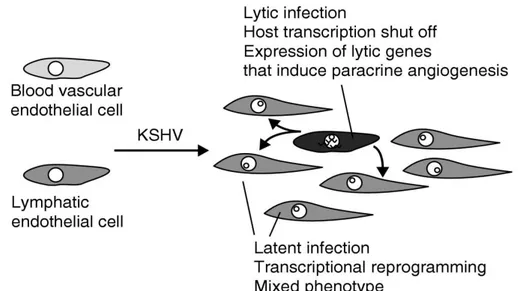
HHV-8 infection of endothelial cells causes changes in cellular phenotype and leads to capillary-like structures formation. This angiogenic activity of HHV-8 is due to the activation of NF-kB and the subsequent induction of MCP-1 synthesis.
MCP-1 is a chemokine produced by macrophages and endothelial cells in response to different stimuli, and is a direct mediator of angiogenesis. The MCP-1 promoter contains an enhancer region with two NF-kB sites. MCP-1 production after HHV-8 infection is accompanied by virus-induced capillary-like structure formation in endothelial cells, and HHV-8-induced angiogenesis is MCP-1 dependent.
Previous studies demonstrate that HHV-8 activates the MCP-1 promoter also in absence of the enhancer region containing the NF-kB binding sites, in fact, mutations in NF-kB sites do not result in a complete loss of MCP-1 transcription. Moreover, treatment with NF-kB-inhibitors does not prevent MCP-1 activation in HHV-8 infected cells.
Therefore, HHV-8 activation of MCP-1 is not completely dependent on NF-kB induction and another cellular factor is involved.
A potential candidate is the cellular activating transcription factor 4 (ATF4), a stress responsive gene. ATF4 is upregulated in several condition, including ER-stress, viral infection (e.g. CMV) and in tumours.
Previous data obtained by gene array in stable Jurkat cell clones transfected with ORF50 gene (major transactivator of HHV-8), indicated that HHV-8 upregulates ATF4.
The aim of the research was therefore to determine whether the viral infection of HHV-8 increases the expression of the transcription factor ATF4 and clarify if this increase is functional for the replication of HHV-8. The results showed that HHV-8
41
(and ORF50) causes a significant increase of ATF4, and that this increase leads to increased virus replication in infected cells.
ATF4 is not able to reactivate HHV-8 from latency, being unable to activate the major gene promoters. This “indirect” effect on HHV-8 activity can be explained by investigation of interactions between ATF4 and MCP-1. In fact, we found that ATF4 activates the MCP-1 promoter, and this activity is NF-kB-independent.
42
MATERIALS AND METHODS
1. Cell cultures
The B cell lines BC-3 and BCBL-1, derived from PEL and chronically infected with HHV-8, were used as representative of HHV-8 target of infection. Lymphoid T (Jurkat cells) and B cell lines were grown in RPMI medium (Gibco) supplemented with 10% inactivated fetal bovine serum (FBS), 2 mM L-glutamine, 100 U/ml penicillin and 100 mg/ml streptomycin.
HeLa cell line (human cervix carcinoma cells) and 293 cell line (human embryonic kidney cells) were grown in Dulbecco’s Modified Eagle medium (Gibco) supplemented with 10% inactivated fetal bovine serum (FBS), 2 mM L-glutamine, 100 U/ml penicillin and 100 mg/ml streptomycin.
All cells were cultured at 37°C in the presence of 5% CO2.
2. Plasmids
Transfection experiments were performed using recombinant plasmids pCR-50sp, pGL-PR57, pGL-PR50, pGL-PRT1.1, PRM, ENH, pGLM-MA1MA2, pRL-SV40 and pCG-ATF4.
Spliced forms of ORF50 were cloned in the expression vector pCR3.1-Uni (Invitrogen) in our laboratory25. Briefly, spliced genes were obtained from TPA-activated BCBL-1 cells by specific retrotranscription of polyA RNA followed by PCR amplification. Amplified fragments were sequenced to verify their integrity and were then inserted into the vector pCR3.1-Uni to obtain the recombinant plasmid pCR-50sp.
43 Figure 19: pCR3.1-Uni map.
44
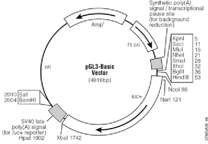
HHV-8 gene promoters were cloned in the reporter vector pGL3-Basic in this laboratory56, containing the Firefly luciferase gene cloned under the transcriptional control of HHV-8 promoters.
The MCP-1 promoter-luciferase constructs (provided by Dr. T. Yoshimura)57 contain the proximal promoter (pGLM-PRM), the proximal promoter and the distal enhancer (pGLM-ENH), the proximal promoter and the distal enhancer in which both NF-kB sites are mutated (pGLM-MA1MA2).
Figure 20: Firefly luciferase pGL3-Basic vector.
pRL-SV40 vector (Promega) was used as an internal transcriptional control, and contains the Renilla luciferase gene cloned under the transcriptional control of the SV40 virus promoter.
45 Figure 21: Renilla luciferase pRL-SV40 vector.
pCG-ATF4 plasmid contains the ATF4 gene cloned under the transcriptional control of the CMV promoter51.
3. Cell transfections
Jurkat, BC-3 and BCBL-1 cells were seeded 24 hours before transfection to obtain optimal cellular density (106 cells/mL) and 106 cell samples were transfected with 1 g of plasmid DNA by electroporation (Nucleofector, Amaxa), following the manufacturer’s instructions. This method permits to obtain high efficiency of transfection, carrying the exogenous DNA into the cells and directly into the nucleus. Efficiency of transfection determined in parallel samples by transfection with pmax- GFP plasmid (Amaxa).
HeLa and 293 cells were seeded in 24-well plates 24 hours prior to transfection to obtain optimal confluence, then were transfected with g of plasmid DNA by GeneJuice Transfection Reagent (Novagen), based on polyamine formulation, following the manufacturer’s instructions.
46
4. Virus purification and cell infection
Cell-free HHV-8 inoculum was obtained by stimulation of BC-3 cells for 3 days with 20 ng/mL 12-O-tetradecanoyl-phorbol-13-acetate (TPA; Sigma). Cells were collected by centrifugation and lysed by rapid freezing and thawing followed by sonication (3 cycles of 5 seconds at medium power with 10-second intervals in a water bath sonicator). Cleared cellular content was added to culture supernatant, and virions were collected by centrifugation for 30 minutes at 20000 xg at 4°C.
Virus particles were purified by density centrifugation on Optiprep self-forming gradients (Sentinel), at 58000 xg for 3,5 hours at 4°C. Purified virions were washed in phosphate buffered saline (PBS) and collected by centrifugation at 20000 xg for 30 minutes at 4°C.
Virions were suspended in sterile PBS containing 0,1% bovine serum albumin (BSA) and stored at -80°C until use. To obtain 1 mL of purified virus, 4^108 BC-3 cells were stimulated. The same ratio between cells and final suspension volume was maintained in all virus preparations to avoid variations in virus concentration between different stocks.
Virus particles were morphologically intact, and the preparation was cell debris-free, as assessed by electron microscope observation.
Prior to use, virus stock was treated with DNase-I and RNase-A, to eliminate free viral nucleic acids eventually present in the preparation.
Infectivity of virus preparation was evaluated by specifically designed infection experiments performed in different cell types, using PCR, rtPCR and immunofluorescence assays to evaluate virus presence, transcription, and expression of antigens.
Quantification of virus genomes present in the stock preparation was obtained by real-time polymerase chain reaction (qPCR)58.
HHV-8 DNA standard was obtained by serial dilutions of pCR-ORF26 plasmid containing a cloned fragment of HHV-8 DNA (ORF26, nucleotides 47127 to 47556). The absence of human gDNA was assessed by amplification of the -actin gene. The purified cell-free virus inoculum contained an average of 4,7^105 copies of viral DNA/L.
47
T and B cells were seeded 24 hours before infection to obtain optimal density of 106cells/mL and then were infected with a m.o.i. (multiplicity of infection, referred to equivalent genomes) of 1:10. After 3 hours of absorption at 37°C, the HHV-8 inoculum was removed, cells were washed with PBS and incubated in fresh medium. Cell samples were harvested at specific time points and processed for DNA or RNA extraction.
5. DNA extraction
Genomic DNA was extracted from 106 cell samples.
Cells were lysed in 500L lysis buffer (10 mM Tris-HCl pH 8.0, 10 mM disodium EDTA pH 8.0, 10 mM NaCl, 0.6% SDS and 100 g/ml proteinase K) and incubated at 37°C for a minimum time of 4 hours. After three cycles of phenol:chloroform:isoamyl alcohol (25:24:1) extractions, DNA was recovered by ethanol precipitation, resuspended in sterile water, and RNase A was added to a final concentration of 100 g/ml. Following 1 hour incubation at 37°C, RNase A was removed by phenol extraction. After precipitation with ethanol, DNA pellets were dissolved in sterile water and stored at -20°C until PCR or qPCR analysis.
DNA concentration was determined by reading optical density at 260 nm.
6. RNA extraction and retrotranscription
Total RNA was extracted with RNAzol B (Tel-Test), following the protocol provided by the manufacturer. Briefly, 106 cell samples were lysed with RNAzol, then, chloroform in ratio 1:5 was added. The mixture was centrifuged 20 minutes at 8500 xg and the aqueous phase was collected and RNA precipitated with isopropanol. After two washes with 75% ethanol and DNAse treatment (4 U/mg RNA, 3 x 20 min. at room temperature), RNA was precipitated with isopropanol 20 minutes at 8500 xg at 4°C. RNA quality was checked by electrophoretic analysis on a 0.8% agarose gel. The absence of contaminating DNA was checked by PCR
48
amplification of human -actin gene before retrotranscription (RT-). Negative PCR results for -actin ensured that the RNA sample was completely free from DNA sequences. First strand cDNA synthesis was carried out with MuLV reverse transcriptase and random hexamer primers (Applied Biosystems), following the manufacturer’s instructions, retrotranscribing 2 g of total RNA from all the samples. The mixture was incubated for 1 hour at 42°C. Efficiency of retrotranscription was assessed by analysis of dilutions of cDNA with PCR specific for human -actin gene (RT+).
7. PCR and rtPCR
The presence and the level of transcription of HHV-8 were analyzed by PCR and reverse transcription PCR (rtPCR) amplification of the ORF26 and ORF50 genes. PCR amplification was performed using 100 ng total DNA or 200 ng total cDNA extracted from infected cells. Amplification of the housekeeping -actin gene was used as a control. The transcription of ATF4 was analysed by rtPCR amplification of the ATF4 gene. Specific primers and PCR conditions are described in Table 2. The absence of contaminating DNA was checked by PCR amplification of human -actin gene before retrotranscription. Negative PCR results for -actin ensured that the RNA sample was completely free from DNA sequences, and that positive amplification after retrotranscription was positively associated to viral transcripts. Particular care was taken to avoid sample-to-sample contamination: different rooms and dedicated equipment were used for DNA extraction and processing, for PCR set-up and gel analyses, all pipette tips had filters for aerosol protection.
49
GENES PRIMERS SEQUENCES AMPLICONS
ORF50 ORF50-Forw ORF50-Rev 5'-TTGGTGCGCTATGTGGTCTG-3‘ 5'-GGAAGGTAGACCGGTTGGAA-3' 420 bps ORF26 ORF26-Forw ORF26-Rev 5'-GCCGAAAGGATTCCACCAT-3‘ 5'-TCCGTGTTGTCTACGTCCAG-3' 232 bps ATF4 ATF4-Forw ATF4-Rev 5’-GTGGCCAAGCACTTCAAACC-3’ 5’-GGAATGATCTGGAGTGGAGG-3’ 414 bps b-actin HACT-Forw HACT-Rev 5’-TCACCCACACTGTGCCCATCT-3’ 5’-GACTACCTCATGAAGATCCTCAC-3’ 674 bps
GENES CONDITIONS CYCLES [MgCl2]
ORF50
94°C 5min
94°C 30 sec, 57°C 1 min, 72°C 1 min + ext. 3 sec/cycle 72°C 10 min, 4°C >>> 1 35 1 2mM ORF26 94°C 5min
94°C 1min, 57°C 1 min, 72°C 1 min + ext. 3 sec/cycle 72°C 10 min, 4°C >>> 1 45 1 1.5 mM ATF4 94°C 5min
94°C 1min, 57°C 1 min, 72°C 1 min + ext. 3 sec/cycle 72°C 10 min, 4°C >>> 1 35 1 2mM b-actin 94°C 5min
94°C 1min, 57°C 1 min, 72°C 1 min + ext. 3 sec/cycle 72°C 10 min, 4°C >>>
1 30
1
1.25mM