Int. J. Mol. Sci. 2021, 22, x. https://doi.org/10.3390/xxxxx www.mdpi.com/journal/ijms Review
MicroRNAs Modulate Signaling Pathways in Osteogenic
Differentiation of Mesenchymal Stem Cells
Chiara Mazziotta 1,†, Carmen Lanzillotti 1,†, Maria Rosa Iaquinta 1,†, Francesca Taraballi 2,3, Elena Torreggiani 1, John Charles Rotondo 1, Lucia Otòn-Gonzalez 1, Elisa Mazzoni 1, Francesca Frontini 1, Ilaria Bononi 1,
Monica De Mattei 1,*, Mauro Tognon 1,* and Fernanda Martini 1,4
1 Department of Medical Sciences, Section of Experimental Medicine, School of Medicine,
University of Ferrara, 64b Fossato di Mortara Street, Ferrara 44121, Italy; [email protected] (C.M.); [email protected] (C.L.); [email protected] (M.R.I.); [email protected] (E.T.); [email protected] (J.C.R.); [email protected] (L.O.-G.); [email protected] (E.M.);
[email protected] (F.F.); [email protected] (I.B.); [email protected] (F.M.)
2 Center for Musculoskeletal Regeneration, Houston Methodist Research Institute, 6670 Bertner Ave, Houston, TX 77030, USA; [email protected]
3 Orthopedics and Sports Medicine, Houston Methodist Hospital, 6565 Fannin Street, Houston, TX 77030, USA
4 Laboratory for Technologies of Advanced Therapies (LTTA), University of Ferrara, 70, Eliporto Street, Ferrara 44121, Italy
* Correspondence: [email protected] (M.D.M.); [email protected] (M.T.) † These authors contributed equally.
Abstract: Mesenchymal stem cells (MSCs) have been identified in many adult tissues and they have
been largely studied in the last years, especially in view of their potential use for treating diseases, damaged tissues and organs. MSCs are capable of self-replication and differentiation into osteo-blasts and are considered an important source of cells in tissue engineering for bone regeneration. Several epigenetic factors are believed to play a role in the osteogenic differentiation of MSCs, in-cluding microRNAs (miRNAs). MiRNAs are small, single-stranded, non-coding RNAs of approxi-mately 22 nucleotides able to regulate cell proliferation, differentiation and apoptosis by binding the 3′ untranslated region (3′-UTR) of target mRNAs, which can be subsequently degraded or trans-lationally silenced. MiRNAs control gene expression in osteogenic differentiation by regulating two crucial signaling cascades in osteogenesis: the transforming growth factor-beta (TGF-β)/bone mor-phogenic protein (BMP) and the Wingless/Int-1(Wnt)/β-catenin signaling pathways. This review provides an overview of the miRNAs involved in osteogenic differentiation and how these miRNAs could regulate the expression of target genes.
Keywords: mesenchymal stem cells; MSCs; microRNA; miRNA; bone; osteogenic differentiation;
osteogenesis; cell differentiation; stem cells; miRNAome; bone regeneration
1. Introduction
Mesenchymal stem cells (MSCs) are multipotent cells widely studied for their poten-tial promising properties in regenerative medicine and in clinical practice [1,2]. Isolated for the first time in 1968 from bone marrow (BM-MSCs) [3], MSCs have been obtained from several body districts and evaluated for their ability to differentiate in vitro into os-teocytes, chondrocytes, and adipocytes [4,5] (Figure 1).
Citation: Mazziotta, C.; Lanzillotti,
C.; Iaquinta, M.R.; Taraballi, F.; Torreggiani, E.; Rotondo, J.C.; Otòn-Gonzalez, L.; Mazzoni, E.; Frontini, F.; Bononi, I.; et al. MicroRNAs Modulate Signaling Pathways in Osteogenic Differentiation of Mesenchymal Stem Cells. Int. J. Mol. Sci. 2021, 22, x. https://doi.org/10.3390/xxxxx Received: date
Accepted: date Published: date
Publisher’s Note: MDPI stays
neu-tral with regard to jurisdictional claims in published maps and institu-tional affiliations.
Copyright: © 2021 by the authors.
Submitted for possible open access publication under the terms and con-ditions of the Creative Commons At-tribution (CC BY) license (http://crea-tivecommons.org/licenses/by/4.0/).
Figure 1. Schematic representation of adult and fetal/neonatal tissue sources of mesenchymal stem cells (MSCs) and their
potential of differentiation in various cell lines. MSCs can be isolated from several tissue sources in the body and they may differentiate into adipocytes, chondrocytes and osteocytes.
MSCs play a key role in the natural events leading to bone repair and healing by differentiating into osteoblasts, which secrete a regenerate bone matrix. Because of their biological features such as sustained self-renewal and expansion, ease of availability, abil-ity to differentiate into osteoblasts, MSCs are also attractive candidates for bone tissue engineering approaches. In addition, they show anti-inflammatory and immunomodula-tory effects and secrete molecules that can initiate or support tissue regeneration/replace-ment [5]. A large number of studies has been carried out on the biological properties of MSCs, signaling pathways involved in their differentiation and factors that might facili-tate MSC osteogenic differentiation [5,6].
Endogenous growth factors, cytokines and signals from the extracellular matrix, mainly via integrin interactions, represent essential regulators in cell differentiation [7,8]. Further, external stimuli, such as mechanical forces or electromagnetic fields, modulate cell behavior during osteogenesis [9,10].
Biomaterials provide the biological structure that supports MSC osteogenic differen-tiation [2,11–18]. The addition of ions has been proved to enhance the osteogenic potential of scaffolds [19]. Biological macromolecules such as growth factors [20] and microRNAs (miRNAs) [21], as well as biophysical stimuli [22] can be also associated with scaffolds to increase cell differentiation.
Growing evidence shows that MSCs differentiation is influenced by several epige-netic processes/factors, including histone methylation and acetylation, DNA methylation [23], non-coding RNA molecules such as long non-coding RNAs (lncRNA) [24] and miR-NAs [25–28].
MiRNAs are small single-stranded, non-coding RNAs consisting of approximately 21–25 nucleotides (nt) and expressed in many organisms from viruses [29–31] to
eukary-ote cells [32]. MiRNAs are able to negatively regulate gene expression at post-transcrip-tional level, by interfering with target messenger RNA (mRNA) translation [33], and their functions have been widely studied in several cellular and molecular processes [34,35], including MSC osteogenic differentiation [5,6]. MiRNAs can positively or negatively reg-ulate osteogenic differentiation by targeting transcription factors (TFs) and genes coding for either negative or positive differentiative modulators [27,36–38]. Different computa-tional tools, such as TargetScan, miRanda and DIANA microT have been developed in order to predict miRNA targets [39]. After prediction, targets have to be experimentally validated in laboratory by PCR [40] and western blot analyses [41] to confirm the miRNA functional activity. Knowledge concerning the role of miRNAs is complex as they modu-late mRNAs acting in concert with other classes of coding RNAs, such as long non-coding RNAs (lncRNA) [27,42]. Further, each miRNA shows multiple activities targeting several mRNAs, thus directly or indirectly modulating the expression of a plethora of genes involved in many cellular functions [43].
This review is addressed to the miRNAs that, by sequence complementarity, bind mRNAs directly regulating the gene expression of molecules involved in the main signal-ing pathways of MSCs osteogenic differentiation, such as the (TGF-β)/bone morphogenic protein (BMP) and the Wingless/Int-1(Wnt)/β-catenin pathway [44,45].
2. MSCs Signaling Pathways in Osteogenic Differentiation
MSC osteogenic differentiation encompasses three phases: the first, also known as the proliferative phase, consists of the recruitment and expansion of osteoprogenitor cells; the second involves the cell maturation in pre-osteoblasts upon extracellular matrix (ECM) synthesis; and the third phase consists of the matrix mineralization [46].
All of these phases are modulated by several signaling pathways [47]. Among them, the TGF-β/BMP and the Wnt/β-catenin pathways [45] play critical interconnected roles and have been largely investigated also for clinical implications in bone healing [48–50]. TGF-β signaling appears mainly involved in the early phase of osteogenesis allowing teoprogenitor cells to differentiate into immature osteoblasts [44], whereas it inhibits os-teoblast maturation, mineralization, and transition into osteocyte [51,52]. Contrariwise, BMP signalling promotes almost every phase during osteoblast differentiation and matu-ration [44]. BMPs have been the first proteins identified as inducing bone formation in vivo [53]. Among over 20 BMP proteins, BMP2, 4, 5, 6, 7 and 9 play important roles in osteogenesis [54,55].
TGF-βs and BMPs work through type I and type II heterodimeric kinase receptors and need Sma and Mad related (SMAD) proteins to transduce the signal inside the cells (Figure 2).
Figure 2. BMPs and TGF-β signaling pathways in osteogenesis. Interaction between TGF-βs and BMPs and receptors
ini-tiate SMAD-dependent and Non-SMAD-dependent signaling pathways. In SMAD-dependent signaling pathway, inter-action between TGF-β and receptor leads to SMAD2/3 (R-SMAD) phosphorylation. Phosphorylated-R-SMAD (SMAD2/3-Pi) binds SMAD4 protein and migrates to the nucleus of the cell where, alongside P300 and CREB-binding protein (CBP) coactivators, controls RUNX2 expression. In the nucleus, SMAD2/3-Pi without SMAD4 recruits HDAC4 and HDAC5 and blocks RUNX2 activity. In BMP signaling, R-SMAD (SMAD1/5/8) binds SMAD4 and migrate to the nucleus inducing
RUNX2 and OSX expression through DLX5 activation. In the Non-SMAD-dependent signaling pathway, TAK1 and TAB1
activate the MKKs -p38 MAPK or -Erk cascades inducing DLX5, RUNX2, and OSX phosphorylation. The interaction be-tween TGF-β and its receptor is blocked by the latent TGF-β binding protein (LTBP), which binds to TGF-β. Similarly, in order to prevent the BMP-receptor interaction, Gremlin, Chordin, Follistatin and Noggin proteins bind to BMP. The trans-location of SMAD2/3 and SMAD1/5/8 into the nucleus is prevented by SMAD7 that, along with SMURF1/2, induce their proteasome-mediated degradation. In addition, the binding between ARKADIA and SMAD6/7 proteins positively regu-lates osteoblast differentiation.
Several type I and type II receptors have been identified in transducing the signal of TGF-βs and BMPs ligands [49,56]. The preferential type I receptor of TGF-βs is TβRI/ALK5 while the main type I receptors of BMPs are BMPR1A/ALK3, BMPR1B/ALK6, and AcvR1/ALK2. The type II kinase receptors of TGF-βs and BMPs are mainly TβRII/TGFBR2 and BMP receptor type II (BMPRII), respectively. The interaction between the ligand and the receptor induces the trans-phosphorylation of a receptor subunit, resulting in the ac-tivation of the SMAD-dependent (canonical) or Non-SMAD-dependent signaling cas-cades, leading to the gene expression regulation and activation of the osteogenic transcrip-tion factors Distal-less Homeobox 5 (DLX5), RUNX2 and the zinc finger Osterix (OSX), also called SP7 (Figure 2) [44,57]. Finally, this results in the expression of specific osteo-blast genes including osteopontin (OPN), osteocalcin (OCN), osteonectin (ON) and colla-gen type 1 (COL1) and cell differentiation [58].
TGF-β and BMP signaling pathways are regulated by different components, which stimulate and/or inhibit osteoblast differentiation. They comprise BMP binding proteins, such as Noggin, Chordin, Gremlin, and Follistatin that sequester ligands preventing bind-ing to receptors, and intracellular inhibitory SMAD proteins, such as SMAD7, and ubiq-uitin-proteasomal degradation pathway (Figure 2) [44,49].
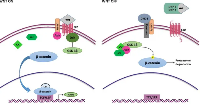
The Wnt/β -catenin or canonical Wnt is another signaling pathway with an essential regulatory role in MSCs self-renewal and differentiation [50]. Wnt/β-catenin signaling pathway occurs in the early stage of osteogenesis allowing the MSCs to differentiate into
osteoblast lineage whilst it is downregulated in differentiated cells [57]. Wnt signaling is initiated upon binding of Wnt ligands with the membrane receptor complex known as Frizzled/low-density lipoprotein receptor-related protein 5/6 (FZD/LRP) [59]. Interactions between Wnt ligands and FZD/LRP5/6 trigger disheveled (DSH), that switching off the glycogen synthase kinase 3 β (GSK-3β) enables β-catenin protein to be translocated into the nucleus, whereby it complexes with the binding T cell factor/lymphoid enhancer bind-ing factor (TCF/LEF) to stimulate the expression of target genes involved in the osteogen-esis including RUNX2 [60,61] (Figure 3).
Figure 3. Wnt/β-catenin signaling pathway in osteogenesis. Wnt ligands interact with FZD and activate Wnt signaling
pathway. FZD binds Dsh that inhibits GSK3-β activity, thus the phosphorylation of β-catenin. β-catenin translocates into the nucleus, where it binds to TCF/LEF and CBP and induces the expression of RUNX2. Interaction between Wnt ligands and SFRP-1/2 and the binding of LRP5/6 and Keremen with DKK-1 antagonist switch off Wnt signaling pathway. In this case, GSK3-β, together with Axin, CK and APC, phosphorylate β-catenin inducing it to proteasome-mediated degradation.
Several Wnt ligands, including Wnt4, Wnt5a, Wnt6, Wnt10a and Wnt10b, are in-volved in initiating the Wnt/β-catenin cascade and stimulation of osteoblast differentia-tion [62–64].
Contrariwise, Wnt3a and Wnt16 have negative effects on MSC osteogenic differenti-ation [65–67]. In addition, extracellular Wnt signaling antagonists can negatively modu-late the Wnt/β-catenin signaling pathway, such as the secreted frizzled-remodu-lated protein 1 and 2 (SFRP1 and SFRP2) [68], that bind Wnt ligands preventing their interaction with receptor [50], and the dickkopf-related protein1 (DKK1) [69], which binds the active site of the LRP5/6 receptors allows proteasome β-catenin degradation through GSK-3β com-plex activation [70,71] (Figure 3). Another relevant Wnt signaling inhibitor is sclerostin (SOST) which binds the first beta propeller (E1) of the LRP5/6 Wnt co-receptors and mainly inhibits the Wnt1 canonical signaling pathway [50,72].
TGF-β/BMP and Wnt/β-catenin signaling pathways strictly interplay in a very com-plex way and affect each other in a positive feedback loop allowing osteoblast differenti-ation in vitro and, thus, a proper skeletal development in vivo. Further, signals from the extracellular (ECM) cooperate with osteogenic signaling pathways, mainly acting through integrins, the mediators of cell adhesion to the ECM and important mechano-transducers involved in both matrix deposition and organization [7,9,73]. Moreover, TGF-β/BMP and
Wnt/β-catenin signaling pathways are components of a more complex molecular network with other different pathways and cytokines such as Hedgehog (Hh), NOTCH, Fibroblast Growth Factor (FGF) and Parathyroid Hormone-related Peptide (PTHrP) that interplay for an appropriate osteoblast differentiation and bone development [44].
3. miRNAs
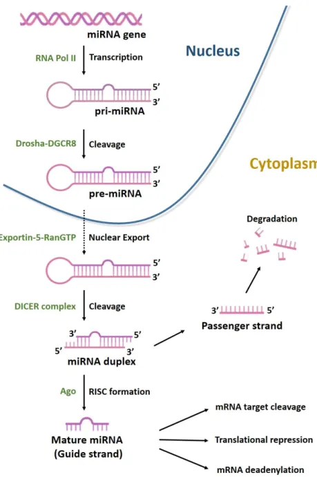
MiRNAs, are a class of non-coding RNAs (ncRNAs), generally 22 nucleotides long, with essential role in gene regulation. They account for 1–5% of the human genome and regulate 30–60% of protein-coding genes [74]. MiRNAs are generated through a multi-step process (Figure 4).
Figure 4. Biogenesis of microRNAs. MiRNAs are transcribed by RNA polymerases II (Pol II) to generate a long precursor
transcript named primary microRNA (pri-miRNA). This RNA molecule folds up into a secondary structure (stem-loop) to form a partial double helix, composed of 100–1000 nt with a 5′-cap. MiRNA maturation process can be divided into three phases. In the first, known as cropping, pri-miRNA is converted into the precursor miRNA (pre-miRNA) via the cutting activity of the Drosha enzyme, a nuclear endoribonuclease III. Pre-miRNA has a hairpin structure (stem-loop) and a length of about 70–80 nt. Following cropping, the pre-miRNA has a 5′P and a 3′OH and 2–3 nt at the protruding end with a single strand. In the second phase, the nuclear export factors Exportin-5 and ras-related nuclear protein (RAN-GTP)
mediate the export of pre-miRNAs from nucleus to cytoplasm. In the third phase, known-as dicing, another type of RNA-endonuclease III, known as Dicer, processes pre-miRNA in the cytoplasm by cleaving it into an 18–22 nt double stranded miRNA (miRNA duplex). Lastly, mature miRNA incorporated into the RNA-induced silencing complex (RISC) is able to bind the 3′UTR region of its target mRNA.
The gene silencing mechanism is determined by the degree and nature of comple-mentarity between the miRNA seed site and the 3′-UTR of its target mRNA. When com-plementarity is complete, the mRNA target undergo degradation, whilst, when it is par-tial, mRNA target protein levels are reduced [75]. Binding to mRNA represents the main activity of miRNA, although it is known they can also regulate gene expression through other molecular mechanisms. MiRNAs can positively or negatively regulate osteogenic differentiation by targeting either osteogenic negative or positive regulatory genes and TFs, as shown below [76–78]. Due to these activities, miRNAs have been proposed as tar-gets for innovative therapeutic approaches in several diseases, including bone related dis-eases [79]. In tissue engineering approaches, miRNA mimics or antago-miRNAs are em-ployed as bioactive factors [80] in combination with stem cells [5] or scaffolds [81] in order to improve bone tissue regeneration.
4. Osteogenic Differentiation by miRNA Regulation
TGF-β/BMP and Wnt/β-catenin cascades are post-transcriptionally modulated by different miRNAs that stimulate or inhibit osteogenesis [45,50]. Specifically, several miR-NAs can directly increase or reduce the expression of the genes coding for components of the signaling pathways and/or the TFs involved in osteogenic differentiation, ultimately exerting both stimulatory and inhibitory effects on osteogenesis, as reported herein (Table 1).
Table 1. MicroRNAs, their direct target genes and cell types in which they play a role during the
osteogenesis.
miRNA Cell Type Target Gene Effect on
Osteogenesis
let-7a-5p PMOP BM-MSCs TGFβR1 −
miR-9 MSCs DKK1 +
miR-9-5p MSCs Wnt3a −
miR-10b hASCs SMAD2 +
miR-16-2-3p hBM-MSCs Wnt5a − miR-17-5p hBM-MSCs SMAD7 + miR-21 BM-MSCs SMAD7 + miR-21-5p BM-MSCs SMAD7 + miR-21a C3H10T1/2 SMAD7 + miR-23a MSCs, PDLSCs, MC3T3-E1, hBM-MSCs RUNX2, LRP6 −
miR-23b MSCs RUNX2 −
miR-24 BM-MSCs, MC3T3-E1 TCF-1 − miR-26a BM-MSCs, hASCs SMAD1, GSK3β −/+ miR-26b BM-MSCs GSK3β + miR-27a hFOB1.19, BM-MSCs APC, SFRP1, GREM1 −/+ miR-29a BM-MSCs, MC3TC-E1 HDAC4 + miR-29b BM-MSCs, MC3TC-E1 HDAC4 + miR-30 MC3T3-E1, MSCs, hASCs, C3H10T1/2 RUNX2, SMAD1 − miR-30a MC3T3-E1 RUNX2, SMAD1 − miR-30b MSCs, MC3T3-E1 RUNX2, SMAD1 − miR-30c MSCs, hASCs, MC3T3-E1 RUNX2, SMAD1 − miR-30d MC3T3-E1 RUNX2, SMAD1 − miR-30e hBM-MSCs LRP6 − miR-34c MSCs, MC3T3-E1 RUNX2 − miR-93-5p MSCs, BM-MSCs BMP2 −
miR-96 Mice AS cells SOST +
miR-98 hMSCs BMP2 −
miR-100 hMSCs BMPR2 −
miR-125b hBM-MSCs BMPR1B − miR-129-5p MC3T3-E1, C57BL6 TCF-4 − miR-132 UC-MSCs β-catenin −
miR-133 C2C12 RUNX2 −
miR-133a DPSCs, C2C12 RUNX2, SMAD5 − miR-133a-5p MC3T3-E1 RUNX2 − miR-133b DPSCs RUNX2, SMAD5 − miR-135 C2C12 SMAD1, SMAD5 − miR-135a MSCs, MC3T3-E1, ATDC5, C2C12,
DPSCs RUNX2, SMAD5 − miR-135a-5p C2C12 RUNX2 − miR-137 MSCs RUNX2 − miR-137-3p BM-MSCs RUNX2 − miR-139-5p hBM-MSCs β-catenin, FZD4 − miR-140-5p hMSCs BMP2 − miR-142-3p hFOB1.19 APC +
miR-143 MSCs RUNX2 −
miR-144-3p C3H10T1/2 SMAD4 − miR-145 MC3T3-E1 CBFB − miR-146a hASCs, AS fibroblast SMAD4, DKK1 −/+
miR-153 hMSCs BMPR2 − miR-154-5p hASCs Wnt11 − miR-155 C2C12, MEF BMPR2 − miR-195-5p PDLSCs BMPR1A − miR-199b-5p hBM-MSCs GSK3β + miR-203 MSCs RUNX2 − miR-203-3p MSCs SMAD1 − miR-204 C3H10T1/2, C2C12, ST2, BM-MSCs RUNX2, BMP2 − miR-205 MSCs, BM-MSCs RUNX2, SATB2 − miR-208a-3p MC3T3-E1 ACVR1/Alk2 − miR-214 BM-MSCs BMP2 − miR-217 MSCs RUNX2, DKK1 −/+ miR-218 hASCs, BM-MSCs, MC3T3, hDPSCs DKK2, SFRP2, SOST,
RUNX2 +/−
miR-221 MSCs RUNX2 −
miR-221-5p MB D-MSCs SMAD3 − miR-222-3p BM-MSCs RUNX2, SMAD5 − miR-335-5p MSCs, C3H10T-1/2, MC3T3-E1, MLO-A5,
-Y4 DKK1 +
miR-338 MSCs RUNX2 −
miR-338-3p BM-MSCs mice RUNX2, FGFR2 − miR-346 hBM-MSCs GSK3β + miR-370 MC3T3-E1 BMP2 − miR-378 MSCs Wnt6, Wnt10a − miR-381 hBM-MSCs Wnt5a, FZD3 − miR-433-3p MSCs, hFOB1.19, ROS17/2.8 DKK1 + miR-505 MC3T3-E1 RUNX2 − miR-590-3p hMSCs APC + miR-590-5p C3H10T1/2, MG63, MC3T3-E1 SMAD7 + miR-628-3p MG63 RUNX2 −
miR-708 MSCs SMAD3 −
miR-1297 hBM-MSCs Wnt5a −
4.1.1. TGF-β/BMP Ligands and Receptors
Few studies have reported on miRNA regulation of TGF-β receptors in osteogenesis. Let-7a-5p targeted the TGFβRI inhibiting the osteogenic differentiation of BM-MSCs in postmenopausal osteoporosis (PMOP) mice [82]. Several miRNAs target both BMP recep-tors and ligands. MiR-100 and miR-153 have been shown to attenuate human MSCs (hMSCs) osteogenic differentiation by targeting BMP receptor type II (BMPR2) [83,84]. Moreover, BMPR2 is a direct target of miR-155, whose inhibitory activity in the osteogenic differentiation has been proven in C2C12 cells and mouse embryonic fibroblasts (MEF) treated with bone morphogenetic protein 9 (BMP9) [85]. MiR-195-5p inhibited osteogene-sis in periodontal MSCs (PDLSCs) from periodontitis patients by targeting the BMP recep-tor type IA (BMPR1A) [86]. In the same study it was also shown that miR-195-5p was reg-ulated by mechanical loading and involved in mechanical loading-induced osteogenic dif-ferentiation. In hBM-MSCs miR-125b binds to the 3′-UTR of the BMP receptor type 1B gene and inhibits differentiation [87]. Furthermore, it has been shown that miR-208a-3p targets ACVR1/Alk2 gene inhibiting osteoblast differentiation in MC3T3-E1 cells and in vivo [88]. For what concerns BMP ligands, miR-93-5p, miR-98 and miR-140-5p inhibit os-teogenic differentiation of hMSCs by direct targeting BMP2 [89–91]. In BMP2-stimulated murine pre-osteoblast MC3T3-E1 cells, miR-370 attenuates osteogenic differentiation by targeting BMP2 and Erythroblastosis virus E26 Oncogene Homolog 1 (Ets1) gene [92]. In hBM-MSCs miR-214 reduces BMP2 expression binding to the 3′UTR and when overexpressed it inhibits osteogenic differentiation [93]. Analogously, direct interaction of miR-204 with BMP2 mRNA reduces differentiation of rat bone marrow MSCs [94].
4.1.2. SMAD Cascade
Several miRNAs target the TGF-β induced SMAD2/3, the BMPs signaling-induced SMAD1/5 and the TGF-β/BMP signaling-signaling-induced common SMAD4 affecting os-teogenic differentiation. In TGF-β signaling SMADs, miR-10b promoted osteogenesis by targeting SMAD2 in hASCs and enhanced bone formation in vivo, while its expression correlated with bone formation marker genes alkaline phosphatases (ALP), RUNX2 and OPN in clinical samples from patients affected by osteoporosis [95]. MiR-221-5p inhibited oste-ogenesis in myeloma bone disease mesenchymal stem cells (MBD-MSCs) by directly tar-geting SMAD3, which in turn negatively affected the PI3K/AKT/mTOR signaling path-way and consequently osteogenic differentiation [96]. SMAD3 is also a direct target of miR-708, which suppressed the osteogenesis and adipogenesis in MSCs, and it was found to be overexpressed in MSCs from patients with steroid-induced osteonecrosis of femoral head (ONFH) [97].
In BMP signaling SMADs, miR-26a, a key player of the skeletal muscle differentiation and regeneration [98], inhibited osteogenesis in hASCs by targeting SMAD1, while it ac-tivated osteogenic differentiation in Wnt signaling-induced BM-MSCs [99]. Members of miR-30 family, such as miR-30a, -30b, -30c, and -30d, inhibited osteogenesis in MC3T3-E1 by targeting SMAD1 and RUNX2, and have been found down-regulated during osteoblast differentiation [100]. Further, miR-222-3p appears as a negative regulator of the osteogenic differentiation, as its overexpression significantly suppressed SMAD5 and RUNX2 pro-tein levels whereas its inhibition increased their expression in human BM-MSCs [101]. Similarly, miR-133 and miR-135 suppressed osteogenesis targeting RUNX2 and SMAD1/5, respectively, in C2C12 mouse mesenchymal progenitors [102]. In agreement, the down-regulation of miR-133a -133b and -135a found in human dental pulp stem cells (DPSCs) grown on titanium disks allowed the target genes RUNX2 and SMAD5 to be ex-pressed, leading to osteogenic differentiation [103]. Tang et al. reported that miR-203-3p targets the 3′-UTR of SMAD1 mRNA inhibiting in vitro and in vivo osteogenesis in dia-betic rats, suggesting miR-203-3p a potential therapeutic target in diadia-betic bones for ame-liorating osteoporosis and fracture healing [104].
Finally, in TGF-β/BMP signaling-induced common SMADs, miR-144-3p negatively regulated osteogenic differentiation and proliferation in embryo cells from C3H mice (C3H10T1/2) cells by directly targeting SMAD4 [105]. Similarly, miR-146a negatively reg-ulated osteogenesis and bone regeneration through interaction with 3′-untranslated re-gion (3′-UTR) of SMAD4 mRNA, both in vitro and in vivo [106].
4.1.3. TGF-β/BMP Signaling Pathway Intracellular and Extracellular Inhibitors: SMAD7, HDAC4, BMP Antagonist Proteins
SMAD7 gene has an antagonistic role in TGF-β/BMP signaling [107] and appears reg-ulated by several miRNAs [108–110]. MiR-17-5p, which is part of the miR-17/92 cluster and has important roles in several cancers, directly targets SMAD7 in human BM-MSCs increasing their osteoblastic differentiation and cell proliferation [111]. MiR-21, largely known for its regulatory role in bone [112], induces BM-MSC bone regeneration through the SMAD7-SMAD1/5/8-RUNX2 pathway [107]. The link between miR-21a and SMAD7 was confirmed by a very recent study, demonstrating that phytol, derived from aromatic plants, is able to promote the osteoblast differentiation in C3H10T1/2 cells by down-regu-lating SMAD7 through mir-21a, thus leading to the increased expression of RUNX2 [113]. Similarly, the expression of miR-21-5p was increased and associated to a reduced expres-sion of SMAD7 also during the Pulsed Electromagnetic Fields (PEMFs)-induced osteo-genic differentiation of human BM-MSCs, stimulating the TGF-β signaling pathway [114]. Further, in a recent study, high levels of 21-5p, as well as of the proosteogenic miR-129-5p and miR-378-5p have been identified in hMSCs differentiated by treatment with serum from runners, after a half marathon. This suggests that miRNAs can regulate MSC osteogenic differentiation also in response to physical exercise [115]. By targeting SMAD7, miR-590-5p indirectly protects and stabilizes the RUNX2 in C3H10T1/2 and MG63 cells [116]. Interestingly, MC3T3-E1 cells exposed to elevated glucose conditions showed in-creased SMAD7 levels with significant downregulation of miR-590-5p and osteoblastic proteins, e.g., Collagen I, RUNX2, and ALP, suggesting that miR-590-5p could be a poten-tial target for treatment of the diabetic osteoporosis [117].
Several extracellular proteins act as antagonist of BMP signaling. They include Grem-lin, noggin, follistatin [118]. To the best of our knowledge, only miR-27a, targeting gremlin 1 (GREM1) has been involved in the regulation of osteogenic and adipogenic differentia-tion in rat BM-MSCs under steroid treatment [119].
HDAC is a family of key epigenetic factors that regulates the expression of genes in concert with DNA methylation and miRNA expression [120–124]. In TGF-β signaling pathway, the HDAC4/5 complex binds the nuclear phosphorylated R-SMAD SMAD2/3 to repress the RUNX2 expression, resulting in osteoblast differentiation inhibition [125]. The overexpression of miR-29a and miR-29b blocked HDAC4, thus inducing osteoblast differ-entiation in primary BM-MSCs and MC3TC-E1 cells [126,127]. Due to the miR-29b ability, the R9-LK15/miR-29b nano-complex that promotes osteogenic differentiation with high transfection efficiency has been recently developed [45]. Further, miR-29b level has been increased following exposure of hBMSCs to external stimuli such as PEMFs [128]. 4.2. Wnt/β-Catenin Signaling Pathway
4.2.1. Wnt Ligands and Receptors
During osteoblast differentiation, the expression of 2-3p, also named miR-16-2*, is significantly decreased in human BM-MSCs, whereas its over-expression impaired the osteogenic differentiation blocking the Wnt signal pathway by directly targeting Wnt5a mRNA [129]. In another study, Wang et al., reported that Wnt5a mRNA can be also a target of miR-1297, which was found highly expressed in sera of osteoporotic patients, and significantly decreased in BM-MSCs after osteogenic induction [130]. Thus, the in-creased expression of miR-1297 may participate in osteoporosis progression by targeting
Wnt5a. In this context, also miR-9-5p promotes the occurrence and progression of osteo-porosis through inhibiting osteogenesis and promoting adipogenesis via targeting Wnt3a in MSCs [131]. Wnt5a and FZD3 expression has been recently found to be inhibited also by miR-38, that binding to the mRNA 3′UTR hampered osteogenic differentiation of BM-MSCs [132]. The same authors also reported the involvement of miR-139-5p in hBM-BM-MSCs osteogenesis by direct targeting β-catenin gene and frizzled 4 (FZD4) [133]. In addition, low-density lipoprotein receptor-related protein 6 (LRP6) and low-density lipoprotein re-ceptor-associated protein 5 (LRP5), critical co-receptors for Wnts, have been shown to be direct target of miR-30e and miR-23a, respectively. It was described that miR-30e recipro-cally regulates the differentiation of adipocytes and osteoblasts by directly targeting LRP6 [134] and miR-23a overexpression inhibited osteogenic differentiation of hBM-MSCs [135].
Other Wnt ligands have been identified as targets of specific miRNAs. Wnt6 and Wnt10a were identified as targets of miR-378. In this study the use of miR-378 mimics could suppress osteogenesis in hMSCs, whereas anti-miR-378 allowed their osteogenic differentiation, thus confirming that miR-378 inhibited osteogenesis via inactivating Wnt/β-catenin signaling [136]. Further, miR-154-5p negatively regulates hASCs osteo-genic differentiation under tensile stress, through the Wnt/PCP pathway by directly tar-geting Wnt11 [137].
4.2.2. Transcription Factors: β-Catenin and TCF/LEF
β-catenin is a fundamental factor in the Wnt/β-catenin signaling pathway which binds to TCF/LEF transcription factors leading to the regulation of genes involved in os-teogenic differentiation [138]. Few miRNAs have been involved in osos-teogenic differentia-tion though binding to β-catenin and TCF/LEF mRNAs, although several miRNA target-ing these genes have been reported in cancer [139]. MiR-132 inhibited the osteogenic dif-ferentiation of umbilical cord MSCs (UC-MSCs) targeting β-catenin and resulting in the downregulation OSX expression [140]. By targeting TCF-1, miR-24 overexpression de-creased the expression of osteogenic differentiation markers [141]. miR-129-5p targets TCF-4 and its downregulation enhanced osteoblast differentiation in MC3T3-E1 cell line, and in C57BL6 mice ameliorated osteoporosis [142]. Further, it has been shown that miR-26a and miR-26b targeting glycogen synthase kinase three beta (GSK3β) lead to Wnt sig-naling activation and promote osteogenic differentiation of BM-MSCs [99,143]. Interest-ingly, miR-26a is increased during osteogenic differentiation induced by PEMFs [128]. 4.2.3. Wnt Cascade Inhibitors
As mentioned above, APC, GSK-3 β, CK1α and Axin form the β-catenin destruction complex, which operates modulating β-catenin levels. MiR-27a and miR-142-3p downreg-ulated APC and therefore activated the osteoblastic differentiation in human fetal osteo-blastic cell line 1.19 (hFOB1.19) through Wnt signaling pathway [144,145]. In hMSCs, miR-590-3p bound to 3′UTR of APC mRNA, leading to osteogenic differentiation via β-catenin stabilization [44]. GSK3β, an osteogenesis suppressor, participates in the silencing of Wnt signaling pathway by phosphorylating cytoplasmic β-catenin. MiR-26a, miR-346 and miR-199b-5p directly target 3′-UTR of GSK3β mRNA and prevent its translation. In agree-ment to their role, these miRNAs are maintained at high expression levels during osteo-blastic differentiation of mouse and human BM-MSCs [99,146,147].
Among the several extracellular Wnt signaling antagonists, DKK1, highly expressed in osteoblasts and MSCs, is a pivotal inhibitor of the Wnt cascade [50]. MiR-9, miR335-5p, miR-433-3p and miR-217 promoted osteoblastic differentiation in mouse and human MSCs targeting the 3′-UTR region of DKK1 mRNA [148–151].
Of note, miR-335-5p was significantly upregulated in bone marrow stem cells during low-magnitude, high-frequency vibration induced osteogenic differentiation [152].
Also, miR-146a inhibited DKK1 expression by directly targeting its 3′UTR mRNA, and was found upregulated in hip capsule tissues from patients affected by ankylosing spondylitis (AS) [153]. MiR-218 has shown stimulatory effects on Wnt signaling pathway, through the downregulation of different inhibitors, including DKK2, SFRP2, and sclerostin (SOST) in mice BM-MSCs, MC3T3, and hASCs [154,155]. Further, miR-96 binds to SOST gene and inhibits its expression. In agreement, the overexpression of miR-96 increase os-teoblast differentiation and bone formation in AS-affected mice [156]. During osos-teoblastic differentiation of hFOB miR-27a was upregulated and targeted the extracellular antago-nist of Wnt signaling pathway SFRP1 [157]. Thus, miR-27a has dual beneficial effects on Wnt signaling pathway by positively regulating it through the downregulation of APC and SFRP1 [144,157].
4.2.4. Transcription Factor RUNX2
CBFA-1/RUNX2 is the master transcription factors for osteogenic differentiation [158]. As reported above, its expression is regulated by several signaling pathways, espe-cially by BMP and Wnt [159]. In addition, RUNX2 can regulate TCF/LEF family genes expression, and the β-catenin-TCF/LEF dimer can bind to the RUNX2 promoter and pro-mote its transcriptional activity. Because of the key role of RUNX2 in the differentiation of MSCs into osteoblasts [160], several studies have been focused on RUNX2 regulation by miRNAs and recently reviewed [159,161].
Zhang et al., found a panel of 11 RUNX2-targeting miRNAs, such as 23a, miR-30c, miR-34c, miR-133a, miR-135a, miR-137, miR-204, miR-205, miR-217, miR-218, and miR-338, expressed in a lineage-related pattern in different mesenchymal cell types. A part miR-218, all RUNX2-targeting miRNAs inhibited osteoblast differentiation in osteo-blastic cell line MC3T3 [162]. Conversely, miR-218 down-regulated RUNX2 expression in undifferentiated human-derived DPSCs [163]. Moreover, computational analyses have been used to identify 23a, 23b, 30b, 143, 203, 217, and miR-221 with potential direct regulation of RUNX2 during the differentiation of MSCs to pre-osteoblasts [164].
Most of the miRNA reported above have been investigated in different studies, con-firming or not their role in osteogenic differentiation [135,151,165–168]. Great interest was shown for miR-23a, which was found over-expressed in mouse non transformed osteo-blastic cells MC3T3-E1, inhibiting osteogenic differentiation [162,169]. In PDLSCs, miR-23a prevented osteogenic differentiation, and its expression was suggested to be a poten-tial biomarker of periodontitis [170]. Differently, Park and collaborators reported the lim-ited role of miR-23a in osteogenesis and bone homeostasis in vivo [171]. The activity of miR-30 in the downregulation of RUNX2 by binding its mRNA 3′-UTR sequence influenc-ing osteogenic differentiation has been confirmed in the mesenchymal stem cell line C3H10T1/2 [172] and more recently in hASCs [173]. Also, the role of miR-133, miR-133a, miRNA-133a-5p and miR-135a as miRNAs targeting RUNX2 and inhibitors of osteogene-sis has been investigated and confirmed in different osteoblastic cell lines including C2C12 cells [162,174] and MC3T3-E1 cells [162,175]. Notably, the expression of miR-135a-5p re-sulted overexpressed in osteoporotic post-menopausal females analyzing the RNA ex-tracted from the bone tissue fragments of postmenopausal women with and without teoporosis [176]. Other miRNAs have been involved in the regulatory mechanisms of os-teogenesis in osteoporosis. MiR-338-3p expression was found down-regulated during the osteoblastic differentiation of BM-MSCs, and increased in BM-MSCs derived from osteo-porosis mice, whereby miR-338-3p inhibited osteogenic differentiation by targeting RUNX2 and fibroblast growth factor receptor2 (FGFR2) [177]. MiR-205 has been involved in altered osteogenic differentiation of human aortic smooth muscle cells (HASMCs) [178]. Further, it inhibits osteogenic differentiation in female mouse model of type 2 diabetes mellitus (T2DM) and osteoporosis (OP) and in agreement high levels of miR-205 are iden-tified in human female patients. This shows the correlation between the in vitro and in vivo activities of this miRNA [179], which negatively regulates osteoblastic differentiation
of BM-MSCs, via inhibition of RUNX2 as well as special AT-rich sequence-binding protein 2 (SATB2) expression [180].
MiRNAs have been investigated in other bone disorders or alteration of the osteo-genic differentiation. Recently, miR-137-3p, directly targeting RUNX2 has been involved in osteonecrosis of the femoral head. It showed the ability to target also CXCL12 coding for SDF-1α, an angiogenic factor and, notably, the silencing of miR-137-3p could favor both osteogenesis and angiogenesis [181]. MiR-204 deficiency has been involved in the increased osteogenic activity associated to calcific aortic valve disease [182]. This result agrees with several studies reporting that in different mesenchymal progenitor cell lines such as C3H10T1/2, C2C12, ST2, and primary BM-MSCs where miR-204, targeting RUNX2, negatively controls osteogenic differentiation [94,183]. The findings of a recent study provided evidence that miR-628-3p is upregulated in patients with atrophic non-union, a serious complication of fractures, and may exert an inhibitory effect on osteogen-esis suppressing its target gene RUNX2. Indeed, in vitro experiments on human blast-like cells (MG63) cells transfected with miR-628-3p mimic led to a decreased osteo-genic differentiation [184].
Different miRNAs can cooperatively regulate a common signaling pathway by af-fecting one specific transcription factor complex. For instance, miR-34c and miR-145 co-operate to inhibit osteoblast differentiation of MC3T3-E1 cells by targeting the RUNX2 and core binding factor beta (CBFB), respectively, hampering the transcription factor com-plex RUNX2/CBFB to be formed and subsequent transcription of RUNX target genes [185]. Recently, miR-505 has been identified as a new regulator of RUNX2 [186]. Specifi-cally, in MC3T3-E1 cells miR-505 resulted downregulated during osteogenic differentia-tion in vitro, whilst its overexpression has been found to inhibit osteogenic differentiadifferentia-tion and suppress osteoblast cells growth. Therefore, miR-505 may be a new potential thera-peutic target for promoting new bone regeneration [186].
5. Conclusions
MSCs are the key players in bone formation and natural bone repair processes as well as in bone tissue engineering approaches. Dysregulation of MSCs activities is involved in several bone disorders, including osteoporosis [187]. Growing evidence shows that MSCs differentiation is regulated by different epigenetic factors, including miRNAs. Since miR-NAs discovery, there have been great advancements in miRNA knowledge by both mech-anistic studies and studies evaluating miRNAs as diagnostic and predictive biomarkers and therapeutics. The present review highlighted miRNAs as powerful post-transcrip-tional regulatory factors of the osteogenic process and was focused on the miRNAs which directly target genes coding for components of the main signaling pathways involved in osteogenesis. Each signaling pathway includes extracellular ligands, membrane receptors and intracellular proteins which acts as mediators of the pathway. Indeed, data collected show that miRNAs positively or negatively regulate osteogenic differentiation by target-ing genes involved in TGF-β/BMP and Wnt/β-catenin signaltarget-ing pathways. A number of miRNAs suppress osteogenesis by directly targeting mRNAs from genes coding for pos-itive osteogenic factors, such as: (i) the extracellular ligands TGF-β, BMP and Wnt; (ii) the TFs RUNX2, TCF-1. On the other hand, miRNAs can promote osteogenesis by directly targeting mRNAs from genes coding for negative osteogenic factors, such as: (i) HDAC4, SMAD7, DKK1, GSK-3β, APC, SFRP1, SFRP2 together with DKK2 and SOST. Interestingly, some miRNAs display both stimulatory and inhibitory effects during osteogenesis. As each miRNA can target different genes, these data indicate that the activity of a miRNA can differ in different MSCs which may be characterized by specific regulatory signaling pathways and molecules including other non-coding RNAs [99,188].
Interestingly, some studies begin to show that miRNAs associated to the main oste-ogenic pathways can be modulated by physical stimuli including electromagnetic field stimulation and mechanical forces [128]. In this context, it has been recently reported that
physical exercise can induce the expression of proosteogenic miRNAs indicating a molec-ular basis for environmental effects on bone [115]. At the cellmolec-ular level, the integrins are important mechano-transducers involved in both matrix deposition and organization. In spite of their relevant role also as modulators of TGF-β/BMP and Wnt/β-catenin signaling pathways, to date only few miRNAs targeting integrins have been identified in osteogenic differentiation [189]. Further studies identifying the biological interaction between integ-rins and miRNAs in osteogenesis are needed [189].
Notably, recent studies begin to identify altered levels of specific miRNAs which tar-get genes of BMP or Wnt pathways in bone disorders including osteoporosis or osteone-crosis [97,176,190]. Since the essential roles of these pathways in the osteogenic process, these results are expected, on the other hand they may be helpful in planning therapeutic approaches.
In conclusion, large evidence supports miRNAs involvement in the osteogenic pro-cess and bone tissue formation, although the related mechanisms remain still poorly un-derstood. Understanding the mechanisms of osteoblast differentiation regulated by spe-cific miRNAs, as well as stimuli able to regulate their expression will be significant to develop new therapeutics to the treatment of bone disorders, and guide lifestyles. Further studies aimed to discover novel miRNAs and to understand their complex molecular ac-tivities on their gene targets are needed. In this context, knowledge on the tissue-specific MSC biology, the deep understanding of the complex relationships among miRNAs, other classes of non-coding RNAs and their target genes are required. Further, crossing data concerning altered miRNAs in specific pathological conditions with their experimental established functional activity may also direct the clinical approaches based on the use miRNA mimics or antago-miRNAs in bone disorders.
Author Contributions: Conceptualization, M.D.M., M.T. and F.M.; Methodology, F.F.; Software,
I.B.; Resources, E.M., L.O.-G.; Writing—Original Draft Preparation, C.M., C.L. and M.R.I.; Writing— Review & Editing, F.T., E.T. J.C.R.; Visualization, E.M., L.O.-G.; Supervision, M.D.M., M.T., F.M.; Project Administration, X.X.; Funding Acquisition, M.T. and F.M. All authors listed have made a substantial, direct and intellectual contribution to the work, and approved it for publication.
Funding: The works of the authors cited in this review was supported, in part, by the University of
Ferrara FAR and FIR grants, Regione Emilia-Romagna POR FESR project “NIPROGEN,” and MIUR PRIN 2017 C8RYSS. University of Ferrara, FAR and FIR grants, Region Emilia-Romagna, FESR POR “NIPROGEN” project and Ministero della Università e della Ricerca, PRIN 2017 project, were the agencies funding this work. These Agencies contributed, in parts, to the work carried out by authors. The open access publication fee of this work is paid by the grants.
Institutional Review Board Statement: . Informed Consent Statement:
Data Availability Statement:
Conflicts of Interest: The authors declare no conflict of interest.
References
1. Chen, Q.; Shou, P.; Zheng, C.; Jiang, M.; Cao, G.; Yang, Q.; Cao, J.; Xie, N.; Velletri, T.; Zhang, X.; et al. Fate Decision of Mesenchymal Stem Cells: Adipocytes or Osteoblasts? Cell Death Differ. 2016, 23, 1128–1139, doi:10.1038/cdd.2015.168.
2. Mazzoni, E.; D’Agostino, A.; Iaquinta, M.R.; Bononi, I.; Trevisiol, L.; Rotondo, J.C.; Patergnani, S.; Giorgi, C.; Gunson, M.J.; Arnett, G.W.; et al. Hydroxylapatite-Collagen Hybrid Scaffold Induces Human Adipose-Derived Mesenchymal Stem Cells to Osteogenic Differentiation in Vitro and Bone Regrowth in Patients. Stem Cells Transl. Med. 2020, 9, 377–388, doi:10.1002/sctm.19-0170.
3. Friedenstein, A.J.; Petrakova, K.V.; Kurolesova, A.I.; Frolova, G.P. Heterotopic of Bone Marrow. Analysis of Precursor Cells for Osteogenic and Hematopoietic Tissues. Transplantation 1968, 6, 230–247.
4. Mortada, I.; Mortada, R. Epigenetic Changes in Mesenchymal Stem Cells Differentiation. Eur. J. Med. Genet. 2018, 61, 114–118, doi:10.1016/j.ejmg.2017.10.015.
5. Iaquinta, M.R.; Mazzoni, E.; Bononi, I.; Rotondo, J.C.; Mazziotta, C.; Montesi, M.; Sprio, S.; Tampieri, A.; Tognon, M.; Martini, F. Adult Stem Cells for Bone Regeneration and Repair. Front. Cell Dev. Biol. 2019, 7, 268, doi:10.3389/fcell.2019.00268.
6. Li, L.; Li, J.; Zou, Q.; Zuo, Y.; Cai, B.; Li, Y. Enhanced Bone Tissue Regeneration of a Biomimetic Cellular Scaffold with Co-Cultured MSCs-Derived Osteogenic and Angiogenic Cells. Cell Prolif. 2019, 52, e12658, doi:10.1111/cpr.12658.
7. Di Benedetto, A.; Brunetti, G.; Posa, F.; Ballini, A.; Grassi, F.R.; Colaianni, G.; Colucci, S.; Rossi, E.; Cavalcanti-Adam, E.A.; Lo Muzio, L.; et al. Osteogenic Differentiation of Mesenchymal Stem Cells from Dental Bud: Role of Integrins and Cadherins. Stem
Cell Res. 2015, 15, 618–628, doi:10.1016/j.scr.2015.09.011.
8. Klontzas, M.E.; Reakasame, S.; Silva, R.; Morais, J.C.F.; Vernardis, S.; MacFarlane, R.J.; Heliotis, M.; Tsiridis, E.; Panoskaltsis, N.; Boccaccini, A.R.; et al. Oxidized Alginate Hydrogels with the GHK Peptide Enhance Cord Blood Mesenchymal Stem Cell Osteogenesis: A Paradigm for Metabolomics-Based Evaluation of Biomaterial Design. Acta Biomater. 2019, 88, 224–240, doi:10.1016/j.actbio.2019.02.017.
9. Da Silva Madaleno, C.; Jatzlau, J.; Knaus, P. BMP Signalling in a Mechanical Context—Implications for Bone Biology. Bone 2020,
137, 115416, doi:10.1016/j.bone.2020.115416.
10. Varani, K.; Vincenzi, F.; Pasquini, S.; Blo, I.; Salati, S.; Cadossi, M.; De Mattei, M. Pulsed Electromagnetic Field Stimulation in Osteogenesis and Chondrogenesis: Signaling Pathways and Therapeutic Implications. Int. J. Mol. Sci. 2021, 22, doi:10.3390/ijms22020809.
11. Barbanti Brodano, G.; Mazzoni, E.; Tognon, M.; Griffoni, C.; Manfrini, M. Human Mesenchymal Stem Cells and Biomaterials Interaction: A Promising Synergy to Improve Spine Fusion. Eur. Spine J. 2012, 21 (Suppl. 1), S3–S9, doi:10.1007/s00586-012-2233-z.
12. Manfrini, M.; Di Bona, C.; Canella, A.; Lucarelli, E.; Pellati, A.; D’Agostino, A.; Barbanti-Bròdano, G.; Tognon, M. Mesenchymal Stem Cells from Patients to Assay Bone Graft Substitutes. J. Cell. Physiol. 2013, 228, 1229–1237, doi:10.1002/jcp.24276.
13. Manfrini, M.; Mazzoni, E.; Barbanti-Brodano, G.; Nocini, P.; D’agostino, A.; Trombelli, L.; Tognon, M. Osteoconductivity of Complex Biomaterials Assayed by Fluorescent-Engineered Osteoblast-like Cells. Cell Biochem. Biophys. 2015, 71, 1509–1515, doi:10.1007/s12013-014-0374-x.
14. Barbanti Bròdano, G.; Griffoni, C.; Nataloni, A.; Manfrini, M.; Giavaresi, G.; Bandiera, S.; Gasbarrini, A.; Terzi, S.; Ghermandi, R.; Tedesco, G.; et al. Biomaterials as Bone Graft Substitutes for Spine Surgery: From Preclinical Results to Clinical Study. J. Biol.
Regul. Homeost. Agents 2017, 31, 167–181.
15. Mazzoni, E.; D’Agostino, A.; Manfrini, M.; Maniero, S.; Puozzo, A.; Bassi, E.; Marsico, S.; Fortini, C.; Trevisiol, L.; Patergnani, S.; et al. Human Adipose Stem Cells Induced to Osteogenic Differentiation by an Innovative Collagen/Hydroxylapatite Hybrid Scaffold. FASEB J. 2017, 31, 4555–4565, doi:10.1096/fj.201601384R.
16. Mazzoni, E.; Mazziotta, C.; Iaquinta, M.R.; Lanzillotti, C.; Fortini, F.; D’Agostino, A.; Trevisiol, L.; Nocini, R.; Barbanti-Brodano, G.; Mescola, A.; et al. Enhanced Osteogenic Differentiation of Human Bone Marrow-Derived Mesenchymal Stem Cells by a Hybrid Hydroxylapatite/Collagen Scaffold. Front. Cell Dev. Biol. 2021, 8, doi:10.3389/fcell.2020.610570.
17. Mazzoni, E.; Iaquinta, M.R.; Lanzillotti, C.; Mazziotta, C.; Maritati, M.; Montesi, M.; Sprio, S.; Tampieri, A.; Tognon, M.; Martini, F. Bioactive Materials for Soft Tissue Repair. Front. Bioeng. Biotechnol. 2021, doi:10.3389/fbioe.2021.613787.
18. Globig, P.; Willumeit-Römer, R.; Martini, F.; Mazzoni, E.; Luthringer-Feyerabend, B.J.C. Optimizing an Osteosarcoma-Fibroblast Coculture Model to Study Antitumoral Activity of Magnesium-Based Biomaterials. Int. J. Mol. Sci. 2020, 21, doi:10.3390/ijms21145099.
19. Sprio, S.; Dapporto, M.; Preti, L.; Mazzoni, E.; Iaquinta, M.R.; Martini, F.; Tognon, M.; Pugno, N.M.; Restivo, E.; Visai, L.; et al. Enhancement of the Biological and Mechanical Performances of Sintered Hydroxyapatite by Multiple Ions Doping. Front. Mater.
2020, 7, doi:10.3389/fmats.2020.00224.
20. Alarçin, E.; Lee, T.Y.; Karuthedom, S.; Mohammadi, M.; Brennan, M.A.; Lee, D.H.; Marrella, A.; Zhang, J.; Syla, D.; Zhang, Y.S.; et al. Injectable Shear-Thinning Hydrogels for Delivering Osteogenic and Angiogenic Cells and Growth Factors. Biomater. Sci.
2018, 6, 1604–1615, doi:10.1039/c8bm00293b.
21. Yang, C.; Liu, X.; Zhao, K.; Zhu, Y.; Hu, B.; Zhou, Y.; Wang, M.; Wu, Y.; Zhang, C.; Xu, J.; et al. MiRNA-21 Promotes Osteogenesis via the PTEN/PI3K/Akt/HIF-1α Pathway and Enhances Bone Regeneration in Critical Size Defects. Stem Cell Res. Ther. 2019, 10, 65, doi:10.1186/s13287-019-1168-2.
22. Fassina, L.; Bloise, N.; Montagna, G.; Visai, L.; Mognaschi, M.E.; Benazzo, F.; Magenes, G. Biomaterials and Biophysical Stimuli for Bone Regeneration. J. Biol. Regul. Homeost. Agents 2018, 32, 41–49.
23. Cakouros, D.; Gronthos, S. Epigenetic Regulators of Mesenchymal Stem/Stromal Cell Lineage Determination. Curr. Osteoporos.
Rep. 2020, 18, 597–605, doi:10.1007/s11914-020-00616-0.
24. Ju, C.; Liu, R.; Zhang, Y.-W.; Zhang, Y.; Zhou, R.; Sun, J.; Lv, X.-B.; Zhang, Z. Mesenchymal Stem Cell-Associated LncRNA in Osteogenic Differentiation. Biomed. Pharmacother. 2019, 115, 108912, doi:10.1016/j.biopha.2019.108912.
25. Martin, E.C.; Qureshi, A.T.; Dasa, V.; Freitas, M.A.; Gimble, J.M.; Davis, T.A. MicroRNA Regulation of Stem Cell Differentiation and Diseases of the Bone and Adipose Tissue: Perspectives on MiRNA Biogenesis and Cellular Transcriptome. Biochimie 2016,
124, 98–111, doi:10.1016/j.biochi.2015.02.012.
26. Fu, G.; Ren, A.; Qiu, Y.; Zhang, Y. Epigenetic Regulation of Osteogenic Differentiation of Mesenchymal Stem Cells. Curr. Stem
Cell Res. Ther. 2016, 11, 235–246, doi:10.2174/1574888x10666150528153313.
27. Lanzillotti, C.; De Mattei, M.; Mazziotta, C.; Taraballi, F.; Rotondo, J.C.; Tognon, M.; Martini, F. Interplay between Long Non-Coding RNAs and Micro RNAs in Osteogenic Differentiation of Mesenchymal Stem Cells. Front. Cell Dev. Biol. 2021.
28. Ostuni, R.; Natoli, G.; Cassatella, M.A.; Tamassia, N. Epigenetic Regulation of Neutrophil Development and Function. Semin.
29. Chirayil, R.; Kincaid, R.P.; Dahlke, C.; Kuny, C.V.; Dälken, N.; Spohn, M.; Lawson, B.; Grundhoff, A.; Sullivan, C.S. Identification of Virus-Encoded MicroRNAs in Divergent Papillomaviruses. PLoS Pathog. 2018, 14, e1007156, doi:10.1371/journal.ppat.1007156. 30. Vojtechova, Z.; Tachezy, R. The Role of MiRNAs in Virus-Mediated Oncogenesis. Int. J. Mol. Sci. 2018, 19,
doi:10.3390/ijms19041217.
31. Rotondo, J.C.; Mazzoni, E.; Bononi, I.; Tognon, M.; Martini, F. Association Between Simian Virus 40 and Human Tumors. Front.
Oncol. 2019, 9, 670, doi:10.3389/fonc.2019.00670.
32. Ledda, B.; Ottaggio, L.; Izzotti, A.; Sukkar, S.G.; Miele, M. Small RNAs in Eucaryotes: New Clues for Amplifying MicroRNA Benefits. Cell Biosci. 2020, 10, 1, doi:10.1186/s13578-019-0370-3.
33. Hensley, A.P.; McAlinden, A. The Role of MicroRNAs in Bone Development. Bone 2021, 143, 115760, doi:10.1016/j.bone.2020.115760.
34. Mirzaei, H.; Fathullahzadeh, S.; Khanmohammadi, R.; Darijani, M.; Momeni, F.; Masoudifar, A.; Goodarzi, M.; Mardanshah, O.; Stenvang, J.; Jaafari, M.R.; et al. State of the Art in MicroRNA as Diagnostic and Therapeutic Biomarkers in Chronic Lymphocytic Leukemia. J. Cell. Physiol. 2018, 233, 888–900, doi:10.1002/jcp.25799.
35. Rotondo, J.C.; Selvatici, R.; Di Domenico, M.; Marci, R.; Vesce, F.; Tognon, M.; Martini, F. Methylation Loss at H19 Imprinted Gene Correlates with Methylenetetrahydrofolate Reductase Gene Promoter Hypermethylation in Semen Samples from Infertile Males. Epigenetics 2013, 8, 990–997, doi:10.4161/epi.25798.
36. Gámez, B.; Rodriguez-Carballo, E.; Ventura, F. MicroRNAs and Post-Transcriptional Regulation of Skeletal Development. J.
Mol. Endocrinol. 2014, 52, R179–R197, doi:10.1530/JME-13-0294.
37. Peng, S.; Gao, D.; Gao, C.; Wei, P.; Niu, M.; Shuai, C. MicroRNAs Regulate Signaling Pathways in Osteogenic Differentiation of Mesenchymal Stem Cells (Review). Mol. Med. Rep. 2016, 14, 623–629, doi:10.3892/mmr.2016.5335.
38. Huang, C.; Geng, J.; Jiang, S. MicroRNAs in Regulation of Osteogenic Differentiation of Mesenchymal Stem Cells. Cell Tissue
Res. 2017, 368, 229–238, doi:10.1007/s00441-016-2462-2.
39. Riffo-Campos, Á.L.; Riquelme, I.; Brebi-Mieville, P. Tools for Sequence-Based MiRNA Target Prediction: What to Choose? Int.
J. Mol. Sci. 2016, 17, doi:10.3390/ijms17121987.
40. Yang, G.; Yang, L.; Wang, W.; Wang, J.; Wang, J.; Xu, Z. Discovery and Validation of Extracellular/Circulating MicroRNAs during Idiopathic Pulmonary Fibrosis Disease Progression. Gene 2015, 562, 138–144, doi:10.1016/j.gene.2015.02.065.
41. Tomasello, L.; Cluts, L.; Croce, C.M. Experimental Validation of MicroRNA Targets: Analysis of MicroRNA Targets Through Western Blotting. Methods Mol. Biol. 2019, 1970, 341–353, doi:10.1007/978-1-4939-9207-2_19.
42. Yoshioka, H.; Yoshiko, Y. The Roles of Long Non-Protein-Coding RNAs in Osteo-Adipogenic Lineage Commitment. Int. J. Mol.
Sci. 2017, 18, doi:10.3390/ijms18061236.
43. Ferronato, S.; Lira, M.G.; Olivato, S.; Scuro, A.; Veraldi, G.F.; Romanelli, M.G.; Patuzzo, C.; Malerba, G.; Pignatti, P.F.; Mazzucco, S. Upregulated Expression of Toll-like Receptor 4 in Peripheral Blood of Ischaemic Stroke Patients Correlates with Cyclooxygenase 2 Expression. Eur. J. Vasc. Endovasc. Surg. 2011, 41, 358–363, doi:10.1016/j.ejvs.2010.11.019.
44. Wu, M.; Chen, G.; Li, Y.-P. TGF-β and BMP Signaling in Osteoblast, Skeletal Development, and Bone Formation, Homeostasis and Disease. Bone Res. 2016, 4, 16009, doi:10.1038/boneres.2016.9.
45. Liu, J.; Dang, L.; Wu, X.; Li, D.; Ren, Q.; Lu, A.; Zhang, G. MicroRNA-Mediated Regulation of Bone Remodeling: A Brief Review.
JBMR Plus 2019, 3, e10213, doi:10.1002/jbm4.10213.
46. Moghaddam, T.; Neshati, Z. Role of MicroRNAs in Osteogenesis of Stem Cells. J. Cell. Biochem. 2019, 120, 14136–14155, doi:10.1002/jcb.28689.
47. Majidinia, M.; Sadeghpour, A.; Yousefi, B. The Roles of Signaling Pathways in Bone Repair and Regeneration. J. Cell. Physiol.
2018, 233, 2937–2948, doi:10.1002/jcp.26042.
48. Poon, B.; Kha, T.; Tran, S.; Dass, C.R. Bone Morphogenetic Protein-2 and Bone Therapy: Successes and Pitfalls. J. Pharm.
Pharmacol. 2016, 68, 139–147, doi:10.1111/jphp.12506.
49. Halloran, D.; Durbano, H.W.; Nohe, A. Bone Morphogenetic Protein-2 in Development and Bone Homeostasis. J. Dev. Biol. 2020,
8, doi:10.3390/jdb8030019.
50. Schupbach, D.; Comeau-Gauthier, M.; Harvey, E.; Merle, G. Wnt Modulation in Bone Healing. Bone 2020, 138, 115491, doi:10.1016/j.bone.2020.115491.
51. Rahman, M.S.; Akhtar, N.; Jamil, H.M.; Banik, R.S.; Asaduzzaman, S.M. TGF-β/BMP Signaling and Other Molecular Events: Regulation of Osteoblastogenesis and Bone Formation. Bone Res. 2015, 3, 15005, doi:10.1038/boneres.2015.5.
52. Aslani, S.; Abhari, A.; Sakhinia, E.; Sanajou, D.; Rajabi, H.; Rahimzadeh, S. Interplay between MicroRNAs and Wnt, Transforming Growth Factor-β, and Bone Morphogenic Protein Signaling Pathways Promote Osteoblastic Differentiation of Mesenchymal Stem Cells. J. Cell. Physiol. 2019, 234, 8082–8093, doi:10.1002/jcp.27582.
53. Sampath, T.K.; Reddi, A.H. Discovery of Bone Morphogenetic Proteins—A Historical Perspective. Bone 2020, 140, 115548, doi:10.1016/j.bone.2020.115548.
54. Yang, J.; Shi, P.; Tu, M.; Wang, Y.; Liu, M.; Fan, F.; Du, M. Bone Morphogenetic Proteins: Relationship between Molecular Structure and Their Osteogenic Activity. Food Sci. Hum. Wellness 2014, 3, 127–135, doi:10.1016/j.fshw.2014.12.002.
55. Martini, F.; Pellati, A.; Mazzoni, E.; Salati, S.; Caruso, G.; Contartese, D.; De Mattei, M. Bone Morphogenetic Protein-2 Signaling in the Osteogenic Differentiation of Human Bone Marrow Mesenchymal Stem Cells Induced by Pulsed Electromagnetic Fields.
56. Gomez-Puerto, M.C.; Iyengar, P.V.; García de Vinuesa, A.; Ten Dijke, P.; Sanchez-Duffhues, G. Bone Morphogenetic Protein Receptor Signal Transduction in Human Disease. J. Pathol. 2019, 247, 9–20, doi:10.1002/path.5170.
57. Li, Z.; Xu, Z.; Duan, C.; Liu, W.; Sun, J.; Han, B. Role of TCF/LEF Transcription Factors in Bone Development and Osteogenesis.
Int. J. Med. Sci. 2018, 15, 1415–1422, doi:10.7150/ijms.26741.
58. Laxman, N.; Mallmin, H.; Nilsson, O.; Kindmark, A. MiR-203 and MiR-320 Regulate Bone Morphogenetic Protein-2-Induced Osteoblast Differentiation by Targeting Distal-Less Homeobox 5 (Dlx5). Genes 2017, 8, 4, doi:10.3390/genes8010004.
59. MacDonald, B.T.; He, X. Frizzled and LRP5/6 Receptors for Wnt/β-Catenin Signaling. Cold Spring Harb. Perspect. Biol. 2012, 4, doi:10.1101/cshperspect.a007880.
60. Houschyar, K.S.; Tapking, C.; Borrelli, M.R.; Popp, D.; Duscher, D.; Maan, Z.N.; Chelliah, M.P.; Li, J.; Harati, K.; Wallner, C.; et al. Wnt Pathway in Bone Repair and Regeneration—What Do We Know So Far. Front. Cell Dev. Biol. 2019, 6, doi:10.3389/fcell.2018.00170.
61. Aulicino, F.; Pedone, E.; Sottile, F.; Lluis, F.; Marucci, L.; Cosma, M.P. Canonical Wnt Pathway Controls MESC Self-Renewal Through Inhibition of Spontaneous Differentiation via β-Catenin/TCF/LEF Functions. Stem Cell Rep. 2020, 15, 646–661, doi:10.1016/j.stemcr.2020.07.019.
62. Chang, J.; Sonoyama, W.; Wang, Z.; Jin, Q.; Zhang, C.; Krebsbach, P.H.; Giannobile, W.; Shi, S.; Wang, C.-Y. Noncanonical Wnt-4 Signaling Enhances Bone Regeneration of Mesenchymal Stem Cells in Craniofacial Defects through Activation of P38 MAPK.
J. Biol. Chem. 2007, 282, 30938–30948, doi:10.1074/jbc.M702391200.
63. Cawthorn, W.P.; Bree, A.J.; Yao, Y.; Du, B.; Hemati, N.; Martinez-Santibañez, G.; MacDougald, O.A. Wnt6, Wnt10a and Wnt10b Inhibit Adipogenesis and Stimulate Osteoblastogenesis through a β-Catenin-Dependent Mechanism. Bone 2012, 50, 477–489, doi:10.1016/j.bone.2011.08.010.
64. Friedman, M.S.; Oyserman, S.M.; Hankenson, K.D. Wnt11 Promotes Osteoblast Maturation and Mineralization through R-Spondin 2. J. Biol. Chem. 2009, 284, 14117–14125, doi:10.1074/jbc.M808337200.
65. Boland, G.M.; Perkins, G.; Hall, D.J.; Tuan, R.S. Wnt 3a Promotes Proliferation and Suppresses Osteogenic Differentiation of Adult Human Mesenchymal Stem Cells. J. Cell. Biochem. 2004, 93, 1210–1230, doi:10.1002/jcb.20284.
66. Jiang, Z.; Von den Hoff, J.W.; Torensma, R.; Meng, L.; Bian, Z. Wnt16 Is Involved in Intramembranous Ossification and Suppresses Osteoblast Differentiation through the Wnt/β-Catenin Pathway. J. Cell. Physiol. 2014, 229, 384–392, doi:10.1002/jcp.24460.
67. Ahmadzadeh, A.; Norozi, F.; Shahrabi, S.; Shahjahani, M.; Saki, N. Wnt/β-Catenin Signaling in Bone Marrow Niche. Cell Tissue
Res. 2016, 363, 321–335, doi:10.1007/s00441-015-2300-y.
68. Rotondo, J.C.; Bosi, S.; Bassi, C.; Ferracin, M.; Lanza, G.; Gafà, R.; Magri, E.; Selvatici, R.; Torresani, S.; Marci, R.; et al. Gene Expression Changes in Progression of Cervical Neoplasia Revealed by Microarray Analysis of Cervical Neoplastic Keratinocytes. J. Cell. Physiol. 2015, 230, 806–812, doi:10.1002/jcp.24808.
69. Corazza, M.; Oton-Gonzalez, L.; Scuderi, V.; Rotondo, J.C.; Lanzillotti, C.; Di Mauro, G.; Tognon, M.; Martini, F.; Borghi, A. Tissue Cytokine/Chemokine Profile in Vulvar Lichen Sclerosus: An Observational Study on Keratinocyte and Fibroblast Cultures. J. Dermatol. Sci. 2020, doi:10.1016/j.jdermsci.2020.09.006.
70. Aulicino, F.; Theka, I.; Ombrato, L.; Lluis, F.; Cosma, M.P. Temporal Perturbation of the Wnt Signaling Pathway in the Control of Cell Reprogramming Is Modulated by TCF1. Stem Cell Rep. 2014, 2, 707–720, doi:10.1016/j.stemcr.2014.04.001.
71. Stamos, J.L.; Weis, W.I. The β-Catenin Destruction Complex. Cold Spring Harb. Perspect. Biol. 2013, 5, a007898, doi:10.1101/cshperspect.a007898.
72. Holdsworth, G.; Roberts, S.J.; Ke, H.Z. Novel Actions of Sclerostin on Bone. J. Mol. Endocrinol. 2019, 62, R167–R185, doi:10.1530/JME-18-0176.
73. Brunner, M.; Mandier, N.; Gautier, T.; Chevalier, G.; Ribba, A.-S.; Guardiola, P.; Block, M.R.; Bouvard, D. Β1 Integrins Mediate the BMP2 Dependent Transcriptional Control of Osteoblast Differentiation and Osteogenesis. PLoS ONE 2018, 13, e0196021, doi:10.1371/journal.pone.0196021.
74. Finotti, A.; Fabbri, E.; Lampronti, I.; Gasparello, J.; Borgatti, M.; Gambari, R. MicroRNAs and Long Non-Coding RNAs in Genetic Diseases. Mol. Diagn. Ther. 2019, 23, 155–171, doi:10.1007/s40291-018-0380-6.
75. O’Brien, J.; Hayder, H.; Zayed, Y.; Peng, C. Overview of MicroRNA Biogenesis, Mechanisms of Actions, and Circulation. Front.
Endocrinol. 2018, 9, doi:10.3389/fendo.2018.00402.
76. Zhang, Y.-C.; Xu, Z.; Zhang, T.-F.; Wang, Y.-L. Circulating MicroRNAs as Diagnostic and Prognostic Tools for Hepatocellular Carcinoma. World J. Gastroenterol. 2015, 21, 9853–9862, doi:10.3748/wjg.v21.i34.9853.
77. Bär, C.; Thum, T.; Gonzalo-Calvo, D. de Circulating MiRNAs as Mediators in Cell-to-Cell Communication. Epigenomics 2019,
11, 111–113, doi:10.2217/epi-2018-0183.
78. Wang, H.; Peng, R.; Wang, J.; Qin, Z.; Xue, L. Circulating MicroRNAs as Potential Cancer Biomarkers: The Advantage and Disadvantage. Clin. Epigenetics 2018, 10, 59, doi:10.1186/s13148-018-0492-1.
79. Desantis, V.; Saltarella, I.; Lamanuzzi, A.; Melaccio, A.; Solimando, A.G.; Mariggiò, M.A.; Racanelli, V.; Paradiso, A.; Vacca, A.; Frassanito, M.A. MicroRNAs-Based Nano-Strategies as New Therapeutic Approach in Multiple Myeloma to Overcome Disease Progression and Drug Resistance. Int. J. Mol. Sci. 2020, 21, doi:10.3390/ijms21093084.
80. Arriaga, M.A.; Ding, M.; Gutierrez, A.S.; Chew, S.A. The Application of MicroRNAs in Biomaterial Scaffold-Based Therapies for Bone Tissue Engineering. Biotechnol. J. 2019, 14, 1900084, doi:10.1002/biot.201900084.
81. Iaquinta, M.R.; Mazzoni, E.; Manfrini, M.; D’Agostino, A.; Trevisiol, L.; Nocini, R.; Trombelli, L.; Barbanti-Brodano, G.; Martini, F.; Tognon, M. Innovative Biomaterials for Bone Regrowth. Int. J. Mol. Sci. 2019, 20, doi:10.3390/ijms20030618.
82. Ma, W.; Dou, Q.; Ha, X. Let-7a-5p Inhibits BMSCs Osteogenesis in Postmenopausal Osteoporosis Mice. Biochem. Biophys. Res.
Commun. 2019, 510, 53–58, doi:10.1016/j.bbrc.2019.01.003.
83. Zeng, Y.; Qu, X.; Li, H.; Huang, S.; Wang, S.; Xu, Q.; Lin, R.; Han, Q.; Li, J.; Zhao, R.C. MicroRNA-100 Regulates Osteogenic Differentiation of Human Adipose-Derived Mesenchymal Stem Cells by Targeting BMPR2. FEBS Lett. 2012, 586, 2375–2381, doi:10.1016/j.febslet.2012.05.049.
84. Cao, Y.; Lv, Q.; Lv, C. MicroRNA-153 Suppresses the Osteogenic Differentiation of Human Mesenchymal Stem Cells by Targeting Bone Morphogenetic Protein Receptor Type II. Int. J. Mol. Med. 2015, 36, 760–766, doi:10.3892/ijmm.2015.2275. 85. Liu, H.; Zhong, L.; Yuan, T.; Chen, S.; Zhou, Y.; An, L.; Guo, Y.; Fan, M.; Li, Y.; Sun, Y.; et al. MicroRNA-155 Inhibits the
Osteogenic Differentiation of Mesenchymal Stem Cells Induced by BMP9 via Downregulation of BMP Signaling Pathway. Int.
J. Mol. Med. 2018, 41, 3379–3393, doi:10.3892/ijmm.2018.3526.
86. Chang, M.; Lin, H.; Fu, H.; Wang, B.; Han, G.; Fan, M. MicroRNA-195-5p Regulates Osteogenic Differentiation of Periodontal Ligament Cells Under Mechanical Loading. J. Cell. Physiol. 2017, 232, 3762–3774, doi:10.1002/jcp.25856.
87. Wang, H.; Xie, Z.; Hou, T.; Li, Z.; Huang, K.; Gong, J.; Zhou, W.; Tang, K.; Xu, J.; Dong, S. MiR-125b Regulates the Osteogenic Differentiation of Human Mesenchymal Stem Cells by Targeting BMPR1b. Cell. Physiol. Biochem. Int. J. Exp. Cell. Physiol. Biochem.
Pharmacol. 2017, 41, 530–542, doi:10.1159/000457013.
88. Arfat, Y.; Basra, M.A.R.; Shahzad, M.; Majeed, K.; Mahmood, N.; Munir, H. MiR-208a-3p Suppresses Osteoblast Differentiation and Inhibits Bone Formation by Targeting ACVR1. Mol. Ther. Nucleic Acids 2018, 11, 323–336, doi:10.1016/j.omtn.2017.11.009. 89. Hwang, S.; Park, S.-K.; Lee, H.Y.; Kim, S.W.; Lee, J.S.; Choi, E.K.; You, D.; Kim, C.-S.; Suh, N. MiR-140-5p Suppresses
BMP2-Mediated Osteogenesis in Undifferentiated Human Mesenchymal Stem Cells. FEBS Lett. 2014, 588, 2957–2963, doi:10.1016/j.febslet.2014.05.048.
90. Zhang, Y.; Wei, Q.-S.; Ding, W.-B.; Zhang, L.-L.; Wang, H.-C.; Zhu, Y.-J.; He, W.; Chai, Y.-N.; Liu, Y.-W. Increased MicroRNA-93-5p Inhibits Osteogenic Differentiation by Targeting Bone Morphogenetic Protein-2. PLoS ONE 2017, 12, e0182678, doi:10.1371/journal.pone.0182678.
91. Zhang, G.-P.; Zhang, J.; Zhu, C.-H.; Lin, L.; Wang, J.; Zhang, H.-J.; Li, J.; Yu, X.-G.; Zhao, Z.-S.; Dong, W.; et al. MicroRNA-98 Regulates Osteogenic Differentiation of Human Bone Mesenchymal Stromal Cells by Targeting BMP2. J. Cell. Mol. Med. 2017,
21, 254–264, doi:10.1111/jcmm.12961.
92. Itoh, T.; Ando, M.; Tsukamasa, Y.; Akao, Y. Expression of BMP-2 and Ets1 in BMP-2-Stimulated Mouse Pre-Osteoblast Differentiation Is Regulated by MicroRNA-370. FEBS Lett. 2012, 586, 1693–1701, doi:10.1016/j.febslet.2012.04.014.
93. Wang, C.-G.; Liao, Z.; Xiao, H.; Liu, H.; Hu, Y.-H.; Liao, Q.-D.; Zhong, D. LncRNA KCNQ1OT1 Promoted BMP2 Expression to Regulate Osteogenic Differentiation by Sponging MiRNA-214. Exp. Mol. Pathol. 2019, 107, 77–84, doi:10.1016/j.yexmp.2019.01.012.
94. Jiang, X.; Zhang, Z.; Peng, T.; Wang, G.; Xu, Q.; Li, G. MiR-204 Inhibits the Osteogenic Differentiation of Mesenchymal Stem Cells by Targeting Bone Morphogenetic Protein 2. Mol. Med. Rep. 2020, 21, 43–50, doi:10.3892/mmr.2019.10791.
95. Li, H.; Fan, J.; Fan, L.; Li, T.; Yang, Y.; Xu, H.; Deng, L.; Li, J.; Li, T.; Weng, X.; et al. MiRNA-10b Reciprocally Stimulates Osteogenesis and Inhibits Adipogenesis Partly through the TGF-β/SMAD2 Signaling Pathway. Aging Dis. 2018, 9, 1058–1073, doi:10.14336/AD.2018.0214.
96. Fan, F.-Y.; Deng, R.; Lai, S.-H.; Wen, Q.; Zeng, Y.; Gao, L.; Liu, Y.; Kong, P.; Zhong, J.; Su, Y.; et al. Inhibition of MicroRNA-221-5p Induces Osteogenic Differentiation by Directly Targeting Smad3 in Myeloma Bone Disease Mesenchymal Stem Cells. Oncol.
Lett. 2019, 18, 6536–6544, doi:10.3892/ol.2019.10992.
97. Hao, C.; Yang, S.; Xu, W.; Shen, J.K.; Ye, S.; Liu, X.; Dong, Z.; Xiao, B.; Feng, Y. MiR-708 Promotes Steroid-Induced Osteonecrosis of Femoral Head, Suppresses Osteogenic Differentiation by Targeting SMAD3. Sci. Rep. 2016, 6, 22599, doi:10.1038/srep22599. 98. Dey, B.K.; Gagan, J.; Yan, Z.; Dutta, A. MiR-26a Is Required for Skeletal Muscle Differentiation and Regeneration in Mice. Genes
Dev. 2012, 26, 2180–2191, doi:10.1101/gad.198085.112.
99. Su, X.; Liao, L.; Shuai, Y.; Jing, H.; Liu, S.; Zhou, H.; Liu, Y.; Jin, Y. MiR-26a Functions Oppositely in Osteogenic Differentiation of BMSCs and ADSCs Depending on Distinct Activation and Roles of Wnt and BMP Signaling Pathway. Cell Death Dis. 2015, 6, e1851, doi:10.1038/cddis.2015.221.
100. Wu, T.; Zhou, H.; Hong, Y.; Li, J.; Jiang, X.; Huang, H. MiR-30 Family Members Negatively Regulate Osteoblast Differentiation.
J. Biol. Chem. 2012, 287, 7503–7511, doi:10.1074/jbc.M111.292722.
101. Yan, J.; Guo, D.; Yang, S.; Sun, H.; Wu, B.; Zhou, D. Inhibition of MiR-222-3p Activity Promoted Osteogenic Differentiation of HBMSCs by Regulating Smad5-RUNX2 Signal Axis. Biochem. Biophys. Res. Commun. 2016, 470, 498–503, doi:10.1016/j.bbrc.2016.01.133.
102. Fakhry, M. Molecular Mechanisms of Mesenchymal Stem Cell Differentiation towards Osteoblasts. World J. Stem Cells 2013, 5, 136, doi:10.4252/wjsc.v5.i4.136.
103. Iaculli, F.; Di Filippo, E.S.; Piattelli, A.; Mancinelli, R.; Fulle, S. Dental Pulp Stem Cells Grown on Dental Implant Titanium Surfaces: An in Vitro Evaluation of Differentiation and MicroRNAs Expression: MicroRNAS Expression during Osteoblasts Differentiation. J. Biomed. Mater. Res. B Appl. Biomater. 2017, 105, 953–965, doi:10.1002/jbm.b.33628.