An Unusual Complication after Infected
Total Knee Arthroplasty
Giuseppe Solarino
1Giuseppe Maccagnano
1Michele Saracino
1Biagio Moretti
1 1Department of Basic Medical Sciences, Neuroscience and SenseOrgans, Orthopaedic Unit, University of Bari“Aldo Moro,” Bari, Italy Joints 2018;6:241–245.
Address for correspondence Giuseppe Maccagnano, MD, PhD, Department of Basic Medical Sciences, Neuroscience and Sense Organs, Orthopaedics Unit, University of Bari“Aldo Moro,” Piazza G. Cesare 11, Bari 70124, Italy (e-mail: [email protected]).
Introduction
Infection is a severe complication after primary total knee arthroplasty (TKA), with an incidence rate of 1 to 4.4%.1,2
One-stage revision TKA involves one surgical procedure to remove the old prosthesis and to implant a new one. How-ever, it also involves aggressive and complete tissue debride-ment. Although one-stage revision TKA can be performed in some cases, the current standard of care is considered to be two-stage revision TKA, including removal of the prosthesis and cement, thorough debridement, placement of an antibi-otic-impregnated cement spacer, a course of intravenous antibiotics, and a delayed two-stage revision TKA.
Nowadays, two types of antibiotic-loaded cement spacers exist: static and dynamic. Static spacers basically create a “temporary arthrodesis” with antibiotic-loaded cement, while dynamic spacers can be created intraoperatively by using different tools or may be prepackaged by the manufacturer.3
Cement spacers have a good infection control rate with successful results reportable up to 96%, though some studies describe related spacer complications such as stiffness, loss of bone stock, and extensor mechanism contracture.4–7
We report a case of a fracture close to the antibiotic-loaded cement spacer.
Case Presentation
A 74-year-old female was admitted to the outpatient depart-ment of our clinic with a 1-year history of right knee pain, inability to bear weight on the right side, and the presence of a secretingfistula localized on popliteal region.
Clinical examination revealed positive thermo tactile sen-sation. Passive and active movement of the right knee was painful and limited (lack of full extension due tofixed flexion deformity of –5°, flexion up to 100°). Initial blood tests revealed raised serum erythrocyte sedimentation rate (ESR)
Keywords
►
fracture
►
spacer
►
tumor-type
prosthesis
Abstract
One-stage or two-stage revision total knee arthroplasty (TKA) in periprosthetic joint
infections has been at the center of scienti
fic debate for many years. As regards
two-stage revision TKA, cement spacers have a good infection control rate with successful
results reportable up to 96%, though some studies describe related spacer
complica-tions such as stiffness and loss of bone stock. We report a case of a fracture close to the
antibiotic-loaded cement spacer in a 74-year-old female patient. Due to the blood tests
and high risk of infection, we performed a hybrid external
fixator. Six months after the
surgery, X-rays did not show signs of fracture consolidation and nonunion was
considered as an impending complication; therefore, the decision was made to
perform tumor-like total knee arthroplasty. The postoperative evolution was
satisfac-tory and return to daily activity without pain. At the 5-year follow-up, the patient
showed a good score of 36-Item Short Form Health Survey and a range of motion from 0
to 90° without pain. The X-rays did not show signs of mobilization, dislocation,
recurrence of infection, or other complications.
received October 24, 2017 accepted after revision August 7, 2019 published online October 11, 2019 DOIhttps://doi.org/ 10.1055/s-0039-1697614. ISSN 2282-4324.
Copyright © 2018 Georg Thieme Verlag KG Stuttgart · New York
THIEME
at 54 and C-reactive protein (CRP) at 16. Three different swabs fromfistula were positive to Staphylococcus lugdunensis.
X-rays revealed a cemented“rotational Knee Joint Prosthe-sis” (Endo-model, Waldemar Link, Hamburg, Germany) (►Fig. 1); this implant is characterized by two cementing stems that increase the security of the prosthetic alignment. The bone scan was positive for septic mobilization of the implant; the decision was to perform a two-stage revision TKA. At thefirst stage, the femoral and tibial prosthetic compo-nents were removed to allow complete removal of the cement; on both the femoral and tibial sides, bony windows were created; they werefixed with multiple metallic wires (►Fig. 2). The tibial and femoral intramedullary shafts were rinsed with 5 L of saline solution, while the antibiotic handmade cement spacer was created.
The intraoperative cultural examinations confirmed the bacterial infection by Staphylococcus lugdunensis. Postoper-atively, the patient was treated with specific antibiotic therapy and immobilized in an above-the-knee bracefixed in full extension; weight bearing was denied.
One month postoperatively, after a fall, the patient was readmitted to the hospital due to pain in the operated limb. X-rays showed a diaphyseal fracture above the spacer and close to the proximal cerclage (►Fig. 3).
Fig. 1 Total knee arthroplasty Endo-Model Waldemar Link.
Fig. 2 Anteroposterior (A) and lateral (B) X-rays show a handmade cement spacer.
Fig. 3 Anteroposterior (A) and lateral (B) X-rays show a fracture at third distal of femur above the spacer.
Blood tests revealed that there had been no reduction in levels of ESR (36) and CRP (72) since thefirst operation.
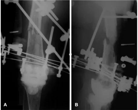
The decision was, therefore, made to perform a closed reduction andfixation of the fracture with a hybrid external fixator (HEF) (Orthofix SRL, Verona, Italy) (►Fig. 4).
Due to the fact that the fracture did not show signs of consolidation, at X-ray, 6 months after HEF, nonunion was an impending complication. Furthermore, the laboratory results revealed a normal ESR at 20 and CRP at 2 (►Fig. 5).
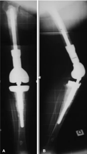
We therefore decided to perform a tumor-like total knee arthroplasty (►Fig. 6).
Intraoperative cultural and histological examinations were negative for infection.
At the 5-year follow-up, the patient showed a range of motion between 0 and 90° without pain.
The X-rays did not show signs of mobilization, dislocation, recurrence of infection, or other complications (►Fig. 7).
Discussion
Antibiotic-loaded cement spacers are used routinely in two-stage revision TKA. The goal of two-two-stage revision TKA is to radically eliminate the infection and to create the healthiest tissue possible for the new implant.8
Wan et al and Choi et al evaluated the use of different spacers in two-stage treatment for infected TKA.9,10Their analyses demonstrated that articulating spacers can provide good results in terms of infection control and functional outcome. However, their studies lack a thorough evaluation of spacer-related problems.
Fig. 4 Anteroposterior (A) and lateral (B) X-rays and clinical images (C) show the Orthofix hybrid external fixation.
Struelens et al retrospectively analyzed 154 patients in whom an articulating cement spacer had been implanted. Only 43% of all spacers were considered optimal, while a total of 12% showed major spacer issues such as fracture of the spacer, spacer dislocation, or knee subluxation.6
Fracture close to the spacer is the object of discussion in this case report.
Although the literature has widely discussed the manage-ment of periprosthetic fractures, there is a lack of information regarding the treatment of the fracture close to the spacer.
The early recovery of function and ambulation is critical in patients with these injuries, and effective surgical strategies to achieve these goals are essential.8,11
The optimum treatment of periprosthetic fractures in the region of the distal femur is undefined. When there is no presence of infection and the implant is well-fixed, the option to retain the implant should be taken. Operative strategies in this context include the use of retrograde intramedullary nail, plates, or externalfixator.
When there is a poor bone stock, comminuted fracture, or loose components, the use of a tumor-type prosthesis shows favorable results with low complication rates and rapid mobilization.12,13
Therefore, in our view, these guidelines can be applied also for the treatment of the fractures close to the spacer.
Conclusion
In our case report, we applied similar guidelines to those for total knee periprosthetic distal femur fractures: the use of megaprosthesis was associated with good functional and satisfactory levels.
We can conclude that the use of megaprosthesis is a reliable option in such cases
Conflict of Interest
None declared.
Fig. 6 Postoperative anteroposterior (A) and lateral (B) radiographs showing a tumor-like total knee arthroplasty (ZSS“Zimmer Segmental System,” Zimmer, Warsaw, Indiana, United States).
Fig. 7 Anteroposterior (A) and lateral (B) X-rays show good posi-tioning of the Zimmer Segmental System at the 5-year follow-up.
References
1 Solarino G, Abate A, Vicenti G, Spinarelli A, Piazzolla A, Moretti B. Reducing periprosthetic joint infection: what really counts? Joints 2016;3(04):208–214
2 Bozic KJ, Kurtz SM, Lau E, et al. The epidemiology of revision total knee arthroplasty in the United States. Clin Orthop Relat Res 2010;468(01):45–51
3 Mazzucchelli L, Rosso F, Marmotti A, Bonasia DE, Bruzzone M, Rossi R. The use of spacers (static and mobile) in infection knee arthroplasty. Curr Rev Musculoskelet Med 2015;8(04):373–382 4 Haleem AA, Berry DJ, Hanssen AD. Mid-term to long-term
follow-up of two-stage reimplantation for infected total knee arthro-plasty. Clin Orthop Relat Res 2004;(428):35–39
5 Park SJ, Song EK, Seon JK, Yoon TR, Park GH. Comparison of static and mobile antibiotic-impregnated cement spacers for the treatment of infected total knee arthroplasty. Int Orthop 2010;34(08):1181–1186 6 Struelens B, Claes S, Bellemans J. Spacer-related problems in two-stage revision knee arthroplasty. Acta Orthop Belg 2013;79(04):422–426 7 Iarikov D, Demian H, Rubin D, Alexander J, Nambiar S. Choice and
doses of antibacterial agents for cement spacers in treatment of
prosthetic joint infections: review of published studies. Clin Infect Dis 2012;55(11):1474–1480
8 Toogood PA, Vail TP. Periprosthetic fractures: a common problem with a disproportionately high impact on healthcare resources. J Arthroplasty 2015;30(10):1688–1691
9 Wan Z, Karim A, Momaya A, Incavo SJ, Mathis KB. Preformed articulating knee spacers in 2-stage total knee revision arthro-plasty: minimum 2-year follow-up. J Arthroplasty 2012;27(08): 1469–1473
10 Choi HR, Malchau H, Bedair H. Are prosthetic spacers safe to use in 2-stage treatment for infected total knee arthroplasty? J Arthro-plasty 2012;27(08):1474–1479
11 Nauth A, Nousiainen MT, Jenkinson R, Hall J. The treatment of periprosthetic fractures. Instr Course Lect 2015;64:161–173 12 Leino OK, Lempainen L, Virolainen P, Sarimo J, Pölönen T, Mäkelä
KT. Operative results of periprosthetic fractures of the distal femur in a single academic unit. Scand J Surg 2015;104(03): 200–207
13 Cannon SR. The use of megaprosthesis in the treatment of peri-prosthetic knee fractures. Int Orthop 2015;39(10):1945–1950