THE
JOURNAL • RESEARCH • www.fasebj.org
Lipoxin A
4
stimulates endothelial miR-126-5p
expression and its transfer via microvesicles
Marilina Codagnone,*,†Antonio Recchiuti,*,†Paola Lanuti,†,‡Anna Maria Pierdomenico,†,‡Eleonora Cianci,*,† Sara Patruno,*,†Veronica Cecilia Mari,† Felice Simiele,*,†Pamela Di Tomo,*,†Assunta Pandolfi,*,†
and Mario Romano*,†,1
*Department of Medical, Oral and Biotechnological Sciences,†Center on Aging Science and Translational Medicine (CeSI-MeT), and
‡Department of Medicine and Aging Science,“G. D’Annunzio” University of Chieti-Pescara, Chieti, Italy
ABSTRACT:The proresolution lipid mediator lipoxin (LX)A4bestows protective bioactions on endothelial cells. We examined the impact of LXA4on transcellular endothelial signalingvia microRNA (miR)-containing microvesicles. We report LXA4inhibition of MV release by TNF-a-treated HUVECs,associated with the down-regulation of 18miR in endothelial microvesicles (EMVs) and the up-regulation of miR-126-5p, both in HUVECs and in EMVs. LXA4 up-regulated miR-126-5p by∼5-fold in HUVECs and promoted a release of microvesicles (LXA4-EMVs) that enhanced miR-126-5p by∼7-fold in recipient HUVECs. In these cells, LXA4-EMVs abrogated the up-regulation of VCAM-1, induced in recipient HUVECs by EMVs released by untreated or TNF-a-treated HUVECs. LXA4-EMVs also reduced by∼40% the expression of SPRED1, which we validated as an miR-126-5p target, whereas they stimulated mono-layer repair in anin vitro wound assay. This effect was lost when the EMVs were depleted of miR-126-5p. These results provide evidence that changes in miR expression and microvesicle packaging and transfer represent a mechanism of action of LXA4, which may be relevant in vascular biology and inflammation.—Codagnone, M., Recchiuti, A., Lanuti, P., Pierdomenico, A. M., Cianci, E., Patruno, S., Mari, V. C., Simiele, F., Di Tomo, P., Pandolfi, A., Romano, M. Lipoxin A4 stimulates endothelial miR-126-5p expression and its transfer via microvesicles. FASEB J. 31, 000–000 (2017). www.fasebj.org
KEY WORDS:proresolving mediators • vascular repair • microRNA • intercellular signaling • inflammation
Endothelial cells are key players in acute inflammation, an innate protective host response to pathogens. They regu-late the initial steps of the inflammatory response that include vasodilation, leakage of plasma proteins, and leukocyte adhesion and transmigration into injured tissues (1). Endothelial cells also control inflammation-associated processes like neoangiogenesis, blood co-agulation and platelet aggregation (2). Ideally, the acute inflammatory response is self-limited, ending with the cessation of leukocyte, mainly polymorpho-nuclear neutrophil (PMN), infiltration, the clearance of
apoptotic PMNs by monocyte-derived macrophages, and tissue repair (3).
Endogenous chemical mediators produced from poly-unsaturated fatty acids are potent regulators of vascular responses during inflammation. For instance, arachidonic acid (AA) (5Z, 8Z, 11Z, 14Z eicosatetraenoic acid) is con-verted into prostaglandin (PG)E2, which promotes edema formation; PGI2(also termed prostacyclin), which induces vasodilation, and thromboxane (TX)A2, which triggers vasoconstriction and platelet aggregation (4, 5). AA can also be converted by 5-lipoxygenase-driven reactions into leukotrienes, which carry potent vasoregulatory actions (6). On the other hand, lipoxygenases catalyze the forma-tion of a series of tetraene-containing compounds termed lipoxins (LXs), which exhibit potent anti-inflammatory and proresolving bioactions (7–9). LX epimers, carrying the C15 alcohol in the R configuration and termed 15-epi-LX, can be produced by endothelial cells during interactions with PMNs in the presence of aspirin (10). Their bio-synthesis occurs in vivo in humans and can be enhanced by pioglitazone or statins (11–14), providing clues for the vasoprotective effects of these drugs. LXA4inhibits PMN chemotaxis, endothelial adherence, and transmigration (15); enhances nonphlogistic recruitment of monocytes (16); and stimulates macrophage phagocytosis (17). It also ABBREVIATIONS:Ann, annexin; AA, arachidonic acid; EGCF,
endothe-lial cell growth factor; EM, endotheendothe-lial medium; EMV, endotheendothe-lial microvesicle; FBS, fetal bovine serum; FPR, formylpeptide receptor; LX, lipoxin; M-CAM, melanoma cell adhesion molecule; miR, microRNA; PECAM, platelet endothelial cell adhesion molecule; PG, prostaglandin; PMN, polymorphonuclear neutrophil; P/S, penicillin/streptomycin; Rv, resolvin; SPRED, sprout-related EVH1 domain-containing protein; VE-cadherin, vascular endothelial cadherin
1Correspondence: Mario Romano, Molecular Medicine Laboratory,
De-partment of Medical, Oral and Biotechnological Sciences, CeSI-Met, “G. D’Annunzio” University, Via Luigi Polacchi 11/13, 66013 Chieti, Italy. E-mail: [email protected]
doi: 10.1096/fj.201600952R
This article includes supplemental data. Please visit http://www.fasebj.org to obtain this information.
regulates endothelial cell migration and angiogenesis by modulating vascular endothelial growth factor (VEGF) and VEGF receptor expression (18, 19). LXA4bioactions depend on the activation of a GPCR termed ALX/for-mylpeptide receptor (FPR)-2 (20), which is expressed in myeloid cells and in endothelial cells (18).
Microvesicles are small cell particles of heterogeneous size (100–1000 nm) that bud off parental cells in homeo-static conditions, upon cell activation. These microstruc-tures contain a variety of proteins, nucleic acids, and lipids that can be delivered, in a paracrine, autocrine, or endo-crine manner, to recipient cells, whereby they regulate functional responses (21). For instance, microvesicles from endothelial progenitor cells activate angiogenesis in vitro and in vivo and protect from kidney ischemia by trans-ferring mRNA or microRNA (miR) (22, 23). Also, human PMN-derived microvesicles block leukocyte–endothelial cell interactions in vivo and in vitro by delivering annexin (Ann)A1 (24). Endothelial microvesicles (EMVs) are pre-sent in inflammatory lesions, such as atherosclerotic plaques or ischemic tissues (25). They may differ in concentration and phenotype between healthy subjects and patients, representing diagnostic markers of en-dothelial dysfunction and associated disorders (26). Microvesicles play a crucial role in inflammation and endothelial physiology, as they can deliver to recipient cells AA, which can be converted into PGI2and TXA2(27), or biogenic precursors of D-series resolvins (Rvs), which limit PMN infiltration and enhance keratinocyte healing (28). miRs constitutes a class of small (;22 nucleotides long), noncoding RNAs that bind to mRNA to regulate post-translational events (29). miRs are tissue- and cell-specific and control a variety of biologic processes, including cell proliferation, differentiation, and tissue development (30). They also play important roles in the inflammatory re-sponse (31) and vascular biology. For instance, the miR-17-92 cluster and miR-27a/b regulate angiogenesis and endothelial cell growth (32, 33). Moreover, miRs are part of the mechanisms triggered by LX and RvD1 to dampen acute inflammation and tissue fibrosis (34–36). miRs can be transferred across cells, packed into microvesicles for RNase protection. Also, significant differences can be observed in relative abundance of miRs between micro-vesicles and their parental cells (37). For instance, changes in miR content of microvesicles have been observed in metabolic and cardiovascular diseases (38), indicating a selective miR“packaging” into microvesicles.
In the present study, LXA4regulated EMV release and activity in inflammatory conditions and modulated miR content in endothelial cells and EMVs. Moreover, micro-vesicles from ECs exposed to LXA4were enriched in miR-126 and had enhanced capability to stimulate endothelial migration.
MATERIALS AND METHODS
Endothelial cell culture and EMV isolation
Umbilical cords were obtained from randomly selected healthy mothers who delivered at the Chieti University
Hospital. All procedures were in agreement with the ethical standards of the Institutional Committee on Human Experi-mentation (Reference Number: 1879/09COET) and with the principles of the Declaration of Helsinki. After approval of the protocol by the Institutional Review Board, a signed informed consent form was obtained from each participating subject. HUVECs were obtained and cultured on a 1.5% gelatin matrix with endothelial medium (EM): DMEM low-glucose (50% vol/vol), M199 (50% vol/vol), penicillin/streptomycin (P/S, 1%), andL-glutamine (2 mM) supplemented with fetal bovine
serum (FBS, 20%), heparin (18 U/ml), and endothelial cell growth factor (ECGF, 50mg/ml). For EMV production, 80% confluent HUVECs between the 3rd and 6th passages were incubated with LXA4(0.1–100 nM) and TNF-a (1 ng/ml) for
24 h in modified EM with 10% FBS without ECGF and heparin, to minimize the influence of these agents on LXA4bioactions.
In some experiments, cells were exposed to the ALX/FPR2 antagonist WRW4(WRWWWW; 10mM, 30 min; Calbiochem,
Billerica, MA, USA). For EMV isolation and purification, the cell medium was collected and centrifuged at 300 g for 10 min, then at 12,096 g for 15 min to remove cell debris. Supernatants were ultracentrifuged (152,000 g, 2 h) to collect EMVs. EMVs and cells were lysed with lysis solution from Norgen Biotek (Thorold, ON, Canada), and materials were stored at220°C until miR isolation.
Flow cytometric analysis of EMVs
For EMV characterization, HUVEC supernatants (5 ml/sample) were added to 200 ml of binding buffer (BD Bio-science, Franklin Lakes, NJ, USA) 2X, to control Ca2+
con-centration, which is relevant for AnnV binding and processed with a“no lyse and no wash” method (39). In brief, samples were stained with AnnV (allophycocyanin-conjugated) and Mitotracker green (Thermo Fisher Scientific, Waltham, MA, USA). After staining (30 min, 4°C in the dark), 500 ml of binding buffer 2X (BD Bioscience) were added to each tube, and 13 106events/samples were acquired by flow cytometry with a FACS Canto cytometer (BD Bioscience) with a 3-laser, 8-color configuration, set at a low threshold (200) on the FITC channel. Signal amplifications for forward scatter and side scatter, as well as for any fluorescence channel, were set in the logarithmic mode. EMV morphology was confirmed by run-ning Megamix beads (Biocytex, Marseille, France). Instrument performances and data reproducibility were checked by the Cytometer Setup & Tracking Module and further validated by the acquisition of Rainbow Beads (BD Bioscience). Fluo-rescence Minus One controls were used to detect nonspecific fluorescence. Compensation was assessed by using Comp-Beads (BD Bioscience) and Mitotracker green single-stained samples. Data were analyzed with the FACSDiva ver. 6.1.3 (BD Bioscience), and FlowJo, ver. 8.8.6 (TreeStar, Ashland, OR, USA) software. EMVs were enumerated by a single-platform counting method with Trucount tubes (BD Bio-science). EMV concentration was calculated with the following formula: (number of events for AnnV in the Tru-count bead region)3 (number of Trucount beads per test/ test volume).
miR measurements
miR were evaluated by real-time PCR. To this end, miR, con-tained in EMVs, were isolated with a purification kit (Norgen Biotek) and quantified using a Nanodrop Spectrophotometer (Thermo Fisher Scientific, Waltham, MA, USA). A hundred nanograms miR was reverse transcribed with miScript II RT Kit (Qiagen, Milan, Italy), and the relative abundance of 86 miR was determined using miScript miR PCR Array (Qiagen). Real-time
PCR was outperformed on a ABI 7900HT thermocycler (Thermo Fisher Scientific), using the manufacturer’s recommended pro-gram. miR expression was determined by the 22DDCtmethod (40) and cel-mir-39 was used to normalize input cDNA.
miR-126-5p inhibition HUVECs (1–1.5 3 106
cells), seeded into a 10 cm culture dish, were transfected with the human serum albumin-miR-126-5p inhibitor (MIN0000444; Qiagen) or a scramble neg-ative control miRNA (200 pmol/dish in 10 ml of complete growth medium) using the Interferin reagent (40ml/dish; Polyplus transfection; Illkirch, France) according to the manufacturer’s protocol. Optimal efficiency was achieved when cells were 50% confluent on the day of transfection. EMVs were collected and analyzed as reported above. EMV uptake by HUVECs and miR-126 expression Conditioned media from untreated or LXA4-treated HUVECs
were incubated with PKH26 (1:250; Sigma-Aldrich, Milan, Italy) and ultracentrifuged for 2 h, as described above. PKH26-labeled EMVs were counted using FACS Verse (BD Biosciences), and 13 106EMVs were added directly to confluent HUVECs cultured in 6-well plates (2 ml). After 4 and 24 h at 37°C, the HUVECs were washed twice with PBS. To assess uptake, the cells were detached and visualized with the Image Stream Amnis (EMD Millipore, Billerica, MA, USA). miR-126 levels in recipient HUVECs were measured as above.
Analysis of VCAM-1 expression
HUVECs (70–80% confluent) were treated with LXA4(10 nM)
and/or TNF-a (1 ng/ml) for 24 h in modified EM with 10% FBS without ECGF and heparin. After 24 h, EMVs were isolated by ultracentrifugation for 2 h and enumerated on the FACS Verse (BD Biosciences) flow cytometer. EMVs (23 106) derived from untreated HUVECs (CTRL-EMV) or from HUVECs exposed to TNF-a (TNFa-EMV), TNF-a plus LXA4(TNFa+LXA4-EMV), or
LXA4(LXA4-EMV), were added to confluent HUVECS cultured
in 6-well plates (2 ml). After a 24 h incubation at 37°C, HUVECs were washed twice with PBS and labeled with an anti-VCAM-1 antibody (PE-A-anti-CD106; BioLegend, San Diego, CA, USA). The cells were analyzed with a FACS Verse flow cytometer (BD Biosciences).
In vitro wound healing
HUVECs were seeded in 6-well plates (23 105cells/well) with basal medium until monolayers reached 80–90% confluence. Wounds were created by manually scraping the monolayers with a 200 ml pipet tip and the plates were then vigorously rinsed with PBS to remove cellular debris. After the wound was created, 2 3 106EMVs from untreated or LXA4-treated
HUVECs were added to each well and incubated at 37°C, in a humidified atmosphere containing 5% CO2 for 6 or 20 h.
HUVECs in 1% or 10% FBS medium were respectively used as negative and positive controls. Wound closure was evaluated at 6 and 20 h with an optical microscope at310 magnification. Two zones of each wound were photographed at different time intervals along the wound boundary, and for each of the 2 im-ages, wound widths were quantified in 5 locations with ImageJ software (National Institutes of Health, Bethesda, MD, USA). Duplicate wells of each condition were examined for each experiment.
Western blot analysis
HUVECs from the wound-healing assay were homogenized at 4°C with RIPA buffer [150 mM NaCl; 1.0% Nonidet P-40; 0.5% deoxycholate; 0.1% SDS, and 50 mM Tris (pH 8.0)] containing 1 mM Na3VO4, 5 mM NaF and a cocktail of protease inhibitors
(Sigma-Aldrich). Protein concentration was determined with the Lowry protein assay (Bio-Rad, Hercules, CA, USA). Equal amounts of proteins (30 mg) were resolved by 12% SDS gel electrophoresis, transferred onto PVDF membranes, and blocked with 10% nonfat dry milk for 1 h. Blots were incubated with a primary anti-sprouty-related EVH1 domain–containing 1 (SPRED1) antibody (1:500 dilution; Thermo Fisher Scientific) or with an anti-GAPDH antibody (Santa-Cruz Biotechnologies, Inc., Dallas TX, USA), followed by the correspondent HRP-conjugated secondary antibodies. Proteins were revealed by chemiluminescence with the ECL kit (Euroclone, Milan, Italy). GAPDH was used as loading control. Chemiluminescence sig-nals were quantitated with an imaging scanner (Alliance 4.7; Uvitec, Cambridge, United Kingdom).
39UTR luciferase reporter assay
Breast cancer MDA-MB-231 cells, grown in DMEM supple-mented with 10% FBS, 1%L-Glu and P/S, were cotransfected with SPRED1 39UTR (pEZX-MT05; Genecopoeia, Rockville, MD, USA), plus the pCMV-miR-126-5p plasmid (Origene, Rockville, MD, USA) or an empty pCMV-MIR plasmid (Origene) as a control (1.5mg each) using Lipofectamine 2000 (12 ml; Thermo Fisher Scientific), according to the manufacturer’s protocol. Twenty-four hours after transfection, cells were lysed, and the luciferase activity was determined as previously reported (41). Statistical analysis
Results are reported as arithmetic means 6 SEM, unless
otherwise indicated. Statistical significance was assessed with the Student’s t test. A value of P , 0.05 indicatied significance. Statistical analysis was performed with Prism 5 (GraphPad, La Jolla, CA, USA).
RESULTS
Impact of LXA4on TNF-a-induced EMV release
and activity
LXA4exerts potent regulatory bioactions on endothelial cells. Therefore, we asked whether LXA4regulates EMV release. To this end, we initially set the appropriate flow cytometric conditions to identify EMVs in conditioned medium, by exposing confluent HUVEC monolayers to TNF-a (1 ng/ml) for 24 h and incubating the condi-tioned medium with Mitotracker Green fluorescent medium. EMVs were detected and distinguished from cell debris based on positive MitoTracker staining (39). They were gated on the basis of their mitochondrial activity and morphologic parameters (Fig. 1A). This method, by avoiding morphologic recognition, maxi-mized the probability of including all EMV events in the analysis and helped to exclude debris, immune com-plexes, and calcium phosphate precipitates.
TNF-a increased EMV release by ;2.2 fold (P = 0.002). This effect was abrogated by LXA4(0.1–100 nM, 24 h) in
a concentration-dependent manner, with maximum inhibition at 10 nM (;80% reduction; P = 0.02) and at
100 nM LXA4(;100% reduction; P = 0.0005; Fig. 1B).
LXA4 itself did not modify the number of released
EMVs (results not shown). To establish receptor de-pendence of this effect, we exposed HUVECs to WRW4, a selective ALX/FPR2 antagonist (42). As illustrated in
Fig. 1C, WRW4suppressed the effect of LXA4on EMV
release (P = 0.004). Moreover, the WKYMVm peptide, another known ALX/FPR2 agonist (43, 44) potently inhibited TNF-a-induced EMV release (Supplemental Fig. 1A). Together, these results indicate that ALX/ FPR2 signaling controls EMV release.
LXA4selectively modulates miR content in
EMVs and HUVECs
Microvesicles contain miRs that exert regulatory effects on cell uptake (22, 23, 38). To determine whether LXA4 also had an impact on the miR profile of EMV, we eval-uated the relative abundance of 86 miRs, selectively expressed by endothelial cells (45), using an miR PCR
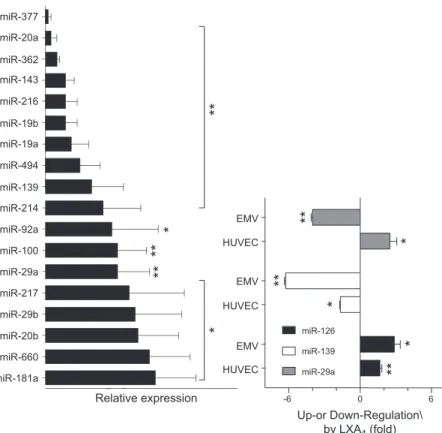
array. LXA4 modulated the contents of several miRs
in EMVs from TNF-a-stimulated HUVECs. Among these, 18 were significantly down-regulated (Fig. 2A), whereas 54 were up-regulated, although only miR-126-5p reached statistical significance (Supplemental
A
2.54 0 FSC-A SSC-A 105 104 103 102 0 105 104 103 102 FITC-A FSC-A 0 102 103 104 105 0 102 103 104 105B
**
C
***
*
0.0EMV Release (Fold of CTRL)
0.1 CTRL 1 100 10 0.5 1.0 1.5 2.0 2.5
**
EMV Release (Fold of CTRL)
0.0 0.5 1.0 1.5 2.0 2.5 -+ -+ + -+ + + + -+
*
***
-+ WRW4 LXA4 TNF-α TNF-α TNF-α + LXA4 [nM]Figure 1. LXA4receptor-dependent regulation of TNF-a-induced EMV release. (A) Left: flow cytometric analysis of MitoTracker+EMV.
Right: EMV morphologic parameters. (B) HUVECs were either left untreated (CTRL) or exposed to TNF-a (1 ng/ml), in the presence or not of the indicated concentrations of LXA4. EMVs were collected after 24 h and enumerated by flow cytometry.
Results are means6SEM of 4 different experiments, each performed in duplicate. *P = 0.02, **P = 0.002, ***P = 0.0005. (C )
HUVECs were exposed for 24 h to TNF-a (1 ng/ml), with or without LXA4(100 nM). When present, WRW4(10mM) was added
to cells alone or 30 min before LXA4. Results are means6SEMof 3 different experiments, each performed in duplicate. *P = 0.04,
Table 1). A comparative analysis of cell-associated vs. EMV-containing miRs showed that LXA4: 1) down-regulated miR-29a in EMVs (P = 0.05), while up-regulating it in cells (P = 0.02); 2) down-regulated miR-139, both in EMVs (P = 0.001) and HUVECs (P = 0.005); and 3) up-regulated miR-126-5p in EMVs (P = 0.02) and cells (P = 0.01) (Fig. 2B). These results show for the first time that LXA4 regulate the rela-tive abundance of miRs and packaging of miR into microvesicles.
Analysis of miR-126-5p up-regulation by LXA4 Recent evidence indicates that miR-126 plays key roles in vascular endothelial cell repair and inflammation (46, 47).Therefore, we analyzed the direct effects of LXA4on this miR. To this end, we evaluated the rela-tive abundance of miR-126 in HUVECs after exposure
to increasing LXA4concentrations (0.01–100 nM). As
illustrated in Fig. 3A, LXA4up-regulated miR-126-5p expression in a concentration-dependent manner, with significant increments at 0.1 nM (;3-fold; P = 0.04), 1 nM (;5-fold; P = 0.00003) and 10 nM (;3-fold; P = 0.02). A time course of exposure to 1 nM LXA4, showed a signifi-cant miR-126-5p increase after 18 h (;3-fold; P = 0.02) and 24 h (;4-fold; P = 0.009; Fig. 3B). In contrast, LXA4did not change miR-126-3p expression (results not shown). Un-like LXA4, the WKYMVm peptide did not modify miR-126-5p expression (Supplemental Fig. 1B).
Impact of LXA4on EMV uptake by HUVECs
EMV uptake by recipient endothelial cells as mechanism of paracrine intercellular signaling has been documented (48).To determine whether LXA4has an impact on this process, HUVECs were exposed to 1 nM LXA4or vehicle for 24 h, and EMVs were isolated, labeled with PKH26, and coincubated with recipient HUVECs for 4 and 24 h. Image Stream flow cytometry showed a significant uptake of PKH26-labeled EMVs by HUVECs. Notably, after 4-h incubation EMVs from LXA4-treated HUVECs (LXA4-EMVs) were incorporated with higher affinity (;1.5-fold; P = 0.0002) compared with EMVs from cells exposed to vehicle (CTRL-EMVs; Fig. 4C). This effect was no longer observed after 24 h. miR-126-5p was unexpectedly more abundant in cells exposed for 4 h to LXA4-EMVs than to either untreated HUVECs (;7-fold increase, P = 0.0004) or cells incubated with CTRL-EMVs (;2 fold increase, P = 0.04; (Fig. 4D). No significant changes in miR-126-3p were instead detected (results not shown). These results indicate that miR-126-5p can be transferred to recipient endothelial cells via EMVs and that such transfer can be enhanced by LXA4.
LXA4-EMVs down-regulate the expression of miR-126-5p targets and stimulate endothelial
migration in vitro
Because LXA4-EMVs enhanced miR-126-5p expression in recipient HUVECs, we next determined whether they also
B
A
miR-126 miR-139 miR-29a -6 0 6**
*
*
**
*
**
HUVEC EMV HUVEC EMV HUVEC EMV miR-181a miR-660 miR-20b miR-29b miR-217 miR-29a miR-100 miR-92a miR-214 miR-139 miR-494 miR-19a miR-19b miR-216 miR-143 miR-362 miR-20a miR-377 Relative expression*
*
**
**
**
Up-or Down-Regulation\ by LXA4 (fold)Figure 2. LXA4 modulates miR profile in
HUVECs and EMVs. Cells (1.5 3 106) were incubated with TNF-a (1 ng/ml) in the presence or not of LXA4(10 nM) for 24 h.
Endothelial-specific miRs were isolated from cells and EMVs and analyzed. (A) LXA4 significantly
down-regulated 18 miRs. *P , 0.01, **P , 0.001.
.Results are expressed as means 6 SEM of 4
different experiments. (B) miR differentially modulated in HUVECs and EMVs by LXA4.
Data are means6SEMof 4 different experiments.
miR-29a: *P = 0.05, **P = 0.002; miR-139: *P = 0.005 **P = 0.001; miR-126: *P = 0.02, **P = 0.01.
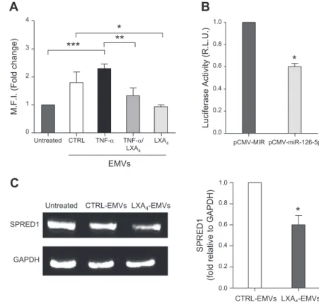
down-regulated select targets, relevant to inflammation and vascular integrity. To this end, we set 2 experimental conditions: one to mimic endothelial inflammation, whereby HUVECs were exposed to TNF-a in the presence or not of LXA4; the other to investigate protective LXA4 bioactions on endothelial cells. For the first condition, we selected VCAM-1 as an miRNA-126-regulated protein (49) and observed that EMVs released by cells exposed to LXA4, alone or with TNF-a, reduced VCAM-1 expression in recipient cells (Fig. 5A). For the second condition, we focused on SPRED1, which was identified as a key effector of miR-126-dependent stimulation of endothelial cells migration (46). We first validated SPRED1 as the miR-126 target gene. We cotransfected SPRED1 39UTR (pEZX-MT05) with an miR-126-5p/luciferase-expressing plasmid (pCMV-miR-126-5p) in MDA-MB231 cells and measured luciferase activity 24 h after transfection. We observed a
significant (;40%; P = 0.0002) reduction in luciferase ac-tivity in MDA-MB-231 cells coexpressing pCMV-miR-126-5p and pEZX-MT05 compared with cells transiently transfected with the empty pCMV-miR vector together with pEZX-MT05 (Fig. 5B), thus demonstrating direct binding of miR-126-5p to SPRED1 39UTR. We then ex-amined SPRED1 expression in recipient HUVECs exposed to CTRL- or LXA4-EMVs. Figure 5C shows that, consistent with previous data (46), CTRL-EMVs reduced SPRED1 expression by;20% in recipient HUVECs and that this inhibition reached;40% with LXA4-EMVs (P = 0.005 vs. CTRL-EMVs).
Subsequently, we examined the impact of LXA4-EMVs on HUVEC migration. For this assay, HUVEC monolayers were manually scraped with a 200ml sterile pipet tip, to obtain a wound of;80 mm width (as described earlier). Cells were then incubated with CTRL- or LXA4-EMVs in the presence of 1% FBS, to inhibit cell proliferation, and wound closure was evaluated after 6 and 20 h. At 6 h, both CTRL- and LXA4-EMVs significantly stimulated wound closure (;1.5-fold; P = 0.006 and P = 0.0094, respectively; Fig. 6A). At this time point, the effect of the LXA4-EMVs was not significantly different from that of CTRL-EMVs. However, after 20 h, LXA4-EMVs stimulated wound clo-sure to a higher extent vs. CTRL-EMVs (P = 0.0072). To obtain more direct evidence of the involvement of miR-126-5p in these phenomena, we transfected HUVECs with an miR-126-5p inhibitor, which yielded miR-126-5p-depleted EMVs (Fig. 6B). miR-126-5p-miR-126-5p-depleted EMVs, either from untreated (126-5pInh CTRL-EMVs) or LXA4-treated (126-5pInh LXA4-EMVs) HUVECs, were unable to stimulate wound closure of the recipient cells (Fig. 6C).
DISCUSSION
Starting from a large body of evidence of the role of LXA4 in vascular biology (8), as well as from previous work showing that proresolving lipid mediators, such as RvD1, control the inflammatory response by modulating miR expression in vitro and in vivo (50), we tested the hypothesis that LXA4controls intercellular pathways of miR delivery. In the present work, we provide evidence that LXA4 in-duces transcellular signaling by exploiting microvesicles as carriers and miR as effectors.
EMVs can be regarded as biomarkers of inflammation and endothelial dysfunction, because they may reflect a balance between cell stimulation, proliferation, and death (26). EMVs are released by endothelial cells upon exposure to inflammatory stimuli and increase in the circulation of patients with cardiovascular disease, renal failure, and metabolic disorders (21, 51). Depending on cell and mi-croenvironmental conditions, EMVs can perturb vascular homeostasis by activating endothelial cells with an auto-crine loop (52), but they can also promote cell survival, induce anti-inflammatory effects, and stimulate endothe-lial regeneration (46). Thus, it is likely that in addition to the number of EMVs, their contents can make the differ-ence between damaging or protective effects. Indeed, EMVs are cargo vectors for material selectively acquired from parental cells, including bioactive lipids, integrins,
Figure 3. Time- and concentration-dependent miR-126-5p up-regulation by LXA4. (A) HUVECs (1.53 105) were incubated
with the indicated concentrations of LXA4for 24 h.
miR-126-5p expression was evaluated by real-time PCR. Results are means6SEM(n = 3). *P = 0.04, **P = 0.02, ***P = 0.00003. (B)
miR-126-5p expression in HUVEC exposed to 1 nM LXA4for
the indicated time. Bars represent means6SEMof 3 different experiments. *P = 0.02; **P = 0.009.
cytokines, enzymes, mRNA and miR, which can repro-gram recipient cells (53). Moreover, they can carry oxygen-reactive species, which are more abundant in EMVs generated by HUVECs subjected to reperfusion injury and promote cardiomyocyte apoptosis (54).
We tested the hypothesis that one of the mechanisms of LXA4bioactions on vascular homeostasis was the regu-lation of EMV release and packaging, with consequences on EMV functional impact on recipient cells. Although LXA4per se did not alter the number of EMVs released under standard culturing conditions, it antagonized TNF-a, significantly reducing the number of microvesicles re-leased by the HUVECs exposed to this proinflammatory cytokine (Fig. 1B). This result is consistent with early data showing that LXA4can counteract proinflammatory re-sponses triggered by cytokines (18, 55) and indicates that modulation of microvesicle biogenesis represents an un-appreciated antagonist function of LXA4. This effect oc-curs through the activation of ALX/FPR2 (Fig. 1C), a
proresolving receptor that intercepts multiple pro-resolution circuits regulated by RvD1 and AnnA1 in addition to LXA4(9). We obtained further evidence of the involvement of ALX/FPR2 in this setting by using the WKYMVm peptide, which binds and activates this receptor (43, 44). This peptide blocked TNF-a-induced EMV release (Fig. 1C), but unlike LXA4, it did not up-regulate miR-126-5p expression (Supple-mental Fig. 1). These results are consistent with dif-ferent interaction domains and signaling of ALX/ FPR2 agonists (56).
Together with biogenesis, the analysis of content is key to understanding the functional significance of EMVs. We focused on miR in light of the emerging role of these small RNA sequences in vascular biology and inflammation (32, 57). We used a top-down approach by analyzing changes in 84 endothelium-specific miRs in EMVs. LXA4 signifi-cantly down-regulated 18 miRs (Fig. 2A) and up-regulated 54, among which only the increase in miR-126-5p reached
Figure 4. LXA4affect EMV uptake by HUVECs. Cells (1.53 105) were incubated with CTRL-EMVs or LXA4-EMVs for 4 (A) or
24 h (B). EMV uptake was monitored byflow cytometry. Representative bright-field (Ch01), PE-fluorescence (Ch03) and side scatter (Ch06) images are shown. Magnification, 340. (C) EMV uptake was quantitated using a specific algorithm to decode fluorescence data. Results are expressed as means 6SEMof 3 different experiments. *P = 0.008; **P = 0.0002. (D) HUVECs were
exposed to CTRL-EMVs or LXA4-EMVs for 4 h and relative abundance of miR-126 was determined by real-time PCR. MFI, mean
statistical significance (Fig. 2B and Supplemental Table 1). Notably, comparing data from measurements of miR content in cells vs. microvesicles, it was clear that LXA4 regulated both miR generation and packaging into EMVs (Fig. 2B), providing evidence that endogenous proresolution lipid mediators selectively regulate miR packaging. Among the LXA4–down-regulated miRs, miR-92a may have negative outcomes on vascular in-tegrity as its inhibition prevents endothelial dysfunction and atherosclerosis (58), whereas miR-19a and miR-20a may be relevant in angiogenesis (59). Thus, overall LXA4 appears to antagonize TNF-a-triggered proinflammatory miR signaling, although further studies are required to support this hypothesis.
Among the 84 miRs evaluated in this study, only miR-126-5p was significantly up-regulated by LXA4, either by itself or in combination with TNF-a, both in HUVECs and EMVs (Fig. 2). Because no changes in miR-126-3p were observed, it is likely that LXA4acts on premiR-126 maturation. Both the 3p and 5p strands of miR-126 ex-ert protective actions on the vasculature, although they may control different pathways. In fact, miR-126-3p delivery via microvesicles released by apoptotic endo-thelial cells, reduces experimental atherosclerosis (60), whereas miR-126-5p, but not miR-126-3p, promotes endothelial cell proliferation and vascular integrity (61). Along these lines, deletion of the gene encoding premiR-126 impairs postischemic neovascularization in mice
(62). Together with previous work showing LXA4
at-tenuation of renal fibrosis by the induction of let-7c in human renal epithelial cells (34), our present data indi-cate that the regulation of selected miR is a relevant
mechanism of action of LXA4, which can be helpful to unveil organ protective pathways relevant in pathobi-ology and pharmacpathobi-ology.
LXA4also modulated properties and paracrine func-tions of EMVs. We observed that LXA4-EMVs had a faster kinetic of uptake by recipient HUVECs compared with the EMVs released in the absence of LXA4. Although an up-take enhancement at 4 h (Fig. 4C) appears to be consistent with an amplification of microvesicle-transferred miR bioactivity (48), the mechanisms underlying this
phe-nomenon are elusive. LXA4did not influence EMV
ex-pression of adhesion molecules (i.e., PECAM-1, M-CAM, VCAM-1 and VE-cadherin), which can potentially control EMV uptake by recipient HUVECs, indicating that other mechanisms may be involved. On the other hand, the LXA4-EMV–induced increase in miR-126-5p expression in recipient HUVECs (Fig. 4D) is likely to be related to the higher miR-126-5p content in LXA4-EMV, although the stimulation of de novo miR-126-5p formation cannot be excluded. This point also should be studied further.
However determined, miR-126-5p up-regulation by LXA4-EMV in recipient HUVECs correlated with the suppression of EMV- or TNF-a-induced EMV up-regulation of VCAM-1, a miR-126 regulated gene and main component of the inflammatory cascade (49) (Fig. 5A). It was also associated with enhanced down-regulation of SPRED1 (Fig. 5C), which we validated as miR-126-5p target (Fig. 5B), as well as with the stimula-tion of wound closure (Fig. 6A). This last effect was lost when EMVs were depleted in miR-126-5p (Figs. 6B, C), emphasizing the role of this miR in EMV-dependent in-tercellular communication routes.
Figure 5. miR-126-5p targets and EMV functions regulated by LXA4. (A) HUVECs were seeded at
1.2 3 105and treated with CTRL-EMVs, TNFa-EMVs, TNFa+LXA4-EMVs, or LXA4-EMVs for
24 h. VCAM-1 was evaluated by flow cytometry. Data are means 6SEM (n = 3). *P = 0.04, **P = 0.02, ***P = 0.0005. (B) MDA-MB-231 cells were transfected with pEZX-MT05 plus pCMV-MIR empty vector or a miR-126-5p-expressing pCMV-MIR vector (pCMV-miR-126-5p). The luciferase activity was normalized for total proteins in cell lysates. Means 6 SEM of 3 independent
experi-ments performed in duplicate. *P = 0.0002. (C ) HUVECs (2 3 105) were exposed to CTRL- or LXA4-EMVs for 20 h, and SPRED1 expression
was evaluated by Western blot analysis. Left: a representative blot; right: densitometric analysis. MFI, meanfluorescence intensity; RLU, relative luminance units. Data are means 6 SEM of 5 different experiments. *P = 0.005.
Together, these results are consistent with early re-ports on the protective functions of miR-126 on the vasculature (46, 47, 59, 61) and indicate that LXA4 anti-inflammatory signaling can propagate by intercellular pathways involving the modulation of microvesicle biogenesis, content, and function. Along these lines, reduced miR-126 content in microvesicles released by endothelial cells exposed to high glucose as well as in peripheral blood microvesicles from diabetic patients has been reported (46), suggesting that the observed LXA4actions on miR-126 could be relevant in disease states.
In summary, our present results highlight the biologic significance of EMVs as autocrine modulators of endo-thelial repair functions. This is a relevant concept that supports the view that cell-to-cell communication is achieved by multiple circuits, involving soluble mediators, as well as signals delivered with microvesicles. In this re-spect, our findings showing that the endogenous
pro-resolution lipid mediator LXA4 can regulate these
mechanisms open new perspectives for the understand-ing of protective circuits that may be relevant in vascular
biology and more generally during the immune– inflammatory response.
ACKNOWLEDGMENTS
The authors thank Sara Di Silvestre (Department of Medical, Oral and Biotechnological Sciences,“G. D’Annunzio University”) for technical assistance with umbilical cords collection and HUVEC isolation. This work was supported, in part, by a grant from the Italian Ministry of Education, University and Research (PRIN 2010YK7Z5K_002; to M.R.). The authors declare no conflicts of interest.
AUTHOR CONTRIBUTIONS
M. Codagnone designed and performed the research and wrote the paper; A. Recchiuti performed the research, analyzed the data, and wrote the paper; P. Lanuti, A. M. Pierdomenico, E. Cianci, S. Patruno, V. C. Mari, F. Simiele and P. Di Tomo performed the research and analyzed the data; A. Pandolfi analyzed the data; and M. Romano designed the research and wrote the paper.
Figure 6. (A) HUVECs were seeded at 2 3 105
and maintained in 1% FBS for 24 h. Monolayers were scraped with a 200-ml pipet tip, and cells were either left untreated or exposed to CTRL-or LXA4-EMVs. Wound closure was quantified at
6 and 20 h after wounding, by measuring the area not filled by cells with image analysis software. Left: representative images obtained by phase-contrast microscopy. Magnification 310. Right: quantitative analysis. Results are expressed as percentage above T0and represent
means 6 SEM of 4 different experiments. 6 h: *P = 0.0094, ***P = 0.006; 20 h: **P = 0.0072. (B) miR-126-5p expression in EMVs from HUVECs transfected with the miR-126-inhibitor, as com-pared with EMVs from cells transfected with the scrambled negative control miRNA (NC). Re-sults are means 6 SE from 2 representative
experiments. (C ) Wound closure of HUVECs exposed to EMVs released by cells transfected with an NC miRNA or the miR-125-5p inhibitor and treated or not with LXA4. Results are means6SE
from 4 separate experiments performed in duplicate. ***P = 0.0008.
REFERENCES
1. Kumar, V., Abbas, A. K., and Aster, J. C. (2014) Robbins and Cotran Pathologic Basis of Disease. 9th ed. Elsevier Health Sciences, Philadelphia, PA
2. Gimbrone, M. A., Jr., Cybulsky, M. I., Kume, N., Collins, T., Resnick, N. (1995) Vascular endothelium: an integrator of pathophysiological stim-uli in atherogenesis. Ann. NY Acad. Sci. 748, 122–131; discussion 131–122 3. Serhan, C. N., Brain, S. D., Buckley, C. D., Gilroy, D. W., Haslett, C., O’Neill, L. A., Perretti, M., Rossi, A. G., and Wallace, J. L. (2007) Resolution of inflammation: state of the art, definitions and terms. FASEB J. 21, 325–332
4. Higgs, E. A., Moncada, S., and Vane, J. R. (1978) Inflammatory effects of prostacyclin (PGI2) and 6-oxo-PGF1alpha in the rat paw. Prosta-glandins 16, 153–162
5. Samuelsson, B. (1983) Leukotrienes: mediators of immediate hypersensitivity reactions and inflammation. Science 220, 568–575 6. Molin, D. G., van den Akker, N. M., and Post, M. J. (2010)‘Lox on
neovascularization’: leukotrienes as mediators in endothelial biology. Cardiovasc. Res. 86, 6–8
7. Samuelsson, B., Dahl´en, S. E., Lindgren, J. A., Rouzer, C. A., and Serhan, C. N. (1987) Leukotrienes and lipoxins: structures, biosynthesis, and biological effects. Science 237, 1171–1176
8. Serhan, C. N., Hamberg, M., and Samuelsson, B. (1984) Lipoxins: novel series of biologically active compounds formed from arachidon-ic acid in human leukocytes. Proc. Natl. Acad. Sci. USA 81, 5335–5339 9. Romano, M., Cianci, E., Simiele, F., and Recchiuti, A. (2015) Lipoxins
and aspirin-triggered lipoxins in resolution of inflammation. Eur. J. Pharmacol. 760, 49–63
10. Cl`aria, J., and Serhan, C. N. (1995) Aspirin triggers previously undescribed bioactive eicosanoids by human endothelial cell-leukocyte interactions. Proc. Natl. Acad. Sci. USA 92, 9475–9479 11. Birnbaum, Y., Ye, Y., Lin, Y., Freeberg, S. Y., Nishi, S. P., Martinez, J. D.,
Huang, M. H., Uretsky, B. F., and Perez-Polo, J. R. (2006) Augmentation of myocardial production of 15-epi-lipoxin-a4 by pio-glitazone and atorvastatin in the rat. Circulation 114, 929–935 12. Chiang, N., Bermudez, E. A., Ridker, P. M., Hurwitz, S., and Serhan,
C. N. (2004) Aspirin triggers antiinflammatory 15-epi-lipoxin A4 and inhibits thromboxane in a randomized human trial. Proc. Natl. Acad. Sci. USA 101, 15178–15183
13. Gutierrez, J., Ramirez, G., Rundek, T., and Sacco, R. L. (2012) Statin therapy in the prevention of recurrent cardiovascular events: a sex-based meta-analysis. Arch. Intern. Med. 172, 909–919
14. Planagum`a, A., Pfeffer, M. A., Rubin, G., Croze, R., Uddin, M., Serhan, C. N., and Levy, B. D. (2010) Lovastatin decreases acute mucosal inflammation via 15-epi-lipoxin A4. Mucosal Immunol. 3, 270–279 15. Fierro, I. M., Colgan, S. P., Bernasconi, G., Petasis, N. A., Clish, C. B.,
Arita, M., and Serhan, C. N. (2003) Lipoxin A4 and aspirin-triggered 15-epi-lipoxin A4 inhibit human neutrophil migration: comparisons between synthetic 15 epimers in chemotaxis and transmigration with microvessel endothelial cells and epithelial cells. J. Immunol. 170, 2688–2694
16. Maddox, J. F., and Serhan, C. N. (1996) Lipoxin A4 and B4 are potent stimuli for human monocyte migration and adhesion: selective in-activation by dehydrogenation and reduction. J. Exp. Med. 183, 137–146 17. Godson, C., Mitchell, S., Harvey, K., Petasis, N. A., Hogg, N., and Brady, H. R. (2000) Cutting edge: lipoxins rapidly stimulate nonphlogistic phagocytosis of apoptotic neutrophils by monocyte-derived macrophages. J. Immunol. 164, 1663–1667
18. Baker, N., O’Meara, S. J., Scannell, M., Maderna, P., and Godson, C. (2009) Lipoxin A4: anti-inflammatory and anti-angiogenic impact on endothelial cells. J. Immunol. 182, 3819–3826
19. Fierro, I. M., Kutok, J. L., and Serhan, C. N. (2002) Novel lipid mediator regulators of endothelial cell proliferation and migration: aspirin-triggered-15R-lipoxin A(4) and lipoxin A(4). J. Pharmacol. Exp. Ther. 300, 385–392
20. Fiore, S., Romano, M., Reardon, E. M., and Serhan, C. N. (1993) Induction of functional lipoxin A4 receptors in HL-60 cells. Blood 81, 3395–3403
21. Mathivanan, S., Ji, H., and Simpson, R. J. (2010) Exosomes: extracellular organelles important in intercellular communication. J. Proteomics 73, 1907–1920
22. Cantaluppi, V., Gatti, S., Medica, D., Figliolini, F., Bruno, S., Deregibus, M. C., Sordi, A., Biancone, L., Tetta, C., and Camussi, G. (2012) Microvesicles derived from endothelial progenitor cells protect the kidney from ischemia-reperfusion injury by microRNA-dependent reprogramming of resident renal cells. Kidney Int. 82, 412–427
23. Deregibus, M. C., Cantaluppi, V., Calogero, R., Lo Iacono, M., Tetta, C., Biancone, L., Bruno, S., Bussolati, B., and Camussi, G. (2007) Endothelial progenitor cell derived microvesicles activate an angiogenic program in endothelial cells by a horizontal transfer of mRNA. Blood 110, 2440–2448
24. Dalli, J., Norling, L. V., Renshaw, D., Cooper, D., Leung, K.-Y., and Perretti, M. (2008) Annexin 1 mediates the rapid anti-inflammatory effects of neutrophil-derived microparticles. Blood 112, 2512–2519 25. Leroyer, A. S., Isobe, H., Les`eche, G., Castier, Y., Wassef, M., Mallat, Z.,
Binder, B. R., Tedgui, A., and Boulanger, C. M. (2007) Cellular origins and thrombogenic activity of microparticles isolated from human atherosclerotic plaques. J. Am. Coll. Cardiol. 49, 772–777
26. Boulanger, C. M. (2010) Microparticles, vascular function and hypertension. Curr. Opin. Nephrol. Hypertens. 19, 177–180
27. Barry, O. P., Pratico, D., Lawson, J. A., and FitzGerald, G. A. (1997) Transcellular activation of platelets and endothelial cells by bioactive lipids in platelet microparticles. J. Clin. Invest. 99, 2118–2127 28. Norling, L. V., Spite, M., Yang, R., Flower, R. J., Perretti, M., and
Serhan, C. N. (2011) Cutting edge: Humanized nano-proresolving medicines mimic inflammation-resolution and enhance wound healing. J. Immunol. 186, 5543–5547
29. Lee, R. C., Feinbaum, R. L., and Ambros, V. (1993) The C. elegans heterochronic gene lin-4 encodes small RNAs with antisense com-plementarity to lin-14. Cell 75, 843–854
30. Bartel, D. P. (2009) MicroRNAs: target recognition and regulatory functions. Cell 136, 215–233
31. Pierdomenico, A. M., Recchiuti, A., Simiele, F., Codagnone, M., Mari, V. C., Dav`ı, G., and Romano, M. (2015) MicroRNA-181b regulates ALX/FPR2 receptor expression and proresolution signaling in hu-man macrophages. J. Biol. Chem. 290, 3592–3600
32. Doebele, C., Bonauer, A., Fischer, A., Scholz, A., Reiss, Y., Urbich, C., Hofmann, W.-K., Zeiher, A. M., and Dimmeler, S. (2010) Members of the microRNA-17-92 cluster exhibit a cell-intrinsic antiangiogenic function in endothelial cells. Blood 115, 4944–4950
33. Urbich, C., Kuehbacher, A., and Dimmeler, S. (2008) Role of microRNAs in vascular diseases, inflammation, and angiogenesis. Cardiovasc. Res. 79, 581–588
34. GENIE Consortium. (2013) Lipoxins attenuate renalfibrosis by in-ducing let-7c and suppressing TGFbR1. J. Am. Soc. Nephrol. 24, 627–637 35. Krishnamoorthy, S., Recchiuti, A., Chiang, N., Fredman, G., and Serhan, C. N. (2012) Resolvin D1 receptor stereoselectivity and regulation of inflammation and proresolving microRNAs. Am. J. Pathol. 180, 2018–2027
36. Recchiuti, A., Krishnamoorthy, S., Fredman, G., Chiang, N., and Serhan, C. N. (2011) MicroRNAs in resolution of acute inflammation: identification of novel resolvin D1-miRNA circuits. FASEB J. 25, 544–560
37. Valadi, H., Ekstr¨om, K., Bossios, A., Sj¨ostrand, M., Lee, J. J., and L¨otvall, J. O. (2007) Exosome-mediated transfer of mRNAs and microRNAs is a novel mechanism of genetic exchange between cells. Nat. Cell Biol. 9, 654–659
38. Diehl, P., Fricke, A., Sander, L., Stamm, J., Bassler, N., Htun, N., Ziemann, M., Helbing, T., El-Osta, A., Jowett, J. B., and Peter, K. (2012) Microparticles: major transport vehicles for distinct microRNAs in circulation. Cardiovasc. Res. 93, 633–644
39. Lanuti, P., Santilli, F., Marchisio, M., Pierdomenico, L., Vitacolonna, E., Santavenere, E., Iacone, A., Dav`ı, G., Romano, M., and Miscia, S. (2012) A novelflow cytometric approach to distinguish circulating endothelial cells from endothelial microparticles: relevance for the evaluation of endothelial dysfunction. J. Immunol. Methods 380, 16–22 40. Livak, K. J., and Schmittgen, T. D. (2001) Analysis of relative gene expression data using real-time quantitative PCR and the 2(-Delta Delta C(T)) Method. Methods 25, 402–408
41. Simiele, F., Recchiuti, A., Mattoscio, D., De Luca, A., Cianci, E., Franchi, S., Gatta, V., Parolari, A., Werba, J. P., Camera, M., Favaloro, B., and Romano, M. (2012) Transcriptional regulation of the human FPR2/ALX gene: evidence of a heritable genetic variant that impairs promoter activity. FASEB J. 26, 1323–1333
42. Shin, E. H., Lee, H.-Y., Kim, S. D., Jo, S. H., Kim, M.-K., Park, K. S., Lee, H., and Bae, Y.-S. (2006) Trp-Arg-Trp-Trp-Trp-Trp antagonizes formyl peptide receptor like 2-mediated signaling. Biochem. Biophys. Res. Commun. 341, 1317–1322
43. Le, Y., Gong, W., Li, B., Dunlop, N. M., Shen, W., Su, S. B., Ye, R. D., and Wang, J. M. (1999) Utilization of two seven-transmembrane, G protein-coupled receptors, formyl peptide receptor-like 1 and formyl peptide receptor, by the synthetic hexapeptide WKYMVm for human phagocyte activation. J. Immunol. 163, 6777–6784
44. Dahlgren, C., Christophe, T., Boulay, F., Madianos, P. N., Rabiet, M. J., and Karlsson, A. (2000) The synthetic chemoattractant Trp-Lys-Tyr-Met-Val-DMet activates neutrophils preferentially through the lipoxin A(4) receptor. Blood 95, 1810–1818
45. McCall, M. N., Kent, O. A., Yu, J., Fox-Talbot, K., Zaiman, A. L., and Halushka, M. K. (2011) MicroRNA profiling of diverse endothelial cell types. BMC Med. Genomics 4, 78
46. Jansen, F., Yang, X., Hoelscher, M., Cattelan, A., Schmitz, T., Proebsting, S., Wenzel, D., Vosen, S., Franklin, B. S., Fleischmann, B. K., Nickenig, G., and Werner, N. (2013) Endothelial microparticle-mediated transfer of MicroRNA-126 promotes vascular endothelial cell repair via SPRED1 and is abrogated in glucose-damaged endo-thelial microparticles. Circulation 128, 2026–2038
47. Wang, S., Aurora, A. B., Johnson, B. A., Qi, X., McAnally, J., Hill, J. A., Richardson, J. A., Bassel-Duby, R., and Olson, E. N. (2008) The endothelial-specific microRNA miR-126 governs vascular integrity and angiogenesis. Dev. Cell 15, 261–271
48. Jansen, F., Yang, X., Hoyer, F. F., Paul, K., Heiermann, N., Becher, M. U., Abu Hussein, N., Kebschull, M., Bedorf, J., Franklin, B. S., Latz, E., Nickenig, G., and Werner, N. (2012) Endothelial microparticle uptake in target cells is annexin I/phosphatidylserine receptor de-pendent and prevents apoptosis. Arterioscler. Thromb. Vasc. Biol. 32, 1925–1935
49. Harris, T. A., Yamakuchi, M., Ferlito, M., Mendell, J. T., and Lowenstein, C. J. (2008) MicroRNA-126 regulates endothelial ex-pression of vascular cell adhesion molecule 1. Proc. Natl. Acad. Sci. USA 105, 1516–1521
50. Recchiuti, A., Codagnone, M., Pierdomenico, A. M., Rossi, C., Mari, V. C., Cianci, E., Simiele, F., Gatta, V., and Romano, M. (2014) Immunoresolving actions of oral resolvin D1 include selective regulation of the transcription machinery in resolution-phase mouse macrophages. FASEB J. 28, 3090–3102
51. Vion, A.-C., Ramkhelawon, B., Loyer, X., Chironi, G., Devue, C., Loirand, G., Tedgui, A., Lehoux, S., and Boulanger, C. M. (2013) Shear stress regulates endothelial microparticle release. Circ. Res. 112, 1323–1333
52. Leroyer, A. S., Rautou, P.-E., Silvestre, J.-S., Castier, Y., Les`eche, G., Devue, C., Duriez, M., Brandes, R. P., Lutgens, E., Tedgui, A., and Boulanger, C. M. (2008) CD40 ligand+ microparticles from human atherosclerotic plaques stimulate endothelial proliferation and angiogenesis a potential mechanism for intraplaque neovascularization. J. Am. Coll. Cardiol. 52, 1302–1311
53. Norling, L. V., and Dalli, J. (2013) Microparticles are novel effectors of immunity. Curr. Opin. Pharmacol. 13, 570–575
54. Zhang, Q., Shang, M., Zhang, M., Wang, Y., Chen, Y., Wu, Y., Liu, M., Song, J., and Liu, Y. (2016) Microvesicles derived from hypoxia/reoxygenation-treated human umbilical vein endothelial cells promote apoptosis and oxidative stress in H9c2 cardiomyocytes. BMC Cell Biol. 17, 25 55. Cezar-de-Mello, P. F., Nascimento-Silva, V., Villela, C. G., and Fierro,
I. M. (2006) Aspirin-triggered Lipoxin A4 inhibition of VEGF-induced endothelial cell migration involves actin polymerization and focal adhesion assembly. Oncogene 25, 122–129
56. Bena, S., Brancaleone, V., Wang, J. M., Perretti, M., and Flower, R. J. (2012) Annexin A1 interaction with the FPR2/ALX receptor: iden-tification of distinct domains and downstream associated signaling. J. Biol. Chem. 287,24690–24697
57. Urbich, C., Kaluza, D., Fr¨omel, T., Knau, A., Bennewitz, K., Boon, R. A., Bonauer, A., Doebele, C., Boeckel, J.-N., Hergenreider, E., Zeiher, A. M., Kroll, J., Fleming, I., and Dimmeler, S. (2012) MicroRNA-27a/b controls endothelial cell repulsion and angiogen-esis by targeting semaphorin 6A. Blood 119, 1607–1616
58. Loyer, X., Potteaux, S., Vion, A.-C., Gu´erin, C. L., Boulkroun, S., Rautou, P.-E., Ramkhelawon, B., Esposito, B., Dalloz, M., Paul, J.-L., Julia, P., Maccario, J., Boulanger, C. M., Mallat, Z., and Tedgui, A. (2014) Inhibition of microRNA-92a prevents endothelial dysfunction and atherosclerosis in mice. Circ. Res. 114, 434–443
59. Chamorro-Jorganes, A., Lee, M. Y., Araldi, E., Landskroner-Eiger, S., Fern´andez-Fuertes, M., Sahraei, M., Quiles Del Rey, M., van Solingen, C., Yu, J., Fern´andez-Hernando, C., Sessa, W. C., and Su´arez, Y. (2016) VEGF-induced expression of miR-17–92 cluster in endothelial cells is mediated by ERK/ELK1 activation and regulates angiogenesis. Circ. Res. 118, 38–47
60. Zernecke, A., Bidzhekov, K., Noels, H., Shagdarsuren, E., Gan, L., Denecke, B., Hristov, M., K¨oppel, T., Jahantigh, M. N., Lutgens, E., Wang, S., Olson, E. N., Schober, A., and Weber, C. (2009) Delivery of microRNA-126 by apoptotic bodies induces CXCL12-dependent vascular protection. Sci. Signal. 2, ra81
61. Schober, A., Nazari-Jahantigh, M., Wei, Y., Bidzhekov, K., Gremse, F., Grommes, J., Megens, R. T., Heyll, K., Noels, H., Hristov, M., Wang, S., Kiessling, F., Olson, E. N., and Weber, C. (2014) MicroRNA-126-5p promotes endothelial proliferation and limits atherosclerosis by suppressing Dlk1. Nat. Med. 20, 368–376
62. Kuhnert, F., Mancuso, M. R., Hampton, J., Stankunas, K., Asano, T., Chen, C. Z., and Kuo, C. J. (2008) Attribution of vascular phenotypes of the murine Egfl7locustothemicroRNAmiR-126.Development135,3989–3993 Received for publication August 16, 2016. Accepted for publication January 3, 2017.
10.1096/fj.201600952R
Access the most recent version at doi: published online January 18, 2017
FASEB J
Marilina Codagnone, Antonio Recchiuti, Paola Lanuti, et al.
via
microvesicles
transfer
stimulates endothelial miR-126-5p expression and its
4
Lipoxin A
Material
Supplemental
http://www.fasebj.org/content/suppl/2017/01/18/fj.201600952R.DC1.htmlSubscriptions
http://www.faseb.org/The-FASEB-Journal/Librarian-s-Resources.aspx is online atThe FASEB Journal
Information about subscribing to
Permissions
http://www.fasebj.org/site/misc/copyright.xhtml
Submit copyright permission requests at:
Email Alerts
http://www.fasebj.org/cgi/alerts
Receive free email alerts when new an article cites this article - sign up at