Review Article
Role of Microbiota and Innate Immunity in Recurrent
Clostridium difficile Infection
Stefano Bibbò, Loris Riccardo Lopetuso, Gianluca Ianiro, Teresa Di Rienzo,
Antonio Gasbarrini, and Giovanni Cammarota
A. Gemelli Hospital, Division of Internal Medicine and Gastroenterology, Department of Internal Medicine, School of Medicine and Surgery, Catholic University, 8, 00168 Rome, Italy
Correspondence should be addressed to Giovanni Cammarota; [email protected] Received 12 February 2014; Accepted 20 May 2014; Published 5 June 2014
Academic Editor: Rossella Cianci
Copyright © 2014 Stefano Bibb`o et al. This is an open access article distributed under the Creative Commons Attribution License, which permits unrestricted use, distribution, and reproduction in any medium, provided the original work is properly cited. Recurrent Clostridium difficile infection represents a burdensome clinical issue whose epidemiology is increasing worldwide. The pathogenesis is not yet completely known. Recent observations suggest that the alteration of the intestinal microbiota and impaired innate immunity may play a leading role in the development of recurrent infection. Various factors can cause dysbiosis. The causes most involved in the process are antibiotics, NSAIDs, acid suppressing therapies, and age. Gut microbiota impairment can favor
Clostridium difficile infection through several mechanisms, such as the alteration of fermentative metabolism (especially SCFAs),
the alteration of bile acid metabolism, and the imbalance of antimicrobial substances production. These factors alter the intestinal homeostasis promoting the development of an ecological niche for Clostridium difficile and of the modulation of immune response. Moreover, the intestinal dysbiosis can promote a proinflammatory environment, whereas Clostridium difficile itself modulates the innate immunity through both toxin-dependent and toxin-independent mechanisms. In this narrative review, we discuss how the intestinal microbiota modifications and the modulation of innate immune response can lead to and exacerbate Clostridium difficile infection.
1. Introduction
Bacteria residing in the intestine consist of a real and essential organ known as commensal flora or microbiota. A morpho-functional entity, composed of intestinal microbiota, intesti-nal epithelium, and mucosal immune system, is responsible for the integrity and homeostasis of gastrointestinal tract. Gut microbial species composition differs greatly among individ-uals. Each person represents a unique collection of bacterial species, which is highly stable over the time. Variability of gut microbiota is based on the host organism’s age, on genetic factors, and on environmental factors [1,2].
Recent molecular techniques have identified 4 major microbial phyla which represent over 90% of the gut micro-biota: Firmicutes, Bacteroides, Proteobacteria, and
Actinobac-teria. The most commensal bacteria present in human fecal
flora are represented by two main groups of Firmicutes, sub-divided in Clostridium coccoides (Clostridium cluster XIVa) and Clostridium leptum (Clostridium cluster IV) that are
butyrate producers, and by the group of the
Cytophaga-Flavobacterium-Bacteroides (CFB) [3,4].
Gut microbiota has metabolic and trophic functions. It has a direct role in the fermentation of dietary residuals and sugar, in the production of substances with antibiotic activity, in the metabolism of proteins, and in the synthesis of vitamins. In addition, it may have a role in the control of proliferation and differentiation of epithelial cells con-tributing to the formation of a protective barrier against pathogenic organisms [5,6]. In particular, the fermentation mechanisms of carbohydrates have an important role in the production of short chain fatty acids (SCFA) that are the main source of energy for the enterocytes and are involved in the proliferation and in the differentiation of these cells.
Carbohydrates that arrive in the colon are, in the great part, fibers, and their degradation leads to the production of gas and SCFA such as acetate, propionate, and butyrate. Human body does not possess the majority of hydrolytic enzymes that are involved in these reactions, which are,
Volume 2014, Article ID 462740, 8 pages http://dx.doi.org/10.1155/2014/462740
however, present in the bacterial species forming the gut microbiota [7,8].
In this review, we will discuss how the intestinal micro-biota modifications (intestinal dysbiosis) and the modulation of innate immune response can lead to and exacerbate
Clostridium difficile infection (CDI).
2. Clinical Aspects of
Clostridium difficile Infection
Clostridium (C.) difficile (Clostridium cluster XI) is a
Gram-positive anaerobic spore-forming bacillus that lives in the environment (soil, water, and animal feces) and in the human gut where it can be a normal commensal [9]. Indeed, some people are carriers of the bacterium but do not develop the symptoms of the infection. We can refer to CDI only in the presence of symptoms [10,11]. The disease is caused by toxin A and B expression that is responsible for gastrointestinal illness with a wide spectrum of severity, ranging from mild diarrhea to pseudomembranous colitis, that may progress to toxic megacolon, sepsis, and death [12].
There are several risk factors for C. difficile-associated diarrhea (CDAD). In particular, factors like the older age, the presence of comorbidities, an increased exposure to the spores of C. difficile during prolonged hospitalizations, and overall protracted and combined antimicrobial therapies can alter gut microbiota and promote CDI [13].
Diagnosis of CDI is based on a combination of clinical presentation signs confirmed by microbiological evidence of
C. difficile toxin in the stools and, in certain cases, by a lower
endoscopic exam that demonstrates pseudomembranous col-itis [14].
Current treatment options for CDI are based on the use of oral antibiotics, fecal microbiota transplantation (FMT), or surgery for severe clinical pictures [15]. The antibiotics commonly used to treat CDI are metronidazole, vancomycin, and fidaxomicin. Patients with fulminant CDI who failed to respond to antimicrobial therapies and progress to systemic toxicity with peritonitis and toxic colonic dilatation require surgical intervention such as total colectomy [16]. In recent years, the restoration of healthy gut microbiota by FMT constitutes a suggestive effective therapeutic option for the management of recurrent CDI [17].
3. Interaction between Commensal Microbiota
and
Clostridium difficile
A great clinical problem related to CDI is the presence of relapses that are more difficult to treat. In fact, sometimes C.
difficile may relapse despite a good adherence to the therapy.
The meaning of this evidence is not well understood. There are many studies which indicate a role of the microbiota and its alteration in the development of the infection and in the resistance to antibiotic therapy [18, 19]. Intestinal dysbiosis may be due to several mechanisms such as the use of medication, diet, and physical and psychological stress [20] (Tables1and2).
Table 1: This table shows the list of the main factors involved in the development of dysbiosis that promotes recurrent Clostridium
difficile infection.
Dysbiosis promoting factors (i) Antimicrobic agents (ii) NSAIDs
(iii) Acid suppressing agents (iv) Age
(v) Diet
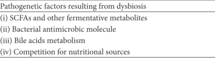
Table 2: This table shows the list of pathogenetic factors generated by dysbiosis.
Pathogenetic factors resulting from dysbiosis (i) SCFAs and other fermentative metabolites (ii) Bacterial antimicrobic molecule
(iii) Bile acids metabolism
(iv) Competition for nutritional sources
Drugs most frequently implicated in the alteration of the intestinal microbiota are antimicrobic agents. It is proved that the administration of various types of antibiotics, in particu-lar clindamycin, second and third generation cephalosporins, fluoroquinolones, and macrolides, can alter the ratio of differ-ent microbial communities. As described in several studies, there is a decrease in carbohydrate-fermenting and butyrate-producing bacteria members of Bacteroides and Firmicutes phyla [21–25].
A reduction of butyrate producers (such as Roseburia and
Ruminococcus) is observed also in NSAIDs users, particularly
in elderly subjects. These subjects, for their natural modifi-cation of the gut microbiota related to the age, have already an increased variability of microbial species and a relative decrease of Firmicutes and Bacteroides regardless of NSAIDs use [26].
Also acid-suppressing agents (H2-receptor antagonists and proton-pump inhibitors) can cause a change in the bacterial flora of the gastrointestinal tract. In particular, there is an increase of gastric and duodenal contamination with a possible minor degradation of Clostridium spores by gastric juices [27, 28]. The significance of this observation in the development of CDI is, however, still controversial. In fact, not all researchers recognize a primary role of acid suppression in establishing conditions that favor the
Clostridium growth [29]. Furthermore, nutrition can have a direct role in modifying the intestinal microbiota and in creating a favorable environment for the growth of C.
difficile. In particular, a prolonged elemental diet, poor in
fibers, which are a substrate for some beneficial bacteria, can support the development of an alteration in the ratio of normal commensal bacteria [24,30].
Overall, these environmental factors and the consequent intestinal dysbiosis disrupt and alter the protective effect exerted by the gut microbiota against recurrent CDI. The loss of this protective barrier allows for the formation of
an ecological niche where C. difficile can develop and better resist to antimicrobial therapies.
This niche concept is even more important if we consider that C. difficile multiplication and development, facilitated by dysbiosis, are necessary for CDAD [31,32]. Consequently, intestinal dysbiosis is very important in the pathogenesis of the disease, especially when specific changes in the composi-tion of the gut microbiota occur. CDI patients have a greater diversity of bacterial species and a reduced concentration of some commensal species, in particular the most represented phyla such as Bacteroides and Firmicutes. Bacteroides, which appear to be extremely reduced in these patients, are mainly responsible for the digestion of carbohydrates in the intestinal lumen, resulting in the production of substrates essential for the homeostasis of colonocytes. The reduced concentration of these commensal bacteria has been therefore associated with a higher frequency of relapse of CDI [23,33,34].
Also, the components of Firmicutes phylum are less represented in CDAD patients with respect to healthy subject. At family level, Lachnospiraceae and Ruminococcaceae, that are important butyrate producers, are significantly unrepre-sented in CDI, whereas Deltaproteobacteria, that are sulfate-reducing bacteria, are depleted. In contrast, several genera are enriched in association with CDI, such as Veillonella,
Enterococcus, and Lactobacillus.
This evident dysbiosis generates an altered production of substrates fermented by the anaerobic gut microbiota, including butyrate, other SCFAs, acetate, and lactate that are critical to the homeostasis of the intestinal epithelial cells [35]. Butyric acid has an important anti-inflammatory molecule and is the preferred source of energy of colonocytes. Other SCFAs are known to decrease intestinal permeability and to increase the production of antimicrobial substances and mucin [36,37]. Furthermore, a direct role of SCFAs in the inhibition of the growth of C. difficile was also assumed. This hypothesis has been confirmed by in vitro experiments, but results of in vivo studies do not seem to fully confirm this hypothesis [38,39].
Higher concentration of some species of Firmicutes such as Ruminococcus gnavus, Ruminococcus hansenii, and
Clostridium nexile was associated with a greater risk of
recur-rence and development of CDI. These bacterial species are producers of a trypsin-dependent antimicrobial substance (ruminococcin A) that has a low activity against C. difficile but can contribute to the disruption of the normal intestinal flora [40]. Another bacterial species that is capable of pro-ducing a substance with antimicrobial activity is the Bacillus
thuringiensis. This bacteria strain produces the Thuricin CD
that in vitro models proved to inhibit the growth of C.
difficile. The efficacy of this molecule is effective as well as
metronidazole [41,42].
A further mechanism that gut microbiota uses against the
C. difficile is the metabolization of bile that is proven to have
a role in both the spores germination and the growth of the vegetative form [19]. Commensal flora plays two important roles in bile transformation. A first mechanism is represented by the action of bile salt hydrolase enzymes produced by bacteria. These enzymes transform bile acids by cleaving their glycine and taurine; the metabolites obtained can stimulate
the germination of spores. A second mechanism is mediated by the enzyme 7-dehydroxylase that is also produced by the bacterial flora; this enzyme converts primary bile acids, cholate, and chenodeoxycholate into secondary biliar acids: deoxycholic and lithocholic acids, respectively. It is not yet well known which bacterial species operate on the transfor-mation of bile acids [43,44].
Deoxycholate is a potent germinant but is highly toxic to vegetative cells; cholate stimulates spore germination and vegetative C. difficile, whereas chenodeoxycholate has a strong inhibitory effect on spore germination. An alteration in the ratio of the different bile acids, caused by a change in the gut microbiota composition, may promote or inhibit the growth of C. difficile [45–47].
In a recent paper, it was demonstrated that the conjugated bile salt taurocholate is able to inhibit C. difficile toxins A and B activities in an in vitro assay. These results suggest that the mechanism of taurocholate-mediated inhibition modulates toxin activity. Indeed, taurocholate does not appear to affect
C. difficile growth and toxin production [48].
An additional mechanism that commensal flora uses against the C. difficile colonization is represented by the competition for energy sources, in particular carbon source, between toxigenic Clostridium and nontoxigenic Clostridium. In animal models, it has been shown that nontoxigenic
Clostridium, prevailing in this competition, crowds out C. dif-ficile by ecological niche preventing its growth. Unfortunately,
little is still known about this interesting aspect [19,49,50].
4.
Clostridium difficile and Innate
Immune Response
Several studies on commensal Clostridia showed that high levels of metabolite products, and their colonization in close proximity to the intestinal mucosa, are able to exert a strong influence on the host immune system [4]. Indeed, it has been shown that Clostridia can promote the development of𝛼𝛽 T-cell receptor intraepithelial lymphocytes (IEL) and immunoglobulin A (IgA-) producing cells in the large intes-tine [51]. IEL, IgA-producing cells within the lamina propria, and intestinal epithelial cells are key players in determining the nature of the immunological response to antigens or pathogens ingested. Umesaki et al. assessed that germ-free mice inoculated with 46 strains of Clostridia singly isolated from conventional mice showed an increase in the ratio of CD4− CD8+ cells to that of CD4+ CD8− in 𝛼𝛽IEL within the large intestine. Conversely, the number and phenotype of IEL were similar to those in conventionally housed mice. The number of IgA-producing cells in the colons of mice treated with Clostridia was slightly increased compared to that in germ-free mice [51]. Thus, Clostridia appear to be involved in the promotion of immunological development [51] in the large intestine, but not in the small intestine. Moreover, commensal Clostridia are able to normalize cecal size when they are associated with germ-free mice [52]. How the immune system fundamentally senses Clostridia remains unclear. In this context, it has been suggested that the presence or gradient of SCFAs and secondary bile acids
produced by Clostridia may be sensed by epithelial cells and, in turn, may be associated with the initiation of immunologi-cal signaling [51], due to the cross-talk between epithelial and immune cells. For example, IL-7 secreted by epithelial cells can activate IL-7 receptor-bearing IEL on their progenitors [53, 54]. Furthermore, IL-6 [55] and transforming growth factor 𝛽 [56] produced by the epithelia during infection can stimulate the development of Peyer’s patches and IgA production [57].
Notably, elevated levels of Clostridium clusters XIVa and IV in mice lead to resistance to allergy and intestinal inflammation in experimental models [58]. Conversely, the microbiota of individuals with chronic inflammation shows lower bacterial diversity and it has been determined that
Clostridium clusters IV, particularly F. prausnitzii, and XIVa
are significantly less abundant in IBD patients compared to healthy subjects [59–61]. It is still unknown whether the decrease in Clostridia is a cause or a consequence of chronic inflammation in IBD patients and in autoimmunity, but we can speculate that they are necessary for immune home-ostasis, contributing to the suppression of autoimmunity and deleterious inflammation in humans.
4.1. Effects of C. difficile Toxins Associated with Acute Colitis.
In animal models the challenge of ileal loops with C. difficile toxin A produces an intense inflammatory response char-acterized by fluid accumulation, edema, increased mucosal permeability, mast cell degranulation, epithelial cell death, and neutrophil recruitment.
Toxins are able to trigger fluid secretion, to induce the production of reactive oxygen intermediates, IL-8 from colonic epithelial cells [62], and to downregulate mucin exocytosis from mucin-producing colon cells [63].
Moreover, toxins lead to the production of multiple proinflammatory cytokines and chemokines including IL-12, IL-18, interferon g (IFN-g), IL-1b, TNF-a, macrophage inflammatory protein 1 a (MIP-1a), MIP-2, IL-8, and leptin [64]. These factors can exacerbate the inflammation and may be responsible for host damage and many of the histopatho-logical features of C. difficile-associated diseases.
Intestinal mast cells also play an important role in the toxin-mediated inflammatory responses. Both toxins A and B lead to activation, degranulation, and the release of inflam-matory mediators from mast cells [65]. The inhibition of mast cell degranulation and the blockade of mast cell-derived histamine were associated with a decrease in inflammatory responses to toxin A [66]. Mast cell-deficient mice show severe inflammation and neutrophilic infiltration compared with wild-type mice in response to C. difficile toxin A [67]. These studies suggest that, like neutrophils, mast cells propagate the inflammatory response in C. difficile-associated diseases. To be noted, a part of the toxin A mediated neutrophil recruitment in rat ileal loops is dependent on mast cell activation [67].
The role of other immune cells, including macrophages, monocytes, and dendritic cells, has generally been extrap-olated from in vitro and ex vivo studies using human and mouse cell lines, human monocytes, and monocyte-derived
dendritic cells. Emerging evidence showed also that C.
difficile toxins can stimulate the release of proinflammatory
cytokines and chemokines from macrophages, monocytes, and dendritic cells with a mitogen-activated protein kinase (MAPK-) and p38-dependent pathway [68]. Furthermore, toxin A leads to NF-𝜅B-mediated IL-8 production from human monocytes [69].
4.2. Effects on the Innate/Adaptative Immune System Predis-posing to Recurrence of CDI. C. difficile is able to
modu-late intestinal innate immune responses and several groups studied this process. Clostridium difficile is able to modulate host innate immunity via toxin-independent and depen-dent mechanisms [70,71]. The innate immune mechanisms against the toxins produced by C. difficile include the endoge-nous microbial flora, the mucus barrier, intestinal epithelial cells, and the mucosal immune system. Furthermore, C.
diffi-cile infection triggers the release of multiple proinflammatory
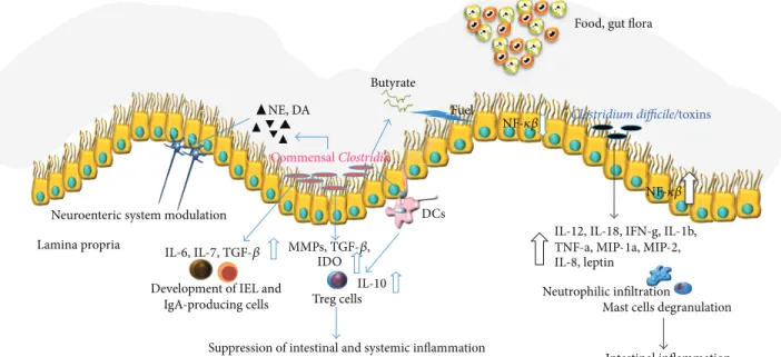
mediators (cytokines, chemokines, and neuroimmune pep-tides) and the recruitment and activation of several innate immune cells (Figure 1).
Interestingly, C. difficile toxins activate both surface and intracellular innate immune sensors, including the inflam-masome and the TLR4, TLR5, and NOD1 signaling path-ways [72]. TLR4- and MyD88-dependent signaling pathways produce an enhanced inflammatory response [73]. The defi-ciency of these pathways increases the bacterial burden and the worsening of the disease [73].
C. difficile shows a proteinaceous cell surface layer, which
is composed of an array of proteins arranged in a crys-talline lattice. The surface layer proteins have the ability to activate proinflammatory signaling through TLR4 expressed on the surface of host cells. Engagement of TLR4 initiates downstream signaling of NF-𝜅B and interferon regulatory factor 3, resulting in subsequent production of inflamma-tory cytokines and immune cell activation. Surface layer proteins induce dendritic cell maturation and activation in
vitro, as demonstrated by increased expression of major
histocompatibility complex class II, CD40, CD80, CD86, and production of IL-12p70, tumor necrosis factor-a, IL-23, and IL-6 [73]. Moreover, surface layer proteins were found to activate NF-𝜅B, but not interferon regulatory factor 3. This indicates that the signaling is myeloid differentiation primary 12 response gene 88 (MyD88)-dependent. In fact, TLR4-deficient and MyD88-TLR4-deficient mice were more susceptible to infection and exhibited greater pathology than wild-type mice [73]. Increased mucosal damage and inflammation in MyD88-deficient mice were attributed to a lack of neutrophil recruitment to the site of infection [74]. Neutrophils were shown to be critical in preventing bacterial dissemination through damaged mucosa [74]. In the case of TLR5 signaling, exogenous stimulation of TLR5 signaling was protective against C. difficile infection [75].
The intracellular innate immune sensors NOD1 and the IL-1b/inflammasome are also activated after C. difficile infection [72]. C. difficile-induced NOD1 activation triggered chemokine production and NOD1-deficient mice have lower chemokine production, less neutrophil recruitment, and
Suppression of intestinal and systemic inflammation Butyratey
Neutrophilic infiltration
Mast cells degranulation
Intestinal inflammation Food, gut flora
Clostridium difficile/toxins DCs Fuel NF-𝜅𝛽 NF-𝜅𝛽 NE, DA Commensal Clostridia
Neuroenteric system modulation Lamina propria
IL-6, IL-7, TGF-𝛽 Development of IEL and
IgA-producing cells Treg cells
IL-10
MMPs, TGF-𝛽, IDO
IL-12, IL-18, IFN-g, IL-1b, TNF-a, MIP-1a, MIP-2, IL-8, leptin
Figure 1: Commensal Clostridia have a peculiar role in modulating gut homeostasis. Establishing a close relationship with gut cells (interfold region), Clostridia spp. exert a strong influence on the host immune system. On the other hand, C. difficile and its toxins lead to the production of multiple proinflammatory cytokines and chemokines including IL-12, IL-18, interferon g (IFN-g), IL-1b, TNF-a, macrophage inflammatory protein 1 a (MIP-1a), MIP-2, IL-8, and leptin [66]. These factors can exacerbate the inflammation and may be responsible for host damage and many of the histopathological features of C. difficile-associated diseases.
more severe disease [72]. In fact NOD1-deficient mice have a higher C. difficile burden [72]. C. difficile toxins stimulate IL-1b release by activating inflammasomes in both mouse macrophages and human colon biopsy specimens [76].
Activation of the innate immune sensors and the release of cytokine and chemokine mediators are followed by an intense local neutrophilic infiltration [77]. This neutrophilic infiltration is one of the major pathological findings after C.
difficile infection. Local recruitment and systemic
prolifera-tion of neutrophils are seen in C. difficile-associated diseases [77]. Indeed, induction of neutropenia in rats was associated with less severe disease [78].
5. Conclusions
In recent years, several studies analyzed the role of gut microbiota in human physiology and in maintaining gut immune homeostasis. One of the most interesting aspects involves CDI and CDAD.
Intestinal dysbiosis and impaired innate immune response are crucial players in triggering C. difficile colonization and related symptoms. In these conditions this Gram-positive anaerobic spore-forming bacillus finds an ecological niche where it can grow and better resist antimicrobial therapies.
In this scenario, gut microbiota modulation and the consequent control of the innate immune response represent a valuable and interesting tool to treat CDI-related diseases.
Conflict of Interests
Stefano Bibb`o, Loris Riccardo Lopetuso, Gianluca Ianiro, Teresa Di Rienzo, and Giovanni Cammarota have no conflict of interests to declare. Antonio Gasbarrini is in the speaker’s bureau of Alfa Wassermann, Bayer, Janssen, Gilead, MSD, BMS, Angelini, and Sanofi.
References
[1] P. B. Eckburg, E. M. Bik, C. N. Bernstein et al., “Microbiology: diversity of the human intestinal microbial flora,” Science, vol. 308, no. 5728, pp. 1635–1638, 2005.
[2] J. Marchesi and F. Shanahan, “The normal intestinal micro-biota,” Current Opinion in Infectious Diseases, vol. 20, no. 5, pp. 508–513, 2007.
[3] P. D. Cani, “Metabolism in 2013: the gut microbiota manages host metabolism,” Nature Reviews Endocrinology, vol. 10, no. 2, pp. 74–76, 2014.
[4] L. R. Lopetuso, F. Scaldaferri, V. Petito, and A. Gasbarrini, “Commensal Clostridia: leading players in the maintenance of gut homeostasis,” Gut Pathogens, vol. 5, no. 1, article 23, 2013. [5] F. Guarner, “Enteric flora in health and disease,” Digestion, vol.
73, supplement 1, pp. 5–12, 2006.
[6] I. Sekirov, S. L. Russell, L. Caetano M Antunes, and B. B. Finlay, “Gut microbiota in health and disease,” Physiological Reviews, vol. 90, no. 3, pp. 859–904, 2010.
[7] A. Bernalier-Donadille, “Fermentative metabolism by
the human gut microbiota,” Gastroenterologie Clinique et
[8] A. Cuervo, N. Salazar, P. Ruas-Madiedo, M. Gueimonde, and S. Gonz´alez, “Fibers from regular diet are directly associated with fecal short-chain fatty acid concentrations in the elderly,”
Nutrition Research, vol. 33, no. 10, pp. 811–816, 2013.
[9] H. Br¨uggemann and G. Gottschalk, “Comparative genomics of clostridia: link between the ecological niche and cell surface properties,” Annals of the New York Academy of Sciences, vol. 1125, pp. 73–81, 2008.
[10] C. Eckert, G. Jones, and F. Barbut, “Diagnosis of Clostridium difficile infection: the molecular approach,” Future
Microbiol-ogy, vol. 8, pp. 1587–1598, 2013.
[11] K. S´anchez-Hurtado and I. R. Poxton, “Enhancement of the cytotoxic activity of Clostridium difficile toxin A by surface-associated antigens,” Journal of Medical Microbiology, vol. 57, part 6, pp. 739–744, 2008.
[12] G. Dobson, C. Hickey, and J. Trinder, “Clostridium difficile colitis causing toxic megacolon, severe sepsis and multiple organ dysfunction syndrome [3],” Intensive Care Medicine, vol. 29, no. 6, p. 1030, 2003.
[13] V. G. Loo, A.-M. Bourgault, L. Poirier et al., “Host and pathogen factors for Clostridium difficile infection and colonization,” New
England Journal of Medicine, vol. 365, no. 18, pp. 1693–1703, 2011.
[14] S. H. Cohen, D. N. Gerding, S. Johnson et al., “Clinical practice guidelines for Clostridium difficile infection in adults: 2010 update by the Society for Healthcare Epidemiology of Amer-ica (SHEA) and the Infectious Diseases Society of AmerAmer-ica (IDSA),” Infection Control and Hospital Epidemiology, vol. 31, no. 5, pp. 431–455, 2010.
[15] S. B. Debast, M. P. Bauer, and E. J. Kuijper, “European Society of Clinical Microbiology and Infectious Diseases: update of the treatment guidance document for Clostridium difficile infec-tion,” Clinical Microbiology and Infection, vol. 20, supplement 2, pp. 1–26, 2014.
[16] C. M. Surawicz, L. J. Brandt, D. G. Binion et al., “Guidelines for diagnosis, treatment, and prevention of Clostridium difficile infections,” The American Journal of Gastroenterology, vol. 108, no. 4, pp. 478–499, 2013.
[17] E. van Nood, M. G. W. Dijkgraaf, and J. J. Keller, “Duodenal infusion of feces for recurrent Clostridium difficile,” The New
England Journal of Medicine, vol. 368, no. 22, pp. 407–415, 2013.
[18] J. Bien, V. Palagani, and P. Bozko, “The intestinal microbiota dysbiosis and Clostridium difficile infection: is there a relation-ship with inflammatory bowel disease?” Therapeutic Advances
in Gastroenterology, vol. 6, no. 1, pp. 53–68, 2013.
[19] R. A. Britton and V. B. Young, “Interaction between the intesti-nal microbiota and host in Clostridium difficile colonization resistance,” Trends in Microbiology, vol. 20, no. 7, pp. 313–319, 2012.
[20] J. A. Hawrelak and S. P. Myers, “The causes of intestinal dysbiosis: a review,” Alternative Medicine Review, vol. 9, no. 2, pp. 180–197, 2004.
[21] D. A. Antonopoulos, S. M. Huse, H. G. Morrison, T. M. Schmidt, M. L. Sogin, and V. B. Young, “Reproducible community dynamics of the gastrointestinal microbiota following antibiotic perturbation,” Infection and Immunity, vol. 77, no. 6, pp. 2367– 2375, 2009.
[22] C. Jernberg, S. L¨ofmark, C. Edlund, and J. K. Jansson, “Long-term ecological impacts of antibiotic administration on the human intestinal microbiota,” ISME Journal, vol. 1, no. 1, pp. 56– 66, 2007.
[23] A. R. Manges, A. Labbe, V. G. Loo et al., “Comparative metagenomic study of alterations to the intestinal microbiota
and risk of nosocomial clostridum difficile-associated disease,”
Journal of Infectious Diseases, vol. 202, no. 12, pp. 1877–1884,
2010.
[24] S. J. D. O'Keefe, “Tube feeding, the microbiota, and Clostridium
difficile infection,” World Journal of Gastroenterology, vol. 16, no.
2, pp. 139–142, 2010.
[25] ˚A. Sullivan, “Effect of antimicrobial agents on the ecological
balance of human microflora,” Lancet Infectious Diseases, vol. 1, no. 2, pp. 101–114, 2001.
[26] H. M¨akivuokko, K. Tiihonen, S. Tynkkynen, L. Paulin, and N. Rautonen, “The effect of age and non-steroidal anti-inflammatory drugs on human intestinal microbiota composi-tion,” The British Journal of Nutrition, vol. 103, no. 2, pp. 227–234, 2010.
[27] M. A. Aldeyab, S. Harbarth, N. Vernaz et al., “Quasiexper-imental study of the effects of antibiotic use, gastric acid-suppressive agents, and infection control practices on the inci-dence of Clostridium difficile-associated diarrhea in hospitalized patients,” Antimicrobial Agents and Chemotherapy, vol. 53, no. 5, pp. 2082–2088, 2009.
[28] S. Dial, J. A. C. Delaney, A. N. Barkun, and S. Suissa, “Use of gastric acid-suppressive agents and the risk of community-acquired Clostridium difficile-associated disease,” Journal of the
American Medical Association, vol. 294, no. 23, pp. 2989–2995,
2005.
[29] M. M. Nerandzic, M. J. Pultz, and C. J. Donskey, “Examination of potential mechanisms to explain the association between proton pump inhibitors and Clostridium difficile infection,”
Antimicrobial Agents and Chemotherapy, vol. 53, no. 10, pp.
4133–4137, 2009.
[30] S. J. D. O'Keefe, “Nutrition and colonic health: the critical role of the microbiota,” Current Opinion in Gastroenterology, vol. 24, no. 1, pp. 51–58, 2008.
[31] Y. C. Ju, D. A. Antonopoulos, A. Kalra et al., “Decreased diversity of the fecal microbiome in recurrent Clostridium
difficile-associated diarrhea,” Journal of Infectious Diseases, vol.
197, no. 3, pp. 435–438, 2008.
[32] K. H. Wilson, “The microecology of Clostridium difficile,”
Clinical Infectious Diseases, vol. 16, supplement 4, pp. S214–S218,
1993.
[33] E. Goldberg, I. Amir, M. Zafran et al., “The correlation between Clostridium-difficile infection and human gut concentrations of Bacteroidetes phylum and clostridial species,” The European
Journal of Clinical Microbiology & Infectious Diseases, vol. 33,
no. 3, pp. 377–383, 2014.
[34] M. J. Hopkins and G. T. Macfarlane, “Changes in predominant bacterial populations in human faeces with age and with
Clostridium difficile infection,” Journal of Medical Microbiology,
vol. 51, no. 5, pp. 448–454, 2002.
[35] V. C. Antharam, E. C. Li, A. Ishmael et al., “Intestinal dysbiosis and depletion of butyrogenic bacteria in Clostridium difficile infection and nosocomial diarrhea,” Journal of Clinical
Micro-biology, vol. 51, no. 9, pp. 2884–2892, 2013.
[36] S. I. Cook and J. H. Sellin, “Review article: short chain fatty acids in health and disease,” Alimentary Pharmacology and
Therapeutics, vol. 12, no. 6, pp. 499–507, 1998.
[37] J. M. Wong, R. de Souza, C. W. Kendall, A. Emam, and D. J. Jenkins, “Colonic health: fermentation and short chain fatty acids,” Journal of Clinical Gastroenterology, vol. 40, no. 3, pp. 235–243, 2006.
[38] R. D. Rolfe, “Role of volatile fatty acids in colonization resistance to Clostridium difficile,” Infection and Immunity, vol. 45, no. 1, pp. 185–191, 1984.
[39] W. J. Su, M. J. Waechter, P. Bourlioux, M. Dolegeal, J. Fourniat, and G. Mahuzier, “Role of volatile fatty acids in colonization resistance to Clostridium difficile in gnotobiotic mice,” Infection
and Immunity, vol. 55, no. 7, pp. 1686–1691, 1987.
[40] F. Marcille, A. Gomez, P. Joubert et al., “Distribution of genes encoding the trypsin-dependent lantibiotic ruminococcin A among bacteria isolated from human fecal microbiota,” Applied
and Environmental Microbiology, vol. 68, no. 7, pp. 3424–3431,
2002.
[41] M. C. Rea, A. Dobson, O. O'Sullivan et al., “Effect of broad- and narrow-spectrum antimicrobials on Clostridium difficile and microbial diversity in a model of the distal colon,” Proceedings of
the National Academy of Sciences of the United States of America,
vol. 108, supplement 1, pp. 4639–4644, 2011.
[42] M. C. Rea, C. S. Sit, E. Clayton et al., “Thuricin CD, a post-translationally modified bacteriocin with a narrow spectrum of activity against Clostridium difficile,” Proceedings of the National
Academy of Sciences of the United States of America, vol. 107, no.
20, pp. 9352–9357, 2010.
[43] J. M. Ridlon, D.-J. Kang, and P. B. Hylemon, “Bile salt bio-transformations by human intestinal bacteria,” Journal of Lipid
Research, vol. 47, no. 2, pp. 241–259, 2006.
[44] K. H. Wilson, “Efficiency of various bile salt preparations for stimulation of Clostridium difficile spore germination,” Journal
of Clinical Microbiology, vol. 18, no. 4, pp. 1017–1019, 1983.
[45] J. L. Giel, J. A. Sorg, A. L. Sonenshein, and J. Zhu, “Metabolism of bile salts in mice influences spore germination in Clostridium
difficile,” PLoS ONE, vol. 5, no. 1, Article ID e8740, 2010.
[46] J. A. Sorg and A. L. Sonenshein, “Bile salts and glycine as coger-minants for Clostridium difficile spores,” Journal of Bacteriology, vol. 190, no. 7, pp. 2505–2512, 2008.
[47] J. A. Sorg and A. L. Sonenshein, “Chenodeoxycholate is an inhibitor of Clostridium difficile spore germination,” Journal of
Bacteriology, vol. 191, no. 3, pp. 1115–1117, 2009.
[48] C. Darkoh, E. L. Brown, H. B. Kaplan, and H. L. DuPont, “Bile salt inhibition of host cell damage by Clostridium difficile toxins,” PLoS ONE, vol. 8, no. 11, Article ID e79631, 2013. [49] M. M. Merrigan, S. P. Sambol, S. Johnson, and D. N.
Gerd-ing, “Prevention of fatal Clostridium difficile-associated disease during continuous administration of clindamycin in hamsters,”
Journal of Infectious Diseases, vol. 188, no. 12, pp. 1922–1927,
2003.
[50] S. P. Sambol, M. M. Merrigan, J. K. Tang, S. Johnson, and D. N. Gerding, “Colonization for the prevention of Clostridium
difficile disease in hamsters,” Journal of Infectious Diseases, vol.
186, no. 12, pp. 1781–1789, 2002.
[51] Y. Umesaki, H. Setoyama, S. Matsumoto, A. Imaoka, and K. Itoh, “Differential roles of segmented filamentous bacteria and clostridia in development of the intestinal immune system,”
Infection and Immunity, vol. 67, no. 7, pp. 3504–3511, 1999.
[52] K. Itoh and T. Mutsuoka, “Characterization of clostridia isolated from faeces of limited flora mice and their effect on caecal size when associated with germ-free mice,” Laboratory Animals, vol. 19, no. 2, pp. 111–118, 1985.
[53] K. Fujihashi, J. R. McGhee, M. Yamamoto, J. J. Peschon, and H. Kiyono, “An interleukin-7 internet for intestinal intraepithelial T cell development: knockout of ligand or receptor reveal differences in the immunodeficient state,” European Journal of
Immunology, vol. 27, no. 9, pp. 2133–2138, 1997.
[54] M. Watanabe, Y. Ueno, T. Yajima et al., “Interleukin 7 is produced by human intestinal epithelial cells and regulates the proliferation of intestinal mucosal lymphocytes,” Journal of
Clinical Investigation, vol. 95, no. 6, pp. 2945–2953, 1995.
[55] D. W. McGee, K. W. Beagley, W. K. Aicher, and J. R. McGhee, “Transforming growth factor-𝛽 enhances interleukin-6 secre-tion by intestinal epithelial cells,” Immunology, vol. 77, no. 1, pp. 7–12, 1992.
[56] J. A. Barnard, G. J. Warwick, and L. I. Gold, “Localization of
transforming growth factor𝛽 isoforms in the normal murine
small intestine and colon,” Gastroenterology, vol. 105, no. 1, pp. 67–73, 1993.
[57] K. W. Beagley, J. H. Eldridge, F. Lee et al., “Interleukins and IgA synthesis. Human and murine interleukin 6 induce high rate IgA secretion in IgA-committed B cells,” Journal of Experimental
Medicine, vol. 169, no. 6, pp. 2133–2148, 1989.
[58] K. Atarashi, T. Tanoue, T. Shima et al., “Induction of colonic regulatory T cells by indigenous Clostridium species,” Science, vol. 331, no. 6015, pp. 337–341, 2011.
[59] D. N. Frank, A. L. St. Amand, R. A. Feldman, E. C. Boedeker, N. Harpaz, and N. R. Pace, “Molecular-phylogenetic character-ization of microbial community imbalances in human inflam-matory bowel diseases,” Proceedings of the National Academy
of Sciences of the United States of America, vol. 104, no. 34, pp.
13780–13785, 2007.
[60] H. Sokol, B. Pigneur, L. Watterlot et al., “Faecalibacterium prausnitzii is an anti-inflammatory commensal bacterium iden-tified by gut microbiota analysis of Crohn disease patients,”
Proceedings of the National Academy of Sciences of the United States of America, vol. 105, no. 43, pp. 16731–16736, 2008.
[61] B. Willing, J. Halfvarson, J. Dicksved et al., “Twin studies reveal specific imbalances in the mucosa-associated microbiota of patients with ileal Crohn's disease,” Inflammatory Bowel
Diseases, vol. 15, no. 5, pp. 653–660, 2009.
[62] Y. R. Mahida, S. Makh, S. Hyde, T. Gray, and S. P. Borriello, “Effect of Clostridium difficile toxin A on human intestinal epithelial cells: induction of interleukin 8 production and apoptosis after cell detachment,” Gut, vol. 38, no. 3, pp. 337–347, 1996.
[63] J.-E. Branka, G. Vallette, A. Jarry, C. Bou-Hanna, P. Lemarre, and C. L. Laboisse, “Early functional effects of Clostridium
difficile toxin A on human colonocytes,” Gastroenterology, vol.
112, no. 6, pp. 1887–1894, 1997.
[64] Y. Ishida, T. Maegawa, T. Kondo et al., “Essential involvement of IFN-𝛾 in Clostridium difficile toxin A-induced enteritis,” Journal
of Immunology, vol. 172, no. 5, pp. 3018–3025, 2004.
[65] G. K. A. Meyer, A. Neetz, G. Brandes et al., “Clostridium difficile toxins A and B directly stimulate human mast cells,” Infection
and Immunity, vol. 75, no. 8, pp. 3868–3876, 2007.
[66] I. Kurose, C. Pothoulakis, J. T. LaMont et al., “Clostridium
difficile toxin A-induced microvascular dysfunction. Role of
histamine,” Journal of Clinical Investigation, vol. 94, no. 5, pp. 1919–1926, 1994.
[67] B. K. Wershil, I. Castagliuolo, and C. Pothoulakis, “Direct evidence of mast cell involvement in Clostridium difficile toxin A-induced enteritis in mice,” Gastroenterology, vol. 114, no. 5, pp. 956–964, 1998.
[68] M. Warny, A. C. Keates, S. Keates et al., “p38 MAP kinase activation by Clostridium difficile toxin A mediates monocyte necrosis, IL-8 production, and enteritis,” Journal of Clinical
[69] K. K. Jefferson, M. F. Smith Jr., and D. A. Bobak, “Roles of intracellular calcium and NF-𝜅B in the Clostridium difficile toxin A-induced up-regulation and secretion of IL-8 from human monocytes,” Journal of Immunology, vol. 163, no. 10, pp. 5183–5191, 1999.
[70] N. V. Jafari, S. A. Kuehne, C. E. Bryant et al., “Clostridium difficile modulates host innate immunity via toxin-independent and dependent mechanism(s),” PLoS ONE, vol. 8, no. 7, Article ID e69846, 2013.
[71] R. Madan and W. A. Petri Jr., “Immune responses to Clostridium
difficile infection,” Trends in Molecular Medicine, vol. 18, no. 11,
pp. 658–666, 2012.
[72] M. Hasegawa, T. Yamazaki, N. Kamada et al., “Nucleotide-binding oligomerization domain 1 mediates recognition of
Clostridium difficile and induces neutrophil recruitment and
protection against the pathogen,” Journal of Immunology, vol. 186, no. 8, pp. 4872–4880, 2011.
[73] A. Ryan, M. Lynch, S. M. Smith et al., “A role for TLR4 in
Clostridium difficile infection and the recognition of surface
layer proteins,” PLOS Pathogens, vol. 7, no. 6, Article ID e1002076, 2011.
[74] I. Jarchum, M. Liu, C. Shi, M. Equinda, and E. G. Pamer, “Crit-ical role for myd88-mediated neutrophil recruitment during
Clostridium difficile colitis,” Infection and Immunity, vol. 80, no.
9, pp. 2989–2996, 2012.
[75] I. Jarchum, M. Liu, L. Lipuma, and E. G. Pamer, “Toll-like receptor 5 stimulation protects mice from acute Clostridium
difficile colitis,” Infection and Immunity, vol. 79, no. 4, pp. 1498–
1503, 2011.
[76] J. Ng, S. A. Hirota, O. Gross et al., “Clostridium difficile toxin-induced inflammation and intestinal injury are mediated by the inflammasome,” Gastroenterology, vol. 139, no. 2, pp. 542–552, 2010.
[77] C. P. Kelly, S. Becker, J. K. Linevsky et al., “Neutrophil recruit-ment in Clostridium difficile toxin A enteritis in the rabbit,”
Journal of Clinical Investigation, vol. 93, no. 3, pp. 1257–1265,
1994.
[78] B. Qiu, C. Pothoulakis, I. Castagliuolo, S. Nikulasson, and J. T. LaMont, “Participation of reactive oxygen metabolites in
Clostridium difficile toxin A-induced enteritis in rats,” The Amer-ican Journal of Physiology: Gastrointestinal and Liver Physiology,
Submit your manuscripts at
http://www.hindawi.com
Stem Cells
International
Hindawi Publishing Corporationhttp://www.hindawi.com Volume 2014
Hindawi Publishing Corporation
http://www.hindawi.com Volume 2014 INFLAMMATION
Hindawi Publishing Corporation
http://www.hindawi.com Volume 2014
Behavioural
Neurology
Endocrinology
International Journal of Hindawi Publishing Corporationhttp://www.hindawi.com Volume 2014
Hindawi Publishing Corporation
http://www.hindawi.com Volume 2014
Disease Markers
Hindawi Publishing Corporation
http://www.hindawi.com Volume 2014
BioMed
Research International
Oncology
Journal ofHindawi Publishing Corporation
http://www.hindawi.com Volume 2014
Hindawi Publishing Corporation
http://www.hindawi.com Volume 2014 Oxidative Medicine and Cellular Longevity Hindawi Publishing Corporation
http://www.hindawi.com Volume 2014
PPAR Research
The Scientific
World Journal
Hindawi Publishing Corporation
http://www.hindawi.com Volume 2014
Immunology Research
Hindawi Publishing Corporation
http://www.hindawi.com Volume 2014
Journal of
Obesity
Journal ofHindawi Publishing Corporation
http://www.hindawi.com Volume 2014
Hindawi Publishing Corporation
http://www.hindawi.com Volume 2014 Computational and Mathematical Methods in Medicine
Ophthalmology
Journal ofHindawi Publishing Corporation
http://www.hindawi.com Volume 2014
Diabetes Research
Journal ofHindawi Publishing Corporation
http://www.hindawi.com Volume 2014
Hindawi Publishing Corporation
http://www.hindawi.com Volume 2014
Research and Treatment
AIDS
Hindawi Publishing Corporation
http://www.hindawi.com Volume 2014 Gastroenterology Research and Practice
Hindawi Publishing Corporation
http://www.hindawi.com Volume 2014