“Amedeo Avogadro”
Department of Pharmaceutical Sciences
Ph.D in Chemistry&Biology
XXIX cycle a.y. 2013-2016
Adult Hippocampal Neural Progenitor Cells:
An Important In vitro Tool for Studying Complex
Mechanisms Regulating Adult Neurogenesis
Drug Discovery and Development
Università degli Studi del Piemonte Orientale
“Amedeo Avogadro”
Department of Pharmaceutical Sciences
Ph.D in Chemistry&Biology
XXIX cycle a.y. 2013-2016
Adult Hippocampal Neural Progenitor Cells:
An Important In vitro Tool for Studying Complex
Mechanisms Regulating Adult Neurogenesis
Drug Discovery and Development
Rita Salem
Supervised by Prof. Mariagrazia Grilli
PhD program coordinator Prof. Domenico Osella
« Il faut savoir s'instruire dans la gaieté.
Le savoir triste est un savoir mort. L'intelligence est joie. »
"Learning should be a cheerful process.
Sad knowledge is dead knowledge. Intelligence is joy.”
7
LIST OF ABBREVIATIONS ... 9
CHAPTER I ... 11
INTRODUCTION ... 11
1. ADULT NEUROGENESIS ... 13
1. ADULT NEUROGENESIS IN HUMAN AND RODENT BRAIN ... 13
2. HIPPOCAMPAL NEUROGENIC NICHE AND NEUROGENESIS PROCESS ... 14
3. ADULT HIPPOCAMPAL NEUROGENESIS MODULATORS ... 16
4. ROLE IN PHYSIOLOGY AND CHANGES UNDER STRESSFUL CONDITIONS ... 22
2. SEROTONIN ... 24
1. SEROTONINERGIC CIRCUIT AND RECEPTOR EXPRESSION IN THE CNS ... 24
2. SEROTONIN AND MOOD REGULATION ... 26
3. SEROTONIN AND PLASTICITY: EMPHASIS ON ADULT NEUROGENESIS ... 30
3. MOOD DISORDERS: FOCUS ON MAJOR DEPRESSIVE DISORDER ... 33
1. DEFINITION AND PATHOPHYSIOLOGY OF MDD ... 33
1. ANTIDEPRESSANTS: CLASSES AND MECHANISMS OF ACTION ... 38
2. ADULT HIPPOCAMPAL NEUROGENESIS,MDD AND ANTIDEPRESSANTS ... 42
4. TRAZODONE ... 45
1. TRAZODONE: PECULIAR MECHANISM OF ACTION ... 45
2. THERAPEUTIC DOSES, EFFICACY AND SIDE EFFECTS... 48
3. TRAZODONE AS A NEUROPROTECTIVE ANTIDEPRESSANT ... 50
5. NF-𝜅B ... 52
1. NF-𝜅B SIGNALING PATHWAY AND SUBUNITS IN THE CNS ... 52
2. NF-𝜅B: RELEVANCE TO PLASTICITY AND ADULT NEUROGENESIS ... 52
8
OUTLINEOFTHETHESIS ... 56
REFERENCES ... 62
CHAPTER III ... 81
CELL AUTONOMOUS AND NON CELL-AUTONOMOUS ROLE OF NF-𝜅B P50 IN ASTROCYTE-MEDIATED FATE SPECIFICATION OF ADULT NEURAL PROGENITOR CELLS. ... 83
ABSTRACT ... 85
INTRODUCTION ... 86
MATERIALS AND METHODS ... 88
RESULTS ... 93
DISCUSSION ... 104
REFERENCES ... 111
CHAPTER IV ... 119
TRAZODONEASANOVELPRONEUROGENICANTIDEPRESSANT ... 121
ABSTRACT ... 122
INTRODUCTION ... 123
MATERIALS AND METHODS ... 125
RESULTS ... 129
... 141
DISCUSSION ... 142
REFERENCES ... 147
CHAPTER V ... 154
DISCUSSION AND CONCLUSIONS ... 154
REFERENCES ... 161
CHAPTER VI ... 165
LIST OF PUBLICATIONS AND POSTERS ... 165
9
List of Abbreviations
5-HT 5-HydroxyTriptamine ACh Acetylcholine
ahNPC adult hippocampal Neural
Progenitor Cells
ahNSC adult hippocampal Neural Stem
Cells
AR Adrenergic receptors
BDNF Brain Derived Neurotrophic Factor BrDU BromoDioxyUridine
CA3 Cornus Ammonis - 3 CNS Central Nervous System DA Dopamine
DAT Dopamine Transporter DCX DoubleCourtin
DDC aromatic amino acid decarboxylase DG Dentate Gyrus
EE Enriched Environnement FST Forced Swim Test
GABA Gamma-AminoButyric Acid GCL Granular Cell Layer
GF Growth Factors
GFAP Glial-Fibrillary Acidic Protein IGF-1 Insulin-like Growth Factor IR Immediate Release
LTP Long Term Potentiation
m-CPP meta-ChloroPhenylPiperazine
MDD Major Depressive Disorder MWM Morris Water Maze NE NorEpinephrine
NET Noradrenaline Transporter NeuN Neuronal Nuclear antigen OB Olfactory Bulb
PCPA DL-P-Chlorophenylalanine PFC PreFrontal Cortex
PSA-NCAM Polysialylated-Neural Cell
Adhesion Molecule
RN Raphe Nucleus SGZ Subgranular Zone
SOX-2 Sry-related HMG box transcription
factor
SVZ Subventricular Zone TPH Tryptophan Hydroxylase TST Tail Suspension Test CMS Chronic Mild Stress
VEGF Vascular Endothelial Growth
Factor
VTA VentroTegmental Area
SSRI Selective Serotonin Reuptake
Inhibitors
SNRI Serotonin and Noradrenaline
Reuptake Inhibitors
SARI Serotonin Antagonist and Reuptake
Inhibitor
TCA TriCyclic Antidepressants MAOI MonoAmine Oxidase inhibitors
11
Chapter I
INTRODUCTION
13
1. Adult neurogenesis
For long time scientists assumed that neurogenesis was restricted to embryonic and early postnatal development period in our central nervous system (CNS). Since two decades several key findings - such as the remodeling of vocal centers in singing birds each year through new born neurons and the isolation and culture of neural cells with undifferentiated cells properties - consolidated the discovery of newly generated and integrated neurons during adult life.
1. Adult neurogenesis in human and rodent brain
Adult neurogenesis is a particular form of neural plasticity described in many brain regions of multiple species such as birds, reptiles, fish and mammalian brain, including human. In adult mammalian brain the subgranular zone (SGZ) of the hippocampal dentate gyrus (DG) and the subventricular zone (SVZ) of the lateral ventricle are the two major niches of neural stem cells where a basal level of neurogenesis is described (Kempermann and Gage 1999, Kempermann et al., 2003, Zhao et al., 2008). In rodent brain thousands of neural progenitors are produced on a daily basis in the SVZ and migrate along the rostral migratory stream (RMS) towards the olfactory bulb (OB) where they differentiate into local interneurons (Malvaut et al., 2016). In the SGZ newly born neurons are integrated in the DG and play a physiological role in hippocampal function (Zhao et al., 2008). Retrospective birth dating by 14C incorporation in CNS neurons showed that one third of human hippocampal neurons are subjected for turn over during life time. The extent of hippocampal neurogenesis between middle aged human and middle aged mice was comparable (Spalding et al., 2013). The importance of adult neurogenesis is different between rodents and human. While it is pronounced in both DG and OB of rodent brain, only DG neurogenesis appears to be substantial in human brain (Bergmann et al., 2012, Spalding et al., 2013). A considerable level of newly generated neurons was also described in other brain regions such as
14 neocortex (Gould et al., 2007) hypothalamus (Kokoeva et al., 2005) and human striatum (Ernst et al., 2014). However, the role of adult neurogenesis outside the SGZ and SVZ remains to be fully described.
2. Hippocampal neurogenic niche and neurogenesis process
Adult CNS “stem cells” exhibit three prime features: (i) they are self-renewing, with the ability to regenerate progeny identical to themselves, (ii) proliferative, undergoing mitosis with long cell cycle and (iii) multipotent, able to generate neuronal and glial lineages cells. Adult hippocampal Neural Stem Cells (ahNSC) reside in a unique microenvironment called neurogenic niche confined within the SGZ hilus region. The neurogenic niche is a complex zone composed of NSC surrounded by ependymal cells, astroglial cells, vessels and mature neurons (Fig. 1). Diffusible molecule and cell-cell contacts within the niche and from outside can influence NSC proliferation, differentiation and survival (Alvarez-Buylla et al., 2004, Quinones-Hinojosa at al., 2006). The generation of new neurons is a complex multistep process (Fig. 2) that involves a series of developmental phases before the complete maturation and functional integration into neuronal networks (Fig. 2, A). Cellular maturation phases could be identified by morphological characteristics as well as the expression of specific molecular markers (Fig. 2, B). In the SGZ, dormant radial glial-like cells have processes spanning the granule layer and the inner molecular layer of the hippocampus. They express glial fibrillary acidic protein (GFAP), Sry-related HMG box transcription factor (Sox2), nestin (nerve cells intermediate filament) (GFAP+, Sox2+, nestin+, S100β−) and are negative for the astroglial marker S100β (Filipov et al., 2003). They are usually referred as type 1 cells or NSC. By asymmetric division, a NSC gives rise to an identical progeny that remains in quiescent state and to a transiently amplifying neural progenitor cell (NPC).
15 NPCs are non-radial cells with short processes positioned tangentially to the granular layer. They are GFAP-, S100β−, Sox2+, Nestin+ highly proliferative and referred to as type 2 cells (Kronenberg et al., 2003). NPCs generate neuronal progenitors expressing doublecortin (DCX) and polysialylated-neural cell adhesion molecule (PSA-NCAM) named neuroblasts type 3 cells, and a population of glial progenitors GFAP+, S100β+ (Ma et al., 2009, Bonaguidi et al., 2012). Immature newborn neurons (DCX+) migrate a small distance in the granule cell layer (GCL) of the DG and start to receive synaptic inputs from existing neuronal circuits. The surviving cells continue their maturation and have more complex apical dendrites connecting the enthorinal cortex and hippocampal cornu ammonis (CA3) subfield. Mature neurons express neuronal nuclear antigen (NeuN) and Calbindin (Fig. 2 B). It takes roughly 1-2 months for newly generated neurons to reach morphological maturity and functionally integrate in neuronal networks (Fig. 2) (Lucassen et al., 2010).
Figure 1. Schematic representation of the cytoarchitecture in the hippocampal neurogenic niche. A. Coronal section of adult mouse brain showing the localization of the subgarnular zone (SGZ) in the hippocampus. B. Cell population in the dentate gyrus (DG) of the mammalian brain. GCL: Granular cell Layer; IML: Inner Molecular Layer. (Modified from
16 In the early 1990s several laboratories concomitantly showed that NSC could be isolated from adult brain and expanded in vitro in presence of growth factors. Isolated stem cells in presence of epidermal growth factors (EGF) formed spheres after days in proliferation and expressed nestin (Colucci-D’Amato et al., 2006; Reynolds and Weiss, 1992). The in vitro model of stem cells deriving from brain tissue and specifically from the hippocampus was validated and well characterized over the years in our and other research groups. It is used as a tool to identify molecular events underlying adult hippocampal neurogenesis and its possible modulators.
3. Adult hippocampal neurogenesis modulators
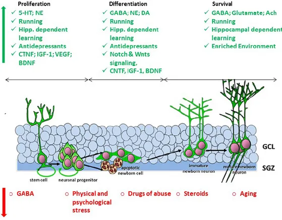
Proliferation of ahNPC, complete maturation, integration into neuronal circuits and spines development of newly generated neurons are plastic processes strongly regulated by neuronal network activity, cell-cell interactions, endogenous and exogenous modulators (Fig. 3).
1. Network activity and neurotransmitters
Enriched environment (EE) and voluntary exercise increase the DG network activity and result in Gamma-Aminobutyric acid (GABA) spill over from local GABAergic synapses into the neurogenic niche. Nestin+ quiescent NSCs in the SGZ respond to spilled GABA through GABAA receptors containing the γ2 subunit. GABA halts the proliferation and favors the differentiation and maturation of new neurons (Dranovsky et al., 2011). One peculiarity of newborn DCX+ young neurons is having high chloride levels due to high expression of chloride importer (NKCC1) that are lost with maturation and replaced by chloride transporter (KCC2).
17
Figure 2. Representative scheme for the phases and hallmarks of adult hippocampal neurogenesis. A. Summary for the different phases of hippocampal neurogenesis. 1. Expansion by activation of radial glia-like cells and proliferation of intermediate adult hippocampal neural progenitor cells (ahNPC). 2. Differentiation toward immature neurons (neuroblasts). 3. Integration into the dentate gyrus networks (Cornu ammonis, CA3) and maturation of young granule neurons. B. Different markers are expressed by ahNPC at different phases of maturation. Immature cells markers (GFAP, Nestin, and Sox-2); neuroblasts markers (DCX, PSA-NCAM and Tuj-1); mature neurons markers (NeuN and Calbindin). C. Time scale of the different steps of maturation. (Modified from Schouten et al., 2012).
18 EE increases the level of stimulatory neurotransmitter glutamate in the synapses. Newly generated neurons express NMDA glutamate receptors with NR2B subunit. This particular feature facilitates long term potentiation (LTP) induced by glutamate that enhances the maturation, integration and survival of new neurons (Ge et al, 2007). In summary, intrinsic cell properties and network activity dictate the quality and/or the quantity of the proliferation of ahNPC and the maturation of young neurons.
Major CNS neurotransmitters such as acetylcholine (ACh), dopamine (DA), norepinephrine (NE) and serotonin (5-HT) modulate neurogenesis. The cholinergic system has a substantial role in the hippocampal functions and is one of the major candidates suggested to influence adult neurogenesis. Indeed the ablation of basal forebrain in adult rats (cholinergic center) decreased the number of double labeled cells for 5-bromo-2-deoxyuridine BrdU (DNA intercalant during proliferation) and NeuN (mature neurons) in the SGZ (Cooper-Kuhn et al., 2004). Dopaminergic axons originate from ventral tegmental area (VTA) and the adjacent substantia nigra to innervate the DG (Mu et al., 2011). The hallmark of Parkinson’s disease is cerebral dopamine depletion. Indirect studies on the brain of patients or rodent models of Parkinson’s disease were used to study the effect of dopamine on adult neurogenesis. The proliferation of ahNPC is reduced in the SGZ in dopamine depleted brains (Höglinger et al., 2004). Noradrenergic innervation of the DG originates from the locus coeruleus. Lesions of NE projecting fibers decreased neurogenesis considerably (Kulkarni et al., 2002). Intra-hippocampal injection of NE and β3-adrenergic receptor (β3-AR) agonist increased the proliferation and the number of nestin+/GFAP+ neural precursors (Jhaveri et al., 2010). A more recent study suggested that NE increase ahNPC proliferation through β2-AR (Masuda et al., 2012). Serotonin is synthesized in the brain stem raphe nuclei and serotonergic fibers project into the DG of the hippocampus where they connect with granule
19 cells and interneurons (Alenina and Klempin, 2015). The role of serotonin in adult hippocampal neurogenesis will be detailed in a separate paragraph (II.3).
2. Astrocytes
With regard to the importance of astrocytes in the CNS, Nedergaard and Goldman stated that "astrocytes might serve neurons not so much as servants but as parents … astrocytes tell neurons what to do beside just cleaning up their mess" (Nedergaard et al., 2003). Astrocytes are the most abundant cell population in the CNS and the major components of the neurogenic niche (Song et al., 2002). Traditionally astrocytes were assigned only a supportive and trophic role in all brain functions (Pixley, 1992). After years of research now astrocytes are acknowledged aside from the trophic role also for their major contribution to embryonic and adult neurogenesis (Gomes et al., 2001). Specifically, astrocytes through the release of diffusible signals and also direct contact to neurons play a role in neuronal differentiation, synapse formation, axonal guidance and electrophysiological properties regulation (Pfrieger and Barres 1997; Lim and Alvarez-Buylla 1999; Song et al. 2002). In particular, hippocampal astrocytes are the energy providers during memory formation. The lactate hydrogenase provided by astrocytes sustains the long term potentiation (LTP) and memory retention in the hippocampus (Suzuki et al. 2011). Astrocytes have intercellular crosstalk between themselves, neurons, microglia cells and endothelial cells. Importantly, astrocytes communicate bidirectionally with neurons by the tripartite synapse, a structure where astrocytes are associated with presynaptic and postsynaptic neurons membrane. The interaction is physical and functional since astrocytes function is dependent on neuronal activity (reviewed in Pekny et al., 2016). Astrocytes employ several molecular mechanisms such as exocytosis, diffusion through cellular membrane and membrane transporters to release numerous neurotransmitters and trophic factors. These elements are signals to neurons and microglial cells that regulate the synaptic and neuronal activity (Newman, 2003). For example,
20 thrombospondin is an extracellular glycoprotein secreted by astrocytes and specifically upregulated after an ischemic injury. Astrocytes through thrombospondin control the post-ischemic angiogenesis and synaptic reformation (Liauw et al., 2008).
In addition to their role in physiological and pathological conditions in the CNS astrocytes are also important contributors to adult neurogenesis specifically in the hippocampus. Song and colleagues showed that co-culture of hippocampal NPCs
Figure 3. Adult hippocampal neurogenesis is modulated by several endogenous and exogenous factors. The different phases of neurogenesis could be increased by several modulators such as neurotransmitters, growth factors, environment and learning (Green factors) or decreased by stress, drugs and aging (Red factors) (Modified from Koehl, 2015).
21 and astrocytes promoted the proliferation of NPCs and favored their differentiation toward the neuronal lineage. An intriguing outcome of this study was that astrocytes originating from the spinal cord did not have neurogenic effect, therefore the neurogenic potential could be niche specific (Song et al., 2002). Another study showed that astrocytes coming from proneurogenic regions secreted cytokines and chemokines such as IL-1β and IL-6 that promoted neuronal differentiation at low concentrations (Barkho et al., 2006). Additionally, astrocytes through Wnt signaling regulate the expression of transcription factors such as: (i) Prox1 that has a role in the proliferation of NPCs and maturation of newly generated neurons and (ii) NeuroD1 that is crucial for survival and maturation of new neurons (Karalay and Jessberger, 2011).
3. Extrinsic and environmental regulators
Neurogenesis is also regulated by behavioral and environmental positive modulators such as voluntary exercise/running, EE, learning and negative modulators such as stress, aging and drug of abuse (Kempermann and Gage, 2000). Running is one of the most potent inducers of neural progenitor cell proliferation. It dramatically increases the number of BrdU positive (BrdU+) cells in the DG of adult mice. Additionally, running increases the survival of newborn neurons after 4 weeks from exercise and synaptic plasticity since it enhances LTP in the DG (Van Praag et al. 1999 a,b). EE has a complementary effect increasing the survival of neurons at a critical stage of their maturation (Kobilo et al., 2011). For example, running itself increases the number of BrdU+ cells. Subsequent learning increases the number of BrdU+/NeuN+ and Tuj-1+ after 4 weeks from running. Hence learning rescues immature neurons from death and favors integration in the DG neuronal circuits (Leuner et al., 2004; Shors, 2008). Increased number and survival of 1- to 3-week-old newborn neurons are influenced by spatial learning and exposure to EE (Kee et al., 2007; Tashiro et al., 2007; Vivar et al., 2013). In
22 contrast, stress and aging are severe negative regulators of proliferation and differentiation (Kempermann, 2000). Proliferation in the DG is drastically reduced in aged (21 month-old) rats compared to middle-aged (6 month-old) rats (Kuhn et al., 1996). Reduced proliferation resulted in a decreased number of new neurons (eight to nine folds) from middle-aged to aged rats, indicating a dramatic decrease in neurogenesis associated with aging (Heine et al., 2004).
4. Role in physiology and changes under stressful conditions
The DG network activity regulates de novo neurogenesis and the post-mitotic neurons contribute to hippocampal dependent learning, memory and behavior (Kempermann et al., 1997, Gould et al., 1999). The dorsal hippocampus has a preferential role in learning and memory, whereas the ventral hippocampus is involved in affective behavior (Bannerman et al., 2004). For example, Morris Water Maze (MWM) learning tasks (conventional paradigm to assess spatial learning) increased the number of BrdU+/DCX+ cells along the SGZ of the DG. These young neurons had more complex and a higher number of DCX+ arborizations (Tronel et al., 2010). Furthermore, learning elicits different influences on neural precursors at different developmental stages. For instance, in contrast to the 7-day old cells, the survival of 3-day old newborn cells is inhibited by MWM training (Dupret et al., 2007). This implicated that there is a selective integration of newborn neurons in spatial learning and memory. Stress reduces neurogenesis and MWM learning capacity. This effect was reversed by housing the mice in EE cages that normalized the number of BrdU+ in the hippocampal DG and restored spatial learning in MWM (Nilsson et al., 1999; Veena et al., 2009). Among the elements of EE, the use of running wheels was the most effective in increasing BrdU+ ahNPC after 24 h and BrdU+/Calbindin+ new neurons after 4 weeks. Increased neurogenesis was correlated with improved performance in MWM tasks (Kempermann et al., 1997). Moreover, running increases the level of brain derived
23 neurotrophic factor (BDNF) and vascular endothelial growth factor (VEGF) and induces LTP in the hippocampus (Vivar et al., 2013; Kronenberg et al., 2003). Supporting the role of active adult neurogenesis in hippocampal-dependent tasks, the age-related reduction of hippocampal neurogenesis was also paralleled with MWM learning and memory deficits (reviewed by Lieberwirth et al., 2016).
Despite its extensive characterization in rodents, the functions of adult hippocampal neurogenesis in human CNS remain a big question. Several researchers suggested that adult hippocampal neurogenesis might have an important role in hippocampal-dependent contextual pattern discrimination. Computational modeling suggests that new stimuli cause the inhibition of old consolidated networks and the activation of new ones. This network remodeling is potentially mediated by newly integrated interneurons that play a role in higher pattern separation capacity between very similar memories and contexts (Bakker et al., 2008; Sahay et al., 2012).
Adult hippocampal neurogenesis (ahNG) is affected diversely in several CNS disorders. It is downregulated in mood disorders, specifically major depression disorder (MDD), epilepsy, stroke, and Parkinson disease (Kempermann et al., 2008). In contrast, cell proliferation was increased in the SVZ and SGZ of Alzheimer’s patients and the SVZ of Huntington’s patients postmortem brains (Zhao et al., 2008). Adult neurogenesis alterations could not explain the entire pathophysiology of neuropsychiatric disorders such as depression, schizophrenia and dementia. However, it was strongly correlated to hippocampal related impairments such as memory loss, difficulties in learning and increased incidence of depression (Kempermann et al., 2008). In 1992, Gould and colleagues were the first to correlate the decrease of ahNPC proliferation in the SGZ and elevated levels of stress hormones in the plasma of adult rats (Gould et al., 1992). Restraint and immobilization used as stressor in rodents inhibited the proliferation of NPC and reduced the survival of newly generated PSA-NCAM cells, therefore resulting in a
24 decrease of the DG volume and GCL thickness (Lee et al, 2009; Pham et al., 2003). The odor of predators, a natural stressor, is also shown to rapidly decrease the number of BrdU-labeled cells in the SGZ. Moreover, there was also a decrease in the percentage of NeuN+ neurons at 3 weeks after the odor exposure (Zhao et al, 2007). Similarly, chronic unpredictable mild stress (UCMS) (a paradigm used to induce depressive-like behavior in rodent) decreased also Ki67+ cells (endogenous marker for proliferation) and young neurons survival in the DG (Heine et al., 2004). In summary, psychological stress that causes depressive state decreased neurogenesis at different levels: proliferation rate of NPC, survival of young neurons (neuroblasts) and the number of mature neurons (Kempermann, 2002).
2. Serotonin
1. Serotoninergic circuit and receptor expression in the CNS
Serotonin (5-hydroxytryptamine, 5-HT) was first discovered in mid-thirties by the pharmacologist V. Erspamer in the gastrointestinal tract and the blood. Serotonin induced smooth muscle fibers contraction, thus it was first described as vasoconstrictor, hence the name serotonin (serum that gives tone) (Rapport et al., 1948). Particularly during early embryonic development 5-HT is secreted by the placenta and enters the fetus blood stream. It is one of the first and essential signaling cues in the embryonic brain developmental stages (Bonnin et al., 2011). In mammals, including human, serotonin is synthesized from the amino acid L
-tryptophan by two enzymes: -tryptophan hydroxylase (TPH) and aromatic amino acid decarboxylase (DDC). TPH enzymes exist in two forms: TPH1, found in several tissues and TPH2 that is a neuron-specific isoform (Walther et al., 2003). The peripheral and central brain serotonin systems are separated but equally important for several physiological functions. Peripherally 5-HT is important for energy balance, food intake, gastrointestinal tract, cardiovascular system. Centrally
25 5-HT is involved in behavioral and neuropsychological processes including mood perception, reward, anger, aggression, appetite, memory, sexuality and attention (Berger et al., 2009).
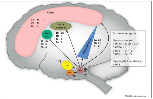
While it was thought that 5-HT is exclusively produced in raphe nuclei (RN) neurons located in the midline of the brainstem, recent in situ hybridization experiments showed that TPH2 is also expressed in the hippocampus and the striatum (Gutknecht et al., 2009). Serotoninergic neurons are the most abundant and complex system in the brain since they innervate almost all brain regions. They are among the earliest differentiated neurons found in a variety of organisms from C. Elegans to vertebrates (Lesch and Waider, 2012). Serotoninergic neurons originating from the rostral-dorsal and medial raphe terminate in the cortical, limbic (hippocampus), midbrain (ventral tegmental area VTA) and hindbrain (cerebellum) regions. Neurons originating from the caudal raphe are descending and innervate the spinal cord (Fig. 4). Indeed Gershone and Tack (2007) reported that “virtually all brain cells are close to at least one serotoninergic fiber”. Dysregulation of the serotoninergic system was implicated mainly in the pathogenesis of neuropsychiatric disorders (Roth et al., 2004).
Figure 4. Central serotoninergic efferent fibers originate from the raphe nucleus to innervate several brain structures. Serotonin is a neurotransmitter produced in neurons originating in the raphe nuclei located in the midline of the brainstem. The most caudal raphe innervates the spinal cord, while the more rostral raphe, the dorsal raphe nucleus and the medial raphe nucleus, innervate the rest of the central nervous system by diffused projections (Dalley and Roiser, 2012).
26 After synthesis in the RN, 5-HT is packed into synaptic vesicles till its release into the synaptic cleft upon stimulatory signals. Extracellular levels of serotonin are regulated by presynaptic 5-HT reuptake transporter (5-HTT or SERT) and degraded by the monoamine oxidase enzymes (MAO) (Fig. 5). Serotonin transporter SERT belongs to the family of monoamine transporters together with noradrenaline transporter (NET) and dopamine transporter (DAT) (Scholze et al., 2008). More insights in the function of 5-HT and its mechanism of action emerged with the cloning of at least 15 receptor subtypes. 5-HT receptors are grouped in 6 classes of G-protein coupled receptors (GPCRs) named 5-HT1,2,4,5,6,7 and one class of ligand-gated ion channel 5-HT3 (Fig. 5) (Kroeze et al., 2002). The expression of 5-HT receptors has been described in the mammalian brain including human. Receptors autoradiography revealed a region specific distribution of different receptors as shown in Fig. 6. Serotonin receptors 5-HT1A, 2A, 2C, 3, 6 and 7 and auto-receptors 5-HT1B/2B were found in the hippocampus (Brichta et al., 2013). Since 5-HT receptors and SERT modulate virtually all brain functions, they became the major focus of CNS drug development, and many current medications modulate serotonin neurotransmission.
2. Serotonin and mood regulation
To study the impact of early and adult life serotonin dysregulations in the brain, scientists relied on genetically modified animal models. Mice lacking SERT (SERT-/-) and MAO (MAO-/-) have constant overexposure to serotonin during early development and adult life. Anatomical studies on these mice showed that they have failed segregation and barrel structure formation of the thalamo-cortical axons. Altered anatomical structure and inhibited cortical neurons migration are under the CTRL of 5-HT1B and 5-HT6 receptors (Cases et al., 1996; Salichon et al., 2001; Meffre et al., 2012). In contrast, mice models lacking TPH2 (TPH2-/-) were depleted for 5-HT in the brain during embryonic and adult life. These mice did not
27 show any anatomical abnormalities in the serotoninergic neurons formation (Gutknecht et al., 2012). In fact the lack of 5-HT during early life is thought to be compensated by 5-HT secreted by the placenta (Gutknecht et al., 2008, Dayer, 2014). In contrast several studies showed that TPH2-/- mice have juvenile and adult fear/aggressive behavior in all experimental paradigms, lack of social interaction and severe maternal neglect to pups (Lerch-Haner et al., 2008, Lesch et al., 2012). The indoleamine hypothesis stated that the vulnerability to either depression or mania was related to the decrease of serotonergic activity. It is attributed to either less serotonin release or to fewer serotonin receptors or impaired serotonin receptor-mediated signal transduction (Mann, 1999). Later studies reported more complication and proved that indeed serotonin dysregulation is associated with impulsive aggression, anxiety, cognitive dysfunction and eating disorders (reviewed in Dayer, 2014). For example, researchers subjected female mice to chronic unpredictable stress and scored their male offspring in forced swim test and glucose consumption to assess their depressive-like behavior and in open field and exploratory paradigms to assess their aversive reactions to environment. The offspring showed depressive- and anxiety-like behavior, aggressiveness and anhedonia (Franklin et al., 2010). Studies on human confirmed that maternal depression could cause cognitive disabilities such as delayed maturation of language discrimination in infants. Within the same study scientists showed that infants from mothers treated with SERT inhibitors antidepressants exhibited more mature language discrimination abilities compared to infants from untreated mothers (Weikum et al., 2012).
28 Functional magnetic resonance imaging (FMRI) studies showed that depression induced by chronic mild stress exposure, reduced the connectivity between the RN and the hippocampus, hence serotonin dependent-functions in the hippocampus was reduced (Gordon and Goelman, 2016). DL-P-Chlorophenylalanine (PCPA), an irreversible inhibitor of TPH2, induces acute 5-HT depletion. Two studies used PCPA and showed that 5-HT depletion in human brain induced depression and cognitive function deficits and more specifically memory impairments. Depressed patients that received PCPA treatment had a transient return of depressed
Figure 5. Serotonin Neuron Synapse. Serotonin (5-HT) is produced from tryptophan by the tryptophan hydroxylase (TPH) and packaged into storage vesicles until its release into the synapse. Multiple postsynaptic 5-HT receptors mediate 5-HT signaling. Serotonin receptors are 7 subfamilies grouped in 2 types as: Ligand-gated ion channel (LGIC) and G-protein coupled receptors (GPCR). 5-HT levels in the synaptic cleft are regulated by SERT or degraded to inactive metabolites by monoamine oxidases (MAO). Presynaptic 5-HT1A and 5-HT1B auto-receptors detect the presence of 5-HT in the synapse and
shut down (-) further 5-HT release. Dopamine receptors D2 and α1-α2- adrenergic receptors
29 symptoms even after treatment with SERT inhibitors antidepressants (Delgado et al. 1990, Shopsin et al. 1976). Therefore antidepressants therapeutic effect required the enhancement of serotoninergic activity.
Taking into account that 5-HT receptors and SERT are distributed diversely in the brain, several studies dissected their role in deleterious effects of stress, onset of depression and response to antidepressants. For instance, partial reduction of SERT expression using silencing RNA (SERT-siRNA), increased the level of 5-HT in the synapse followed by a reduced expression of 5-HT1A autoreceptor. Silencing SERT in mice brains caused decreased immobility in the tail suspension test that is used to evaluate antidepressant-like activity (Ferrés-Coy et al., 2013). Presynaptic and
postsynaptic 5-HT1A receptors are described as the major mediators of antidepressant activity. For example, 5-HT1A autoreceptor agonists inhibited the feedback loop that usually leads to 5-HT reuptake from the synapse. Thus the increased availability of 5-HT in the synaptic cleft induced an anxiolytic and
Figure 6. Serotonin receptors distribution in mammal’s brain. (Brichta et al.,
30 antidepressant effect (Baldwin and Rudge, 1995). Moreover 5-HT by binding the postsynaptic 5-HT1A reduced the firing in the post-synaptic neurons inducing and anxiolytic effect (Chilmonczyk et al., 2015). Receptors binding studies as well as autoradiography studies showed that depressed patients have an increased 5-HT2A binding and activity (Ferrier et al. 1986; McKeith et al. 1987).
3. Serotonin and plasticity: emphasis on adult neurogenesis
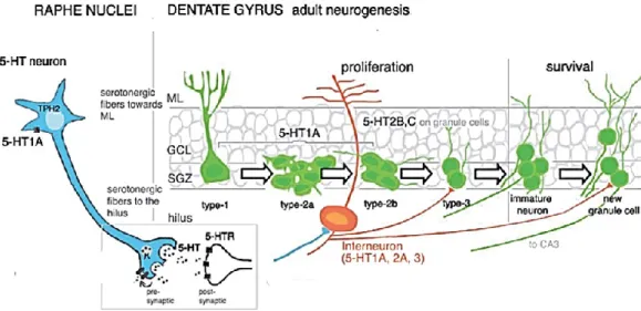
As highlighted previously, 5-HT is an essential neurotransmitter that promotes neuronal development during embryonic as well as adult life. Particularly the medial raphe provides substantial inputs to various interneurons throughout the entire hippocampus and in the hilus (Fig. 7). While the dorsal raphe projections terminate preferentially in the SGZ region of the DG, with small axonal terminations that appear to be positioned for diffused release of 5-HT (Djavadian, 2004). The dorsal projections are positioned the best to modulate directly adult hippocampal neurogenesis, however also medial projections could modulate the neurogenic process through indirect effect on interneurons and network activity (Fig. 7) (Gordon and Goelman, 2016). The ablation of the RN (by 5,7-dihydroxytryptamine injection in the medial/dorsal raphe) or PCPA treatment depleted 5-HT levels in the brain, including the hippocampus. This depletion caused a drastic reduction of BrdU+ and PSA-NCAM+ cells in the DG (Brezun et al., 2000). A RN graft restored the levels of 5-HT, the number of proliferative ahNPCs and newly generated neurons (neuroblasts) (Brezun and Daszuta, 2000). Interestingly, TPH2-/- mice do not show a drastic reduction in the number of proliferative cells, but a severe deficient transition from type 1 stem cells to type 2 progenitor cells and increased cell death rate. Moreover, DG neurogenesis in TPH2-/- mice did not increase after voluntary wheel running, a well-known positive modulator of neurogenesis (Klempin et al., 2013).
31 Several studies investigated the different contribution of 5-HT receptors in the synaptic plasticity and neurogenesis specifically in the hippocampus. For example, 5-HT1A is involved in dendrites and spine formation in the hippocampus. 5-HT1A antagonist decreased the number of dendritic spines of granule neurons and the total dendritic length of DG. This deleterious effect could be reversed by administrating 5-HT1A agonist (Yan et al., 1997). Interestingly 5-HT1A is expressed in type 1 (NSC) and type 2 (NPC) in the SGZ (Fig. 7). Blocking postsynaptic 5-HT1A in vivo by a selective antagonist caused a 30% reduction of BrdU+ in the SGZ of treated vs. not treated rats (Radley and Jacobs 2002). Depleting 5-HT secretion by PCPA followed by treatment with postsynaptic 5-HT1A agonist (8-OH-DPAT)
Figure 7. Illustration of serotonin mode of action during adult hippocampal neurogenesis. Serotonergic neurons located in the brainstem raphe nuclei synthesize TPH2 to be released in the synaptic cleft. Tracts of serotonergic fibers terminate in the hippocampus projecting into the molecular layer (ML) and the hilus, where they make synapses with principle neurons (Blue) and interneurons (in orange). Interneurons in the hilus in turn contact immature and newly generated granule cells in the SGZ and GCL. Several 5-HT receptors are expressed by the cell populations of the DG. 5-HT1AR in type-1 to type-2b ahNPC in the SGZ; 5-HT2B and 2C receptors in adult granule
32 rescued the number of proliferative ahNPC BrdU+ in the SGZ (Banasr et al, 2003). Hippocampal NPC grown in vitro in proliferation expressed TPH2 and produced 5-HT in the medium. 5-5-HT1A antagonist and PCPA treatment counteracted the proliferation of ahNPC in vitro as well as in vivo. Proliferation was rescued by administrating exogenous 5-HT or 5-HT1A agonist (Benninghoff et al., 2010). Moreover, 5-HT1A KO mice, even though they do not have lower baseline neurogenesis, were not responsive to 5-HT1A agonist (Santarelli et al., 2003). Therefore, genetic animal model further confirmed that 5-HT1A is usually required for positive modulation of neurogenesis.
Serotonin receptors 5-HT2 are also expressed in the hippocampus. In details 5-HT2A expression is very dense in the hilus neurons and interneurons, while 5-HT2C mostly marked the granular layer cells. Type 2 NPC in the SGZ expressed 5-HT2B/2C (Fig. 7) (Alenina and Klempin, 2015). Stimulation of 5-HT2C in vivo decreased mRNA and protein levels of BDNF, known to increase adult neurogenesis; vice versa blocking 5-HT2C restored the levels of BDNF and increased the number of proliferative cells (BrdU+) in the SGZ (Vaidya et al., 1997; Klempin et al. 2010). Another group also investigated the implication of 5-HT2 in the regulation of ahNPC proliferation and neuronal differentiation in the SGZ. Adult rats were treated with DOI (5-HT2A/2C agonist) or ketanserine (5-HT2A/2C antagonist). DOI did not change the percentage of proliferative cells, but ketanserine produced a decrease in the number of BrdU-labeled cells (Banasr et al, 2003). In addition to the highlighted role of 5-HT1A and5-HT2C, also 5-HT4 could play a role in adult hippocampal neurogenesis. For example chronic treatment with RS6733, a 5-HT4 agonist, increased the number of newborn DCX+ cells, and more specifically the ones with a more mature profile determined by more developed dendrites (Mendez-David et al., 2014). Altogether these studies showed that despite the opposite role of 5-HT receptors in neurogenesis, serotonin remains necessary for the response to positive modulators of neurogenesis such as exercise and antidepressants (Klempin et al., 2013).
33
3. Mood disorders: focus on Major Depressive
Disorder
Mood disorders are complex pathologies of the brain where the mood spectrum involves both depressed and manic episodes. Unipolar, bipolar and major depression disorders (MDD) are the most complex and prevalent mood disorders. Major depressive disorder is the second leading cause of disability in patients aged between 15-44 years. In addition to being ranked as the second in global disease burden, MDD is also projected to be the leading cause worldwide by 2030 (World Health Organization, 2012; Manji et al., 2001). Despite the discovery of first and second generation medications for MDD, at least 50% of patients who begin therapy with newer-generation of medications either fail to respond or discontinue therapy due to intolerable side effects. Thus, there remains an ongoing need for antidepressants that are more effective and/or better tolerated (Berton and Nestler, 2006; Hunot et al., 2007).
1. Definition and pathophysiology of MDD
MDD is a pathology characterized by multiple functional and structural impairments in the CNS and behavior. The most commonly listed characteristics are:
1. Emotional/motivational symptoms such as sadness, hopelessness, misery, anhedonia (loss of interest and joy), guilt and suicidal thoughts.
2. Cognitive symptoms such as negative cognition of self and the world (worthlessness).
3. Decreased mental productivity such as memory loss and learning disabilities.
4. Somatic symptoms such as loss of appetite, lack of energy, sleep difficulties and weight loss. Under stress the hippocampus and the
hypothalamic-34 pituitary-adrenal axis (HPA) are activated and stress hormones such as corticosteroids are released by adrenal glands. Stress hormones are the main inducers to the somatic symptoms of depression (Eisch and Petrick 2012).
To make an MDD diagnosis, clinicians rely on 3 obligatory symptoms: deep sadness, loss of interest and loss of energy that last at least two weeks in the first episode (Mill and Petronis 2007). Among others, one animal model of depression stood out as the most reliable and closes to human MDD. This animal model helped to study the physiological basis of depression. Chronic mild stress (CMS) procedure was developed in 1980s and used to induce depressive-like behavior in rodents. It is considered reliable since the conditions are realistic, reversible (specifically by antidepressant treatment) and the time course is suitable for chronic drug treatments (Willner, 2005). CMS could be induced by several factors such as: early maternal isolation, adult social isolation, and restraint/immobilization episodes. Depressive-like behavior could be assessed by behavioral tests such as: the forced swim test (FST), tail suspension test (TST) and hedonic reactivity. In fact animals with depressed-like behavior have longer immobility times in the FST, TST and have decreased palatable (sucrose) food intake (anhedonia) compared to naïve animals. These parameters are indications for helplessness and loss of interest. Additionally, MWM test is commonly used to assess the hippocampal-dependent spatial memory that is impaired by stress (Ehninger and Kempermann, 2005). Moreover, the advantage of this model is to predict in vivo the mechanisms of action of known or putative antidepressants and their effect on depressive symptoms and adult neurogenesis (Willner, 2005; Mirescu and Gould, 2006). The complex pathophysiology of MDD involves many combined factors such as genetic susceptibility, anatomical and morphological alterations and neurochemical unbalance.
Genetic susceptibility. Studies on twins of families with mood disorder history
35 disorder during their life. Additionally genetic studies showed that a number of chromosomal abnormalities were associated with the development of MDD. Most of the genes were linked to neurotransmitters or neurotrophic systems such as: 5-HT2A receptor, SERT, TPH2 and BDNF (Marchand et al., 2005; Fakhoury, 2015). Genetic factors increased the risk of developing MDD when combined with environmental factors such as stress and traumatic experience. For example, a study showed that patients holding two short (S) alleles instead of 1 or 2 long (L) alleles of SERT locus were more sensitive to stressful life events, exhibited more severe depressive symptoms and higher number of depressive episodes (Kendler et al., 2005).
Anatomical changes. MDD is accompanied or could cause impaired connectivity
among neuroanatomical structures involved in the regulation of mood and stress response. For example, physical connections between the hippocampus, amygdala and prefrontal cortex (PFC) are disrupted physically and chemically in patients with MDD. Moreover, the PFC and hippocampus have smaller volumes in patients with MDD compared to healthy individuals (Bremner et al., 2000, Cole et al., 2011, McKinnon et al., 2009; Boldrini et al., 2012). Emotional and cognitive aspects of depression such as worthlessness, hopelessness, guilt and memory loss could be caused by the loss of these connections.
Morphological changes. Exposure to stress causes atrophy of neurons, reduced
number of synapses (specifically glutamatergic synapses), loss of neurons and glia in the PFC and the hippocampus in human and rodent brain (Fig. 8) (Duman et al., 2012). Moreover, postmortem studies on brain tissue showed that the number of neurons in the GCL of the DG is reduced in patients with untreated MDD, which could explain the reduced volume of the hippocampus in these patients (Boldrini et al., 2012).
36
Neurotrophic factor downregulation. Neurotrophins are considered key regulators
of neuroplasticity hence they underlie the compromised plasticity aspects of MDD (Fig. 9). In rodents, acute and chronic immobilization stress as well as chronic pain stimuli resulted in decreased BDNF mRNA expression in rat hippocampus. This study suggested also that probably depression and pain have similar pathways (Maletic et al., 2007).
Decreased BDNF is followed by a low CREB activity, reduced arborization and spine formation (Fig. 9) (Duman and Monteggia, 2006). Brains of depressed patients have a severe reduction in the number of synapses specifically in the PFC and hippocampus (Maletic et al., 2007). A postmortem study on human serum
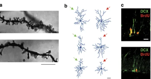
Figure 8. Stress alters several forms of neuroplasticity including adult neurogenesis. a. Post-weaning social isolation stress for 8 weeks significantly reduced dendritic spines in the prefrontal cortex (PFC) (visualized by Golgi-Cox staining) (Upper panel CTRL vs. Lower panel stressed). b. Chronic restraint stress reduced the length and complexity of cortical dendrites (Reconstitution of CTRL left panel vs. stressed right panel). c. Chronic unpredictable stress reduced the number of proliferative cells in the hippocampal SGZ. Visualized by bromodeoxyudridine BrdU and doublecortin (DCX) labeling markers of neuroblasts (CTRL upper panel vs. stressed lower panel).
37 showed that BDNF levels are reduced in samples from depressed patients, and antidepressant therapy restored brain BDNF levels to the normal range simultaneously with improvement in depressive state (Shimizu et al., 2003). Mice lacking for BDNF did not show depressive-like symptoms in the FST. The infusion of BDNF into the hippocampus and the RN region of depressed wild type mice mimicked the effects of antidepressants (Eisch et al., 2003). Similarly, fibroblast growth factor (FGF) is downregulated in the hippocampus of patients with MDD. Additionally insulin-like GF- I (IGF-I) is increased in the hippocampus by antidepressants treatment. IGF-I infusion into the brain has an anti-depressant like effect similarly to BDNF (reviewed in Duman et al., 20-16).
Neurochemical unbalance: emphasis on monoamine deficiency. Back in 1960,
European and American researchers simultaneously suggested that depression was caused by deficiency in the catecholamine norepinephrine (NE) and the indoleamine 5-HT in the brain (Fig. 9). In fact NE from the locus coeruleus (LC) and 5-HT from the RN are the most dominant innervations in the CNS regions involved in MDD (Berton and Nestler, 2006). The involvement of the monoaminergic system was initially postulated after the finding that monoamine depleting agents such as “reserpine” (anti-hypertension drug) could produce depressive symptoms. Animal research studies showed that reserpine depleted 5-HT and NE from neurons. Simultaneously, patients showed severe depressive state and sedative behavior. In contrast monoamine oxidase inhibitors (MAOI) increased the levels of monoamines in the synapse and antagonized the effect of reserpine (Manji et al., 2001; Lopez-Munoz and Alamo, 2009). Subsequently, pharmacological studies showed that 5-HT receptors might add more complication to antidepressants effect. Serotonin receptors 5-HT1A, 5-HT2A/2C and 5-HT4 emerge as candidates to mediate antidepressant response. For instance, they regulate normal development of dendritic spine density, synapse formation of pyramidal and granule cells in the DG, long-term plastic changes that decrease anxiety-like behavior and mediate ahNPC maturation at the late phases (Djavadian, 2004). For
38 example, 5-HT1A is overexpressed in the hippocampus of MDD patients. This compensatory mechanism is a response to decreased levels of 5-HT (Santarelli et al., 2003). In contrast, increased binding on 5-HT2A/2C receptors is responsible for adverse side effects related mainly to weight changes and sleep deprivation (De Vry and Schreiber, 2000). Activated 5-HT4 increases the levels of BDNF and ahNPC proliferation in the DG through cAMP signaling cascade (Djavadian, 2004; Mattson et al., 2004; Ferrés-Coy et al., 2013).
Similarly to 5-HT, low levels of NE were specifically associated with cognitive dysfunction, fatigue and apathy. These associations solicited scientist to study more deeply the role of NE in cognitive and psychiatric disorders that will not be detailed in the context of this work. Many other neurotransmitters systems were found to be involved in MDD but to a less extent compared to 5-HT and NE. For example, the synthesis and release of GABA are reduced in response to acute and chronic stress whereas the glutamate levels are high after stress. Likewise increased levels of histamine in the limbic system have been associated with depression and sleeping problems in patients with MDD (reviewed in Marchand et al., 2005).
2. Antidepressants: classes and mechanisms of action
During the 1950s, while carrying out trials on a new medication for tuberculosis, researchers noticed that the medication had also a mood improving effect on tuberculosis patients. Clinicians showed that anti-tuberculosis drug improved the depressive state of patients (raised mood, weight gain, better interpersonal capacity, increased interest in themselves and surroundings). Scientists showed that “iproniazid” had indeed MAO inhibitory activity (thus the name MAOI, Fig. 10 A). It increased the levels of 5-HT and NE in the synaptic cleft by inhibiting their degradation (Sandler, 1990). MAOI caused severe side effects thus their use became limited.
39 At the same year of MAOI discovery in 1957 tricyclic (TC) molecules were characterized chemically similar to antihistaminic drugs. Therefore, they were tested for their potential antihistaminic and sedative effects. R. Kuhn, a famous psychiatrist at that time, showed that they improved the symptoms in patients with depressive psychosis and raised the first suggestion of potential antidepressant effect. TC molecules started to be used as antidepressants (hence the name TCA, Fig. 10 A). TCA such as imipramine showed anti-histaminic activity as well as NE and 5-HT reuptake inhibition (NRI and SRI). TCA had low MAOI activity therefore they had safer profile in patients (Lopez-Munoz and Alamo, 2009). In
Figure 9. Synaptic connectivity and chemical balance is lost in stressed brain. A. Normal state: healthy hippocampal pyramidal neurons receive physiological levels of monoamines (5-HT, NE), glutamate and BDNF (Thick arrows). B. Severe stress increases the levels of glucocorticoids that, in turn inhibit CREB signaling causing reduction of BDNF, monoamines and stimulatory glutamate thus inducing radical loss of dendritic arborizations and spines (Thin arrows). C. Antidepressants, by activating CREB signaling or by increasing monoamines restore BDNF levels, dendritic arborizations and spine number. (Modified from E. Nestler et al., 2002)
40 summary, the first-generation of antidepressants was effective because it enhanced serotonergic or noradrenergic mechanisms or both. Unfortunately, even TCA were still non-selective drugs with considerable side effects due to: anti-histaminic activity (sedation, sleep), cholinergic activity (tremors), anti-muscarinic M1 (constipation, dry mouth, urinary retention) and α1-adrenergic receptor sites (orthostatic hypotension, sexual dysfunction) (Duman et al., 2016). Moreover, TCA and MAOI had a fast chemical effect on the amine levels in the synaptic cleft, but the therapeutic effects did not take place before 4 weeks of treatment (Pittenger and Duman, 2008). It is important to point out that these initial drugs were approved and prescribed merely upon clinical observation without real knowledge of the mechanism of action. Since then, rational drug design (relying on the knowledge of the biological target and its role) led to the introduction of selective, faster, and better tolerated antidepressants.
The second generation of antidepressants included: selective serotonin reuptake inhibitors (SSRI), serotonin and norepinephrine reuptake inhibitors (SNRIs) and noradrenaline reuptake inhibitors (NARI) (Fig. 10 B). Selective SSRI had similar efficacy as TCA but lacked the anti-muscarinic activity, thus they had less cardiovascular side effects. Their major side effects were headaches, sexual dysfunction, and weight gain due to 5-HT2C agonistic activity. SNRI were more balanced on both receptors system, had the same efficacy as TCA and SSRI yet they lacked the muscarinic activity and α1 AR blockade, thus their major side effect was hypertension. NARI had mainly analgesic activity but they dangerously increase the arterial blood pressure, hence they were subjected to prescription restrictions (Berton and Nestler, 2006).
41 As part of the effort to address the unmet need of better tolerated antidepressants, attention has been given to multimodal acting drugs and new mechanisms of action for depression treatment. Multifunctional or multimodal drugs could be used in multiple disorders and have less severe side effects controlled by dosage regulations. As an example (i) serotonin antagonist and reuptake inhibitors (SARI) (Fig. 10, B) such as trazodone and nefazodone that have the same efficacy as TCA and SSRI with additional anxiolytic and hypnotic activity (Lopez-Munoz and Alamo, 2009); (ii) agomelatine that is melatonergic receptor agonist and 5-HT2C receptor antagonist (Soumier et al., 2009); (iii) vortioxetine that is a 5-HT3,7,1D antagonist, 5-HT1B partial agonist, 5-HT1A agonist and SERT inhibitor (Guilloux et al., 2013). Several studies are focusing on understanding how multimodal drugs induce their antidepressant effect, their off-label therapeutic use and their side effects.
Figure 10. Antidepressant Mechanism of action. A. Monoamine oxidase inhibitors (MAOI) inhibit mitochondrial oxidation of monoamines in the presynaptic neuron. Tricyclic antidepressants (TCA) inhibit 5-HT transporter (SERT) and/or noradrenaline transporter (NET) B. Serotonin Antagonist and Reuptake Inhibitors (SARI) antagonize serotonin receptor 2 (5-HT2)
and block SERT; Selective serotonin reuptake inhibitors (SSRI) block serotonin reuptake transporter (SERT) on the presynaptic neuron; Serotonin and Noradrenaline Reuptake Inhibitors (SNRI) block SERT and NET. All mechanisms increase the level of 5-HT and NE in the synaptic cleft. (Modified from Brigitte Schiott, Biomodeling, Semper Ardens Research project, 2016)
B
A
42
3. Adult hippocampal neurogenesis, MDD and antidepressants
Environmental interventions such as learning tasks, enriched environment and physical activity stimulate adult neurogenesis and have an antidepressant-like effect on behavior (Sahay and Hen, 2007). By the same token scientists suggested that antidepressants might also increase adult hippocampal neurogenesis. “The neurogenic hypothesis of depression” postulated that decreased production of new granule cells in the dentate gyrus of the hippocampus is linked to the pathophysiology of depression and that the increase in hippocampal neurogenesis and plasticity are required for the behavioral effect of antidepressant treatment (Eisch and Petrick, 2012).
The ablation of the SGZ by X-ray irradiation inhibited the onset of behavioral improvement mediated by fluoxetine (SSRI) on non-human primate animal model of MDD (Perera et al., 2011). Boldrini and colleagues showed that the number of ahNPC in the hippocampal SGZ of patients with MDD is increased in SSRI antidepressant-treated versus non-treated patients (Boldrini et al., 2009). Fluoxetine accelerated synaptogenesis (increase spines formation) and enhanced LTP in the hippocampal neurogenic niche. These improvements were accompanied with improved depressive behavior (Wang et al., 2008). Moreover, several studies showed that chronic (14 days or more) treatment with fluoxetine upregulates NPC proliferation rate (increase the number of BrdU+ cells in the SGZ) and immature neuron survival rate (Malberg et al, 2000; Kodama et al, 2004; Huang and Herbert, 2006; Marcussen et al, 2008). Similar results have been seen in experiments using citalopram and escitalopram (SSRI). In details, scientists induced a depressive state by olfactory ablation in rats. Rats were treated with citalopram in short and long term paradigms to assess the proliferation and survival of newly generated neuron. They showed that citalopram increases the number of proliferative BrdU+ cells and restore the number of PSA-NCAM+ surviving new neurons to levels compared to control rats (Jaako-Movits et al, 2006). Another study used CMS paradigm and
43 assessed the effect of escitalopram on ahNPC proliferation and differentiation. They showed that, similarly to fluoxetine and citalopram, also long term treatment with escitalopram increased the neurogenesis in the SGZ with simultaneous increase in glucose consumption (hedonic behavior) as a sign of alleviated depressive state (Jayatissa et al, 2006). Citalopram and reboxetine (NARI) may potentially upregulate neurogenesis through BDNF signaling since serum and hippocampal regions BDNF levels are increased following treatment (Russo-Neustadt et al, 2004). Several studies used chronic treatments with antidepressants such as SSRI, TCA, NRI and MAOI to enhance several steps of the neurogenic process in the DG of rodents and non-human primates. Notably, the increase of neurogenesis was simultaneous to the onset of therapeutic effect of antidepressants (Malberg et al., 2000, Santarelli et al., 2003, Airan et al., 2007). Neurotrophic factors such as BDNF and IGF-1 activate ERK in neurons to induce neuroprotection and in ahNPC to increase proliferation and differentiation (Yamada et al. 2001; Shelton et al. 2004). Similarly, mood stabilizers such as lithium and valproate activate ERK and enhance both proliferation and survival of ahNPC in the SGZ (Hao et al, 2004; Hanson et al, 2011a). Low serum levels of BDNF were associated with depression in patients. Treatment with antidepressant and, specifically, with SSRI was associated to normalized levels of BDNF (Molendijk et al., 2011). Agomelatine that is a multimodal antidepressant also increased the survival of ahNPC via melatonergic receptor agonistic and 5-HT2C antagonistic activities accompanied with increased BNDF signaling (Soumier et al., 2009). Similarly, acute and repeated dosing of vortioxetine (multimodal antidepressant) significantly increased the number of BrdU+ cells and DCX+ with tertiary dendrites (more mature cells). Vortioxetine increased proliferation, cell survival and stimulated maturation of immature granule cells in the hippocampal SGZ (Guilloux et al., 2013).
Our group also participated in consolidating the neurogenic hypothesis of depression and the role of antidepressants. Pregabalin (PGB) and gabapentin (GBP)
44 are clinically relevant anticonvulsant, analgesic and anxiolytic drug. Interestingly, PGB and GBP are also prescribed to MDD patients with post-traumatic stress and anxiety (Stein et al., 2008). Our group showed that PGB and GBP increase, in a concentration-dependent manner, the number of mature and immature neurons generated from the in vitro model of ahNPC. They act through α2-δ1 subunit of the voltage-gated calcium channels expressed by hippocampal NPC (Valente et al., 2012). More interestingly, this effect was blunted by inhibiting the nuclear translocation of NF-κB p50/p65 subunits to the nucleus (Valente et al., 2012). Within the same study PGB interestingly decreased depressive-like behavior in mice that received unpredictable CMS, since it reduced immobility time in both TST and FST. Thus the antidepressant activity of PGB and GBP could be potentially mediated by neurogenesis (Valente et al., 2012). In another study, our group focused on Acetyl-L-carnitine (ALC) that can (i) cross the blood brain barrier, (ii) improve mood disorders in human particularly in elderly and (iii) have an analgesic effect in rodent studies (Zanardi et al., 2006; Cuccurazzu et al., 2013; Bortolotto et al., 2014). ALC is an acetyl group donor to proteins including NF-𝜅B p65 subunit (Chiechio et al., 2006), but it was not known how ALC can have an antidepressant effect. Our group hypothesized that the antidepressant activity of ALC could be due to a proneurogenic effect mediated by NF-𝜅B signaling pathway. ALC showed a potent pro-neurogenic activity on ahNPC in vitro. This effect was mediated by the activation of NF-𝜅B pathway and subsequent upregulation of metabotropic glutamate receptors 2 (mGlu2) expression in vitro and in vivo. These cellular mechanisms were inhibited by blocking p65 translocation to the nucleus. In vivo ALC reversed depressive-like behavior in mouse model of MDD (Cuccurazzu et al., 2013). The antidepressant activity described by our group was also confirmed by another study on genetic rat model of depression. Moreover they compared ALC to TCA and showed that rats had reduced immobility in FST and increased palatable food consumption. More interestingly in their model the onset of ALC effect required three days of
45 treatment while TCA required 14 days of treatment (Nasca et al., 2013). Altogether our group and literature studies suggested that increasing neurogenesis might be a new mechanism of action for antidepressants. It is still not known if multimodal antidepressants such as trazodone (SARI) could have an effect on neurogenesis and by which mechanisms.
4. Trazodone
Trazodone hydrochloride (TZD) is a dose dependent multifunctional drug that has advantages over other antidepressants in terms of therapeutic use. It is prescribed as antidepressant at high doses and has multiple off-label uses mainly for sleep and anxiety disorders at low doses. TZD detailed mechanism of action and diverse therapeutic benefits are still not fully characterized.
1. Trazodone: peculiar mechanism of action
Trazodone hydrochloride is a phenylpiperazine-triazolopyridine compound first discovered by Angelini research laboratories in Italy in the 1960s. Trazodone, similarly to SSRI and their derivatives, was introduced as the second generation of antidepressants after TCA and MAOI. Trazodone had comparable efficacy to TCA and SSRI antidepressants and yet was more tolerated. Early studies in rat brain showed that given acutely or chronically with doses similar to other SSRI antidepressants, trazodone appeared inactive as an inhibitor of serotonin reuptake by SERT but showed 5-HT2A/C antagonistic activity (Fuller et al., 1984). Affinity binding studies showed that Trazodone has 50 to 100 fold higher affinity to 5-HT2A/C than SERT (Fig. 12 A). To elicit a therapeutic effect trazodone is administrated at a dose that recruits both the weaker binding to SERT and the strong binding 5-HT2A/C receptor (Fig. 11, B) (Knight et al., 2004; Stahl et al., 2009). Together these properties annotated trazodone as a dose-dependent 5-HT2A/C antagonist and serotonin reuptake inhibitor (SARI). Based on the knowledge of
46 both mechanisms of action outcome, trazodone mechanisms are suggested to act synergistically (Stahl et al., 2009). On one hand antagonizing 5-HT2A/C, trazodone similarly to TCA antidepressants, increase the release of dopamine in the cortex inducing antidepressant activity (Chagraoui et al., 2015).
On the other hand, by blocking SERT, trazodone increases the availability of serotonin in the synapse and allows the activation of 5-HT1A resulting in hyperpolarization and reduced neuronal excitability and firing (Chilmonczyk et al., 2015). The hyperpolarization makes the neurons less excitable and leads to antidepressant and anxiolytic activity. Additionally, trazodone has different binding affinities to various receptors such as α1 and α2 adrenergic receptors, histamine
Figure 11. Chemical structure of trazodone and its antidepressant mechanism. A. Chemical structure of phenylpiperazine-triazolopyridine, trazodone B. Schematic representation of trazodone mechanism of action. Trazodone is a serotonin antagonist and reuptake inhibitor (SARI). It inhibits serotonin reuptake by blocking SERT on the presynaptic membrane allowing serotonin to activate it 5-HT1A receptor and antagonizes 5-HT2A receptor. (Stahl, Trends in
Psychopharmacology, 2009)