Università degli Studi di Ferrara
DOTTORATO DI RICERCA IN
SCIENZE BIOMEDICHE
CICLO: XXIV
COORDINATORE: Prof. Silvano Capitani
Molecular cytogenetic investigations in chronic B-cell lymphoproliferative disorders: novel mitosis stimulators and clonal evolution
Settore Scientifico Disciplinare: BIO/16
Dottorando Tutore
Dott. Abass Awad Elkareem Prof. Capitani Silvano
_________________________ ______________________
(firma) (firma)
Tutore
Prof. Cuneo Antonio
_____________________
(firma)
Acknowledgment
The work of the present thesis was performed at section of Hematology- Department of Biomedical Sciences- Ferrara University.
First of all I would like to thank the members of the doctorate selection committee- Ferrara University for giving me a chance to do PhD research at this prestigious institute.
I’m grateful to Prof. Alessandro Martini for his guide and help. My thanks also extend to Prof. Silvano Capitani for his continuous help, sincere guide and support.
I heartily thank Prof. Antonio Cuneo; who have introduced me to the research and offered me many opportunities. I appreciate all his efforts, support, guide and help.
I would like also to thank Dr. Gian Matteo Rigolin and Dr. Lara Rizzotto for their technical advisement, constructive talks, and keeping me in touch with molecular cytogenetic science. Special Thank to Dr. Francesco Cavazzini, Dr. Antonella Badri, Dr .Sara Martinelli, Dr. Luisa Ferrari, and Dr. Diana Campioni for their help and continuous cooperation.
Beyond strictly scientific matters, I’m heartily grateful to all those peoples for their sincere friendships.
Table of Contents
1.0. Introduction & Literature Review ---03
1.1. Cytogenetics: Historical Background ---03
1.2. Overview of Cancer Cytogenetics ---05
1.3. Genetic analysis of B-CLL---07
1.3.1. Chromosome banding analysis ---07
1.3.2. Fluorescence in situ hybridization ---08
1.3.3. Comparative genomic hybridization analysis---11
1.3.4. Molecular genetic techniques---12
1.4. Novel mitogenic stimulaters in CLL ---13
1.4.1. Application of CD40---14
1.4.2. Application of Oligonucleotides and IL-2---14
1.5. CLL Cytogenetics ---17
1.5.1. Incidence of genomic aberrations in CLL---18
1.5.2. Cytogenetic profile of CLL ---19
1. 6. Clonal Evolution in CLL---24
2.0. Rationale---28
3.0. Objectives---30
4.0. Material & Methods---31
4.1. Study area ---31
4.2. Study design ---31
4.3. Patients and Samples ---31
4.4. Methods ---31
4.4.1. Clinicobiologic investigations ---31
4.4. 2.Cytogenetic analysis ---32
4.5. Statistical analysis ---35
5.0. Results---36
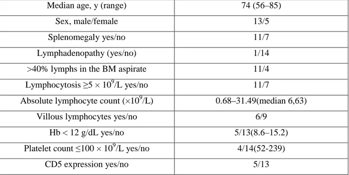
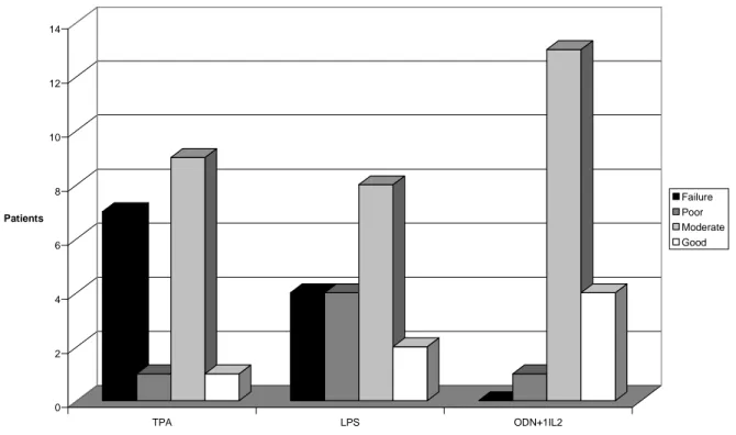
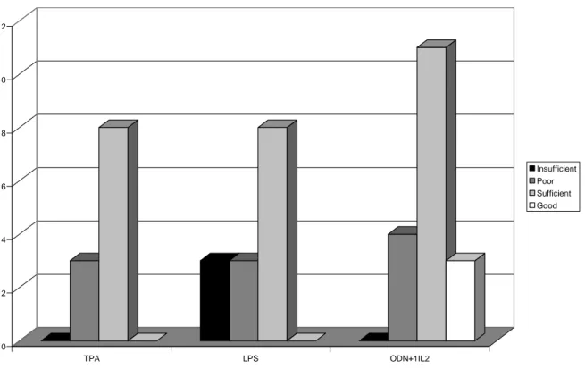
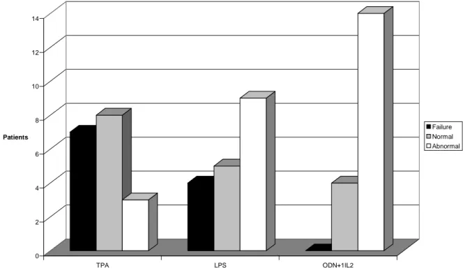
5.1. Novel mitogenic stimulators for SMZL samples ---36
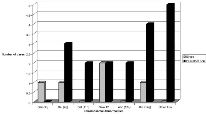
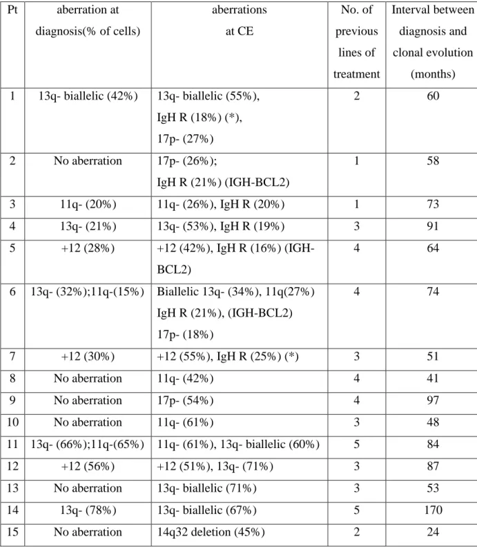
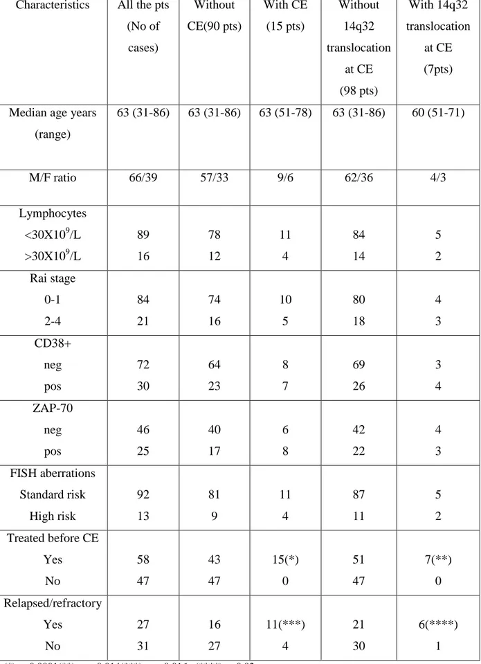
5.2. Clonal evolution in CLL---37
5.3. Correlation between CE, hematologic and clinical parameters in CLL---37
6.0. Discussion---51
7.0. Conclusion---55
References---56
I. Introduction & Literature Review
1.1. Cytogenetics: Historical Background:
Cytogenetic analysis has become an integral part of the diagnosis and management of many malignancies. Theodor Boveri was the first to suggest that malignant tumours could be due to an abnormal chromosome constitution (Sandberg, 1979). He also postulated the existence of enhancing or suppressing chromosomes, suggesting that malignant growth would result from loss of suppressing chromosomes or the predominance of enhancing chromosomes. The term chromosome was first coined by Waldeyer in 1888. The birth of cytogenetics is generally dated from Tjio and Levan’s identification of the true chromosome complement in human cells, as it is from this time that abnormalities of chromosome number and subsequently chromosome structure were reported (Tjio & Levan, 1956). Their discovery was made possible by a number of advances. In his delightful book, Hsu divided the study of human cytogenetics into four periods: the pre-hypotonic period (or Dark Ages), the period from 1952 to 1959 that included the discovery of hypotonic solution pretreatment for cytological preparations, the third period (1959–1969) during which time chromosome abnormalities were linked to clinical syndromes, and the post-banding (modern) period (Hsu, 1979). During the pre-hypotonic era, chromosomes were studied in mouse and rat cancers and camera lucida drawings of metaphases suggested the presence of many more chromosomes than normal and of structural abnormalities within these chromosomes.
The drawings were taken from squash preparations; a technique that was used to flatten metaphase spreads of chromosomes into a two-dimensional configuration, but still resulted in crowded overlapping aggregations of chromosomes that were very difficult to count. Colchicine, an extract of the autumn crocus, was used to arrest the cells in the metaphase stage of the cell cycle and increase the number of mitoses available for analysis.
The use of a hypotonic pre-treatment method was an enormous step forward in the production of analysable chromosome preparations. The chromosomes could now be separated and viewed individually. Counting chromosomes was simplified and gross structural abnormalities could be discerned. Hsu describes the discovery of the utility of a hypotonic pre-treatment as a laboratory accident, the perpetrator of which never owned up to the error; thus, a major discovery in the history of cytogenetics was apparently made by an unknown technician. It was in this era, in 1956, that Tjio and Levan finally answered the question that had been plaguing investigators for more than 30 years, when they reported that there were 46
chromosomes in the human cell rather than 48. Subsequently, a number of researchers were able to identify chromosome abnormalities that appeared specific for clinical syndromes. Lejeune and his colleagues published the chromosomal nature of Down syndrome in 1959 (Lejeune et al, 1959). Their observation of an extra G group chromosome in patients with a specific congenital malformation syndrome showed for the first time that cytogenetic analysis could be used to diagnose a human condition. The study of the constitutional karyotype was aided by the discovery that phytohemagglutinin (PHA) could induce peripheral blood lymphocytes to divide (Nowell, 1960). This method was adopted by Moorhead et al. for the study of human chromosomes and remains one of the mainstays of modern cytogenetic analysis (Moorhead et al, 1960).
The confirmation of the correct chromosome number in human cells led in the late 1950s and early 1960s to a flood of publications describing numerical and structural abnormalities of chromosomes. The resulting confusion in the literature made clear that there was a need for a common nomenclature to describe these rearrangements in a manner that was intelligible to other workers in the field. Thus, a small group met in Denver, CO, to establish a system of describing chromosome abnormalities. They published the results of their deliberations in a report entitled “A Proposed Standard System of Nomenclature of Human Mitotic Chromosomes,” also known as the Denver Conference (1960), and this report has formed the basis for all subsequent nomenclature reports, now published as An International System for Human Cytogenetic Nomenclature (ISCN) (Shaffer et al, 2009).
The advent of banding enabled chromosomes to be individually identified and the normal homologues paired. Initially, banding patterns along the length of each chromosome were induced by preparations stained with quinacrine mustard and visualized via a fluorescence microscope (Caspersson et al, 1968) or depended upon a method whereby slides were incubated in warm saline or buffer solutions prior to staining by Giemsa. The initial Giemsa staining method required 3 days for completion. Seabright’s rapid banding technique was therefore embraced as the whole procedure could be carried out at room temperature using air-dried slides and producing G-banded chromosomes ready for observation within 10 minutes (Seabright, 1971). Once banding became available, there were numerous publications describing recurrent chromosome abnormalities that appeared to be found in specific tumour types.
1.2. Overview of Cancer Cytogenetics:
The history of cancer cytogenetics is not a long one but it has been eventful and much knowledge has been accumulated in the 50 years since Peter Nowell and David Hungerford published their finding of a small marker chromosome in the chromosome complement of cells cultured from seven patients with chronic myeloid leukaemia (CML) (Nowell & Hungerford, 1960). Nowell and Hungerford made their landmark discovery in 1960, only just beating another group from Edinburgh who had also noted the same marker chromosome in their CML patients (Baikie et al, 1960). Although the hypothesis that malignant cells were derived from normal tissue cells that had acquired abnormal chromatin content was first proposed by Boveri, it was not until Nowell and Hungerford’s description of the Philadelphia chromosome that a revolution in our understanding of the processes underlying the development of malignancy began. Nowell and Hungerford called the marker chromosome the Philadelphia chromosome 1, Ph1, after the city in which they worked and the number 1 superscript signalled that they fully expected that there were many more cytogenetic markers of cancer to be discovered. This was an exciting finding and an exciting time that effectively launched the field of cancer cytogenetics but the following years were frustrating as abnormalities were observed in various cancers but the inability to identify specific chromosomes by any method other than their basic shape limited researchers’ abilities to link abnormalities with different morphological subtypes of haematological or solid tumours. All this changed with the advent of banding techniques. Banding allowed the chromosomes to be clearly distinguished from one another and, most importantly, revealed the nature of structural abnormalities: balanced translocations of material between chromosomes, deletion of part of a chromosome, duplication of another segment, or an inversion of a chromosome segment. In 1973, Janet Rowley reported that a reciprocal translocation between chromosomes 9 and 22 resulted in the Philadelphia chromosome (Rowley, 1973). Since then, hundreds of rearrangements have been identified including not only translocations but also deletions and additions of part or all of chromosomes and also inversions of genetic material within chromosomes. From the descriptions of chromosome rearrangements, long before the Human Genome Project shone a light on the location of genes strung along our chromosomes, the molecular biologists were able to discover critical genes at the breakpoints of translocations. Banding continues to allow us to identify new chromosome abnormalities in both haematological and solid tumours and these abnormalities provide the sign posts to the critical genetic changes that underlie the transformation of normal cells into cancer cells. The first cytogenetic abnormality to have its genetic secrets unlocked was the 8;14 translocation which
characterizes Burkitt lymphoma/leukaemia. Researchers identified that the translocation caused two genes, MYC on 8q24 and the immunoglobulin heavy chain gene, IGH on 14q32, to come together (Dalla-Favera et al, 1982, Taub et al, 1982). It is now known that two classes of translocations are found in malignancies. The first type is epitomized by the t(8;14) in Burkitt lymphoma, one gene which is already actively transcribed in the cell type, such as IGH in B lymphocytes, is juxtaposed to a gene such as MYC which is, by virtue of its resulting proximity to IGH, up-regulated. Other translocations such as the t(8;21) in acute myeloid leukaemia (AML), also described by Janet Rowley in 1973 (Rowley et al, 1973), and the t(9;22) in CML, form fusion genes with a “new” gene product which incorporates part of the normal genes broken at the sites of translocation.
It took time for the medical and scientific worlds to realize the importance of the cytogenetic discoveries of the 1960s and 1970s. It was necessary to convince clinicians that the chromosome changes being described in the marrow and peripheral blood of their patients with a variety of malignancies could provide valuable information about the type and prognosis of these disorders. Many important clinical correlations were either identified or confirmed in the International Workshops on Chromosomes in Leukaemia. These constituted gatherings of physicians and scientists from around the world who brought together case studies of chromosome analyses together with clinical and laboratory data relating to each case. The first of these was held in Helsinki, Finland in August 1977 (Rowley & de la Chapelle, 1978). Laboratories participated from Belgium, Finland, Sweden, England, Germany, and the USA and the participants reviewed the data of 223 patients with Ph1-positive CML and 279 patients with acute non-lymphocytic leukaemia. A number of further workshops were held and information regarding the incidence and prognostic significance of rearrangements in CML, AML, acute lymphoblastic leukaemia (ALL), and myelodysplastic syndromes (MDS) provided by these workshops formed the basis for all future studies. Subsequently, national and multinational clinical trial groups have incorporated cytogenetic studies into their prospective trials and provided a wealth of data to show that cytogenetic analysis is of diagnostic and prognostic importance in most haematological malignancies and a number of solid tumours. It has only been by the careful observation of chromosome abnormalities and their correlations with clinical features that true insights have been obtained as to the underlying genetic basis of malignancy.
However, although the basic cytogenetic methods used in laboratories around the world today are very similar to those first described in the 1960s and 1970s, there are areas where improvements have been made. Mitogens were introduced into cultures to induce chronic
lymphoid malignancy cells to divide in the late 1970s (Gahrton et al, 1980) but further refinements and combinations of mitogens are still being discovered.
1.3. Genetic analysis of B-CLL:
1.3.1. Chromosome banding analysis (metaphase cytogenetics):
In the 1960s and 1970s most cytogenetic studies of B-CLL were performed on blood lymphocytes stimulated with phytohemagglutinin and the vast majority of tumors exhibited a normal karyotype. A review by Mitelman & Levan (Mitelman & Levan, 1978) on chromosome aberrations in human neoplasms associated no specific aberration with B-CLL. Specific chromosome aberrations in CLL were not identified before the late 1970s when B-cell mitogens were shown to induce leukemic B-cells from B-CLL tumors to proliferate in culture (Robèrt et al, 1978; Autio et al, 1979; Hurley et al, 1980; Gahrton et al, 1980a; Gahrton et al, 1980b). Among the mitogens that have been used are tetradecanoyl-0-phorbol-13-acetate, Epstein-Barr virus, lipopolysaccharide, pokeweed mitogen, cytochalasin B, anti- human IgM, B-cell growth factor, calcium ionophore (Ca2+), and an anti-CD40 antibody (Döhner et al, 1993; Crawford & Catovsky, 1993; Oscier et al, 1994). Despite the use of these mitogens, conventional chromosome banding analysis has remained difficult in CLL even with improved culture techniques clonal genomic abnormalities can be detected in only 40– 50% of CLL cases (Juliusson et al, 1990; Juliusson et al, 1991). In the cases without clonal abnormalities mitotic cells often stem from nonleukemic T-lymphocytes, as shown by the study of (Autio et al, 1986) using the technique of sequential immunophenotyping and karyotype analysis.
Specific chromosomal abnormalities have been shown to affect the overall survival of patients with acute leukemia. Juliusson et al assessed this possibility; blood mononuclear cells from 433 patients with B-cell CLL in five European centers were cultured with B-cell mitogens (Epstein–Barr virus, lipopolysaccharide from Escherichia coli, 12-O-tetradecanoylphorbol-13-acetate, cytochalasin B, pokeweed mitogen, and phytohemagglutinin) according to the protocols of the individual institutions and banded metaphases were studied.
They evaluated 391 patients cytogenetically, and 218 had clonal chromosomal changes. The most common abnormalities were trisomy 12 (n = 67) and structural abnormalities of chromosome 13 (n = 51; most involving the site of the retinoblastoma gene) and of chromosome 14 (n = 41). Patients with a normal karyotype had a median overall survival of more than 15 years, in contrast to 7.7 years for patients with clonal changes. Patients with single abnormalities (n = 113) did better than those with complex karyotypes (P<0.001).
Patients with abnormalities involving chromosome 14q had poorer survival than those with aberrations of chromosome 13q (P<0.05). Among patients with single abnormalities, those with trisomy 12 alone had poorer survival than patients with single aberrations of chromosome 13q (P = 0.01); the latter had the same survival as those with a normal karyotype. A high percentage of cells in metaphase with chromosomal abnormalities, indicating highly proliferative leukemic cells, was associated with poor survival (P<0.001). Cox proportional-hazards analysis identified age, sex, the percentage of cells in metaphase with chromosomal abnormalities, and the clinical stage of the disease (Binet classification system) as independent prognostic variables (Juliusson et a, 1990).
1.3.2. Fluorescence in situ hybridization (FISH):
FISH is a process by which specific fragments of DNA are labeled with a fluorochrome (a dye that emits visible light when irradiated with ultraviolet light) and are then allowed to attach to a particular location on a chromosome. The FISH technique is now well established and generally robust and reliable. However, the correct interpretation of the results is not always obvious, and there are some factors that need to be considered.
The chromosomes obtained in studies of malignancy are often of poor morphology, and tend to be involved in complex and subtle rearrangements; in such cases, it is often impossible to define the entire karyotype by simply using G-banding. There is a variety of other newer genetic techniques that are available. These include polymerase chain reaction (PCR) assays that can detect specific gene rearrangements (including translocations) but not gains or losses of chromosome material, and array comparative genomic hybridization (CGH) that can detect minute gains and losses but not balanced rearrangements. FISH has some of the attributes of both assays and forms a very powerful combination with karyotype studies. In particular, FISH can compensate for one of main restrictions of karyotype studies, the need for metaphase divisions: clinically useful information can often be obtained from FISH studies of interphase nuclei (Tkachuk et al, 1990). The impression is sometimes given that FISH studies are simply a matter of buying a kit with the right DNA probe, following the supplier’s instructions, and reading a simple positive or negative result. In practice, getting a reliable result from a FISH study requires experience, time spent in testing to determine the precise local conditions needed for optimum hybridization, and time spent in assessing and scoring positive and negative controls to determine local baseline levels.
The development of FISH in the late 1980s and early 1990s provided a very powerful tool for the detection of chromosome aberrations in tumors, Lichter (Lichter et al, 1990), especially in
CLL. Delineation of specific DNA sequences in the cells by the technique of in situ hybridization is the basis for this molecular cytogenetic approach, by wich the genomic abnormalities can be detected on the single cell level in interphase nuclei or metaphase spreads. Therefore, this approach is also referred to as ‘interphase cytogenetics’ (Cremer et al, 1986). Numerical and structural chromosome aberrations that involve changes in the copy number in tumor cells are identified by aberrant signal numbers per cell, while translocation breakpoints are detected by the pattern of spatial distribution of the fluorescence signals. The sensitivity of detection is determined by the probe and the target size. While chromosome banding analysis detects only gross aberrations, i.e., rearranged or deleted subregions several megabasepairs in size, FISH identifies aberrant regions as small as the sequences targeted by the DNA probe(s). To achieve a sufficient hybridization efficiency the cloned DNA fragment should be at least 30–40 kb in size, i.e., DNA fragments cloned in cosmid vectors (Lichter et al, 1995). Routine diagnosis of heterogeneous tumor material using interphase cytogenetics generally require the DNA probes to be of even higher complexity, i.e., DNA fragments of 80 kb to several hundred kilobases cloned in P1, PAC, BAC, and YAC vectors. The diagnostic potential of these molecular cytogenetic techniques is not restricted to the study of metaphase chromosomes but, most importantly, includes the analysis of interphase nuclei, referred to as interphase cytogenetics (Joseph et al, 1984; Cremer et al, 1986).
At the molecular level, many critical genomic regions have only very recently been characterized in CLL and in contrast to PCR-based methods no sequence information of the region under investigation is required. Another major advantage of FISH compared to conventional banding analysis (CBA) or comparative genomic hypridization (CGH) is a higher spatial resolution for the detection of genomic aberrations resulting in a higher sensitivity. Therefore, the different findings regarding the spectrum and frequency of genomic abnormalities seen in various FISH studies compared to banding studies are not surprising (Do¨hner et al, 1999). Using the molecular cytogenetic FISH approach with a comprehensive probe set, today genomic aberrations are detected in approximately 80% of CLL cases (Do¨hner et al, 2000).
To assess the frequency and clinical relevance of genomic aberrations in CLL, Dohner et al. designed a comprehensive set of DNA probes for evaluating genomic changes in CLL by interphase cytogenetics. They evaluated 325 cases over a period of eight years. Of these cases, 268 (82 percent) exhibited abnormalities. In 175 patients there was one aberration, 67 patients had two aberrations, and 26 patients had more than two aberrations. Among the 178 patients with 13q deletion, the deletion was the sole abnormality in 117 (66 percent). In the remaining
61 patients (34 percent), 13q deletion was accompanied by 11q deletion (28 patients), 12q trisomy (13 patients), 11q deletion and 12q trisomy (1 patient), 17p deletion (8 patients), or other abnormalities (11 patients). An 11q deletion occurred as the sole aberration in 19 of 58 patients (33 percent), 12q trisomy in 22 of 53 patients (42 percent), 17p deletion in 4 of 23 patients (17 percent), and 6q deletion in 6 of 21 patients (29 percent). All deletions were monoallelic except for the 13q14 region: in 43 of the 178 patients with 13q deletions (24 percent), there were biallelic or concomitant monoallelic and biallelic deletions. In all cases, biallelic deletion affected the D13S25 locus, and in 2 of the 43 patients there was also biallelic RB1 deletion. Of the 12 patients with the translocation t(14q32), 7 had t(14;18), and the rest had t(14q32) with an unidentified partner (Dohner et al, 2000).
They reported that the most frequent abnormality was a deletion involving chromosome band 13q14, which occurred in 55 percent of cases. The second-most-frequent change was a deletion in 11q (found in 18 percent of patients). The third most frequent aberration was 12q trisomy, which was long considered the most frequent chromosomal abnormality in chronic lymphocytic leukemia.
These aberrations are among the most important factors in predicting survival. Patients with 17p deletions had by far the worst prognosis, followed by patients with 11q deletions, those with 12q trisomy, and those with normal karyotypes, whereas patients with 13q deletions as the sole abnormality had the longest estimated survival times. These observations parallel the more frequent finding of advanced disease at enrollment in patients with 17p or 11q deletions. In a smaller series of patients, extensive lymphadenopathy was particularly striking in patients with an 11q deletion (Döhner et al, 1997). In the multivariate analysis, both 17p deletion and 11q deletion provided statistically significant prognostic information, with 17p deletion being the strongest predictor of poor survival. The poor prognosis of patients with 17p deletion or p53 mutation has been reported in only a few studies (El Rouby et al, 1993; Döhner et al, 1995; Geisler et al, 1997). El Rouby et al. found that mutation of p53 was the strongest independent prognostic factor (El Rouby et al, 1993). In a prospective study using chromosome banding, abnormality of chromosome 17 was associated with poor survival, and it was the only cytogenetic finding with independent prognostic value (Geisler et al, 1997). Neilson et al. found that 11q deletions were associated with rapid disease progression and shorter survival times (Neilson et al, 1997). The prognostic effect of 12q trisomy has been controversial (Juliusson et al, 1990; Oscier et al, 1990; Escudier et al, 1993; Juliusson & Merup, 1998). Döhner et al. study revealed that patients with 12q trisomy have shorter survival than those who have a 13q deletion as the sole aberration (Döhner et al, 2000).
1.3.3. Comparative genomic hybridization analysis:
In contrast to FISH, the novel technique of CGH (Kallioniemi et al, 1992) allows comprehensive screening for the presence of chromosomal imbalances in a tumor genome and does not depend on the knowledge of candidate regions that are altered in a specific tumor (Kallioniemi et al, 1992; Du Manoir et al, 1993; Joos et al, 1993). For CGH the whole genomic DNA of the tumor of interest is hybridized as probe to well-defined (normal) metaphase cells under suppression conditions (reverse in situ hybridization, reverse painting). Hybridization of genomic DNA results in a more or less homogeneous staining of all chromosomes. Chromosome regions that are overrepresented (e.g., trisomies or DNA amplifications) or underrepresented (e.g., monosomies or deletions) in the test genome can be detected by a stronger or a weaker staining of the respective target regions in the metaphases. Because signal inhomogeneities can also be caused by experimental parameters, an internal standard is introduced by cohybridization of normal genomic DNA. Signal inhomogeneities of diagnostic relevance are identified by comparison of the differentially visualized signal intensities of the test and control DNAs along the chromosomes. Dohner’s (Dohner et al., 1999) study on CGH of 28 patients with chronic B-cell leukemia shows the potential of this method for diagnosis of genetic alterations in B-CLL (Bentz et al, 1995). Whereas many of the aberrations detected by CGH were already well known to occur in this disease, a gain of material on chromosome arm 8q that was identified in 3 of 28 patients had not been described before. Comparison of the CGH data with banding results revealed a high proportion of cases (13 of 28) in which additional imbalances were detected by CGH. CGH has proven to be a sensitive method for the detection of high-level DNA amplifications, and high incidences of such amplifications have recently been reported in various subtypes of lymphoproliferative disorders (Houldsworth et al, 1996; Joos et al, 1996; Monni wt al, 1996; Bentz et al, 1996a; Werner et al, 1997). In addition to identifying amplified sequences, CGH also provides information for localization within the genome. The involvement of candidate proto-oncogenes mapping to the respective bands has been tested in the two B-CLL tumors by Southern blot and interphase FISH analyses. These studies demonstrated amplification of the proto-oncogene MYC (8q24) and the cell cycle gene encoding cyclin D2 (CCND2; 12p13) (Bentz et al, 1995; Werner et al, 1997). Delineation of critical genomic regions by CGH has provided important information for selecting locus-specific DNA probes to be used for rapidly screening large numbers of B-CLL cases by interphase cytogenetics.
1.3.4. Molecular genetic techniques:
Deletion screening detecting loss of heterozygosity (LOH) by quantitative Southern blot or microsatellite analyses and mutation analyses of genes by single strand conformational polymorphism (SSCP) or DNA sequence analysis have long been limited in CLL due to the lack of candidate genes. Recently, the elucidations of the pathogenic role of p53 and ATM (in a subset of CLL patients) have made molecular genetic screening possible.
The identification and sequencing of clonal VDJ rearrangements will be of growing importance to further evaluate the prognostic impact of the VH gene mutational status as biological risk factor in CLL. Chronic lymphocytic leukemia is a monoclonal expansion of antigen-stimulated mature CD5+ B lymphocytes, expressing functional, rearranged immunoglobulin genes. During normal B-cell maturation in primary lymphoid organs, rearrangements of immunoglobulin heavy (IGH) variable (IGHV) – diversity (IGHD) – junction (IGHJ) genes and immunoglobulin kappa (IGK)/lambda (IGL) V-J genes provide the basis for the structure of the B-cell receptor (BCR). Upon antigen engagement, at least two maturation pathways can be followed by BCR-expressing antigen-inexperienced B cells in the context of secondary lymphoid tissues: (i) a T cell-dependent antigenic stimulation, usually occurring in germinal centres (GC) of secondary lymphoid organs, which needs a close cooperation with GC T cells, and is always associated with the induction of somatic mutations in B cell immunoglobulin variable genes; this process, known as somatic hyper-mutation (SHM), is finalized to a better antigen recognition by BCR; (ii) a T cell-independent antigenic stimulation, usually occurring outside GC, which does not need a cooperation with T cells, and may or may not induce somatic mutations in B cell immunoglobulin variable genes (Dal-Bo et al, 2011). Different antigens have been reported to be responsible for a T cell-dependent or T cell-independent antigenic stimulation of mature B cells. In the case of CLL, tumour cells from more than half of the cases have a significant number of point mutations in IGHV genes, whilst IGHV genes from the remaining cases present minimal or no somatic mutations (Dal-Bo et al, 2011). This observation has been demonstrated to harbour clinical relevance, as shown by two seminal studies from the late 1990s, concordantly demonstrating the role of
IGHV mutational status as an independent prognosticator in CLL (Damle et al, 1999;
Hamblin et al, 1999). In particular, unmutated (UM) CLL, i.e. bearing a BCR with less than 2% somatic mutations in IGHV genes, had a more aggressive clinical course compared to CLL characterized by a BCR with mutated (M) IGHV genes. Despite this clinical correlation, studies of gene expression profiles and extensive immunophenotypes comparing CLL cells expressing UM or M IGHV genes with subpopulations of normal B cells concordantly
demonstrate a CLL profile more related to memory, i.e. antigen-experienced, than naïve, i.e. antigen-inexperienced, B cells, regardless of the IGHV gene mutational status(Dal-Bo et al, 2011). Thus, CLL can be considered as a disease derived from antigen-experienced B lymphocytes differing in the immunoglobulin variable gene mutational load (Dal-Bo et al, 2011).
Because of the prognostic relevance of IGHV mutational status in patients with CLL, the diagnostic procedure for the detection of mutated VH genes has to become less labor- and costintensive. Currently a set-up consisting of a multiplex PCR from genomic DNA with a mixture of family specific unlabeled 3′-JH primers and fluorochrome-labeled 5′-VH primers can be used.
The PCR product is subsequently subjected to a genescan analysis through which the VH gene family involved in the clonal VDJ rearrangement can be identified. The product of the initial multiplex PCR is then directly sequenced with the unlabeled primer corresponding to the VH family involved in the clonal VDJ rearrangement of the respective case (Kro¨ ber, et al, 2002).
1.4. Novel mitogenic stimulators in CLL:
Similar to other hematological malignancies, cytogenetic alterations are prognostically highly relevant in B-CLL. In contrast to acute leukemias or myelodysplastic syndromes, only a few studies have investigated chromosome banding analysis in CLL due to the low in vitro proliferative activity even in the presence of B-cell mitogens. Either the generation of metaphases was completely hampered or clonal aberrations were detectable in 40–70% of cases only – due to the poor quality of the metaphases or as the normal hematopoietic cells showed in vitro proliferation while the CLL cells did not (Juliusson et al, 1990; Oscier et al, 2002). Therefore, other methods were necessary to determine these recurrent abnormalities in CLL. FISH and CGH on interphase nuclei do not require proliferating cells. Interphase FISH analysis has become the standard technique for diagnosis in CLL. Mostly, probes are used for the detection of trisomy 12 and deletions of 6q21, 11q22.3, 13q14, and 17p13. This allows the detection of aberrations in ~80% of CLL samples. However, both CGH and FISH have shortcomings. CGH detects genomic imbalances but misses balanced translocations. Interphase FISH is restricted to the genes/loci for which probes were selected. Thus, new approaches were needed to identify specific cultivation techniques capable of generating sufficient metaphases in CLL. Stimulation of CLL cells with CD40 ligand (a B-cell mitogen) or a combination of CpG-oligodeoxynucleotides and IL-2 was demonstrated to overcome the
problem of established cultivation techniques in this disorder (Buhmann et al, 2002; Mayr et al, 2006).
1.4.1. Application of CD40:
In contrast to CBA, which gives an overview of all microscopically visible aberrations without previous knowledge of the affected regions, interphase FISH is able to detect limited patterns of aberrations only. Thus, efforts continue to improve the karyotyping technique in CLL. Conventional chromosome banding techniques used the B-cell mitogen 12-O-tetradecanoyl-phorbol-13-acetate (TPA). With this approach, the number and quality of metaphases were low in CLL cases. In lymph node proliferation centers in CLL, the environment protects the lymphatic cells from apoptotic and cytotoxic triggers. This environment is missing in in vitro culture. Prolonged in vitro CD40 stimulation of B-CLL cells was shown to result in upregulation of antiapoptotic Bcl-xL, A1/Bfl-1, and Mcl-1 proteins, and to mediate resistance to various classes of drugs, e.g., fludarabine or bortezomib. Thus, addition of CD40 was able to induce an antiapoptotic profile in CLL which was similar to the effects of BCR-ABL1 in chronic myeloid leukemia (Hallaert et al, 2008). Using these antiapoptotic effects of CD40 application, generation of metaphases is markedly improved in CLL.
The B-cell mitogen CD40-ligand (CD40L) was previously compared to conventional mitogens for metaphase induction in CLL, and these results were compared with those from standard FISH analysis (Buhmann et al, 2002; Mayr et al, 2006). CD40L stimulation induced metaphases in 93% of cases, versus 78% with conventional methods. Even more important, CD40L stimulation resulted in the detection of aberrations in 89% of cases versus only 22% by conventional methods (Buhmann et al, 2002) and confirmed all aberrations detected by FISH (Buhmann et al, 2002). In addition, so-called complex aberrations (that is, 3 or more aberrations), were detected in 41% of cases. Due to the need for a labor-intensive, cellular coculture system, CD40L-enhanced cytogenetics is hardly applicable for routine diagnostics. More recently, the combination of an immunostimulatory CpG oligonucleotide combined with interleukin-2 (IL-2) has been introduced (Dicker et al, 2006; Mayr et al, 2006; Haferlach et al, 2007).
1.4.2. Application of Oligonucleotides and IL-2:
B-CLL cells are arrested in G0/early G1 phase of the cell cycle and are characterized by a marked hyporesponsiveness toward a variety of polyclonal B-cell activators. Regulation of
early cell cycle progression differs between B-CLL cells and normal B-cells. Costimulation with CpG-oligonucleotides and IL-2 can overcome this proliferative defect (Decker et al, 2002). Thus, another approach to induce in vitro stimulation of B-CLL cells is combination of the CpG-oligonucleotide (DSP30) and interleukin-2 (IL-2). Briefly, for metaphase induction, peripheral blood mononuclear cells are cultured in RPMI 1640 medium with 20% fetal calf serum (FCS) in the presence of the immunostimulatory CpG oligonucleotide DSP30 and interleukin 2 (IL-2). After 48 h, colcemid is added for another 24 h before chromosome preparation. The above procedure results, in most cases, in a resolution of 200–300 bands per haploid karyotype (Dicker et al, 2006). This method has now become the standard technique in several laboratories and is very robust in a routine setting.
With combination of CpG-oligonucleotide DSP30 and IL-2 stimulation of metaphases was successful in ~95% of cases (Haferlack & Ulrick, 2011). Chromosomal aberration rates were detected in >80% of cases. This corresponded to an almost twofold increase when compared with earlier chromosomal banding studies and was comparable to the aberration rate detected by FISH analyses (Haferlach et al, 2007; Dicker et al, 2009). Also, cytogenetic alterations detected in ~30% of cases with an apparently normal karyotype as assessed by FISH (Haferlach & Ulrick, 2011). Only the use of CD40-ligand as a B-cell stimulus produced comparable results with respect to metaphase generation and chromosomal aberrations (Buhmann et al, 2002; Mayr et al, 2006). Importantly, three studies demonstrated that CpG/IL-2 does not induce clonal cytogenetic changes. The detected abnormalities hence represent true CLL-associated aberrations. Wu et al. cultured blood of healthy donors and did not observe clonal chromosome aberrations (Wu et al, 2008). Dicker et al. also showed that normal karyotypes were obtained after CpG/IL-2 stimulation of samples of some CLL patients and of healthy individuals and that sequential analysis yielded the same aberrations in two tested cases (Dicker et al, 2006). Accordingly, Put et al. did not detect clonal aberrations in samples from five healthy donors, and most abnormalities in patient samples were found in both CpG/IL-2 and TPA cultures (Put et al, 2009). Moreover, nonclonal aberrations were not more prevalent after CpG/IL-2 cultures compared with TPA cultures.
Until now, only two studies reported comparative data on stimulation with TPA and CpG/IL-2, showing an increase in detection rate of 9% (Struski et al, 2009 ) versus 13% ( Put , et al, 2009).
Interestingly, Put et al. detected most cases of CLL aberrations by CpG/ IL-2 (n =33) not TPA (n = 5). They reported that CpG/IL-2 cultures resulted in a higher detection rate of translocations (P = 0.027) and del (13q) (P = 0.0059) (Put et al., 2009). Most of the
aberrations that were undetected at the karyotypic level but detected by FISH were small deletions involving 13q14 (n =71/80), which often are cytogenetically cryptic. While del(13q) is easily detected by FISH, translocations which represent an indicator of poor prognosis in CLL (Mayr et al, 2006; Van Den Neste et al, 2007) can escape FISH detection. This illustrates the importance of performing conventional cytogenetics in addition to FISH.
Stimulation with CpG/IL-2 resulted in better quality of banding and a higher proliferation capacity; however, these results were not significant (Struski et al, 2009; Put et al, 2009). Indeed, detection rate of clonal chromosomal abnormalities, especially of translocations and del (13q), is superior after CpG/IL-2 stimulation compared with TPA.
Chromosome banding analysis might contribute to prognostic predictions in CLL, and thus may lead to an improved understanding of the biological background of this heterogeneous disorder: one example was the description of reciprocal rearrangements in 34% of CLL patients by Mayr et al. Breakpoints clustered in the regions previously described as being deleted in CLL, e.g., 13q14, 11q21–1125, or 14q32. The occurrence of reciprocal translocations was an unfavourable prognostic parameter (Mayr et al, 2006). Previous studies demonstrated that increasing numbers of clonal cytogenetic abnormalities were associated with shorter survival (Juliusson et al, 1990; Mayr et al, 2006). Complex aberrant karyotype is defined by the combined occurrence of at least three clonal aberrations in accordance with the definition in other hematological malignancies and the cited studies in CLL (Juliusson et al, 1990; Schoch et al, 2005). In contrast to FISH data, which were able to identify complex aberrant karyotypes in only ~3% of cases, complex aberrant karyotypes were detected in ~20% of CLL cases with the above culture techniques. Similar to acute myeloid leukemia, there was an association between TP53 deletion and the number of chromosome aberrations (Dicker et al, 2009). Further, complex aberrant karyotypes were associated with an unmutated IGHV status. Therefore, FISH analysis seems to underestimate the complexity and heterogeneity of cytogenetic aberrations in CLL when compared to modern chromosomal banding techniques (Dicker et al, 2006, Haferlach et al, 2007).
As an additional aspect, chromosome banding can reveal new subgroups within defined cytogenetic aberrations. In cases with a 13q deletion (according to interphase FISH), chromosomal banding can differentiate between interstitial deletions and reciprocal translocations. Also, additional cytogenetic abnormalities were identified with chromosomal banding in >30% of cases which seemed to have a sole del(13q) with interphase FISH (Haferlach et al, 2007). Furthermore, clonal evolution can easily be followed by chromosome analysis. Finally, the use of automated procedures, such as pipetting or relocation of
metaphases, allows improving the technical quality of chromosomal banding analysis, e.g., due to higher numbers of metaphases which can be screened for further evaluation. These novel options of automation in cytogenetics are able to contribute as well to the success of chromosomal banding in CLL cases. In conclusion, stimulation with the oligonucleotide DSP30 and IL-2 is an easy and efficient stimulus for metaphase generation in CLL. Two recently published studies confirmed that this novel cultivation technique leads to an optimization of chromosome analysis in CLL (Struski et al, 2009; Put et al, 2009). This results in a more comprehensive genetic characterization of CLL and improved insights in the whole genome. As demonstrated by the subgroup of complex aberrant karyotype in CLL, new clinically relevant subgroups can be identified in CLL with chromosome banding. A field of active interest is therefore the prognostic value of chromosome banding analysis in direct comparison to other clinically relevant parameters, e.g., interphase FISH or the IGHV mutational status (Rigolin et al, 2012). Finally, it remains to be clarified which combination of techniques is necessary to optimize prognostic predictions in CLL.
1.5. CLL Cytogenetics:
Chronic lymphocytic leukemia has a highly heterogeneous clinical course, with time to progression varying from months to many years. The classical clinical staging systems for CLL that were introduced 3 decades ago by Rai (Rai et al, 1975) and Binet (Binet et al, 1981) have been extremely useful in guiding disease management and treatment decisions. However, these staging systems have failed to predict the clinical course for individual patients at early-stage disease and to identify patients with poor prognosis. More recently, the analysis of chromosomal aberrations has provided significant prognostic information (Juliusson et al, 1985; Juliusson et al, 1990). The use of metaphase cytogenetics, however, has turned out to be problematic, due to the low mitotic index of most CLL cells even in the presence of B-cell mitogens (Gahrton et al, 1980). But even when metaphases could be generated, the quality was often poor and aberrations escaped detection. Thus, clonal aberrations were detected in only 40% to 50% of cases (Juliusson et al, 1990). Regardless of these problems, metaphase cytogenetics and, later, CGH (Bentz et al, 1995) could define the most common cytogenetic aberrations in CLL. Based on these results, the analysis of aberrant chromosomal regions with specific DNA probes by FISH, which can be applied to interphase cells, resulted in the detection of clonal aberrations in more than 80% of CLL patients (Do¨hner et al, 2000).
1.5.1. Incidence of genomic aberrations in CLL:
In conventional chromosome banding studies, trisomy 12 was the first recurrent chromosome aberration described in the late 1970s and early 1980s (Robe`rt et al, 1978; Autio et al, 1979; Hurley et al, 1980; Gahrton et al, 1980). Several investigators confirmed trisomy 12 as frequent aberration in CLL in the following years (Morita et al, 1981; Han et al, 1984; Pittman & Catovsky, 1984; Juliusson et al, 1985; Nowell et al, 1986; Ross & Stockdill, 1987; Han et al, 1988; Bird et al, 1989; Oscier et al, 1990). Deletions and less frequently translocations involving chromosome band 13q14 were another recurrent aberration identified by several different groups in the late 1980s (Ross & Stockdill, 1987; Fitchett et al, 1987; Zech & Mellstedt, 1988; Peterson et al, 1992). Further genomic aberrations detected in varying frequencies by conventional cytogenetics included deletions of chromosome bands 11q (Callen DF, Ford et al, 1983; Pittman & Catovsky, 1984; Juliusson et al, 1985; Ross & Stockdill, 1987; Zech & Mellstedt, 1988), 6q14 (Pittman & Catovsky, 1984; Juliusson et al, 1985; Bird et al, 1989; Oscier et al, 1990) and 17p,34 partial or total trisomy 3q (Pittman & Catovsky , 1984; Ross & Stockdill, 1987) and translocations involving band 14q32 (Autio et al, 1979; Van den Berghe et al, 1979; Gahrton et al, 1980; Morita et al, 1981; Bloomfield et al, 1983; Pittman & Catovsky , 1984; Ueshima et al, 1985; Nowell et al, 1986; Bird et al, 1989; Oscier et al, 1990). This breakpoint was most frequently the result of a t(11;14)(q13;q32), today considered a hallmark of mantle cell lymphoma (MCL). (Raffeld & Jaffe et al, 1991). Therefore, many of these cases most likely represented leukemic MCL variants, rather than bona fide CLL cases. In 1990 and 1991 the largest CLL banding series were reported by the First and Second International Working Party on Chromosomes in CLL (IWCCLL) (Juliusson et al, 1990; Juliusson et al, 1991). Of 662 cases compiled in the Second IWCCLL, 604 were cytogenetically evaluable. Clonal genomic aberrations could be identified in 311 of these CLL cases, with trisomy 12 (19%) being the most frequent abnormality followed by aberrations of chromosome 13 (10%), 14 (8%), 11 (8%), 6 (6%), and 17 (4%) (Juliusson et al, 1991). However, in 351 cases no clonal abnormality was found. Due to the methodological problems of conventional chromosome banding studies, it became necessary to reassess the incidence of genomic aberrations in CLL with the aid of novel molecular cytogenetic techniques. Based on conventional cytogenetic analyses and CGH data, a comprehensive DNA probe set was developed allowing the evaluation of the incidence and prognostic significance of the most important CLL associated genomic aberrations. Dohner et al analyzed 325 CLL cases by FISH for deletions in the chromosome regions 6q21, 11q22-q23, 13q14, 17p13, for trisomies of bands 3q26, 8q24, 12q13 and for translocations involving
the immunoglobulin heavy chain locus on band 14q32. The prevalence of specific genomic aberrations in this large cohort was 82% (268 of 325 cases) and therefore almost twice as high as assumed from chromosome banding studies. The most common aberration was deletion 13q14 (55%), followed by deletion 11q22-q23 (18%), trisomy 12q13 (16%) deletion 17p13 (7%) and deletion 6q21 (7%) (Do¨hner et al, 2000). In multivariate analysis, the 17p and 11q deletions gave significant prognostic information showing that genomic aberrations are important independent predictors of disease progression and survival in CLL (Do¨hner et al, 2000).
1.5.2. Cytogenetic profile of CLL:
13q-
Structural aberrations involving the long arm of chromosome 13 were initially reported in smaller chromosome banding studies in the late 1980s (Ross & Stockdill, 1987; Fitchett et al, 1987; Zech & Mellstedt, 1988; Oscier et al, 1990; Peterson et al, 1992). While in the beginning trisomy 12 was often identified at a higher frequency, in more recent banding series deletions involving 13q turned out to be the most common abnormalities. The clinical significance of 13q14 aberrations was first shown by the multicenter studies of the First and Second IWCCLL, where patients with structural abnormalities of chromosome 13 seemed to have a more favorable prognosis exhibiting survival probabilities similar to those with a normal karyotype (Juliusson et al, 1990; Juliusson et al, 1991). These findings were supported by the results of the unicentric interphase FISH study by (Do¨hner et al, 2000). This deletion occurs in the hemizygous state in approximately 75-80% of the cases and in the homozygous state in the remaining 20-25 %( Migliazza, et al, 2001). Patients with homozygous deletion of chromosome 13q14 normally show higher lymphocyte growth kinetics than patients with hemizygous deletions. Calin and colleagues identified a minimal deleted region of 29 Kb on 13q14, between the exon 2 and 5 of the LEU2 gene containing two micro RNA genes, miR15A and miR16- 1 (Calin et al, 2002), the expression of which is significantly deregulated in a fraction of CLLs. The deletion of these micro-RNA genes was recently confirmed by other investigators using a high resolution CGH array (Grubor et al, 2009). The application of high resolution techniques in the study of CLL patients revealed multiple, discrete genomic alterations in 13q region including other genes such as RB and NUDT1 (Stilgenbaueret al, 1995; Calin et al, 2002) the role of which in the transformation process has not been defined yet.
+12
Trisomy 12 was reported as the first recurrent chromosome aberration in CLL and was the most common chromosome aberration in many chromosome banding studies (Gahrton et al, 1980; Morita et al, 1981; Han et al, 1984; Pittman & Catovsky, 1984; Juliusson et al, 1985; Nowell et al, 1986; Ross & Stockdill, 1987; Han et al, 1988; Bird et al, 1989; Oscier et al, 1990). The frequency ranged from 7% to more than 25% according to the different investigations (Juliusson et al, 1990). Some CLL cases carrying a partial trisomy of chromosome 12 were reported (Einhorn et al, 1989). The duplicated segment extended between chromosomal bands 12q13 and 12q21.2, suggesting that this region may harbor genes important in the pathogenesis of CLL, although still unidentified. Among the considered hypothesis, the MDM2 gene, mapping at 12q14.3- q15 may be upregulated in CLL bearing trisomy 12 (Watanabe et al, 1994). MDM2 is a gene whose product acts as a major regulator of the tumor suppressor gene p53 (de Oca Luna et al, 1996) and its overexpression may correspond to a functional deletion of the TP53 product.
Trisomy 12 was assessed by interphase FISH by numerous groups (Perez et al, 1991; Anastasi et al, 1992; Raghoebier et al, 1992; Escudier et al, 1993; Que et al, 1993; Do¨hner et al, 1993; Criel et al, 1994; Arif et al, 1995; Matutes et al, 1996; Hjalmar et al, 1998) determining the frequency of this aberration between 10% and 20%. The clinical significance of trisomy 12 was shown in the First and Second IWCCLL, where patients with this aberration had the shortest survival times among patients with single chromosomal abnormalities (Juliusson et al, 1990; Juliusson et al, 1991). Interphase cytogenetics showed an association between trisomy 12 and an increased percentage of atypical lymphocytes or prolymphocytes within the leukemic cell population, as well as an atypical immunophenotype (Que et al, 1993; Criel et al 1994; Matutes et al, 1996, Hjalmar et al, 1998). A sequential FISH analysis of trisomy 12 in CLL showed over a 4-year period similar requirement for treatment and similar overall survival for patients with and without FISH defined trisomy 12 (Auer et al, 1999). A long-term follow-up of a FISH study that initially reported increased need of therapy and reduced survival in patients with trisomy 12 found no statistical significant difference in survival between patients with and without chromosome 12 aberrations after a median observation time of 87 months (Hjalmar et al, 2001).
11q-
The commonly deleted segment includes the ataxia teleangiectasia mutated (ATM) gene which is involved in the signal transduction pathway activated in response to DNA breaks
(Löbrich & Jeggo, 2005). The remaining ATM allele is mutated in up to 36% of CLLs with 11q- and those patients with homozygous ATM defects may show a more aggressive disease than patients with 11q only (Austen, et al, 2007). The Second IWCCLL reported abnormalities of the long arm of chromosome 11 in 49 of 604 (8%) cytogenetically evaluable cases (Juliusson et al, 1991). Most of these resulted from the translocation t(11;14) (q13;q32) and aberrations involving 11q other than translocations at 11q13 were found in less than 5% of CLL cases. Evidence for the significance of 11q22-q23 aberrations came from smaller chromosome banding studies (Callen & Ford, et al, 1983; Pittman S, Catovsky, 1984; Juliusson et al, 1985; Ross & Stockdill, 1987; Zech & Mellstedt, 1988; Fegan et al, 1995). First evidence for a prognostic role of 11q aberrations in CLL came from two banding studies showing a correlation between 11q deletions and progressive disease with reduced survival times (Fegan et al, 1995; Neilson et al, 1997). Patients with 11q deletion exhibit extensive lymphadenopathy and have a more rapid disease progression as shown by a shorter treatment-free interval and reduced overall survival (Do¨hner et al, 1997).
11q- represented an independent adverse prognostic factor predicting for a shorter progression free survival (PFS) in several clinical trials. Anyway, patients with 11q- treated upfront with fludarabine and cyclophosphamide plus rituximab (FCR) showed a 100% overall response rate (complete remission rate (CR) 88%) with 77% relapse free survival at 3 years (Tsimberidou et al, 2009). Likewise, a good overall response rate (87% CR + Partial Remission (PR) ) was described using the monoclonal antibody anti CD52 alemtuzumab upfront (Hillmen et al, 2007).
17p-
Structural aberrations of chromosome 17 most commonly resulting from loss of the short arm were only observed in 4% of CLL cases by the multicenter study of the Second IWCCLL (Do¨hner et al, 2000). The 17p- anomaly is frequently associated with additional aberrations and a complex karyotype by conventional banding analysis (Haferlach et al, 2007). Over 70% of CLLs with del 17p13 may carry mutations in the remaining TP53 allele. TP53 gene mutations may occur in patients with inferior prognosis in the absence of 17p deletion (Oscier et al, 2002). 17p-/TP53 mutation confer genetic instability, this gene being involved in the maintenance of effective DNA damage checkpoints, in cooperation with the DNA double-strand break sensitive ATM gene (Pettitt et al, 2001). The clinical outcome in 17p- is severe (Dohnerr et al, 2000; Zenz et al, 2002). Only 2 out of 8 patients (25%) with 17p- achieved CR using the FCR regimen upfront, as compared with a 72% CR rate in the remaining patients
(Tam et al, 2002). Alemtuzumab as single agent could induce responses in 7 out of 11 (64%) patients treated upfront (Hillmen et al, 2007). Flavopriridol was able to induce a partial response in 7/18 relapsed/refractory patients in a recent study (Phelps et al, 2008). Non myeloablative allogeneic transplantation may induce prolonged disease free survival in this subset of CLL (Caballero et al, 2005).
6q-
The Second IWCCLL found structural aberrations of chromosome 6 in 6% of the evaluable tumors and described chromosome bands 6q15 and 6q23 as most commonly involved (Juliusson et al, 1991). In small lymphocytic lymphoma (SLL), the lymphomatous counterpart of CLL, especially deletions of 6q21-q23 were identified (Offit et al, 1994). Recently, in CLL the proximal location of the minimally deleted region was confirmed by Merup et al, who identified a critical deletion region spanning markers D6S283 to D6S270 on chromosome band 6q21 in 6% of CLL cases (Merup et al, 1998). The 6q- chromosome may be frequently associated with other chromosome changes. A subset of patients, however, may carry 6q- as single or early chromosome abnormality (Cuneo et al, 2004). The incidence of 6q- was found to be higher in patients requiring treatment or in relapsed/ refractory patients than in patients with indolent disease. Patients with 6q- show distinct hematological features, consisting of high WBC count at presentation, atypical morphology, CD38+, unmutated IGHV gene in 60% of the cases, shorter treatment free interval and survival as compared with CLL with favorable cytogenetic aberrations (i.e. 13q-, normal).
14q32 translocations
The Second IWCCLL reported aberrations of chromosome 14 clustering in band 14q32, the locus of the immunoglobulin heavy chain (IgH) gene, in 8% of evaluable cases (Juliusson et al, 1991). Recurrent partner chromosomes include 18q21/BCL2 and 19q13/BCL3; other partners occasionally identified were 2p12/BCL11A; 2p13; 4p16; 4q31; 5q31; 6p21/CCND3; 7q21/CDK6, 8q11; 9q34; 17p11. The classical t(11;14) (q13;q32), indistinguishable from the translocation associated with mantle cell lymphoma, was documented in CLL by several groups: these cases are likely to represent an atypical form of CLL sharing some features with leukemic mantle cell lymphoma (Cuneo et al, 1997). The clinical outcome of CLL with 14q32 translocations may be worse as compared with CLL with favorable karyotype (Nelson et al, 2007; Cavazzini et al, 2008).
Other translocations (balanced and unbalanced)
Following the introduction of effective stimulation methods, a previously undescribed 34% incidence of balanced or unbalanced chromosome translocations was documented in CLL (Mayr et al, 2006). Some recurrent breakpoints were observed at 1p32, 1q21, 2p11, 6p11, 13q14, 14q32 and 18q21 and chromosome translocations were frequently found in the context of complex karyotype. A recurrent translocation t(1;6) (p35.3;p25.2) was documented in 8 patients in association with other chromosome lesions (i.e. 11q-, 17p-) and with unmutated IGHV and unfavorable outcome (Michaux etal, 2005). Chromosome translocations represented the strongest predictor of an inferior clinical outcome in a study (Mayr et al, 2006).
Novel aberrations
Up to 21% of CLL investigated by automated array-based genomic profiling (matrix CGH) could be shown to harbor genomic imbalances in regions not covered by conventional FISH probe-set (Gunn et al, 2008), including recurrent submicroscopic deletions of chromosome 22q11 in 15% of the cases (Gunn et al, 2008). Trisomy of a small segment at 2p24 with upregulation of the MYCN gene mapping at this region was documented (Schwaene et al, 2004). Novel regions of deletion were documented in >10% of the patients at 9q, 10q, 15q, 22q (Novak et al, 2002). Imbalances reflecting gain of chromosome material and/or amplification occurring in >10% of the cases involved 1p, 2q, 4q, 6q, 8q, 10p, 11p, 16q, 17q, 18q, 19q, 21q, 22q. Four distinct regions on chromosome 12 showed the highest rate of allelic imbalance and, interestingly, a gene (CLLU1) upregulated in CLL with aggressive clinical course was identified at 12q22 (Buhl et al, 2006; Josefsson et al, 2007). High density single nucleotide polymorphism (SNP) arrays were employed to study LOH and allelic loss without loss of gene dosage, also referred to as uniparental disomy (UPD) (Tsimberidou et al, 2009). In 4 out of 70 cases with unmutated IGHV a 3,5 Mb gain at 2p16 including BCL11A and REL was detected. In 3/11 patients with homozygous 13q14 deletion, UPD of a large segment on 13q, distal to the 13q14 deleted region was identified. Using a highly sensitive technique (Lucito et al, 2003), the presence of additional allelic losses or gains in the CD38+ lymphocytes vs. CD38– lymphocytes was observed in 3 cases, suggesting that genetically unstable subclones spend most of the time in the activated CD38+ status.
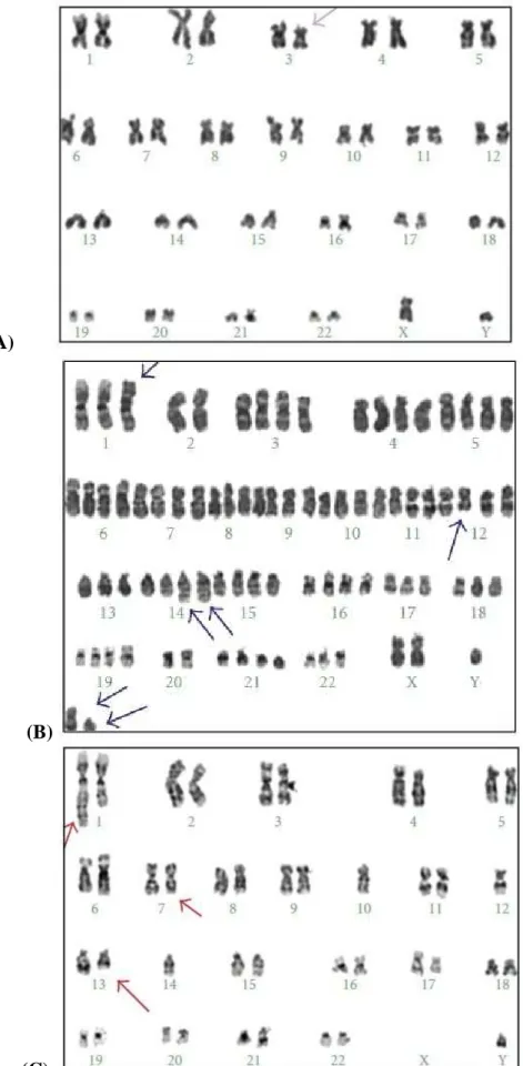
1.6. Clonal Evolution in CLL:
Clonal evolution of genomic aberrations has been documented in CLL. In a conventional cytogenetic analysis carried out by Oscier et al karyotypic evolution was seen in 18 out of 112 patients (16%), but there was no correlation between the incidence of clonal evolution and disease progression (Oscier et al, 1991). In two other chromosomes banding studies a significant association between the presence of ongoing karyotype changes and disease progression was seen (Fegan et al, 1995; Finn et al, 1998). In these investigations 6q and 11q deletions were the most commonly acquired secondary chromosome aberrations associated with a shorter progression-free survival. Using a molecular cytogenetic approach Leupolt et al performed a sequential interphase cytogenetic study applying FISH on 55 CLL patients over a median observation time period of 42 months (Leupolt et al, 2001). Clonal evolution was seen in nine out of 55 patients (16%) with 17p deletion (four cases), 6q deletion (three cases), 11q deletion (one case) and evolution from mono- to biallelic 13q deletion (three cases) being the acquired aberrations. Stilgenbauer et al found a significant association between the presence of clonal evolution and progressive disease. Only 20% of the patients with a stable karyotype have died compared to two-thirds of those exhibiting clonal evolution. In consideration of these studies the sensitive detection of genomic aberrations by interphase FISH provides a basis for a more accurate correlation of genomic aberrations with clinical features in CLL. Interphase FISH and molecular genetic techniques represent excellent tools for a better characterization of the critical genomic regions and have allowed the identification of candidate genes involved in pathogenesis or disease progression of CLL (Stilgenbauer et al, 2002).
Recently the clonal stability of malignant cells in CLL has been focused for intensive investigation. Several studies suggest that some patients with no detectable chromosomal abnormalities on initial analysis can acquire new abnormalities that may correlate with more aggressive disease behavior (Finn et al, 1998; Fegan et al, 2003). This raises a question of whether retrospective studies on the prognostic utility of FISH relate to cytogenetic abnormalities present at diagnosis or acquired during the course of the disease. Early studies using conventional cytogenetic analysis suggested such clonal evolution was rare (Nowell et al, 1988; Oscier et al, 1991), but subsequent studies suggested it may be more common than originally believed (Finn et al, 1988; Fegan et al, 2003).
Sequential chromosome banding analyses indicated the acquisition of genomic aberrations over time (clonal evolution) as an infrequent phenomenon in CLL (Han et al, 1986, Nowell et al, 1988, Juliusson et al, 1990, Oscier et al, 1991). However, only limited data are available
from FISH studies of interphase cells, a more sensitive method for the detection of genomic aberrations in CLL (Raghoebier et al, 1992; Escudier et al, 1993; Cuneo et al, 1994; Auer et al, 1999; Hjalmar et al, 2001; Chevallier et al, 2002; Byrd et al, 2006; Shanafelt et al, 2006; Grever et al, 2007). Auer et al. and Hjalmar et al. found the acquisition of trisomy 12 in none of 41 and 2 of 77 CLL cases, respectively (Auer et al, 1999; Hjalmar et al, 2001). Chevalier et al. observed additional genomic aberrations with an extended probe set in 13/31 (42%) CLL cases after a median time of 83 months (Chevallier et al, 2002). There was no association between clonal evolution and CD38 expression or disease progression but the acquisition of del(17p13) was associated with death in 7/11 cases.
More recent studies on CE detected by FISH in CLL and its relation to other prognostic markers such as CD38, ZAP-70 and IGHV gene mutation status have inconsistent results. Shanafelt et al. reported the increasing occurrence of CE after more than 5 years of observation (27 incidence, n= 63) and concluded that ZAP-70 positive patients may be more likely to experience CE (n=159; CE in 13/31 ZAP-70 positive v 3/29 ZAP-70 negative, p=0.008; statistically relevant association with CD38 positivity or unmutated IGHV gene not found) (Shanafelt et al, 2006). Stilgenbauer et al. observed CE only in CLL patients with unmutated IGHV genes (n=64, p=0.002; ZAP-70 and CD38 had not been examined) and presented CE as an independent adverse prognostic factor (Stilgenbauer et al, 2007).
In a study done by Cavazzini et al.; immunophenotypic studies, FISH and conventional karyotyping were used to define the clinicobiological significance of 14q32 translocations involving the immunoglobulin gene locus (14q32/IGH) in 252 chronic lymphocytic leukaemia (CLL) patients. The panel of probes included: 13q14, centromere 12, 6q21; 11q22/ ATM; 17p13/TP53, 14q32/IGH. Patients were classified as group 1 (favourable, i.e. 13q-single or normal), group 2 (intermediate risk, i.e. +12, 6q-, 1–2 anomalies), group 3 (unfavourable, i.e. 17p-, 11q-, complex karyotype), or group 4 (14q32/IGH translocation). They found that one hundred and twenty patients had a clonal chromosome aberration in the karyotype; 31 had a chromosome translocation, of which 15 had either complex karyotype and/or 11q- or 17p-. Half of the patients (n = 110) were allocated to the favourable risk group (group 1); 99 to the intermediate risk group (group 2); 25 to the unfavourable risk group (group 3) and 18 to the 14q32/IGH translocation group (group 4). Two patients with 14q32/IGH rearrangements were included in group 3 because they had 3 or more chromosome changes in the karyotype. The presence of an additional 17p- was noted at relapse after first line treatment in one patient included in group 4. 14q32 rearrangement did not occur as a secondary change in their series. IGH probe splitting occurred in 20– 99% of
the cells (median 70%) in 18 patients. The IGH translocation was the sole anomaly in eight patients; of the remaining 10 patients a concurrent 13q14 deletion was detected in nine cases and +12 in one case. Chromosome 14q32 translocation partners were identified in eight patients: in five cases BCL2/IGH rearrangement was detected; whereas fusions of IGH with BCL11A at 2p12 (63% of the cells), with CCND3 at 6p21 (71% of the cells) and CDK6 at 7q21 (30% of the cells) were seen in one case each.