Abstract - Farnesyltransferase inhibitors (FTIs)
are a class of oral anti-cancer drugs currently tested
in phase I-II clinical trials for treatment of
hematological malignancies. The in vitro effects of
various
FTIs
(alpha-hydroxyfarnesylphosphonic
acid, manumycin-A and SCH66336) were tested on
CD34
+KG1a cell line and in primary acute myeloid
leukemia (AML) cells from 64 patients. By cell
viability and clonogeneic methylcellulose assays,
FTIs showed a significant inhibitory activity in
CD34
+KG1a and primary bone marrow (BM)
leukemic cells from 56% of AML patients. FTIs also
induced activation of caspase-3 and Fas-independent
apoptosis, confirmed by the finding that inhibition of
caspase-8 was not associated with the rescue of
FTI-treated cells. We concluded that other cellular events
induced by FTIs may trigger activation of caspase-3
and subsequent apoptosis, but the expression of
proapoptotic molecules, as Bcl-2 and Bcl-XL, and
antiapoptotic, as Bcl-X(s), were not modified by
FTIs. By contrast, expression of inducible nitric
oxide synthase (iNOS) was increased in FTI-treated
AML cells. Our results suggest a very complex
mechanism of action of FTIs that require more
studies for a better clinical use of the drugs alone or
in combination in the treatment of hematological
malignancies.
Keywords: acute myeloid leukemia, farnesyltransferase inhibitors, apoptosis, nitric oxide
I. INTRODUCTION
In recent years, several target therapies have revolutionized the approach to various hematological malignancies [1-3]. Farnesyltransferase inhibitors (FTIs), a class of oral active anti-cancer drugs, can competitively inhibit the farnesyltransferase (FTase) enzyme [4-9]. FTIs have been tested in several myeloid hematological diseases, as acute and chronic myeloid leukemia (AML, CML), because of their overexpression of Ras, MAPK, AKT and other FTase-dependent proteins involved in proliferation, survival and apoptosis [10-21]. The Ras
proteins mediate signals from multiple cell surface receptors to the nucleus via different effector cascades [16-21]. Cytoplasmic Ras proteins need to be prenylated for fully biological functions. Farnesylation by the housekeeping enzyme FTase is the first required step in ras prenylation, enabling membrane anchor, ras activation and Ras-mediated activation of its downstream effectors [7-8,20-23]. FTase enzyme transfers a lipophilic farnesyl group from farnesyl diphosphate (FDP) to thiol group of cystein terminal portion of Ras, known as "CAAX- tetrapeptide motif", where "C" iscysteine, “A” is typically an aliphatic amino acid, and “X” is a serine or methionine as substrates of FTase. The other two additional steps of Ras prenylation are mediated by geranylgeranyl-transferase type I and type II enzymes (GGTase I and II) [7-8,19-23]. As other G proteins, also Ras activation is mediated by membrane guanine nucleotide exchange factors (GNEF), which allow Ras to cycle between an inactive guanosine diphosphate(GDP)- to an active guanosine triphosphate (GTP)-binding status [24]. Besides, the intrinsicGTPase activity of Ras can revert the molecule backto its GDP-inactive form. Mutations in N- and K-ras genes, documented in 15-30% of AML patients, can constitutively active Ras, which appears to be also involved in leukemogenic transformation by bcr/abl in CML [25-29].
In the last years,many FTIs have been identified, grouped into three main classes [30-31], and several of these have been evaluated preclinically. FDP analogues, as alpha-hydroxyfarnesyl phosphonic acid, beta-hydroxyphosphonic acid derivatives, PD 169451 or RPR 130401, can compete with FDP for the binding to FTase, but no specific antitumor effects have been described in vivo. CAAX competitive inhibitors, as L-778123, SCH66336 and R115777, are effective and selective FTase inhibitors in vitro and are currently testing in vivo. Bisubstrate inhibitors, as BMS 186511 and BMS-214662, combine the structural motifs of FPP and the CAAX tetrapeptide, linked by a phosphinyl or phosphonyl binding [30-31]. In particular, SCH66336 and R115777 oral compounds are testing in phase I and II clinical trials in haematological malignancies, including AML, CML, juvenile myelomonocytic leukemia (JMML), myelodysplastic syndromes (MDS) and multiple myeloma (MM), myelofibrosis with myeloid metaplasia and elderly high-risk AML [25-29,32-38]. These preclinical trials have been shown a Ras-independent FTI activity [30-31,39], probably because inhibition of Ras isoprenylation
In Vitro Apoptotic Effects of Farnesyltransferase blockade
in Acute Myeloid Leukemia Cells
V Giudice
1*, P Ricci
2*, L Marino
1, M Rocco
1, G Villani
1, M Langella
1, L Manente
1, E
Seneca
1, I Ferrara
1, L Pezzullo
1, B Serio
1, C Selleri
11Department of Medicine and Surgery, University of Salerno, Baronissi, Italy
2 Department of Clinical Medicine and Surgery, Federico II University of Naples, Naples, Italy
may be not necessary for FTI antineoplastic effects [40-41].
The in vitro cytotoxic effects of various FTIs have been previously described, including the NO-mediated apoptotic effects of SCH66336 in CML cells [42-43], but FTIs alone have shown only limited activity in AML and MDS patients [44-46]. The aim of the present study was to clarify the mechanism of action of FTIs, in order to explain the not enthusiastic results obtained in in vivo experiments. Consequently, we studied the in vitro effects of FTIs on apoptosis and growth of AML primary cells by blocking the downstream and upstream proteins of Ras-mediated apoptosis pathways.
II. METHODOLOGY Compounds blocking farnesyltransferase
FTIs tested were: a nonpeptidic tryciclic competitive FTI SCH66336 (SCH) (a gift from Schering-Plough Research Institute, Kenilworth, NJ); alpha-hydroxy-farnesylphosphonic acid (α-HFPA) and manumycin-A (Man-A) (Sigma-Aldrich, St Louis, MO). These FTI compounds were stored at –20°C as 10 mM stocks in dimethyl sulfoxide(DMSO, Sigma-Aldrich). Cell lines and AML patient samples collection
KG1a cells (American Type Culture Collection, Manassas, VA), were grownin RPMI 1640 supplemented with 10% heat-inactivated fetal calfserum (FCS), 2 mM L-glutamine, 100 U/mL penicillin, and 0.1 mg/mL streptomycin (complete medium, CM) at 37°C in 5% CO2.
Heparinized BM samples were obtained from 11 normal BM transplantation donors and during diagnostic procedures from 64 patients (median age, 52 years; range, 19-82; male/female, 28/36) with de novo (n=38) AML and AML with MDS-related features (n=26). Before sampling, informed consent was obtained according to the Declaration of Helsinki [47] and the procedures outlined by the ethicalcommittee of our institution. Bone marrow mononuclear cells (BMMNCs) were isolated by density gradient centrifugation. After washing with phosphate-bufferedsaline (PBS; Life Technologies, Carlsbad, USA), BMMNCs were resuspended in Iscove’s modified Dulbecco’s Medium (IMDM) supplemented with 10% heat-inactivated FCS. RPMI 1640, IMDM, PBS, FCS, L-glutamine, penicillin, streptomycin, and lymphocyte separation medium were purchased from Life Technologies (Gaithersburg, MD).
Suspension cultures
KG1a and primary AML cells were placed in 24-well plates in RPMI containing 0.1% FCS for 12 and 2 hours, respectively, before exposure to FTIs. For functional experiments, AML cells werepreincubated for 2 hours with the following reagents: caspase-3 inhibitor Z-DEVD-FMK, caspase-8 inhibitor IETD-FMK, (all used at 50 µM and purchasedfrom Alexis, San Diego, CA), Fas-receptor triggering inhibitor Fas:Fc (50 µM) (Alexis), iNOS inhibitor NG-monomethyl-arginine (500 µM)
(γ-MM-arg; Calbiochem, San Diego, CA). All experiments were repeated at least 3 times and each experimental condition was repeated at least in duplicate wells in each experiment. All the incubations were conducted at 37°C with 5% CO2.
Proliferation assay
In vitro sensitivity of KG1a and primary BM AML cells to FTIswas determined by plating 5 x 10⁵ cells
in RPMI 0.1% FCS and several dilutions of FTIs in 24-well plates. Controls were performed using identical concentrations of the solvent used for FTIs. After 48h incubation, cell viability was determined by MTS (3-(4,5- dimethylthiazole- 2yl)- 5- (3- carboxymethoxyphenyl)- 2- (4- sulfophenyl)- 2H-tetrazolium, inner salt) and PES (phenazine ethosulfate) assay (CellTiter 96 AQueous One
Solution Reagent) provided by Promega (Madison, WI, USA). Briefly, 20 l of CellTiter 96 AQueous One Solution
Reagent was added to each well and incubated for 4hours. The optical density (OD) was measured at 490 nm usinga microplate spectrophotometer (Titertek Multiscan, BioRad, Hercules, CA). FTI effects were measured as percentage of inhibitionof cell viability by the following equation: 1-[(ODtreated well/mean OD control wells) x 100], after background correctionusing blank wells. Each experiment was performed in triplicate. Values were expressed as mean value ± standard error of mean (SEM). Theresults were considered evaluable only if the control wells containedat least 70% of live AML cells after 48 h culture.
Hematopoietic colony assay
Short-term clonogenic progenitors were measured in methylcellulose (Stem Cell Technologies, Vancouver, CA). Isolated BMMNCs were plated in methylcellulose at 1 x 10³ cells/ml concentration (35 mm
dishes; 1 ml of medium/dish) in the presence of the following cocktail: 10 ng/mL IL-3, 50 ng/mL G-CSF, 50 ng/mL GM-CSF, 20 ng/mL SCF, and 2 U/mL EPO. Anti-Fas antibody (clone CH11, Amac) was used at concentrations ranging from 20-1000 U/ml to 1 µg/ml. Myeloid and erythroid colonies and clonogeneic aggregates of less than 50 cells, were counted as granulocyte-macrophage colony forming unit (CFU-GM), erythroid-burst forming unit (BFU-E) and clusters, respectively, after 14-day incubation. Each experiment was done in quadruplicate. Values were expressed as mean value ± standard error of mean (SEM).
Apoptosis assays
DNA fragmentation was measured after low molecular weight (LMW) DNA extraction from 2 x 10⁶
cells as previously described [48]. The high molecular weight DNA fraction was precipitated for 6 h in the presence of 5 mol/L NaCl and pelletted by high-speed centrifugation. The fragmented DNA was extracted, precipitated and electrophoresed as previously described [48].
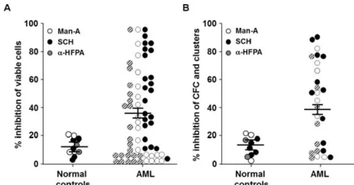
Figure 1. FTI-induced inhibition of AML cell viability and clonal growth.
Viability and cluster/colonies of AML cells were detected by MTT (A) and methylcellulose assays (B) measured by colony
forming cell (CFC) assays. Each dot (subject studied) represents the percentage of inhibition of cell viability and colony formation by each FTI at the IC50 described in the text
(control cultures were considered 100%). Horizontal bars are the mean values, and vertical bars are SEMs. Cumulative mean inhibition of cell viability and CFC ± SEM, as well as statistical
analysis, are reported in the corresponding results section. Abbreviations, see text.
Flow cytometric analysis of cellular DNA was performed using propidium iodide (PI) staining as previously described [48], adding 100 U/mL RNAse A (Boehringer Mannheim, Mannheim, Germany) during the incubation step. A minimum of 60,000 events was counted per sample. Apoptotic cell nuclei containing hypodiploid (fragmented) DNA were counted as a percentageof total population.
Fas-R/FasL flow cytometry analysis
Phycoerythrin (PE)-conjugated - CD34 (clone HPCA-1; Becton Dickinson [BD], San Jose, CA) was used to identify CD34+ cells. A fluorescein isothiocyanate
(FITC)-conjugated - fragment of a murine anti-human CD95 (clone UB2; Amac, Westbrook, ME) combined with a PE-conjugated - CD34 was used to determine the expression of Fas-R on CD34+ cells. For Fas-L
expression, BM cells were incubated with metalloproteinase inhibitor KB8301 (10 µmol/L, Pharmingen, San Diego, CA), PE-conjugated - CD34 (BD) combined with a purified mouse anti-human Fas-L (IgG1, Nok1; Pharmingen) for 1 hour at 4°C. Then cells were incubated with FITC-conjugated goat anti-mouse IgG secondary antibody for 30 minutes at 4°C. Proper isotypic controls were used in all experiments.
Detection of caspases 3 and 8 activities
Activation of intracellular caspases was performed by flow cytometry using fluorogenic caspase-specific substrates (DEVD-AFC for caspase-3, Alexis; and IETD-AFC for caspase-8, Pharmingen). Cells (1 x 10⁶) were treated with FTIs in the presenceor absence of
caspase-specific inhibitors for 24 hours, washed withPBS, and then resuspended in 50 µL of substrate buffer containing 10 mmol/L dithiothreitol (DTT) and 10 µL of the fluorogenic caspase-specific substrate supplemented with 5 µL of FCS. After centrifugation at 800g for 5 minutes, cells were incubated at 37°C for 60 minutes. After incubation, cells were washed and acquired. At least 10,000 events were analysed. Results were measured as fold increasein fluorescence relative to untreated control cells.
Reverse transcription-polymerase chain reaction (RT-PCR) for human iNOS RNA
Total RNA was extracted from BMMNCs in TRIzol (Invitrogen, Carlsbad, CA), as previously described [48]. Human iNOS cDNA, after reverse transcription using an oligo d(T)16 primer, was amplified using the following primer pairs: 5' CGGTGCTGTATTT
CCTTACGAGGCGAAGAAGG-3' and
5'-GGTGCTGCTTGTTAGGAGGTCAAGTAAAGGGC-3'. Human glyceraldehyde-3-phosphate dehydrogenase (GAPDH, 5’-TTCACCACCATGGAGAAGGCT-3’ and 5’-ACAGCCTTGGCAGCACCAGT-3’) was used as housekeeping gene. Reagents supplied in the Amplimer kit (Perkin Elmer, Foster City, CA) were used for the amplification reaction. PCR products were electrophoresed in 1.2% agarose gels and the bands were
visualized after staining with ethidium bromide and UV-light exposure.
Immunoblotting of iNOS, Bcl2, BclXL and p53
Treated AML cells were washed and lysed as previously described [48]. Concentrations were measured by colorimetric method (Biorad, Richmond, CO). A total of 100 µg of cell lysate, together with molecular weight markers (Amersham, Little Chalfont, UK) and iNOS positive mouse macrophages lysate (Transduction Laboratories, Lexington, KY) were fractionated by 7.5% SDS-polyacrylamide gel electrophoresis. PVDF membranes were prepared as previously described [48] and incubated with 2 µg/ml of mouse iNOS, anti-Bcl2, anti-p53, anti-BclXL and anti-BclXS antibodies (Transduction Laboratories, Lexington, KY) overnight at 4°C. The reaction was revealed by incubating filters with horseradish peroxidase-conjugated goat anti-rabbit antibodies (BioRad, Richmond, CO) and developed by ECL (Amersham, Little Chalfont, UK) according to manufacturer’s specifications.
Statistical analysis
Two-tailed Student t-test was used for flow cytometric analysis and tissue culture experiments. p values 0.05 were considered statistically significant.
III. RESULTS
FTI-induced growth inhibition of AML cells is partly related to apoptosis
We first investigated the effects of FTIs on growth of KG1a AML cell line. In cell viability tests performed in logarithmic growth phase, KG1a cell survival was inhibited by all three FTIs in a dose
dependent manner. By linear regression analysis, we determined the 50% inhibitory concentration (IC50) after 48 hour exposure to SCH, Man-A and α-HFPA. On an equimolar concentrations, SCH displayed more toxicity than Man-A (IC50 mean, 5 µM vs 50 µM, respectively; range, 1-20 µM vs 25-150 µM, respectively) and α-HFPA (IC50 mean, 100 µM; range, 75–150). The observed mean IC50 concentrations were used for next experiments.
The inhibitory effects of SCH, Man-A and α-HFPA were significantly strongerin primary AML cells than normal marrow cells (mean inhibition % ± SEM, 35±6 vs 13±5, respectively; p=0.03). We documented a FTI-mediated inhibition of cell viability >25% in 56% of AML patients (Fig. 1A). Similarly, by methylcellulose clonogeneic assays, colony and cluster growth inhibition >25% was observed in 64% of AML patients, whereas colony cells from normal BM cells were only weakly affected by FTIs exposure (mean inhibition % ± SEM, 47.7±7 vs 15.5±1, respectively; p<0.001) (Fig. 1B). By allele-specific PCR, the presence of oncogenic N-Ras and K-Ras mutations were also investigated in primary cells from 30 AML patients. Oncogenic mutations of N-Ras were detected in 27% of AML subjects; by contrast, we did not observe K-Ras mutations in any AML cell sample. Indeed, FTI-mediated inhibition of cell growth was observed in both AML with and without N-Ras mutations (data not shown).
To define whether the FTI-mediated growth inhibition of primary AML cells was associated to
apoptosis, BM AML cells sensitive to FTI inhibition were exposed for 48 h to FTIs at IC50. By LMW DNA fragmentation analysis, AML cells showed the characteristic DNA ladder suggestive of apoptosis (Fig. 2A). Flow cytometric detection of apoptotic hypodiploid DNA peak derived from treated BM AML cells confirmed the FTI-enhanced apoptotic cytotoxic effect (Fig. 2B). Indeed, percentages of apoptotic AML cells were significantly lower than those with cytolysis measured by cell viability assay (mean % ± SEM of cytolysis, 55.7±10; p=0.03). The parallel measurement of the number of apoptotic AML cells and viable cells from 10 AML patients carried by flow cytometry and cell viability assays showed that the percentages of apoptotic AML cells were significantly lower than those undergoing cytolysis (mean % ± SEM of apoptotic and viable AML cells, 32 ± 7 vs 51 ± 11, respectively; p = 0.03) (Fig. 2C). Caspase-3 and Fas-independent pathways are involved in FTI-mediated apoptosis of AML cells
FTIs could induce caspase-3 activation by flow cytometric measurement of MFI after cleavage of the specific fluorogenic substrate DEVD-AFC (mean percentage of positive cells after 6 and 24h culture, 25±4% and 38±4% vs 9±1% and 18±5%, FTI-treated and control AML cells, respectively; mean of 5 experiments). In addition, pre-incubation with caspase-3 inhibitor Z-DEVD-FMK allowed to partially abrogate FTI-mediated caspase activation (mean % ± SEM after 24 h exposure to
Figure 2. FTI-induced inhibition of AML cell proliferation is partly related to apoptosis.
Agarose gel stained with ethidium bromide after electrophoresis of low molecular weight DNA from 12 representative BM AML patients exposed for 48 h to FTIs at the IC50 (A). A representative flow cytometric detection of apoptic hypodiploid DNA
peak stained with PI from BM AML patient exposed to SCH (B). Percentages of viable cells and apoptotic cells simultaneously measured from 10 representative BM AML patients after exposure for 48 h to FTIs (C). Cumulative percentage of mean inhibition of cell viability and apoptotic cells, as well as statistical analysis, are reported in the corresponding results section.
FTI, 22±6; p=0.03) (Fig. 3A-B), and also to partially prevent FTI-mediated apoptosis (mean % of apoptosis ± SEM after FTI treatment, 36±4 vs 16.3±5, in absence and in presence of Z-DEVD-FMK respectively; p=0.01) (Fig. 3C).
Fas-R and Fas-L expression on AML cells exposed to FTIs was analysed by flow cytometry, but no
variations were observed (21±4% and 23±8% in the absence and presence of FTIs, respectively) (Fig. 4A). Moreover, FTI–sensitive and FTI-resistant AML cells displayed similar expression of Fas-R (mean % ± SEM, 39±4 vs 49±6, respectively; p=0.5) and Fas-L (mean % ± SEM, 49±4 vs 52±3, respectively; p=0.7) (Fig. 4B). No modifications of caspase-8 activity were detected in AML Figure 3. FTI-induced apoptosis in AML cells is mediated by activation of caspase-3.
FTI induced activation of caspase-3. Caspase-3 activity in BM AML cells from a representative patient, cultured for 24 h in medium alone (untreated) or treated with FTI or with FTI + caspase-3 inhibitor Z-DEVD-FMK (50 µM), were measured by flow cytometry using the fluorogenic caspase-3 specific substrate DEVD-AMC (A). Caspase-3 is activated after FTI exposure in AML
cells. Caspase-3 production in BM AML cells from a representative case, cultured for 6 and 24h in control medium (-FTI) or treated with FTI (+ FTI), was measured by flow cytometry using the cell-permeable FITC-labeled peptide FAM-VAD-FMK in combination with PI to differentiate dead from live cells (B). After 6 and 24 h of FTI exposure, 33% and 55% of live AML cells
(B, bottom right panels) are positive for FAM-VAD-FMK, respectively. (Panel C) FTI-mediated apoptosis of AML cells is partially reverted by caspase-3 inhibition. Flow cytometric detection of apoptosis from a representative AML patient who showed
in vitro susceptibility to FTI (SCH 1 µM) (C). Abbreviations, see text.
Figure 4. FasR/FasL pathway is not involved in FTI-induced apoptosis in AML
cells.
Flow cytometric analysis of FasR and FasL expression in CD34+ AML cells from a representative case, untreated and treated with FTI (A). FTI-mediated inhibition of AML cell viability does not correlate with FasR and FasL expression on
CD34+ AML cells. Cumulative mean ± SEM CD34+ Fas-R+ and C34+ Fas-L+ are reported (B).
Caspase–8 is not activated by FTIs. AML cells untreated and treated with FTI after 48 h of culture.
(C). AML cells were grown for 48 h with FTIs at IC50 and with FTIs + Fas-receptor triggering inhibitor Fas:Fc or caspase-8 inhibitor IETD-FMK
(all used at 50 µM). AML cell viability was measured by colorimetric MTT assay. Columns represent cumulative mean ± SEM of cell viability
cells after 48 h FTIs exposure by flow cytometry and intracellular caspase staining (Fig. 4C). To further exclude the involvement of Fas-R/Fas-L system in FTI-mediated apoptosis, prior to exposure to FTIs, AML cells were preincubated for 2 h with selective Fas-R/Fas-L inhibitors. As expected, no variations were observed in FTI-mediated inhibition of cell growth and apoptosis after pretreatment with Fas-receptor antagonist and caspase-8 inhibitor (mean % ± SEM of cell growth after FTI exposure, 41.6±3 and 43.3±3 without and with Fas:Fc vs 38±2 and 39±1 without and with IETD-FMK vs 42.3±3 and 46.6±3 without and with ZYVAD-FMK, respectively) (Fig. 4D). FTIs trigger iNOS expression in AML cells
Unstimulated primary total AML BM cells expressed iNOS mRNA by PCR detection. Because iNOS expression in total BM AML cells might be related to non-leukemic cells, immature CD34+ KG1a cells were
tested for the expression of iNOS mRNA after 48 h culture. A stronger amplification of iNOS was detected, and also after FTIs stimulation (Fig. 5A). Similarly, immunoblot of cell lysates showed lower levels of iNOS at baseline, and a 10-fold increased after FTIs stimulation in both primary total BM AML cells and KG1a cells (Fig. 5B). Usingthe cell-permeable fluorescent indicator DAF-2 DA, NO levelsincreased 40% after FTI exposure from
basal levels detected in AML BM cells cultured in control medium without FTI (p<0.001), and γ -MM-arg partially blocked FTI-mediated NO production in CML cells (Fig 5C). Inhibition of NO synthesis by pretreatment of AML cells with γ-MM-arg (500 µM), a competitive inhibitor of iNOS, did not affect the inhibitory effect on cell growth and apoptosis of FTIs (data not shown). However, when FTIs were used at IC10, γ-MM-arg partially prevented FTI effects (mean % ± SEM cell growth and apoptosis after FTI exposure, 31±2 and 21±4 vs 46±1 and 31±3 in absence and in presence of γ- MM-arg, respectively; p=0.01 and p=0.03) (Fig. 5C).
FTI exposure may modify p53 but not Bcl-2 pathway in AML cells
To explore whether FTI-induced apoptosis could be mediated by decreased expression of anti-apoptotic 2 and X(L) or increasing in proapoptotic Bcl-X(S) proteins, AML cells were exposed for 48 hours to FTIs or control medium and Bcl-2 or Bcl-X(L)/(S) were measured. Quantification of protein banding by densitometry did not document changes in Bcl-2 protein expression in KG1a as well as in four primary BM AML samples after FTI exposure as compared to control cultures (Fig. 6A). Similarly, the expression of Bcl-X(L)
and Bcl-X(S) did not show variations after FTI exposure
Figure 5. FTI-induced inhibition of CML cell viability is mediated by NO pathway.
FTIs induce iNOS mRNA signal amplification in KG1a and AML cells cultured for 48h. Ethidium bromide RT-PCR products (iNOS and GAPDH) are shown (A). FTIs cause increased expression of iNOS protein in KG1a cell line and 4 AML patients.
Actin is used as control (B). γ-MM-arg partially preventes FTI-mediated apoptosis. Flow cytometric detection of apoptic hypodiploid DNA peak stained with PI from a CML patient treated with FTI (C).
(Fig. 5B). By contrast, in KG1a cells and few AML cases, an enhancement of p53 expression after FTI exposure was detected (Fig. 6C).
IV. DISCUSSION
Despite the improvements in achieving prolonged remission for adult AML after cytotoxic induction chemotherapy or BM transplantation, the overall survival remains unsatisfactory, particularly for older patients [1-3]. For this reason, new treatment approaches are required to improve the outcome in these patients. The upregulation of the classic mitogenic Ras/Raf/MAPK cascade has been documented in more than 30% of AML cases, due to mutations, overexpression or amplification of other oncogenic proteins with tyrosine kinase activity that may influence Ras pathways [10-21]. When farnesylation of Ras is blocked by FTIs in vitro, Ras is unable to anchor to cell membrane and its function is impaired (Fig. 7A-B) [16-21]. Various FTIs have already been evaluated in phase I-II and III clinical trials for the
treatment of hematological malignancies, but contrasting results were reported, even though when associated to cytotoxic agents [25-29,32-38].
Here, the in vitro effects of FTIs were studied in AML cells. We have shown that FTIs showed a significant inhibitory activity on cell viability in CD34+
KG1a cell line and primary BM cells from 56% of AML patients. Indeed, only half of our AML patients showed increased sensitivity of cluster and colony growth after FTI exposure compared to normal marrow progenitor cells. Furthermore, the lack of response in the remaining half of AML patients could be related to the presence of mutations in FTase or other markers of resistance to FTIs, as p53 mutations, but more studies are needed to confirm this hypothesis [49-51].
Preliminary data have excluded that FTI-mediated inhibition of cell growth was Ras-mutations dependent, as FTIs induced inhibition of viability in AML cells with and without N-Ras mutations (Selleri et al., personal communication). As previously reported for CML cells, FTI-mediated cytotoxic effects in AML cells were partially related to enhanced apoptosis [48]. Although it has been reported that Ras-transformed cells showed Fas-R upregulation and higher Fas-related apoptosis after FTI-exposure [52-53], we documented a Fas-independent FTI-mediated apoptosis in AML cells. Moreover, FTI-mediated apoptosis seemed to be caspase-3 dependent but caspase-8 independent as inhibition of caspase-8 was not associated with the rescue of FTI-treated cells. These findings suggest that other cellular events induced by FTIs may trigger caspase-3 activation and subsequent apoptosis in AML cells. As the main event for caspase-3 activation is the release of cytochrome c from mitochondria, the expression of potential molecules modulating apoptosis via mitochondrial pathways were studied [54-55]. Expression of proapoptotic Bcl-2 and Bcl-X(L) and antiapoptotic Bcl-X(S) proteins were not modified by FTIs, except for the involvement of Bcl-2 pathway in FTI-induced apoptosis in human AMLcells.
Another known mechanism that can concur in the inhibition of tumor growth is the macrophage-mediated NO release in the site of inflammation or in tumor environment, causing mitochondrial release of cytochrome c and apoptosis. In addition, it has been described that Ras inhibitors can increase NO-induced cell death [56-62]. In a similar manner, FTIs can induce apoptosis in CML cells, through cytochrome c release and caspase-3 activation, as previously documented [59,62]. As expected, FTIs also can induce iNOS expression in AML cells. Moreover, inhibition of NO synthesis partially abrogated the effects of FTIs on apoptosis suggesting that iNOS cascade may be only one of the possible mechanisms of FTI-mediated apoptosis in AML cells (Fig. 7 C-D). The enhancement of p53 expression in KG1a cell line and some AML cases suggested a complex mechanism of action of FTIs. Indeed, Moasser et al. has already reported a more susceptibility to FTIs in p53 wild-type breast cancer cell lines [63].
Figure 6.
FTI exposure may modify p53 but not Bcl-2 pathway in AML cells
Immunoblotting of Bcl2, BclXL and BclXs from KG1a cells and total marrow cells of four AML patients cultured for 48 h in absence or in presence of FTIs, excludes the involvement of
Bcl-2 pathway in FTI-induced apoptosis in human AML cells (A-B). Immunoblotting of p53 from KG1a cells and total marrow cells of four AML patients cultured for 48 h in absence
or in presence of FTIs, shows the enhancement of p53, as quantified by densitometry scanning, in 2 out of 4 AML cases
Because caspases, iNOS and p53 play an important role in several intracellular pathways and can be triggered by several different extracellular signals, it is hard to clearly understand the FTIs mechanisms, also due to their aspecific action [54-62]. Several pathways are deregulated in cancers, as JAK/STATs or Syk/Btk, allowing proliferation and survival of tumor cells [64-67]. Indeed, it has been reported that over-activity of growth factor signaling pathways in breast cancer did not correlate with sensitivity to FTIs [63]. On the other hand, FTIs have shown contradictory results in MDS an AML patients, particularly in elderly and poor-risk AML [68-72], but synergized actions were reported when FTIs were combined to tyrosine kinase inhibitors (TKIs) or other drugs, as simvastatin [73-77]. These findings suggest that FTIs can induce growth inhibition not only through apoptosis, but also interfering with proliferation pathways, enhanced when TKIs are used. Besides, the efficacy of ruxolitinib, a JAK1/2 inhibitor, or ibrutinib, a Btk inhibitor, has been described in several hematological malignancies, and more studies are ongoing [78-79]. For these reasons, to improve the therapeutic potential of FTIs alone or in combination with standard chemotherapy or new targeting therapies, it is important to better understand their mechanisms of action, as to investigate how they could impact on other pathways or vice versa.
V. CONCLUSION
As already reported, in vitro FTIs effects on cell viability and apoptosis are caspase-3 and iNOS dependent in AML cells, and Fas-R/Fas-L or Bcl2 independent. But the involvement of p53 suggests a more complex mechanism in the susceptibility of FTI-mediated apoptosis in AML cells. It is also possible that FTIs may have only additive anti-neoplastic effects, requiring the combination with other anti-cancer agents to be effective in AML therapy. Our data open new questions about FTIs mechanism of action and their use in myeloid malignancies as single agents or in combination, in order to improve the outcome of hematological patients who cannot be treated with more aggressive treatments.
ACKNOWLEDGMENT
*These authors have contributed equally to this work. This study was supported in part by grants from the Ministero dell’Università e della Ricerca Scientifica e Tecnologica (MURST).
REFERENCES
[1] Jabbour E, Cortes J, Ravandi F, O'Brien S, Kantarjian H. Targeted therapies in hematology and their impact on Figure 7. Models for FTI-mediated Ras inhibition (A-B) and NO-induced apoptosis (C-D) in AML.
patient care: chronic and acute myeloid leukemia. Semin Hematol. 2013;50(4):271-283.
[2] Bunting KD, Qu CK, Tomasson MH. Molecular-Targeted Therapies for Hematologic Malignancies. Adv Hematol. 2012;2012:606423.
[3] Podhorecka M, Markowicz J, Szymczyk A, Pawlowski J. Target Therapy in Hematological Malignances: New Monoclonal Antibodies. Int Sch Res Notices. 2014;2014:701493.
[4] Cox AD, Der CJ. Farnesyltransferase inhibitors and cancer treatment: targeting simply Ras? Biochim Biophys Acta. 1997;1333:51-71.
[5] Pan J, Yeung SC. Recent advances in understanding the antineoplastic mechanisms of farnesyltransferase inhibitors. Cancer Res. 2005;65(20):9109-9112.
[6] Oliff A. Farnesyltransferase inhibitors: targeting the molecular basis of cancer. Biochim Biophys Acta. 1999;1423:19-30.
[7] Sebti SM, Hamilton AD. Farnesyltransferase and geranylgeranyltransferase I inhibitors and cancer therapy: lessons from mechanism and bench-to-bedside translational studies. Oncogene. 2000;19:6584-6593. [8] Houssin R, Pommery J, Salaün MC, Deweer S, Goossens JF, Chavatte P, Hénichart JP. Design, synthesis, and pharmacological evaluation of new farnesyl protein transferase inhibitors. J Med Chem. 2002;45(2):533-536. [9] Prendergast GC. Farnesyltransferase inhibitors: antineoplastic mechanism and clinical prospects. Curr Opin Cell Biol. 2000;12:166-173.
[10] Majewski M, Nieborowska-Skorska M, Salomoni P, Slupianek A, Reiss K, Trotta R, Calabretta B, Skorski T. Activation of mitochondrial Raf-1 is involved in the antiapoptotic effects of Akt. Cancer Res. 1999;59:2815-2819.
[11] Di Cristofano A, Niki M, Zhao M, Karnell FG, Clarkson B, Pear WS, Van Aelst L, Pandolfi PP. p62(dok), a negative regulator of Ras and mitogen-activated protein kinase (MAPK) activity, opposes leukemogenesis by p210(bcr-abl). J Exp Med. 2001;194:275-284.
[12] Ding H, McDonald JS, Yun S, Schneider PA, Peterson KL, Flatten KS, Loegering DA, Oberg AL, Riska SM, Huang S, Sinicrope FA, Adjei AA, Karp JE, Meng XW, Kaufmann SH. Farnesyltransferase inhibitor tipifarnib inhibits Rheb prenylation and stabilizes Bax in acute myelogenous leukemia cells. Haematologica. 2014;99(1):60-69.
[13] Pellicano F, Simara P, Sinclair A, Helgason GV, Copland M, Grant S, Holyoake TL. The MEK inhibitor PD184352 enhances BMS-214662-induced apoptosis in CD34+ CML stem/progenitor cells. Leukemia. 2011;25(7):1159-1167.
[14] Gómez-Benito M, Marzo I, Anel A, Naval J. Farnesyltransferase inhibitor BMS-214662 induces apoptosis in myeloma cells through PUMA up-regulation, Bax and Bak activation, and Mcl-1 elimination. Mol Pharmacol. 2005;67(6):1991-1998.
[15] Dai Y, Rahmani M, Pei XY, Khanna P, Han SI, Mitchell C, Dent P, Grant S. Farnesyltransferase inhibitors
interact synergistically with the Chk1 inhibitor UCN-01 to induce apoptosis in human leukemia cells through interruption of both Akt and MEK/ERK pathways and activation of SEK1/JNK. Blood. 2005;105(4):1706-1716. [16] Reuter CW, Morgan MA, Bergmann L. Targeting the Ras signaling pathway: a rational, mechanism-based treatment for hematologic malignancies? Blood. 2000;96:1655-1669.
[17] Rowinsky EK, Windle JJ, Von Hoff DD. Ras protein farnesyltransferase: A strategic target for anticancer therapeutic development. J Clin Oncol. 1999;17:3631-3652.
[18] Adjei AA. Blocking oncogenic Ras signaling for cancer therapy. J Natl Cancer Inst. 2001;93:1062-1074. [19] Shannon K. The Ras signaling pathway and the molecular basis of myeloid leukemogenesis. Curr Opin Hematol. 1995;2:305-308.
[20] Campbell SL, Khosravi-Far R, Rossman KL, Clark GJ, Der CJ. Increasing complexity of Ras signaling. Oncogene. 1998;17:1395-1413.
[21] Reuther GW, Der CJ. The Ras branch of small GTPases: Ras family members don't fall far from the tree. Curr. Opin Cell Biol. 2000;12:157-165.
[22] Karp JE, Lancet JE, Kaufmann SH, End DW, Wright JJ, Bol K, Horak I, Tidwell ML, Liesveld J, Kottke TJ, Ange D, Buddharaju L, Gojo I, Highsmith WE, Belly RT, Hohl RJ, Rybak ME, Thibault A, Rosenblatt J. Clinical and biologic activity of the farnesyltransferase inhibitor R115777 in adults with refractory and relapsed acute leukemias: a phase 1 clinical-laboratory correlative trial. Blood. 2001;97:3361-3369.
[23] Gelb MH. Protein prenylation, et cetera: signal transduction in two dimensions. Science. 1997;275(5307):1750-1751.
[24] Karp JE. Farnesyltransferase inhibitor as targeted therapies for hematological malignancies. Semin Hematol. 2001;38(suppl. 7):16-23.
[25] Cortes J, Albitar M, Thomas D, Giles F, Kurzrock R, Thibault A, Rackoff W, Koller C, O'Brien S, Garcia-Manero G, Talpaz M, Kantarjian H. Efficacy of the farnesyl transferase inhibitor R115777 in chronic myeloid leukemia and other hematologic malignancies. Blood. 2003;101(5):1692-1697.
[26] Pellicano F, Copland M, Jorgensen HG, Mountford J, Leber B, Holyoake TL. BMS-214662 induces mitochondrial apoptosis in chronic myeloid leukemia (CML) stem/progenitor cells, including CD34+38- cells, through activation of protein kinase Cbeta. Blood. 2009;114(19):4186-4196.
[27] Reichert A, Heisterkamp N, Daley GQ, Groffen J. Treatment of Bcr/Abl-positive acute lymphoblastic leukemia in P190 transgenic mice with the farnesyl transferase inhibitor SCH66336. Blood. 2001;97:1399-1403.
[28] Keating A. Chronic myeloid leukemia: current therapies and the potential role of farnesyltransferase inhibitors. Semin Hematol. 2002;39(suppl. 2):11-17. [29] Borthakur G, Kantarjian H, Daley G, Talpaz M, O'Brien S, Garcia-Manero G, Giles F, Faderl S, Sugrue M,
Cortes J. Pilot study of lonafarnib, a farnesyl transferase inhibitor, in patients with chronic myeloid leukemia in the chronic or accelerated phase that is resistant or refractory to imatinib therapy. Cancer. 2006;106(2):346-352. [30] Byrne JL, Marshall CJ. The molecular pathophysiology of myeloid leukaemias: Ras revisited. Br J Haematol. 1998;100(2):256-264.
[31] Prendergast GC, Du W. Targeting farnesyltransferase: is Ras relevant? Drug Resist Updat. 1999;2(2):81-84.
[32] Frassanito MA, Cusmai A, Piccoli C, Dammacco F. Manumycin inhibits farnesyltransferase and induces apoptosis of drug-resistant interleukin 6-producing myeloma cells. Br J Haematol. 2002;118:157-165. [33] Stieglitz E, Ward AF, Gerbing RB, Alonzo TA, Arceci RJ, Liu YL, Emanuel PD, Widemann BC, Cheng JW, Jayaprakash N, Balis FM, Castleberry RP, Bunin NJ, Loh ML, Cooper TM. Phase II/III trial of a pre-transplant farnesyl transferase inhibitor in juvenile myelomonocytic leukemia: a report from the Children's Oncology Group. Pediatr Blood Cancer. 2015;62(4):629-636.
[34] Lancet JE, Duong VH, Winton EF, Stuart RK, Burton M, Zhang S, Cubitt C, Blaskovich MA, Wright JJ, Sebti S, Sullivan DM. A phase I clinical-pharmacodynamic study of the farnesyltransferase inhibitor tipifarnib in combination with the proteasome inhibitor bortezomib in advanced acute leukemias. Clin Cancer Res. 2011;17(5):1140-1146.
[35] Jabbour E, Kantarjian H, Ravandi F, Garcia-Manero G, Estrov Z, Verstovsek S, O'Brien S, Faderl S, Thomas DA, Wright JJ, Cortes J. A phase 1-2 study of a farnesyltransferase inhibitor, tipifarnib, combined with idarubicin and cytarabine for patients with newly diagnosed acute myeloid leukemia and high-risk myelodysplastic syndrome. Cancer. 2011;117(6):1236-1244.
[36] Epling-Burnette PK, Loughran TP Jr. Suppression of farnesyltransferase activity in acute myeloid leukemia and myelodysplastic syndrome: current understanding and recommended use of tipifarnib. Expert Opin Investig Drugs. 2010;19(5):689-698.
[37] Ravoet C, Mineur P, Robin V, Debusscher L, Bosly A, André M, El Housni H, Soree A, Bron D, Martiat P. Farnesyl transferase inhibitor (lonafarnib) in patients with myelodysplastic syndrome or secondary acute myeloid leukaemia: a phase II study. Ann Hematol. 2008;87(11):881-885.
[38] Jabbour E, Kantarjian H, Cortes J. Clinical activity of tipifarnib in hematologic malignancies. Expert Opin Investig Drugs. 2007;16(3):381-392.
[39] Du W, Lebowitz PF, Prendergast GC. Cell growth inhibition by farnesyltransferase inhibitors is mediated by gain of geranylgeranylated RhoB. Mol Cell Biol. 1999;19(3):1831-1840.
[40] Zhang B, Prendergast GC, Fenton RG. Farnesyltransferase inhibitors reverse Ras-mediated inhibition of Fas gene expression. Cancer Research. 2002;62:452-458.
[41] Liu Ax, Du W, Liu JP, Jessell TM, Prendergast GC. RhoB alteration is necessary for apoptotic and antineoplastic responses to farnesyltransferase inhibitors. Mol Cell Biol. 2000;20(16):6105-6113.
[42] Liu Ax, Cerniglia GJ, Bernhard EJ, Prendergast GC. RhoB is required to mediate apoptosis in neoplastically transformed cells after DNA damage. Proc Natl Acad Sci. USA. 2001;98(11):6192-6197.
[43] Sawyers CL, McLaughlin J, Witte ON. Genetic requirement for Ras in the transformation of fibroblasts and hematopoietic cells by the Bcr-Abl oncogene. J Exp Med. 1995;181(1):307-313.
[44] Faderl S, Talpaz M, Estrov Z, O'Brien S, Kurzrock R, Kantarjian HM. The biology of chronic myeloid leukemia. N Engl J Med. 1999;341(3):164-172.
[45] Zou X, Calame K. Signaling pathways activated by oncogenic forms of Abl tyrosine kinase. J Biol Chem. 1999;274:18141-18144.
[46] Skorski T, Wlodarski P, Daheron L, Salomoni P, Nieborowska-Skorska M, Majewski M, Wasik M, Calabretta B. BCR/ABL-mediated leukemogenesis requires the activity of the small GTP-binding protein Rac.Proc Natl Acad Sci. USA 1998;95:11858-11862. [47] World Medical Association. World Medical Association Declaration of Helsinki: ethical principles for medical research involving human subjects. JAMA. 2013;310(20):2191-2194.
[48] Selleri C, Maciejewski JP, Montuori N, Ricci P, Visconte V, Serio B, Luciano L, Rotoli B. Involvement of nitric oxide in farnesyltransferase inhibitor-mediated apoptosis in chronic myeloid leukemia cells. Blood. 2003;102(4):1490-1498.
[49] Adjei AA, Davis JN, Erlichman C, Svingen PA, Kaufmann SH. Comparison of potential markers of farnesyltransferase inhibition. Clin Cancer Res. 2000;6:2318-2325.
[50] Raz T, Nardi V, Azam M, Cortes J, George Q. Daley GQ. Farnesyl transferase inhibitor resistance probed by target mutagenesis. Blood. 2007;110(6):2102–2109. [51] Zhang B, Groffen J, Heisterkamp N. Resistance to farnesyltransferase inhibitors in Bcr/Abl-positive lymphoblastic leukemia by increased expression of a novel ABC transporter homolog ATP11a. Blood. 2005;106(4):1355–1361.
[52] Selleri C, Maciejewski JP, Pane F, Luciano L, Raiola AM, Mostarda I, Salvatore F, Rotoli B. Fas-mediated modulation of Bcr/Abl in chronic myelogenous leukemia results in differential effects on apoptosis. Blood. 1998;92:981-989.
[53] Selleri C, Sato T, Del Vecchio L, Luciano L, Barrett AJ, Rotoli B, Young NS, Maciejewski JP. Involvement of Fas-mediated apoptosis in the inhibitory effects of interferon-alpha in chronic myelogenous leukemia. Blood. 1997;89:957-964.
[54] Tsujimoto Y.Bcl-2 family of proteins: life-or-death switch in mitochondria. Biosci Rep. 2002;22:47-58. [55] Sanchez-Garcia I, Grutz G. Tumorigenic activity of the BCR-ABL oncogenes is mediated by BCL2. Proc Natl Acad Sci. USA. 1995;92:5287-5291.
[56] Boyd CS, Cadenas E. Nitric oxide and cell signaling pathways in mitochondrial-dependent apoptosis. Biol Chem. 2002;383:411-423.
[57] Arnoult D, Parone P, Martinou JC, Antonsson B, Estaquier J, Ameisen JC. Mitochondrial release of apoptosis-inducing factor occurs downstream of cytochrome c release in response to several proapoptotic stimuli. J Cell Biol. 2002;159:923-929.
[58] Selleri C, Maciejewski JP. Nitric oxide and cell survival: megakaryocytes say "NO". J Lab Clin Med. 2001;137:225-230.
[59] Pervin S, Singh R, Gau CL, Edamatsu H, Tamanoi F, Chaudhuri G. Potentiation of nitric oxide-induced apoptosis of MDA-MB-468 cells by farnesyltransferase inhibitor: implications in breast cancer. Cancer Res. 2001;61:4701-4706.
[60] Maciejewski JP, Selleri C, Sato T, Cho HJ, Keefer LK, Nathan CF, Young NS. Nitric oxide suppression of human hematopoiesis in vitro. Contribution to inhibitory action of interferon-gamma and tumor necrosis factor-alpha. J Clin Invest. 1995;96:1085-1092.
[61] Kolb JP. Mechanisms involved in the pro- and anti-apoptotic role of NO in human leukemia. Leukemia. 2000;14:1685-1694.
[62] Suzuki N, Urano J, Tamanoi F. Farnesyltransferase inhibitors induce cytochrome c release and caspase 3 activation preferentially in transformed cells. Proc. Natl. Acad. Sci. USA 1998;95:15356-15361.
[63] Moasser MM, Rosen N. The use of molecular markers in farnesyltransferase inhibitor (FTI) therapy of breast cancer. Breast Cancer Res Treat. 2002;73(2):135-144.
[64] Sonoyama J, Matsumura I, Ezoe S, Satoh Y, Zhang X, Kataoka Y, Takai E, Mizuki M, Machii T, Wakao H, Kanakura Y.Functional cooperation among Ras, STAT5, and phosphatidylinositol 3-kinase is required for full oncogenic activities of BCR/ABL in K562 cells. J Biol Chem. 2002;277:8076-8082.
[65] Hoover RR, Gerlach MJ, Koh EY, Daley GQ. Cooperative and redundant effects of STAT5 and Ras signaling in BCR/ABL transformed hematopoietic cells. Oncogene. 2001;20:5826-5835.
[66] Cummings M, Siitonen T, Higginbottom K, Newland AC, Allen PD. p53-mediated downregulation of Chk1 abrogates the DNA damage-induced G2M checkpoint in K562 cells, resulting in increased apoptosis. Br J Haematol. 2002;116:421-428.
[67] Hendriks RW, Yuvaraj S, Kil LP. Targeting Bruton's tyrosine kinase in B cell malignancies. Nat Rev Cancer. 2014;14(4):219-232.
[68] Erba HP, Othus M, Walter RB, Kirschbaum MH, Tallman MS, Larson RA, Slovak ML, Kopecky KJ, Gundacker HM, Appelbaum FR. Four different regimens of farnesyltransferase inhibitor tipifarnib in older, untreated acute myeloid leukemia patients: North American Intergroup Phase II study SWOG S0432. Leuk Res. 2014;38(3):329-333.
[69] Burnett AK, Russell NH, Culligan D, Cavanagh J, Kell J, Wheatley K, Virchis A, Hills RK, Milligan D;
AML Working Group of the UK National Cancer Research Institute. The addition of the farnesyl transferase inhibitor, tipifarnib, to low dose cytarabine does not improve outcome for older patients with AML. Br J Haematol. 2012;158(4):519-522.
[70] Karp JE, Flatten K, Feldman EJ, Greer JM, Loegering DA, Ricklis RM, Morris LE, Ritchie E, Smith BD, Ironside V, Talbott T, Roboz G, Le SB, Meng XW, Schneider PA, Dai NT, Adjei AA, Gore SD, Levis MJ, Wright JJ, Garrett-Mayer E, Kaufmann SH. Active oral regimen for elderly adults with newly diagnosed acute myelogenous leukemia: a preclinical and phase 1 trial of the farnesyltransferase inhibitor tipifarnib (R115777, Zarnestra) combined with etoposide. Blood. 2009;113(20):4841-4852.
[71] Harousseau JL, Lancet JE, Reiffers J, Lowenberg B, Thomas X, Huguet F, Fenaux P, Zhang S, Rackoff W, De Porre P, Stone R; Farnesyltransferase Inhibition Global Human Trials (FIGHT) Acute Myeloid Leukemia Study Group. A phase 2 study of the oral farnesyltransferase inhibitor tipifarnib in patients with refractory or relapsed acute myeloid leukemia. Blood. 2007;109(12):5151-5156. [72] Lancet JE, Gojo I, Gotlib J, Feldman EJ, Greer J, Liesveld JL, Bruzek LM, Morris L, Park Y, Adjei AA, Kaufmann SH, Garrett-Mayer E, Greenberg PL, Wright JJ, Karp JE. A phase 2 study of the farnesyltransferase inhibitor tipifarnib in poor-risk and elderly patients with previously untreated acute myelogenous leukemia. Blood. 2007;109(4):1387-1394.
[73] Edamatsu H, Gau CL, Nemoto T, Guo L, Tamanoi F. Cdk inhibitors, roscovitine and olomoucine, synergize with farnesyltransferase inhibitor (FTI) to induce efficient apoptosis of human cancer cell lines. Oncogene. 2000; 19:3059-3068.
[74] van der Weide K, de Jonge-Peeters SD, Kuipers F, de Vries EG, Vellenga E. Combining simvastatin with the farnesyltransferase inhibitor tipifarnib results in an enhanced cytotoxic effect in a subset of primary CD34+ acute myeloid leukemia samples. Clin Cancer Res. 2009;15(9):3076-3083.
[75] Copland M, Pellicano F, Richmond L, Allan EK, Hamilton A, Lee FY, Weinmann R, Holyoake TL. BMS-214662 potently induces apoptosis of chronic myeloid leukemia stem and progenitor cells and synergizes with tyrosine kinase inhibitors. Blood. 2008;111(5):2843-2853. [76] Radujkovic A, Topaly J, Fruehauf S, Zeller WJ. Combination treatment of imatinib-sensitive and -resistant BCR-ABL-positive CML cells with imatinib and farnesyltransferase inhibitors. Anticancer Res. 2006;26(3A):2169-2177.
[77] Miyoshi T, Nagai T, Ohmine K, Nakamura M, Kano Y, Muroi K, Komatsu N, Ozawa K. Relative importance of apoptosis and cell cycle blockage in the synergistic effect of combined R115777 and imatinib treatment in BCR/ABL-positive cell lines. Biochem Pharmacol. 2005;69(11):1585-1594.
[78] Roskoski R Jr. Janus kinase (JAK) inhibitors in the treatment of inflammatory and neoplastic diseases. Pharmacol Res. 2016;111:784-803.
[79] Routledge DJ, Bloor AJ. Recent advances in therapy of chronic lymphocytic leukaemia. Br J Haematol. 2016;174(3):351-367.