Journal section: Oral Surgery Publication Types: Case Report
Large-sized pleomorphic adenoma of the cheek treated with
Nd:Yag laser: report of a case and review of the literature
Federica Veneri 1, Marco Meleti 2, Luigi Corcione 3, Elena Bardellini 4, Alessandra Majorana 5, Paolo Vescovi 6 1 DDS. Postgraduate student of the Pediatric Dentistry Program - Department of Medical and Surgical Specialties, Radiological
Sciences and Public Health, Dental School, University of Brescia, Italy
2 DDS, PhD. Researcher in Oral Medicine and Surgery – Centro Universitario di Odontoiatria – Department of Medicine and
Sur-gery – University of Parma, Italy
3 MD. Department of Human Histopathology, Academic Hospital of Parma, Italy
4 DDS, PhD. Associate Professor - Department of Medical and Surgical Specialties, Radiological Sciences and Public Health,
Dental School, University of Brescia, Italy
5 MD, DDS. Full Professor – Director of the Postgraduate Pediatric Dentistry Program - Department of Medical and Surgical
Spe-cialties, Radiological Sciences and Public Health, Dental School, University of Brescia, Italy
6 DDS, PhD. Associate Professor in Oral Medicine and Surgery - Centro Universitario di Odontoiatria - Department of Medicine
and Surgery, University of Parma, Italy
Correspondence:
Piazzale Spedali Civili 1, 25123, Brescia, Italy [email protected]
Received: 09/09/2019 Accepted: 28/05/2020
Abstract
Pleomorphic adenoma (PA) mostly involves parotid glands, while extra-parotid localizations are relatively uncom-mon. Particularly, PAs of the cheek minor salivary glands with a size larger than 4 cm are exceedingly rare, with only few cases reported. Surgical treatment of PA usually consists in radical excision. However, despite a presump-tive radicality, recurrences, sometimes followed by malignant transformation, may occur. Here we report a case of a large-sized (6 cm) PA of the cheek minor salivary glands in a 70 year-old female patient, successfully treated through a conservative approach, based on the use of Nd:YAG Laser (λ=1064 nm). No recurrences were observed after a 2-year follow-up. A concise review of the literature, describing the features of 14 cases is also provided. Advantages of laser treatment include a precise cut, reduction of trauma on surrounding tissues, the possibility of a very good intraoperative hemostasis. Such features may sometimes allow to avoid general anesthesia, even for removal of big lesions. Post-operative course, in terms of pain and swelling, is usually better for intervention per-formed with laser, when compared to traditional surgery.
Key words: Oral surgery, oral pathology, pleomorphic adenoma, laser surgery, minor salivary glands, salivary
glands tumors.
doi:10.4317/jced.56274
https://doi.org/10.4317/jced.56274
Veneri F, Meleti M, Corcione L, Bardellini E, Majorana A, Vescovi P. Large-sized pleomorphic adenoma of the cheek treated with Nd:Yag laser: report of a case and review of the literature. J Clin Exp Dent. 2020;12(9):e883-7.
Article Number: 56274 http://www.medicinaoral.com/odo/indice.htm © Medicina Oral S. L. C.I.F. B 96689336 - eISSN: 1989-5488
eMail: [email protected] Indexed in: Pubmed Pubmed Central® (PMC) Scopus DOI® System
Introduction
Pleomorphic adenoma (PA) is the most common benign neoplasm of salivary glands, accounting approximately for 70% of all salivary neoplasms. PA occurs between the 3rd and the 6th decade of life with a slight fema-le predifema-lection. Only 8% of PAs affect minor salivary glands, the most common intra-oral sites being the pala-te (60-65%), followed by the lip (10%) and buccal mu-cosa. PAs affecting minor salivary glands of the cheek are very rare, accounting for less than 1% of all PAs (1). Surgical excision with clear margins is the treatment
Case Gender Age Size Histopatological features Type of treatment Follow-up
and Recurrences Khandekar S.
et al.
(2015)
F 70 4.5 x 3 cm Epithelial spindle or oval cells arranged
in sheets; duct-like structures filled with chondromyxoid areas. Pleomorphic and hyperchromatic cells with few mitotic figures; few clear cells showing squa-mous metaplasia, and osteoid-like tissue and necrotic areas
Traditional surgery Local anesthesia Intra-oral approach Wide local excision
N/D
Vlepula N. et
al. (2015) M 40 4 x 3 cm Glandular epithelium in the form of salivary acini and mesenchymal tissue, mucoid material, foci of hyalinization, myoepithelial cells arranged in cords and nests
Traditional surgery Local anesthesia Extra-oral approach Wide local excision
6 months No recurrences Jayaram R. et
al. (2015) F 37 2.2 x 1.8 x 1.4 cm Ductal, myoepithelial, and stromal ele-ments, intact pseudocapsule, perineural involvement of a small nerve
Traditional surgery Intra-oral approach Wide local excision
9 months No recur-rences Jagadishkumar
K. et al. (2014) F 9 2 x 2 cm Ductal cells arranged in sheets, glandu-lar and acinar pattern in a background of abundant fibromyxoid areas
Traditional surgery General anesthesia Intra-oral approach Wide local excision
1 year No recurrences Verma P. et al.
(2014) F 42 2.2 x 1.2 x 1.6 cm Epithelial glandular structures lined by myoepithelial cells. No mitotic figures or necrosis
Traditional surgery Local anesthesia Wide local excision
3 years No recurrences Panigrahi RG.
et al. (2013) F 16 3.8 x 3.2 x 3.6 cm epithelial and myoepithelial cells ar-ranged in trabecular, tubular, solid, cys-tic and papillary architecture in a con-nective tissue background of myxoid in nature
Traditional surgery General anesthesia Intra-oral approach Wide local excision
N/D
Sharma A. et
al. (2013) M 40 4 x 4 cm N/D Traditional surgeryLocal anesthesia
Intra-oral approach Wide local excision
N/D
Ananthaneni A.
et al. (2013) F 12 2 x 2 cm Round-to-oval cells having eccentric nuclei in tubular and nest structures, ad-mixed with
mucous cells. Ducts lined by cuboidal cells with central eosinophilic coagu-lum, in mucoid stroma, with few hyalin-ised and myxoid areas.
Bands and fascicles of spindle cells with foci of squamous metaplasia and focal capsular penetration. Diagnosed as cel-lular variant of pleomorphic adenoma
Traditional surgery Local anesthesia Intra-oral approach Wide local excision
6 months No recur-rences
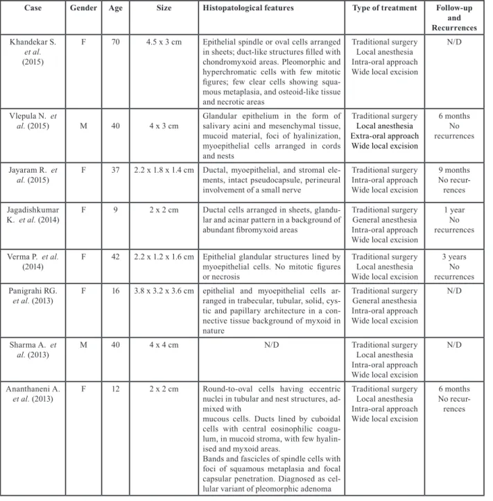
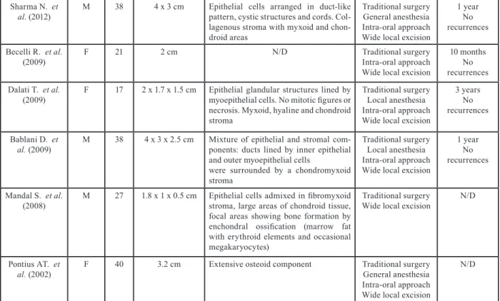
of choice. To the best of our knowledge, extra-parotid PA of the minor salivary glands within the cheek and mucobuccal fold has been described only in a few ca-ses, thus representing a very unusual finding. (1-14). Here we report a rare case of a large-sized PA arising from minor salivary glands of the cheek, successfully treated through Neodimium: Yttrium Aluminium Gar-net (Nd:YAG) Laser, altogether with a concise review of the literature of the last 30 years, focused on PAs of minor salivary glands larger than 1 cm (1990 to 2019) (Table 1, 1 cont.).
Table 1: Review of the literature: clinico-pathological features of 14 cases of pleomorphic adenoma of minor salivary glands of the cheek and mucobuccal fold, larger than 1 cm, identified from January 1990 to July 2019.
Sharma N. et
al. (2012) M 38 4 x 3 cm Epithelial cells arranged in duct-like pattern, cystic structures and cords. Col-lagenous stroma with myxoid and chon-droid areas
Traditional surgery General anesthesia Intra-oral approach Wide local excision
1 year No recurrences Becelli R. et al.
(2009) F 21 2 cm N/D Traditional surgeryIntra-oral approach
Wide local excision
10 months No recurrences Dalati T. et al.
(2009) F 17 2 x 1.7 x 1.5 cm Epithelial glandular structures lined by myoepithelial cells. No mitotic figures or necrosis. Myxoid, hyaline and chondroid stroma
Traditional surgery Local anesthesia Intra-oral approach Wide local excision
3 years No recurrences Bablani D. et
al. (2009) M 38 4 x 3 x 2.5 cm Mixture of epithelial and stromal com-ponents: ducts lined by inner epithelial and outer myoepithelial cells
were surrounded by a chondromyxoid stroma
Traditional surgery Local anesthesia Intra-oral approach Wide local excision
1 year No recurrences Mandal S. et al.
(2008) M 27 1.8 x 1 x 0.5 cm Epithelial cells admixed in fibromyxoid stroma, large areas of chondroid tissue, focal areas showing bone formation by enchondral ossification (marrow fat with erythroid elements and occasional megakaryocytes)
Traditional surgery
Wide local excision N/D
Pontius AT. et
al. (2002) F 40 3.2 cm Extensive osteoid component Traditional surgeryGeneral anesthesia Intra-oral approach Wide local excision
N/D Table 1 cont.: Review of the literature: clinico-pathological features of 14 cases of pleomorphic adenoma of minor salivary glands of the cheek and mucobuccal fold, larger than 1 cm, identified from January 1990 to July 2019.
Case report
A 70-year-old female patient was referred for a pain-less, slow-growing swelling within the right cheek, which was present since the last 15 years and signifi-cantly increased during the last year. Extraoral clinical examination revealed asymmetry of the face caused by the presence of a mobile, hard-elastic mass. Intraoral evaluation showed a swelling covered by intact muco-sa, with a dimension on the major size of approximately 5 cm. At magnetic resonance imaging (MRI), the mass showed well-defined margins (most likely associated to the presence of a capsule), smooth borders, inhomoge-neous aspect and a high-intensity T2-weighted signal. The mass was adherent to Bichat’s fat pad, delimited by the Stensen’s duct and masseter muscle, the upper fornix and lower fornix posteriorly and the facial artery ante-riorly (Fig. 1).
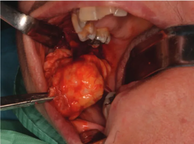
On the basis of a presumptive diagnosis of a benign tu-mor of minor salivary glands or of connective tissue, sur-gical excision trough Nd:YAG laser (λ=1064 nm, Fidelis Plus®, Fotona – Slovenia - 3.5W, 60Hz, PD 488,281W/ cm2 ) was performed under local anesthesia (Fig. 2). At macroscopic examination the mass measured 6 x 3.5 x 3 cm, with a whitish and glistening cut surface and multiple translucent cystic areas. Histopathological exa-mination showed a mixture of epithelial and stromal tis-sue. Ducts lined by inner epithelial and outer myoepithe-lial cells were surrounded by a chondromyxoid stroma,
Fig. 1: MRI. Axial T2-weighted images showing a mass with well-defined margins, smooth borders, slight inhomogeneous pattern and high-intensity signal; the mass seems encapsulated.
consistent with pleomorphic adenoma. Being the tumor mass not associated with the parotid gland or duct, it was considered to be of minor salivary gland origin. The
Fig. 2: Surgical excision. Intra-oral incision of the overlying mucosa performed with Nd:YAG laser (Fidelis Plus®, Fotona – Slovenia -
3.5W, 60Hz, PD 488,281W/cm2) and isolation of the tumor from the
surrounding tissues.
microscopic examination therefore confirmed the diag-nosis of PA; all margins were free from neoplastic infil-tration (Fig. 3). No pain or swelling were present after 2 weeks. No clinical and MRI signs of recurrence were noticed after 2-year follow-up.
Fig. 3: Microscopic view. pleomorphic adenoma showing a mixture of glandular epithelium and myoepithelial cells within a mesen-chyme-like background, with a mixed mucoid, myxoid or chondroid appearance (Haematoxilyn and Eosin staining – 10X magnification).
Discussion
Most of patients with PAs of minor salivary glands com-plain for the presence of a small, painless, nodule which slowly increases in size, sometimes showing intermit-tent or accelerating growth (3). Most cases are asympto-matic and there is usually a long period of time (ranging from few days up to 15 years) between the appearance of the lesion and the diagnosis (12). Differential diagno-sis of PA of cheek mucosa should include odontogenic and non-odontogenic buccal abscesses, salivary glands
infections and other inflammatory conditions such as sialolithiasis. Further non-oncologic causes of swelling include dermoid and sebaceous cysts as well as reactive lymph nodes. Among neoplasms, diagnostic hypotheses should include benign tumors such as lipomas, neuro-fibromas and Warthin’s tumor as well as malignancies like mucoepidermoid and adenoid cystic carcinomas, polymorphous adenocarcinoma and carcinoma ex pleo-morphic adenoma (9).
While Velpula and other authors performed ultrasono-graphy, fine needle aspiration cytology (FNAC) and/or computed tomography (CT) prior to MRI, we performed MRI (1,2,4,14). Being the clinical presentation strongly suggestive for a benign tumor of soft tissues, MRI was considered to be appropriate as first line imaging inves-tigation, thus limiting invasiveness and unnecessary ex-posure to radiation (10). At MRI, benign salivary gland tumors typically show well-defined borders and hyper-intense signal on T2-weighted images. On the contrary, ill-defined borders, invasion into surrounding tissues, low T2 signal, heterogeneous enhancement, cystic chan-ges and central necrosis are sugchan-gestive of malignancy (1,10). In the present case, MRI features were consistent with a benign tumor, probably of minor salivary gland origin. In fact, axial T2-weighted images showed we-ll-defined and smooth borders and high-intensity signal. A slight inhomogeneous aspect could also be observed, probably due to the large size of the mass and to a dege-neration induced by its long persistence. These findings were similar to the majority of the cases reviewed, whe-re imaging information was available (1,2,7,10,14). From the pathologic macroscopic point of view, PAs of minor salivary glands are often grossly encapsula-ted and they usually range from 1 to 4 cm in size (2). Larger lesions may show necrotic or cystic areas. Their consistency varies from hard to rubbery to soft, some-times fluctuant, swellings. The cut surface of the mass is solid and the color may vary from gray blue, yellow to brown. Gritty, gelatinous or glistening areas may be present when there is a cartilaginous or myxochondroid differentiation (4). Mandal et al. reported a peculiar case of PA showing bone and bone marrow formation within chondroid tissue (13). In the present case, both epithelial and myoepithelial cells, typical of PAs, were present; such a finding, may be useful, to some extent, to rule out mucoepidermoid carcinoma. The absence of peri-neural invasion and mitotic figures reduced the chances of polymorphic adenocarcinoma and carcinoma ex pleo-morphic adenoma. Nevertheless, Jayaram et al. reported a case of perineural involvement of a small nerve asso-ciated with a benign PA (5).
PAs might sometimes recur, either due to seeding or ina-dequate removal (1,12). PA does not spread through the lymphatic or blood system and does not have the ability to induce locoregional or distant metastasis. However,
in few cases a malignant epithelial tumor has been do-cumented within the context of a PA, being known as carcinoma ex PA (12).
Moreover, PAs show sometimes signs of extracapsular spread at postoperative histopathologic examination (9). Such a feature may account for some of the recurrences observed in cases of presumptive radical excision (12). On the basis of the impossibility to precisely assess the presence of malignant cells or microscopic extracap-sular spread, the preferred treatment for PA consists in wide local excision (0.5 cm free margins) and follow-up for at least 3-4 years (4). This procedure was, in fact, performed in the totality of the reviewed cases, where no recurrences were reported.
In our case, Nd:YAG laser was chosen for its precise and minimally invasive cut, which allows radical excision in most cases. Nd:YAG laser wavelength also provides a powerful haemostatic activity (15).
The energy transferred by the laser beam to biological tissues results in tissue heating. The mean thickness of epithelial, stromal and vascular changes induced by Nd:YAG laser, as detected at microscopic evaluation, is 363.7, 459.1 and 169.85 micron, respectively (15). It has been demonstrated that Nd:YAG laser causes gross changes in small mucosa specimens (less than 7 mm in the main size), independently from laser se-ttings. On the other hand, the heat-induced damage is negligible in specimen larger than 7 mm. In these cases, it is usually possible to perform a precise histological evaluation of the margins (15). We also hypothesize that another advantage of laser surgery is the carbonization of possible residual PA cells on the excision margins. All cheek PA cases, described in the literature considered here, underwent traditional surgery and approximate-ly 30% of them (4 of the cases, where the information was available) was carried out under general anesthesia (6,7,10,14).
Nd:YAG laser also shows a strong antimicrobial acti-vity, which reduces the need for antibiotics and the rate of infectious complication, even in cases of second in-tention wound healing (15). In the present case, the use of laser gave the possibility to perform the surgical in-tervention within an outpatient setting, allowing to save time and economic resources, if compared with an inter-vention performed under general anesthesia followed by hospitalization.
References
1. Velpula N, Annam SR, Pallepati SR, Kumar R, Kumar A. Pleomor-phic Adenoma of Cheek Masquerading as Fibrolipoma - Case Report with Review. J Clin Diagn Res. 2015;9:ZD13-5.
2. Bablani D, Bansal S, Shetty SJ, Desai R, Kulkarni SR, Prasad P, et al. Pleomorphic adenoma of the cheek: a case report and review. J Oral Maxillofac Surg. 2009;67:1539-42.
3. Verma P, Sachdeva SK, Verma KG, Sachdeva K. Pleomorphic ade-noma of cheek: A rare case report and review of literature. Indian J Dent Res. 2014;25:122-124.
4. Khandekar S, Dive A, Munde P, Wankhede ND. Pleomorphic adenoma of the buccal salivary gland. J Oral Maxillofac Pathol. 2015;19:111.
5. Jayaram R, Patel D, Santhanam V. Benign pleomorphic adenoma of minor salivary gland showing perineural invasion: a rare entity. Br J Oral Maxillofac Surg. 2015;53:81-2.
6. Jagadishkumar K, Anilkumar MG, Krishna Kumar HC, Maggad R. Pleomorphic adenoma of the cheek in a child: A case report. Dent Res J. 2014;11:522-4.
7. Panigrahi RG, Sahoo SR, Panda S, Lenka S, Padhiary SK, Bhuyan R, et al. Juvenile pleomorphic adenoma of masticator space: The first case report. Contemp Clin Dent 2013;4:527-30.
8. Sharma A, Deshmukh S, Shaikh A, Dabholkar J. Pleomorphic adenoma of the minor salivary gland of the cheek. Singapore Med J. 2013;54:e183-4.
9. Ananthaneni A, Undavalli SB. Juvenile cellular pleomorphic adeno-ma. BMJ Case Rep. 2013;24:bcr2012007641.
10. Sharma N. Pleomorphic adenoma of the buccal salivary gland: magnetic resonance imaging findings with differential diagnoses. J Investig Clin Dent. 2012;3:228-31.
11. Becelli R, Quarato D, Matarazzo G, Renzi G, Morello R. Surgical treatment of an extraparotid pleomorphic adenoma of minor salivary glands of the cheek. J Craniofac Surg. 2009;20:1604-6.
12. Dalati T, Hussein MR. Juvenile pleomorphic adenoma of the cheek: a case report and review of literature. Diagn Pathol. 2009;22:32. 13. Mandal S, Dhingra K, Roy S, Khurana N. Extensive bone with marrow formation in pleomorphic adenoma. Report of a case. ANZ J Surg. 2008;78:323-4.
14. Pontius AT, Myers LL. Pleomorphic adenoma of the buccal space. Otolaryngol Head Neck Surg. 2002;126:695-6.
15. Vescovi P, Corcione L, Meleti M, Merigo E, Fornaini C, Manfredi M, et al. Nd:YAG laser versus traditional scalpel. A preliminary his-tological analysis of specimens from the human oral mucosa. Lasers Med Sci. 2010;25:685-91.
Conflict of Interest
All Authors declare that they have no conflicts of interest with people or organizations that could inappropriately influence this work.