UNIVERSITA’ DEGLI STUDI DI PISA
FACOLTA’ DI MEDICINA E CHIRURGIA
DIPARTIMENTO DI PSICHIATRIA, NEUROBIOLOGIA,FARMACOLOGIA E BIOTECNOLOGIE
DOTTORATO DI RICERCA
NEUROBIOLOGIA E CLINICA DEI DISTURBI
AFFETTIVI
Tesi
“Vascular dysfunction in a mouse model of
Rett Syndrome and effects of Curcumin
treatment”
SSD BIO/11Relatori:
Prof.ssa Claudia Martini
Dr. Mario Costa
ANNO 2011
Candidata:
Riassunto
Mutazioni nel gene legato al cromosoma X, MeCP2 (Methyl CpG-binding protein) sono presenti in circa l’80% dei pazienti affetti da Sindrome di Rett, una delle più comuni cause di ritardo mentale nelle bambine e per cui ancora oggi non esistono efficaci trattamenti farmacologici. Un aspetto rilevante, ma ancora poco esplorato, nei pazienti con sindrome di Rett è la riduzione della circolazione periferica.
Per investigare la relazione tra la perdita di MeCP2 e questo aspetto clinico abbiamo utilizzato per gli studi funzionali, farmacologici e comportamentali, topi mancanti del gene MeCP2, maschi (MeCP2y/-) e femmine (MeCP2+/-). Gli studi funzionali condotti su branche dissezionate delle arteriole dell’albero mesenterico montate su microcannule di vetro in un miografo a pressione, hanno mostrato una drammatica riduzione della reattività vascolare endotelio dipendente nei topi MeCP2+/- rispetto ai controlli sani. Le arteriole preincubate con inibitori delle NOS o con acido ascorbico hanno rilevato una ridotta biodisponibilità di Ossido Nitrico (NO) e un aumento nelle specie reattive dell’ossigeno (ROS). Inoltre, l’animale Rett presenta bassi livelli di espressione sia dell’mRNA che del peptide eNOS nelle arteriole e alti livelli di stress ossidativo. Da un punto di vista comportamentale i topi knockout per MeCP2 mostrano comportamenti stereotipati e si osserva una riduzione del tempo di riposo.
Il trattamento cronico delle femmine MeCP2+/- con curcumina è in grado di ripristinare il normale fenotipo e di migliorare la sintomatologia comportamentale della Rett, diminuendo i caratteristici movimenti stereotipati e aumentando il tempo di riposo degli animali.
Questi dati indicano che la mancanza del gene MeCP2 modifica la circolazione periferica alterando la reattività vascolare, riducendo i livelli di espressione di eNOS e di NO. Inoltre, i nostri risultati supportano sia dal punto di vista funzionale/molecolare che comportamentale l’utilizzo della curcumina nella terapia dei pazienti affetti da sindrome di Rett.
Abstract
Mutations in the coding sequence of the X-linked gene MeCP2 (Methyl CpG– binding protein), are present in around 80% of patients with Rett Syndrome, a common cause of mental retardation in female and to date without any effective pharmacological treatment. A relevant, and so far unexplored feature of RTT patients is a marked reduction in peripheral circulation.
To investigate the relationship between loss of MeCP2 and this clinical aspect, we used a MeCP2 null mouse model, male (MeCP2y/-) and female (MeCP2+/-), for functional, pharmacological and behavioural studies. The functional studies performed on dissected branches of mesenteric arterial tree mounted on glass microcannule in a pressurized myograph, demonstrated a dramatic endothelial-dependent vascular reactivity impairment in MeCP2+/- compared to control littermate. The mesenteric arteriole preincubation with NOS inhibitors or ascorbic acid indicate a decrease Nitric Oxide (NO) availability and the increased presence of Reactive Oxygen Species (ROS). Consistently, the RTT mouse model exhibited a decreased expression in both mRNA and peptide eNOS in the arterioles and a higher systemic oxidative level. MeCP2 knockout mice show stereotyped movements and less resting time when compared to control littermates.
Chronic curcumin treatment of female MeCP2+/- mice was able to reverse this vascular phenotype and ameliorate the mouse RTT behavioural symptomatology by decreasing stereotyped movements and by increasing resting time.
These data indicate that in the absence of MeCP2 peripheral circulation is impaired by an altered vascular reactivity and decreased arteriolar eNOS expression and NO production. Further, they provide a physiological/molecular rational for the use of curcumin as a treatment to improve the health of RTT patients.
INDEX
1 INTRODUCTION
………...………...…….. 11.1
Pervasive developmental disorders: an overview
..… 21.2
Rett Syndrome
………..……..……...… 41.2.1
Clinical features of Rett Syndrome…………..………...….. 51.2.2
The autonomic nervous system………..……...…...……. 81.2.3
Clinical Criteria For Typical Rett Syndrome……….……...….. 91.2.4
Atypical Forms of Rett Syndrome…...………...… 111.2.5
Genetic Origin Of Rett Syndrome………..………..…...…. 121.2.6
Effect Of XCI………..………..……… 131.2.7
Genotype/Phenotype Correlations…...…………..………...…… 131.2.8
MeCP2 Mutations In Boys………..…………..……...…. 141.3
MECP2 GENE: Structure and Function
..……...…. 151.3.1
Tissue expression of MeCP2……...……….………....…. 211.3.2
MeCP2 genes target………... 251.4
Rett Mouse Models
………...………....…. 281.5
Oxidative stress in Rett Syndrome
………... 321.6
Vascular Function
………...…..…. 341.7
Curcumin
………... 381.7.1
Metabolism and Bioavailability…...………..……...… 401.7.2
Biological activity………..………..…...….. 413
MATERIALS AND METHODS
……….…….. 483.1
Animals and genotyping
... 493.2
Curcumin treatment
... 503.3
Pressure Myograph System
... 513.4
Determination of Superoxide Anion Production in
the Mesenteric Vessels
……….….… 533.5
Measurements of Plasma Malonyldialdehyde
Levels
... 543.6
RNA extraction and Real Time PCR
... 553.7
Immunostaining of eNOS
………....… 563.8
Rotating rod test (Rotarod)
... 583.9
Behavioural observation
... 593.10 Open Field test
... 603.11 Data Analysis
... 624
RESULTS
………..………..…. 634.1
Vascular reactivity on WT and Rett mice
………..…... 644.2
Endothelial function: effect of curcumin
……...…….... 674.3
Vascular superoxide generation: effect of
curcumin
…..………...…… 684.4
Plasma MDA levels: effect of curcumin
………. 694.5
eNOS expression in mesenteric vessels and aorta
…... 704.6
Vascular eNOS immunostaining
…..….………...….. 714.7
Body weight variation
………...…. 724.9
Stereotyped movements: effect of curcumin
…….….... 744.10 Open Field test
………..…...…. 755
DISCUSSION
………..……….. 786
BIBLIOGRAFY
……….……..………... 86FIGURE INDEX
Figure 1: Pervasive Developmental Disorders (PDD). Current Classification in DMS-IV.
Figure 2: Onset and Progression of RTT Clinical Phenotypes. Figure 3: Revised diagnostic criteria for Rett Syndrome. Figure 4: Transcriptional repression by MeCP2.
Figure 5: Alternative splicing forms of the MECP2: MECP2-e1 and MECP2-e2. Figure 6: MeCP2 function domains. MBD methyl-binding domain; TRD transcriptional repression domain; C-ter C-terminal domain and NLS nuclear localization signals.
Figure 7: MeCP2 as a transcriptional repressor. MeCP2 binds to methylated CpG upstream of the transcriptional start site of a MeCP2 target gene.
Figure 8: MeCP2 as a transcriptional activator. MeCP2 recruits a transcriptional coactivator to cause the transcriptional upregulation of a target gene.
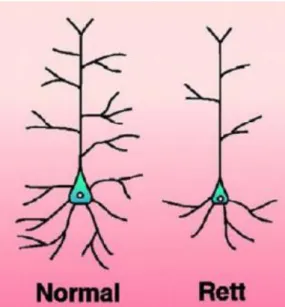
Figure 9: Schematic representation of the spatial and temporal distribution of MeCP2 during human (A) and mouse (B) development. Wg, weeks of gestation. Figure 10: Schematic representation of pyramidal neurons from control and Rett brains.
Figure 11: MeCP2 Target Genes.
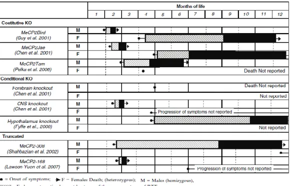
Figure 12: Lifespan and progression of symptoms of selected mouse models. Figure 13: Rett mice phenotype.
Figure 14: Nitric oxide in the regulation of vasodilation.
Figure 16: Chemical structure of the major curcuminoids. Figure 17: Characteristic phenotype of Rett female mouse.
Figure 18: Representation of mouse third-order branch of mesenteric arterioles isolated and mounted on two glass microcannule.
Figure 19: Schematic representation of pressure myograph system.
Figure 20: Representative diagram of the three phases of the solution: upper colorless aqueous phase contains RNA; white interphase and lower red organic phase contain DNA and protein.
Figure 21: Schematic Rotarod experimental apparatus.
Figure 22: Open Field arena square used for testing anxiety and exploratory drive.
Figure 23: Endothelium-dependent relaxations elicited by Acetylcholine in mesenteric resistance arteries without (saline) or with L-NAME or Vit C from WT female (A), Rett female (B) and P50 Rett male mice (C). Each point represents the mean of 6 experiments ± SEM. *P<0.05, **P<0.01.
Figure 24: Inhibition by L-NAME on maximal response to acetylcholine in vessels from WT, MeCP2+/− , and curcumin-treated MeCP2+/− mice, without (saline) or with ascorbic (asc) acid. Results are given as the mean of 6 experiments ± SEM. *P<0.05, **P<0.01. Figure 25: Vasorelaxation endothelium independent. Vascular response to increasing concentrations of sodium nitroprusside in WT and Rett female and male mice. **P<0.01. Figure 26: Endothelium-dependent relaxations elicited by Acetylcholine in mesenteric resistance arteries from Rett control (A) and curcumin treated (B) female mice without (saline) or with L-NAME or Vit.C. Each point represents the mean of 6 experiments ± SEM. **P<0.01.
Figure 27: Representative DHE staining and quantification (bar graph) of the red signal in mesenteric arteries (magnification X 40) from WT and MeCP2+/− mice feed with or without curcumin. Results are given as the mean of 6 experiments ± SEM. *P<0.05, **P<0.01.
Figure 28: Oxidative stress parameter MDA. Mean±SEM Malondialdehyde (MDA) in the plasma of WT and MeCP2+/− mice feed with or without curcumin. **P<0.01.
Figure 29: Quantitative RT–PCR analysis of mRNA expression for eNOS in Mesenteric vessels (A) and Aorta (B) of Wild-Type and Rett animals treated or not three weeks with 5% curcumin. eNOS has been normalized to the expression of GAPDH. Data represent the mean±SEM (*P<0.05, **P<0.01).
Figure 30: Representative photomicrograph of immunostaining for eNOS in small mesenteric arteries from WT and MeCP2+/− mice. A strong, specific eNOS staining is detected in endothelium of control vessels, while mesenteric arterioles from MeCP2+/− mice show only a slight eNOS immunostaining at level of the outer vascular smooth muscle cells, without any appreciable amount in the endothelial cell layer.
Figure 31: Curcumin effect on the animal weight, normalized at 1.0.
Figure 32: The averages (±SEM) for time spent on rotarod across five test trials for MeCP2+/- mice curcumin treated and untreated.
Figure 33: Behavioral observation. Percentage of time spent to perform stereotyped movements (A) and to rest or sleep (B) by Wild-Type and Rett female mice untreated (ctrl) and treated with 5% curcumin (curc). Data represent the mean±SEM (*P<0.05, ***P<0.001).
Figure 34: Time in seconds spent in the "center" of the arena for each animal in each group. In addition to individual scores shows the average for the treated group (n = 5) and controls (n = 5) with SEM.
Figure 35: Time spent in the "corners" of the arena for each animal in each group. In addition to individual scores shows the average for the treated group (n = 5) and for controls (n = 5) with SEM.
Figure 36: Percentage of time spent moving for each animal in each group. The bars indicate SEM.
Figure 37: Example of a path drawn by an curcumin treated animal (A) and a control one (B). The red line indicates the route taken by the animals.
2
1.1Pervasive developmental disorders: an overview
Pervasive developmental disorders (PDD), also called autism spectrum disorders, comprise a complex and heterogeneous group of pathological conditions characterized by abnormalities of brain development and function, appearing within the first three years of life. Diagnostic and Statistical Manual of Mental Disorders, Fourth Edition (DMS-IV) identify as PDD five pathology: autism, Rett syndrome, childhood disintegrative disorder, Asperger disorder and PDD not otherwise specified under the spectrum of PDD (PDD-NOS) (American Psychiatric Association., 2000) (Figure 1).
Figure 1: Pervasive Developmental Disorders (PDD). Current Classification in DMS-IV.
Although the detailed causal mechanisms are not known, autism is likely to have multiple etiologies including genetic factors. Children with PDD share the following characteristics:
i) impairments in social interaction; ii) inability to imaginative activity;
3 iv) stereotyped behaviors;
v) cognitive deficits;
vi) limited number of interests and activities.
Children affected by PDD have difficult in talking, playing with other children, and relating to others, including their family.
The incidence of these disorders has risen dramatically over the past two decades mainly because of the use of broader diagnostic criteria and the increased attention of the medical community (Levy et al, 2009) and is estimated 60-70/10000 children. PDD is 4/5 times more frequently in boys, with the exception of Rett syndrome, which is found mainly in girls. Great interest exists in the elucidation of the causes and pathogenesis of autism. Although autism is recognized to be the common endpoint of neurological dysfunction of varying etiologies, common disease mechanisms may underlie the phenotypes shared by RTT and autism, and advances in understanding of RTT may also shed some light into the pathogenesis of autism.
4
1.2 Rett Syndrome
Rett syndrome (RTT) is a neurological disorder that predominantly affects girls (Hagberg et al, 1985; Hagberg & Hagberg, 1997). It represents the second most common cause of mental retardation in females with an incidence of 1:10000 (Percy, 2002). The disease has been described for the first time by the Austrian neurologist Andreas Rett in 1966, but the scientist community recognized the pathology only 17 years later when the Swedish neurologist Dr. B. Hagberg described 35 new cases of RTT (Hagberg et al, 1983).
In 1954, the Viennese pediatrician Dr. Andreas Rett observed two girls in his waiting room displaying the same repetitive hand-washing motions. Looking at their clinical charts, he realized they shared similar clinical and developmental histories. Dr. Rett soon recognized that six other girls in his practice had similar behaviors, and reasoned that these girls must have the same disorder, which at that time was new to the medical field. In fact, their distinctive behavioral patterns indicated to him that this new condition was not a simple mental retardation syndrome, but rather a complex condition that affected several facets of neural function. Unfortunately, during the 1960’s in Europe the medical community was hesitant to recognize “new” conditions in the absence of a metabolic abnormality. This led Dr. Rett to examine several physiological parameters in his affected girls, where he found elevated ammonia content in blood. In 1966, Dr. Rett published his observations in a leading German medical journal, where he described the condition as one of cerebral atrophy and hyperammonia in girls, characterized by autistic behavior, dementia, and apraxia of gait (Rett, 1966). The condition stayed mostly unrecognized in the English language literature until 1983, when Bengt Hagberg described 35 patients, all girls from three countries (France, Portugal and
5 Sweden), with a uniform and striking, progressive encephalopathy. At this point, he revisited Rett’s unusual mental retardation syndrome and suggested the condition bear his name (Hagberg et al, 1983). In 1991, Bruck and colleagues described a set of monozygotic female twins with Rett syndrome (Bruck et al, 1991).
The genetic cause of RTT remained evasive until quite recently, largely because the inheritance pattern was chiefly sporadic. In 1999, Amir and colleagues (Amir et al, 1999) discovered that mutations in the gene encoding Methyl-CpG-binding protein 2 (MeCP2) are associated both with rare familial cases of RTT as well as with the more common sporadic occurrences of typical RTT. This mutation is responsible of 95% of RTT cases (Amir et al, 1999; Van den Veyver & Zoghbi, 2000; Wan et al, 1999; Xiang et al, 2000). Further, mice lacking MeCP2 display Rett-like symptoms, and this phenotype can be reversed with the reintroduction of MeCP2 (Guy et al, 2007).
1.2.1 Clinical features of Rett Syndrome
Girls with RTT born after an apparently quiet pregnancy. They are normal at the birth and achieve expected developmental milestones until 6-18 months of age. Nevertheless, some studies revealed subtle behavioral abnormalities soon after birth. There is a wide variability in the clinical presentation, classic Rett syndrome follows a well recognized pattern. Not all symptoms compare at the beginning of the disease but can be distinguish four different stages (Figure 2).
6
Figure 2: Onset and Progression of RTT Clinical Phenotypes.
In the development of the disorder, the first indicator of neurological involvement is deceleration of head growth, leading to microcephaly by the second year of life. This decreased head growth is due to decreased neuronal growth. The acquired microcephaly is accompanied by general growth retardation, weight loss, and a weak posture brought on by muscle hypotonia.
Subsequently there is a period of rapid regression and patients lose purposeful use of their hands and instead develop stereotypic hand wringing or washing movements, and in some cases clapping, flapping, and mouthing of the hands. Social withdrawal and loss of language become apparent in addition to irritability and self-abusive behavior. Other autistic features also manifest, including expressionless face, hypersensitivity to sound, lack of eye-to-eye contact, indifference to the surrounding environment, and unresponsiveness to social cues (Nomura, 2005). The onset of mental deterioration is accompanied by loss of
7 motor coordination and the development of ataxia and gait apraxia. The earliest autonomic perturbation is hyperventilation during wakefulness. Most girls with RTT suffer additional breathing anomalies, including breath-holding, aerophagia, forced expulsion of air and saliva, and apnea. One of the most arduous features of RTT is the occurrence of seizures, which range from easily controlled to intractable epilepsy, with the most common types being partial complex and tonic-clonic seizures (Jian et al, 2006). The seizures tend to decrease in severity after the teenage years and into adulthood, presenting minor problems after the age of forty.
The third stage of the pathology is characterized by an amelioration of the social component of the autistic-like behavior occurs sometime between 5 to 10 years of age. Behavioral abnormalities during this post regression phase include teeth grinding, night laughing or crying, screaming fits, low mood, and anxiety episodes elicited by distressful external events (Mount et al, 2001). Patients suffer devastating motor deterioration, generalized rigidity, dystonia, and worsening of scoliosis. Most girls with RTT lose mobility, and are often wheelchair-bound during the teenage years. Sleep problems are common in Rett syndrome (in over 80% of cases),specifically has been shown disrupted sleep patterns at night and an increase in total and daytime sleep (Young et al, 2007). Additional autonomic abnormalities include hypotrophic, severe constipation, oropharyngeal dysfunction, and cardiac abnormalities, including tachycardia, prolonged corrected QT intervals, sinus bradycardia and cold blue feet. Despite their excellent appetites, individuals with RTT commonly present eating problems (Reilly & Cass, 2001) and not have a correct intake of calories in the diet, especially during pre-school and early school years. This can be caused by
8 problems in coordinating movements of the mouth and throat, muscle spasms and involuntary movements, the expenditure of large amounts of energy when breathing and the inability of the intestine to absorb nutrients. They have difficult to co-ordinate breathing and swallowing. Patients continue to lose weight and many suffer from osteopenia, scoliosis, and rigidity as they age. As patients get older they often develop Parkinsonian features (Hagberg, 2005; Roze et al, 2007). The condition reaches a plateau and some patients survive up to the sixth or seventh decade of life in a severely debilitated physical condition.
1.2.2 The autonomic nervous system
Certain physical functions including the regulation of heart rate, blood pressure, peripheral blood circulation, respiration and digestion are governed by the autonomic nervous system. In Rett syndrome there are varying degrees of dysfunction in the control of the central autonomic nervous system.
Abnormal breathing patterns affecting pulmonary and cardiovascular function are characteristic of Rett syndrome. They can be divided into three categories: forceful breathing, abnormally shallow breathing and apneustic breathing (a series of slow deep inspirations). Valsalva breathing (attempting to forcibly exhale while the epiglottis is closed) is a common, complication to the breathing abnormalities characteristic of Rett syndrome, and affects the autonomic nerve system and brain stem functions. The consequences for the individual of Valsalva breathing depend on the category of breathing abnormality he or she manifests(Julu et al, 2001). Impaired balance of central autonomic control may also result in cold, bluish, clammy feet due to peripheral vasomotor disturbances. Hydrotherapy, and
9 physiotherapy to the extremities is often used to regain proper circulation and helps to keep their extremities limber (Stearns et al, 2007).
The motility of the gastrointestinal tract (peristalsis) shifts the food along the tract and is controlled and coordinated by the autonomic nervous system. It is common that these movements are impaired and that the passage of food through the tract is unusually slow, leading to a number of symptoms of varying degrees of severity. The girls have problems swallowing and may swallow air resulting in an extended, painful stomach, and vomiting. Painful inflammation in the lower esophagus (esophagitis) can occur from an early age if the lower esophageal sphincter leaks. Severe constipation is very common.
1.2.3 Clinical criteria for typical Rett Syndrome
In 1994 Hagberg and Skjeldal suggested a model of inclusion and exclusion criteria for the diagnosis of RTT that relaxed the international criteria originally drawn up in Vienna in September 1984 (Hagberg & Skjeldal, 1994). In 2002 the same authors updated the previous diagnostic criteria (Hagberg et al, 2002). In 2010 researchers reviewed 2002 diagnostic criteria (Figure 3) to clarify and simplify the diagnosis of typical, or classic, RTT. They emphasized that it remains a clinical diagnosis, since not all RTT patients have MECP2 mutations and not all patients with MeCP2 mutations have Rett syndrome. They limited the necessary criteria to the presence of a period of clear developmental regression plus four main criteria that are absolutely required for the diagnosis of typical RTT: loss of purposeful hand skills, loss of spoken language, gait abnormalities, and stereotypic hand movements. After the period of regression, a stage of stabilization and potentially even improvement ensues, with some individuals
10 partially regaining skills. This potential for some skill recovery emphasizes the importance of the acquisition of a careful history to determine the presence of regression. Furthermore they eliminated postnatal deceleration in head growth from the necessary criteria because this feature in not found in all individuals with typical RTT. However, because it is a clinical feature that can alert a clinician to the potential diagnosis and it is a distinctive feature in the disorder, this has been included as a preamble to the criteria as a feature that should raise suspicion for the diagnosis. In these new criteria, history of regression and all of the necessary and exclusion criteria must be met to make the diagnosis of typical RTT, without exception (Neul et al, 2010).
Figure 3: Revised diagnostic criteria for Rett Syndrome.
11
1.2.4 Atypical forms of Rett Syndrome
In addition to the classic form of RTT, five distinct categories of variants have been delineated on the bases of clinical criteria (Hagberg et al, 2002; Trevathan & Naidu, 1988). These variants show some, but not all diagnostic features of RTT and can be milder or more severe. They include:
i) the preserved speech variant (PSV), is the best characterized, has well-defined clinical features, and mutations in MECP2 have been found in the majority of cases; in this variant girls recover the ability to speak in single word or third person phrases and display an improvements of purposeful hand movements at stage III of disease progression (Zappella et al, 2001);
ii) the early seizures variant (Hanefeld Variant) , in which the normal perinatal period is soon followed by the appearance of seizures preceding the regression period (Hanefeld, 1985);
iii) the “forme fruste” with a milder, incomplete and protracted clinical course (Hagberg & Rasmussen, 1986; Hagberg & Witt-Engerstrom, 1986);
iv) the congenital variant, where patients show RTT features straight from birth (Rolando, 1985);
v) the late regression variant, which is characterized by normal head circumference and gradual loss of acquired speech and fine motor skills in late childhood (Gillberg, 1989).
Furthermore, it has been described a “highly functioning PSV” associated with acquisition of more complex language function including the use of first person phrases. In this variant, girls acquire a better control of their hands and they are
12 able to draw figures and write simple words (Zappella et al, 2003). The degree of mental retardation is milder then in PSV with the I.Q that can be as high as 50. Recent works have found mutations in different loci associated with congenital and Hanefeld variants, with mutations in FOXG1 found in congenital variant cases (Ariani et al, 2008) and mutations in CDKL5 found in early seizure variant cases (Bahi-Buisson et al, 2008).
1.2.5 Genetic origin of Rett Syndrome
Early reports postulated an X-linked dominant mode of inheritance with fateful consequences in homozygous males. Using information from rare familial cases, exclusion mapping identified the Xq28 candidate region, and subsequent screening of candidate genes in RTT patients revealed mutations in MECP2 (Amir et al, 1999). Mutations in MECP2 are found in more than 95% of classic RTT cases, most arise de novo in the paternal germ line and often involve a C to T transition at CpG dinucleotides. The spectrum of mutation types includes missense, nonsense, and frameshift mutations, with over 300 unique pathogenic nucleotide changes described (Christodoulou & Weaving, 2003), as well as deletions encompassing whole exons (Archer et al, 2006; Pan et al, 2006; Ravn et al, 2005). Eight missense and nonsense mutations account for 70% of all mutations, while small C-terminal deletions account for another 10%, and complex rearrangements constitute 6%.
13
1.2.6 Effect of XCI
In each body cell (somatic cell) of the developing baby girl, one of the X chromosomes becomes very shortened and condensed so that most of its genes are not able to be ‘read’ by the cells.
This system of inactivation in the body cells known as X-chromosome inactivation (XCI) is totally normal in female and is usually random so that women’s bodies have a mixture of cells in regard to the inactivated X chromosome, although certain genes on the silenced chromosome may still be expressed. Some cells will have the X chromosome switched off that came from their mother (an inactive maternal X chromosome); other cells will have the paternal X chromosome inactivated. The relative proportion of cells with an active maternal or paternal X chromosome varies from female to female (even between identical twins) because the process is usually random.In rare situations, females can show nonrandom or skewed X-inactivation with the preferential inactivation of one X-chromosome in most or all of the cells in the body. In these cases, females may be protected from expression of linked diseases if the X-chromosome with the abnormal gene is inactivated. XCI only occurs in the somatic cells, since both X chromosomes need to be active in the egg cells for their normal development.
Of course this process occurs also in Rett patients and can produce a pattern favoring the expression of the X chromosome containing the normal MECP2 gene, or mutated MECP2 gene. Asymptomatic carriers of RTT can have highly skewed inactivation patterns of XCI favoring the normal MECP2 allele. Sisters with identical MECP2 mutations can have extremely discordant phenotypes as a result of XCI skewing. One sister displayed classic Rett Syndrome while the other
14 was a “highly functioning” preserved speech variant as a result of favourable XCI skewing (Scala et al, 2007). XCI patterns may interact with mutation type, so that Rett patients with severe early truncating mutation present a milder phenotype than would be expected. The process of XCI also occurs in Mecp2-deficient female mice models (Young & Zoghbi, 2004). In Mecp2-heterozygotes, XCI patterns generally favour the wild-type allele which may in part explain the mild phenotype observed (Young & Zoghbi, 2004).
1.2.7 Genotype/Phenotype correlations
Several studies have addressed the relationship between genotype and phenotype directly using scoring systems and statistical methods to compare the severity of individual clinical features, with different types of MeCP2 mutations (missense mutations vs. nonsense mutations, early truncations vs. late truncating mutations, MBD mutations vs. TRD mutations). They evidenced that RTT patients present a large phenotypic variability associated with different MeCP2 mutations. Recent genotype-phenotype studies showed that severity of RTT phenotype depends on the type of the mutation, the genetic background and the X-chromosome inactivation (XCI) balance.
Patients carrying mutations that truncate the protein in the C-terminal domain (late truncating mutations) present milder phenotype and are less typical of classical Rett Syndrome than those carrying missense or early truncating mutations. Jian reported in 2005 that R270X mutation (X representing here a stop codon) is associated with elevated mortality; whereas Wan showed that girls carrying the same mutations could sometimes present different phenotypes. This
15 observation is consistent with an important role for the XCI balance (Jian et al, 2005; Wan et al, 1999).
1.2.8 MeCP2 Mutations In Boys
At the beginning, RTT was considered to be an X-linked dominant disorder girls with Rett syndrome would be heterozygous for the defective gene (one normal X-chromosome and one X-X-chromosome with the defective gene) and males, who only have one X-chromosome, would not survive the gene defect.
In 1999, Jan and colleagues identified males with Rett syndrome (Jan et al, 1999). Interestingly, males with MeCP2 mutations can be classified in three ways:
1) MeCP2 mutations normally observed in girls with Rett syndrome result in a more severe clinical phenotype in hemizygous males (Hoffbuhr et al, 2001; Villard et al, 2000; Wan et al, 1999)
2) Classical Rett syndrome and nonfatal neurodevelopmental disorder with similarities to Rett syndrome have been reported in an XXY Klinefelter’s male with a T158M MeCP2 mutation (Hoffbuhr et al, 2001) and a boy showing somatic mosaicism for 2 bp deletion in the MeCP2 gene (Clayton-Smith et al, 2000)
3) A subset of novel MeCP2 mutations (A140V, Q406X, G428S, E137G, R167W, P399L, R453Q), not identified in girls with Rett syndrome, has been found in several familial cases and five sporadic cases of nonspecific X-linked mental retardation in boys (Couvert et al, 2001; Imessaoudene et al, 2001; Meloni et al, 2000; Orrico et al, 2000).
16
1.3 MECP2 GENE: Structure and Function
The MECP2 gene is located in Xq28 between the IRAK and RCP genes. The MeCP2 protein is a chromatin-associated protein identified and purified for the first time in 1992 by Dr Adrian Bird on basis of its capacity to bind methylated DNA (Lewis et al, 1992) (Figure 4). MeCP2 is the “founding member” of the methylated DNA binding protein (MBP) family. In order it is also the first MBP found to interact with HDAC-containing complexes, linking two epigenetic repression mechanism: DNA methylation and histone deacetylation (Nan et al, 1998). It is required for maturation of neurons and is developmentally regulated (Swanberg et al, 2009).
Figure 4: Transcriptional repression by MeCP2.
The protein is present in all vertebrates, including the sea lamprey, a primitive jawless vertebrate, but no MeCP2 ortholog has been detected in invertebrate animals or in plants. Among mammals the MeCP2 protein is highly conserved. Sequences from human and mouse, for example, which diverged from a common ancestor ∼75 million years ago, are 95% identical at the amino acid level. Divergences between mammalian MeCP2 and amphibian or fish MeCP2
17 are more extensive (33% amino acid identity between human and zebrafish), but conserved domains are present.
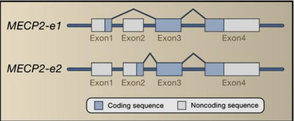
MECP2 consists of four exons that code for two different isoforms of the protein produced by alternative splicing of a short segment at the extreme N-terminus of the protein (Figure 5).
Figure 5: Alternative splicing forms of the MECP2: MECP2-e1 and MECP2-e2.
The MeCP2-e2 isoform was the first identified variant of MeCP2 and therefore the best characterized, but the MeCP2-e1 isoform is more abundant in the brain of both mouse and human (Mnatzakanian et al, 2004).
The MECP2-e1 isoform contains 24 amino acids encoded by exon 1 and lacks the 9 amino acids encoded by exon 2, whereas the start site for the MECP2-e2 isoform is in exon 2 (Dragich et al, 2007; Kriaucionis & Bird, 2004; Mnatzakanian et al, 2004). The two splice variants differ in translation efficiency and are expressed at different relative amounts in different tissues. MECP2-e1 is more abundant in the brain, thymus and lung and during neuronal differentiation. However, recent studies show that both of these isoforms co-localize to heterochromatic regions in murine fibroblastic cells (Kumar et al, 2008). These
18 two MeCP2 isoforms present differences in structure and distribution, but not in function. MeCP2 protein levels are low during embryogenesis and increase progressively during the postnatal period of neuronal maturation (Balmer et al, 2003; Cohen et al, 2003; Kishi & Macklis, 2004; Mullaney et al, 2004; Shahbazian et al, 2002b). Both MeCP2 isoforms are nuclear and co-localize with methylated heterochromatic foci in mouse cells. A recent report suggests that MeCP2 translocates to the nucleus upon neuronal differentiation (Miyake & Nagai, 2007).
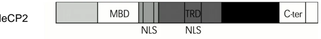
Since MeCP2 is expressed in mature neurons and its levels increase during postnatal development, MeCP2 may play a role in modulating the activity or plasticity of mature neurons. Consistent with this, MeCP2 mutations do not seem to affect the proliferation or differentiation of neuronal precursors. Although the mechanisms that regulate the complex MeCP2 expression patterns are unknown. Recent studies identified the core promoter and several cis-regulatory elements that drive MeCP2 expression (Liu & Francke, 2006). These regulatory sequences may dictate the spatial and temporal patterns of MeCP2 expression. MeCP2 is composed of three domains: the methyl-binding domain (MBD), the transcriptional repression domain (TRD), and a C-terminal domain, in addition to two nuclear localization signals (NLS) (Figure 6).
19 MeCP2
Figure 6: MeCP2 function domains. MBD methyl-binding domain; TRD transcriptional repression domain; C-ter C-terminal domain and NLS nuclear localization signals.
The MBD, an 85 amino acids domain is located on exons 3 and 4 (from amino acid 78 to 162 in MeCP2), and specifically binds to methylated CpG dinucleotides, with preference for CpG sequences with adjacent A/T-rich motifs (Klose et al, 2005). MBD also binds to unmethylated four-way DNA junctions with a similar affinity (Galvao & Thomas, 2005), implicating a role for the MeCP2 MBD in higher-order chromatin interactions. The TRD is a 104 amino acids domain, situated on exon 4 (from amino acid 207 to 310), important in generating a physical association with the transcriptional corepressor Sin3a, which recruits the histone deacetylases HDAC1 and HDAC2. These histone deacetylases remove acetyl groups from histones, resulting in a compact chromatin structure that represses local gene expression (Jones et al, 1998; Nan et al, 1997). The C-terminal region of MeCP2 is not yet well characterized, it is clearly essential for protein function as evidenced by the numerous RTT-causing mutations that involve deletion of this domain, and the fact that a mouse model lacking the MeCP2 C-terminus reproduces many RTT phenotypes (Shahbazian et al, 2002a). The C-terminal region has been described as containing a WW binding domain important for MeCP2 interactions with splicing factors (Buschdorf & Stratling, 2004). WW domains are characterized by the presence
20 of 2 tryptophan residues (W) that are separated by 20-22 amino acids and that recognize proline residues of interacting ligands.
The function of MeCP2 as a transcriptional repressor was first suggested on the basis of in vitro experiments in which MeCP2 specifically inhibited transcription from methylated promoters (Nan et al, 2007). When MeCP2 binds to methylated CpG dinucleotides of target genes via its MBD, its TRD recruits the corepressor Sin3A and HDAC 1 and HDAC 2 (Jones et al, 1998; Nan et al, 1998)( Figure 7).
Figure 7: MeCP2 as a transcriptional repressor. MeCP2 binds to methylated CpG upstream of the transcriptional start site of a MeCP2 target gene.
The transcriptional repressor activity of MeCP2 involves compaction of chromatin by promoting nucleosome clustering, either through recruitment of HDAC and histone deacetylation or through direct interaction between its C-terminal domain and chromatin (Nikitina et al, 2007). Additional MeCP2-interacting proteins include the catalytic component of the SWI/SNF chromatin- remodeling complex Brahma (at least in NIH 3T3 cells), the DNA methyltransferase DNMT1, the histone methyltransferase Suv39H1, the
21 transcription factors TFIIB and PU.1, the corepressors c-Ski and N-CoR, LANA, and the SWI2/SNF2 DNA helicase/ATPase responsible for a-thalassemia/ mental retardation syndrome X-linked (ATRX) (Harikrishnan et al, 2005; Kaludov & Wolffe, 2000; Kimura & Shiota, 2003; Nan et al, 2007).
In 2005, has been demonstrated that MeCP2 in addition to its role as a global repressor, acts as a splicing regulator (Young et al, 2005). The authors identified the RNA-binding protein Y box-binding protein 1 (YB1), a principal component of messenger ribonucleoprotein particles that controls multiple step of mRNA processing, as a MeCP2 binding partner. The functional significance of this interaction was investigated by determining whether the MeCP2-YB1 complex affects mRNA processing and splice-site selection. It has been shown that in MeCP2-deficient neurons, the splicing is altered, and aberrantly spliced transcripts can be produced (Young et al, 2005).
Given that MeCP2 interacts with other proteins, chromatin, DNA, and RNA, it is clearly a multifunctional protein, with roles in chromatin remodeling and RNA splicing.
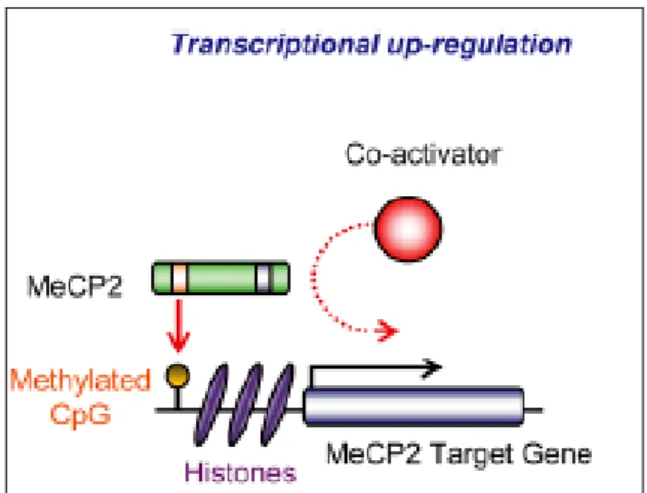
In contrast to the data showing that MeCP2 is only a transcription inhibitor, in 2008, Chahrour and colleagues carried out a study to examine gene expression patterns in the hypothalamus of MeCP2-null and transgenic mice. Surprisingly, authors found that the majority of genes displaying altered expression were up-regulated rather than down-up-regulated by MeCP2 (Figure 8), and that, in these neurons, MeCP2 protein was directly associate with the transcriptional activator CREB1 (Chahrour et al, 2008). An increased level of CREB1 induces miR132 microRNA and represses MeCP2 translation suggesting a negative regulatory loop between MeCP2 and CREB1 (Klein et al, 2007). These results confirm
22 another study, in which, by using ChIP-on-chip analysis in SH-SY5Y cells, MeCP2 has been found associated more frequently with promoters that are also associated with RNA polymerase II (Yasui et al, 2007). All these data suggest that MeCP2 would be a "transcriptional modulator" rather than a repressor.
Figure 8: MeCP2 as a transcriptional activator. MeCP2 recruits a transcriptional coactivator to cause the transcriptional upregulation of a target gene.
1.3.1 Tissue expression of MeCP2
Several studies, carried out in rodents (Cassel et al, 2004; Mullaney et al, 2004; Shahbazian et al, 2002b) monkeys (Akbarian et al, 2001) and human (Shahbazian et al, 2002b), were aimed to analyze the expression profile of MeCP2. From these studies appears that MeCP2 is expressed in many tissues. Analysis on mice tissue samples revealed that the MeCP2 protein is not abundant in liver, stomach and small intestine, moderately expressed in kidney and heart and highly abundant in brain, lung and spleen (Shahbazian et al, 2002b). Within the cerebral tissue, MeCP2 is not abundant in astrocytes
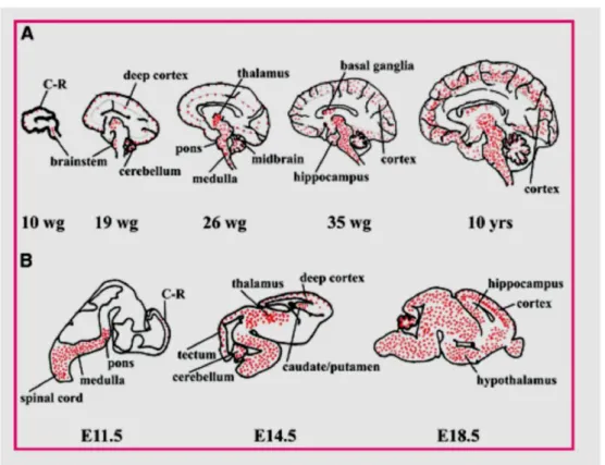
23 (Maezawa et al, 2009) and immature neurons, but is preferentially expressed in mature neurons. The timing of its appearance correlated with the ontogeny of central nervous system. MeCP2 expression followed a similar time course in both mouse and human, with its early expression in the spinal cord and brainstem, and late expression in the hippocampus and cerebral cortex (Figure 9).
Figure 9: Schematic representation of the spatial and temporal distribution of MeCP2 during human (A) and mouse (B) development. Wg, weeks of gestation.
Direct quantification in adult mouse brain has estimated ∼16 million molecules of MeCP2 per nucleus in neurons with almost an order of magnitude less in glial cells and 30-fold less in liver cells (Skene et al, 2010). The neuronal MeCP2 level is relatively low at birth but, in the mouse, increases greatly during the first
24 3 weeks of life before reaching a plateau (Kishi & Macklis, 2004; Skene et al, 2010). Because neurogenesis is largely complete before birth, the increase is due to up-regulation of MeCP2 expression within a constant number of neurons. These neurons are, however, developmentally active, undergoing synaptogenesis at this time. MeCP2 expression varies between neuronal populations from different regions and different structures within the central nervous system (LaSalle et al, 2001). MeCP2 expression is regulated in a developmental stage and cell type specific manner. Few things are known about molecular mechanisms implicated in this regulation. It was shown that the MeCP2 gene contains multiple transcription starting sites embedded in a region that is GC rich and contains CpG islands. In this study has been showed that the mouse MeCP2 promoter does not contain any canonical boxes like TATA or CAAT. They also identified a promoter region (-677/+56) that is responsible for the expression of MeCP2 in neuronal cells. In this region, there is a positive regulatory element of 19 bp (-64 to +46) that controls the major activity of the promoter region (Adachi et al, 2005).
Anatomically, MeCP2 deficiency causes reduced brain size. This may be due to size decreases in major brain regions such as the frontal and temporal lobes, caudate nucleus, thalamus, midbrain, and cerebellum, all of which have been documented in RTT patients (Armstrong et al, 2001; Reiss et al, 1993; Saywell et al, 2006; Subramaniam et al, 1997). At the cellular level, the neuronal soma is smaller in the absence of MeCP2, and cells are more densely packed, with decreased dendritic complexity (Armstrong, 2005; Chen et al, 2001; Kaufmann & Moser, 2000; Taneja et al, 2009). Importantly, degeneration and atrophy are not observed, establishing the notion that RTT is a postnatal developmental disorder,
25 rather than a neurodegenerative disorder (Armstrong, 2005; Jellinger et al, 1988; Jellinger & Seitelberger, 1986).
More specific abnormalities have been observed at the synapses. Postmortem brain samples from RTT patients or MeCP2-deficient mice present postsynaptic morphological defects such as reduced dendritic branching, reduced dendritic spine density, and defects in spine morphology (Armstrong et al, 1995; Armstrong et al, 2001; Belichenko et al, 1994; Chapleau et al, 2009; Fukuda et al, 2005; Kishi & Macklis, 2004; Schule et al, 2008; Smrt et al, 2007) (Figure 10).
Figure 10: Schematic representation of pyramidal neurons from control and Rett brains.
Presynaptically, lack of MeCP2 is associated with an abnormal number of axons (Belichenko et al, 2009a) and a defect in axonal targeting (Belichenko et al, 2009b; Matarazzo et al, 2004; Palmer et al, 2008).Overall, the structural defects described at the synapse would suggest that loss of MeCP2 triggers alterations in the functioning of the synapses and, consequently, of the neuronal networks.
26 Analysis of neurotransmission associated with loss of MeCP2 provides further evidence for synapse dysfunction. Postmortem analysis in RTT brains showed altered levels of neurotransmitters such as glutamate and biogenic amines as well as changes in the abundance of some neurotransmitter receptors.In mice, reduced levels of serotonin (5- hydroxytryptamine), adrenaline, and dopamine have been found in the MeCP2-null brain (Ide et al, 2005; Isoda et al, 2010; Samaco et al, 2009; Santos et al, 2010). Analysis of spontaneous miniature excitatory and inhibitory postsynaptic currents indicated a shift in the excitatory/inhibitory (E/I) balance, with increased excitatory and decreased inhibitory neurotransmission in the hippocampus and cortex (Chao et al, 2007; Dani et al, 2005; Nelson et al, 2006; Wood & Shepherd, 2010; Zhang & Minassian, 2008). This is supported by data showing pre- and postsynaptic defects of GABA and therefore inhibitory, neurotransmission in the brainstem (Medrihan et al, 2008). Consistent with the idea of disturbance of the E/I balance, long-term potentiation (LTP) is also altered in the hippocampus of symptomatic MeCP2-deficient mice (Asaka et al, 2006; Guy et al, 2007; Weng et al, 2011). These data, added to the morphological studies, imply that loss of MeCP2 causes malfunction of numerous synapses throughout the brain, which creates less efficient neuronal networks and gives rise to RTT-like phenotypes.
1.3.2 MeCP2 genes target
The identification of MeCP2 genes target has always been one of the main objectives of many researchers. This will allow you to better understand the correlation between the presence of MeCP2 mutations and clinical manifestations of RTT and to think about how to make up for excess or deficiency of certain
27 proteins in these patients.Many studies with microarrays, with which it is possible to simultaneously evaluate the expression of a large number of genes, have contributed to uniquely identify a few target genes (Chahrour & Zoghbi, 2007) (Figure 11).
Figure 11: MeCP2 Target Genes.
In 2003, BDNF has been identified as the first MeCP2 target in mammals (Chen et al, 2003; Martinowich et al, 2003). BDNF is a neurotrophin essential for survival, growth, differentiation and maintenance of neurons during development (Ghosh et al, 1994). In the brain, it is active in the hippocampus, cortex, and basal forebrain-areas vital to learning, memory, and higher thinking (Yamada & Nabeshima, 2003). BDNF itself is important for long-term memory (Bekinschtein et al, 2008).
28 The role of BDNF in the pathogenesis of RTT is still unknown and it is unknown how the absence of MeCP2 results in a malfunction of BDNF and if this contributes to the neurological patients phenotype. In cultured neonatal cortical neurons, basal BDNF transcription is repressed by MeCP2 in the absence of neuronal activity, but activity-dependent upregulation of BDNF is unaffected by MeCP2 deletion (Chen et al, 2003).Chang and colleagues have shown that BDNF protein level in the whole-brain lysate in MeCP2 mutant mice was decreased to about 70% of the wild-type level (Chang et al, 2006). To further investigate the in vivo role of BDNF in RTT, BDNF expression has been manipulated in the postnatal brains of MeCP2-deficient mice and discovered that deleting BDNF from the MeCP2 mutant brain resulted in an earlier onset/accelerated disease progression, whereas overexpressing BDNF in the MeCP2 mutant brain led to later onset/slower disease progression. So they demonstrated in vivo a functional interaction between MeCP2 and BDNF and suggested that RTT may be a human disease that is partially mediated through BDNF.
Recently, Zhou and colleagues demonstrated that the phosphorylation of a specific amino acid residue S421 of MeCP2 controls the ability of the protein to regulate dendritic patterning, spine morphogenesis and the activity dependent induction of Bdnf transcription (Zhou et al, 2006). These findings suggest that, by triggering MeCP2 phosphorylation, neuronal activity regulates a program of gene expression that mediates neuronal connettivity in the nervous system.
29
1.4 Rett Mouse Models
With the discovery of the gene that causes the Rett Syndrome, several MeCP2 deficient mice models of RTT have been developed. Their creation has allowed examine the relationship between molecular and anatomical changes of the disease and associated behavioral abnormalities. These models also represent an extraordinary tool to test therapeutic interventions that may lead to improved an eventual cure for this devastating disease (Stearns et al, 2007).
Several mouse models currently exist (Figure 12).
Figure 12: Lifespan and progression of symptoms of selected mouse models.
Three constitutive knockout models result in the loss of functional MeCP2 protein and were created using the technique of Cre-loxP recombination (Nagy, 2000). In Dr Bird’s laboratory, mice with deletions of exons 3 and 4 of MeCP2 (Mecp2tm1-1Bird ,which comprises nearly the entire protein) were created (Guy et al, 2001). Dr Jaenisch’s lab produced a MeCP2-deficient mice model in which was
30 deleted only exon 3 (Mecp21lox), which contains a large part of the MBD domain (Chen et al, 2001). Both models produce null mice and MeCP2-heterozygotes, and result in similar phenotypes. MeCP2-null male mice are apparently healthy at birth until 3 weeks of age. After this period, mice begin to show neurological symptoms like those observed in RTT patients: stiff and uncoordinated gait, hind limb clasping, and irregular breathing. Uneven wearing of the teeth and misalignment of the jaws are also observed. Testes of MeCP2-null males were always internal. Symptoms progression leads to weight loss and early death around 54 days. Brain architecture in null mice is grossly normal, although a slight decrease in the size and weight can be noticed in comparison with wild-type littermates. This is due to neurons compaction in hippocampus, cerebral cortex and cerebellum. Total brain weight was reduced 9 to 13% compared to wild-type controls. Cortical brain volume was decreased between 7 to 11% and hippocampal volume was decreased 8% (Belichenko et al, 2008).By comparison, an autopsy study of Rett girls found brain weight to be reduced 12 to 34% relative to age-matched controls (Jellinger et al, 1988). MeCP2+/− females mice are viable, fertile and grow normally until adulthood. At 3 months, they start showing hind limb clasping and by 6 months they show RTT phenotypes such as breathing irregularity and inertia. These results show that MeCP2-null mice can be a good model to study RTT because of delayed onset of neurological symptoms affecting posture, gait, breathing and spontaneous movements. In 2006, Dr Patrick Tam generated mice with a targeted deletion of the methyl binding domain (MBD) resulting in complete loss of MeCP2 protein (Mecp2tm1Tam, (Pelka et al, 2006). MeCP2tm1Tam phenotype is comparable with that of the Jaenisch and
31 Bird’s mice phenotype. In addition, at 8 to 10 weeks after birth, they display reduced level of anxiety, locomotors activity and cerebellar learning (Figure 13).
Figure 13: Rett mice phenotype.
In the fourth model, the MeCP2 protein is truncated after codon 308 (Mecp2308), retaining several key functional domains (Shahbazian et al, 2002a). In these mice the phenotype is milder than those seen previously. MeCP2308/y are normal until 6 weeks and then they develop symptoms like tremors, kyphosis, learning and memory deficits, social behavior abnormalities, etc. Heterozygote females MeCP2308/+ have impaired motor features starting at 35-39 weeks after birth. As in the case of human female patients, these mice show phenotypic variability due to the XCI. MeCP2-deficient models are reminiscent of RTT in that autonomic misregulation occurs (tremor, breathing irregularities, weight abnormalities), smaller brain and neuronal size is observed, posture and motor coordination are abnormal, and seizures may be present (Chen et al, 2001; Guy et al, 2001; Shahbazian et al, 2002a). Additionally, Rett mice show a similar developmental progression to Rett girls, appearing normal at birth, achieving motor and developmental milestones early on, and showing a delayed onset of symptoms. Mecp2-deficient mice show similar neurochemistry to Rett girls; cholinergic markers are low in the brains of Rett girls and Rett mice show phenotypic improvement in response to dietary cholinergic supplementation (Nag &
Berger-32 Sweeney, 2007; Wenk, 1997). Also, studies on Rett mice models are consistent with the view that RTT is mainly a synaptic disorder. Synaptic plasticity is impaired in Rett mice models, dendritic spines, the principal site of excitatory neurotransmission, are greatly reduced, and there appears to be an overall imbalance between excitation and inhibition (Armstrong, 2005; Dani et al, 2005). Recently neuron-specific or regional MeCP2 deletion studies reproduce some aspects of Rett syndrome. Has been demonstrated that loss of MeCP2 from dopaminergic neurons causes motor incoordination whereas loss from serotoninergic neurons leads to increased aggression (Samaco et al, 2009); furthermore the loss of MeCP2 from amygdale impairs amygdale-dependent learning and memory (Adachi et al, 2009) and MeCP2 deletion in hypothalamic Sim1-expressing neurons leads to alterations in feeding behavior, aggression and stress response (Fyffe et al, 2008). Postnatal loss of MeCP2 from forebrain excitatory neurons produces motor incoordination, increased anxiety-like behaviours and impaired fear conditioning and social behavior (Gemelli et al, 2006). In the last year Lioy and colleagues showed that in globally MeCP2-deficient mice, reexpression of MeCP2 preferentially in astrocytes significantly improved locomotion and anxiety levels, restored respiratory abnormalities to a normal pattern, and greatly prolonged life span compared to globally null mice. Furthermore, restoration of MeCP2 in the mutant astrocytes exerted a non-cell-autonomous positive effect on mutant neurons in vivo, restoring normal dendritic morphology and increasing levels of the excitatory glutamate transporter VGLUT1. Finally, they concluded their study showed that glia, like neurons, are integral components of the neuropathology of Rett syndrome, and supported the targeting of glia as a strategy for improving the associated symptoms.
33
1.5 Oxidative stress in Rett Syndrome
The antioxidant enzymatic system is one of the most important free radical detoxification mechanisms. The enzymes act in equilibrium, and any unbalance of this system may provoke oxygen-derived free radical generation. Free radicals are extremely reactive molecules that can disrupt lipid cell membranes, destroy cell enzyme functions, alter DNA, and lead to cell death (Shadid et al, 1998; Thomas & Aust, 1985). Free radicals appear to play a role in the pathogenesis of Rett syndrome (Sofic et al, 1987).
There is an increasing amount of experimental evidence that oxidative stress caused by oxygen free radicals is involved in the neuropathology of several neurodevelopmental and neurodegenerative disorders, as well as in stroke and seizures (Coyle & Puttfarcken, 1993).
A growing body of evidence supported the association between oxidative stress and stereotyped movements in different animal models and in patients affected by neurological disorders such as obsessive compulsive disorders or autism (Ghanizadeh, 2011; Ghanizadeh, 2012; Guldenpfennig et al, 2011).
Elevated cerebrospinal fluid glutamate has been found in Rett syndrome (Hamberger et al, 1992). The neurotransmitter glutamate can provoke oxidative stress in cells (Coyle & Puttfarcken, 1993). Moreover, a postmortem study of a case with RTT showed a severe reduction of ascorbic acid and reduced glutathione levels in most brain regions (Sofic et al, 1987). Ascorbic acid and glutathione play an important role in the antioxidant control system, preventing excessive accumulation of free radicals in living tissue. The authors suggested that the impairment of these defense mechanisms could indicate a process of progressive neurological illness with mental retardation and delayed motor
34 development. High levels of malondialdehyde reflect peroxidative damage of bio-membranes that might contribute to progressive dementia, impaired motor function, behavioral changes, and seizures (Buss & Winterbourn, 2002; Dalle-Donne et al, 2003). Any relationship was found between malondialdehyde levels and the clinical stages of pathology in a large number of patients. However, malondialdehyde levels were significantly lower in patients with moderate phenotype compared with patients with severe clinical phenotype. These results indicate that free radicals generated from oxidation reactions might contribute to the pathogenesis of Rett syndrome.
Formichi et al. (Formichi et al, 1998) found low serum vitamin E levels in nine of 28 patients with RTT. These results indicated that the oxidative free radical metabolism might be impaired in some patients with Rett syndrome.
Interestingly, Brain-derived neurotrophic factor (BDNF), a MeCP2 gene target, that is down-regulated both at the RNA and protein levels in MeCP2-null mice, can protect neurons against oxidative damage resulting from different neuropathologic insults(Mattson, 2002). Is reasonable to assume that oxidative stress detected in Rett syndrome is at least partly due to decreased levels of BDNF. Likewise, the cAMP-responsive-element binding protein (CREB) is recently reported as another molecular target of MeCP2 (Chahrour et al, 2008), can produce a lipid peroxidation-induced differential regulation thought to play a role in neurodegenerative disease such as Alzheimer disease (Pugazhenthi et al, 2006).
35
1.6 Vascular Function
While the neurological phenotype of the Rett syndrome has been well-characterized in animal models and in humans, few studies focused on alterations in the cardiovascular system of MeCP2 deficient mice (Bissonnette et al, 2007; McCauley et al, 2011), but nobody specifically investigated the vascular functions in this pathology.
An important role in the vascular functions is played by the endothelium: a thin layer of cells that serves not merely as a passive barrier between flowing blood and the vascular wall but uses this strategic location to maintain vascular homeostasis. The healthy endothelium is able to respond to physical and chemical signals by production of a wide range of factors that regulate vascular tone, cellular adhesion, thromboresistance, smooth muscle cell proliferation, and vessel wall inflammation.The importance of the endothelium was first recognized by its effect on vascular tone. Pioneering experiments by Furchgott and Zawadzki1 showed that the presence of intact endothelium was essential for acetylcholine (ACh) to induce dilation of isolated arteries. In contrast, if the endothelium was removed, the arteries constricted in response to ACh. Subsequent studies revealed that ACh stimulated the release of a potent vasodilating substance by the endothelium, identified as nitric oxide (NO) (Furchgott, 1996; Ignarro et al, 1987). NO is probably the most important and the best characterized mediator, and its intrinsic vasodilator function is commonly used as a surrogate index of endothelial function. It is generated from L-arginine by the action of endothelial NO synthase (eNOS) in the presence of cofactors such as tetrahydrobiopterin (Forstermann & Munzel, 2006). NO produces vasodilation primarily by activating guanylyl cyclase in vascular smooth muscle cells, which increases intracellular
36 concentrations of cyclic-3’,5’-guanosine monophosphate (cGMP) (Arnold et al, 1977). cGMP in turn acts as a second messenger, activating cGMP-dependent protein kinase, which decreases cytosolic calcium concentration and modulates ion channel function leading to relaxation of vascular smooth muscle cells (Lincoln & Cornwell, 1993). NO inhibits platelet aggregation and adhesion by a guanylyl-cyclase mechanism (Radomski et al, 1990). Endothelium-devived NO is an inhibitor of leucocyte adhesion at the vessel wall (Kubes et al, 1991), and has anti-mitogenic effects on vascular smooth muscle cells (Garg & Hassid, 1989) (Figure 14). Thus, it is clear how in normal vascular physiology, NO plays a key role to maintain the vascular wall in a quiescent state by inhibition of inflammation, cellular proliferation, and thrombosis. Normal NO release opposes the vasoconstrictor responses to clinically relevant stimuli including catecholamines and serotonin (Golino et al, 1991; Vita et al, 1992). NOS inhibition is associated with increased systemic blood pressure, decreased blood flow responses to exercise and local ischemia (Gilligan et al, 1994; Meredith et al, 1996), and shortened bleeding time.
37 Failure of endothelium to elicit NO-mediated vasodilation is caused by reduced bioavailability of endothelium-derived NO due to either decreased formation or accelerated degradation. A large body of evidence shows that accelerated degradation of NO by reactive oxidant species (ROS) accounts for a large proportion of reduced NO bioavailability and endothelial dysfunction in vascular disease states. Both excess generation of ROS including superoxide anion and oxidized LDL cholesterol and decreased antioxidant defense mechanisms contribute to enhanced degradation of NO (Kojda & Harrison, 1999).
Superoxide anion can directly inactivate NO by reacting very rapidly to form peroxinitrite, which is vasoinactive and a powerful, damaging oxidant more stable than either superoxide or NO (Gryglewski et al, 1986; Rubanyi & Vanhoutte, 1986). In human blood vessels, increased superoxide production is also associated with impaired NO-mediated vasorelaxation (Landmesser et al, 2000).
Another consequence of increased production of ROS in the vasculature is lipid peroxidation. Oxidized LDL is cytotoxic to endothelial cells. It can react with NO directly and eliminate its biological activity or interfere with signal transduction and receptor-dependent stimulation of NOS activity and with activation of guanylyl cyclase.
Given the relationship between increased oxidative stress in the vasculature and impaired endothelial vasodilator function, researchers considered the possibility that augmenting antioxidant defenses would have a beneficial effect. For example, Vitamin E supplementation improves the bioactivity of endothelium-derived NO in hypercholesterolemia and in diabetes mellitus (Andersson et al, 1994; Keegan et al, 1995). Vitamin C improves endothelium-dependent vasodilation by restoring nitric oxide activity in essential hypertension (Taddei et al, 1998).
38 Reduced NO formation may also contribute to endothelial dysfunction. Although the NO-producing enzyme eNOS is constitutively expressed by endothelial cells, its expression is subject to modulation by shear stress, atherogenic lipoproteins, and cytochines. Altered eNOS expression may affect NO synthesis. Several pathways can regulate eNOS expression and function.
In vitro data demonstrate a selective and differential presence of MeCP2 at
promoter level of eNOS endothelial cells and vascular smooth muscle cells (Chan et al, 2004; Fish et al, 2005). These findings are in line with data in humans, reporting elevated plasma levels of oxidative stress in MeCP2-RTT patients, together with a decreased superoxide dismutase activity, and increased markers of lipid peroxidation (De Felice et al, 2009; Sierra et al, 2001).