La Sapienza Università Rome, Italy
DOTTORATO DI RICERCA IN BIOCHIMICA CICLO XXXI
(2015-2018)
Cross Talk Between Impaired Brain Glucose
Metabolism and Tau hyperphosphorylation
An Implication for Alzheimer Disease
PhD Candidate
Nidhi Sharma
Supervisor PhD Coordinator Prof. Marzia Perluigi Prof. Stefano Gianni
Eugenio Barone
ii
Disclosure
This PhD dissertation has been reviewed by:
Prof David Allan Butterfield 249 Chemistry-Physics Building
University of Kentucky US
Prof Paola Gamba San Luigi Gonzaga Hospital, regione
Gonzole 10, Orbassano (TO), Italy
The work described in this thesis was carried out at LRBN group (Laboratory of Redox Biochemistry in Neuroscience), Faculty of Biochemical Science, La Sapienza University, Rome, 00181, Italy.
iii
To My Family
(Alla mia famiglia)
iv
Nothing in Life is to be feared
It is only to be understood
Now is the time to understand more
So that we may fear less
v
Table of Contents
Title Page no.
Chapter 1: Introduction
1.1 Alzheimer: What do we know? ... 1
1.2 Molecular basis of Alzheimer pathology ... 5
1.2.1 APP and Tau in physiology and pathology ... 6
1.2.2 Glycogen synthase kinase 3-β: A hallmark kinase in AD ... 10
1.2.3 GSK-3β and oxidative stress ... 13
1.2.4 GSK-3β and Insulin resistance (IR) ... 14
1.3 Oxidative stress and Brain: A staircase to AD development ... 16
1.3.1 Oxidative stress ... 16
1.3.2 Oxidative stress in AD brain ... 18
1.3.3 BVR-A/HO-1 System... 21
1.3.4 Role of BVR-A in the regulation of cell stress response ... 23
1.4 Glucose metabolism in the brain ... 28
1.4.1 impaired glucose metabolism, OS and Alzheimer ... .30
1.4.2 Cross talk between O-GlcNAcylation and Phosphorylation ... 33
1.4.3 O-GlcNAcylation of Tau and APP in Alzheimer disease ... 34
Chapter 2: Background and Aim ...
39Chapter 3: Project 1
3.1 Material and Methods ... 443.1.1 Animal ... 44
3.1.2 Human sample ... 44
vi
3.1.4 MTT assay ... 46
3.1.5 Slot blot analysis ... 46
3.1.6 Sample preparation ... 47 3.1.7 Western blot ... 48 3.1.8 Immunoprecipitation ... 49 3.1.9 Immunofluorescence ... 50 3.1.10 Statistical analysis ... 51 3.2 Results ... 52 3.3. Discussion ... 71
Chapter 4: Project 2
4.1 Material and Methods ... 784.1.1 Animals ... 81
4.1.2 Sample preparation ... 79
4.1.3 Two-dimensional (2D) electrophoresis and 2D blots ... 79
4.1.4 Image analysis ... 80
4.1.5 In-gel trypsin digestion/peptide extraction ... 81
4.1.6 MS/MS characterization of peptides ... 82 4.1.7 MS data analysis ... 82 4.1.8 Immunofluorescence ... 83 4.1.9 OGA assay ... 84 4.1.10 Western blot ... 84 4.1.11 Immunoprecipitation ... 85 4 .1.12 Statistical Analysis ... 86 4.2 Result ... 87 4.3. Discussion ... 101
vii
Chapter 5: Conclusion
... 113Appendix
... 117Reference
... 120viii GLOSSARY
APOE Apolipoprotein
APP Amyloid precursor protein ARE Antioxidant-responsive elements Aβ Beta Amyloid
ACH Amyloid cascade hypothesis AD Alzheimer Disease
BR Bilirubin
BVR Biliverdin reductase BV Biliverdin
CAT Catalase
DMEM Delbecco’s modified Eagle’s medium EAD Early-onset AD
FAD Familial Alzheimer disease GSK-3β Glycogen Synthase 3β
HBP Hexose biosynthesis pathway HBSP Hexosamine biosynthetic pathway HEK Human embryonic kidney
HNE 4-hydroxynonenal HO Heme-oxygenase H2O2 Hydrogen peroxide
IGF Insulin-like growth factor IRK Insulin receptor kinase
iNOS Inducible nitric oxide synthase IPL Inferior parietal lobule
LAD Late-onset AD
MARK Microtubule-affinity-regulating kinase MARE Maf recognition element
MCI Mild Cognitive impairment MLSP Maximum lifespan potential MMSE Mini Mental State Examination MS Mass Spectrometry
NOX NAPDH oxidase Nrf2 NF-E2-related factor 2 OS Oxidative stress OGA O-GlcNAcase
OGT O-GlcNAc transferase PBS Phosphate buffer saline
ix PC Protein carbonyl
PCAD Preclinical AD PD Parkinson disease
PET Positron emission tomography PHF Pre-helical filaments
PP2A Protein phosphatase 2A PS-1 Presenilin
PTM Post-translation modifications PUFA Polyunsaturated fatty acid RNS Reactive nitrogen species ROS Reactive oxygen species SiRNA short interfering RNA SOD Superoxide dismutase SREs Stress responsive element T2DM Type 2 diabetes mellitus WT Wild Type
3-NT 3-nitrotyrosine 3×Tg-AD Triple transgenic
1
Chapter 1
Introduction
1.1 Alzheimer: What do we know?
“Time is the hallmark of aging, that fades away your memories with the greatest challenge”
The word Alzheimer, first time coined by a Bavarian psychiatrist and neuropathologist “Alois Alzheimer” in 1907 when his 51 years old female patient diagnosed with unfamiliar psychiatric symptoms and given a description of this pathological syndrome. Eventually this clinico-pathological symptom defines later a new neuroclinico-pathological phenotype of the brain. He described it as a “peculiar disease of cortex” and found the strange alteration of the “neurofibrils” and “Foci (Focal deposits)” which are build up by peculiar substance and spread over the entire cortex along with the aging. (Alzheimer 1907).
In 1980, ninety years later structural evidence of those microscopic lesions confirmed as a “senile (Amyloid) plaques” and “neurofibrillary tangles (NFTs) “by Dr. Alzheimer. In the area of biochemical pathology, George Glenner first isolated and partially characterized the amyloid β-protein from the brains of patients who had died with Alzheimer disease or Down syndrome in 1984 (Glenner, G. G., 1984a; Masters, C. L., 1985). Two years later, several laboratories had identified the microtubule-associated protein Tau, as the principal constituent of the neurofibrillary tangles (Brion,
J. P., 1985; Grundke-Iqbal, I., 1986; KoSIK, K. S., 1986; Nukina, N., 1986;
2
Alzheimer is the most common form of the neurodegenerative disease that affects the vast area of cerebral cortex and hippocampus leads to dementia within the aging (Masters, C. L., 2015).
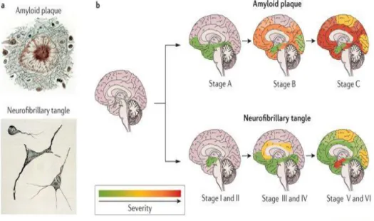
Figure 1: Progression of AD at Aβ deposition (Stage A-C) and neurofibrillary tangles (Stage
I-VI, adopted from Braak and Braak)
Surprisingly AD affects 5.3 million Americans and is the 6th leading cause of death in the US. Alzheimer has been projected as a socially and economically burden on the society, and it reduces the quality of life for the patients who are suffering from this neurodegenerative disease. Prevalence of AD is 3 % for age 65-74 and 18.7% for 75-84 years and 47.2 % for those over 85 years old (Evans, D. A., 1989). On the broad spectrum, this disease has been classified into two categories, based on their occurrence in the
3
Population; 1) familial AD (FAD) affects the people younger than 65 ages and 2) Sporadic affects the people at the age of 65 and older than this. In less than 1% of cases of AD population are the FAD and caused by a specific point mutation in all three proteins; i) APP ii) Presenilin 1(PS-1) and iii) Presenilin 2 (PS-2) (Campion, D., 1999) known as a membrane protein. Some of these mutations inherit to next generations that leads to Aβ and Tau pathology by altering signal transduction probably for protein phosphatase 2A (PP2A) and glycogen synthase-3β (GSk-3β) (Iqbal, K.,2005) but not always this alteration shows significant changes or instead decrease Aβ generations, which have been observed in longitudinal studies (Blanchard, J., 2010). However, for instance, some individuals from FAD/sporadic AD cases are characterized by APOE polymorphism at the genetic level with three alternative allele expressions (ε2, ε3, ε4). Apolipoprotein E (APOE) is lipolipoprotein controls synaptic repair in case of tissue injury and maintains the synaptic structure at the tissue level (Poirier, J., 1995, Soininen, H., 1995) but also considered as a risk factor (other than age) for AD development (Strittmatter, W. J., 1993).
To provide the preventive attention and care , It is important to define the risk factors for AD and they are mostly, aging (Evans, D. A., 1989), presence of APOE4 alleles (Strittmatter, W. J., 1993), a family history for AD dementia (Henderson, A. S., 1986), head injury ( Mortimer, J. A., 1985), low educational attainment (Zhang, M.,1990) and low linguistic ability early in life (Snowdon, D. A., 1996). The very first manifestation of a dementing disorder is behavioral symptoms those appear before the cognitive declines (Rubin, E. H., 1989) and later during the course of illness (Rubin, E. H., 1989; Petry, S., 1988). Early detection of these symptoms is extremely important as it does cause the impairment in daily living activities (Lyketsos, C. G., 1997; Mok, W. Y., 2004), accelerate cognitive decline (Stern, Y.,1987)
4
and worsen patient's quality of life (González‐Salvador, T., 2000). In the symptomatic assessment, episodic memory lost very early because at the earliest of AD usually neurofibrillary changes occur in medial temporal lobe structures e.g., hippocampus, entorhinal cortex (Braak, H., & Braak, E., 1991) which is critical for episodic memories and thus a deficit in the ability to learn and remember new information (i.e., anterograde amnesia) which is something not surprising but a clinical hallmark of earliest AD (Warren, J. D., 2012). However, the short-term or working memory is maintained until very late in the course of MCI or AD development (Perry, R. J., 1999; Chen, P., 2001).
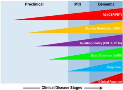
Based on diagnosis stages, Alzheimer disease has categorized into preclinical AD (PCAD), amnestic mild cognitive impairment (MCI), early-onset AD (EAD), and a late-stage AD (LAD) respectively. MCI is often referred to as the first stage or transition stage of PCAD to AD, and it is the stage in which the patient and those around them begin first to notice signs of memory loss (Jicha, G. A., 2006). Aβ load and NFT density increases in the number and the concentration through the progression of MCI to LAD. LAD presenting the highest amounts and subsequently the lowest neuron density and highest cognitive impairment and ultimately death. However, PCAD is an unusual stage in which the patients have significant levels of Aβ and NFT yet perform normally in cognitive testing and activities of daily living (Bradley, M. A., 2010).
5
Figure 2: Different stages of AD progression with different biomarkers
Because of limited availability of diagnostic tools, a definitive diagnosis cannot be made without neuropathological observation so only post -mortem analysis can reveal the condition of patient’s death, whether it was the AD or some other neurodegenerative disorders.
1.2 Molecular Basis of AD Pathology
At neuropathological level, neurofibrillary tangle (NFT) formation begins in the entorhinal cortex (universal over the age of 65 and considered as a part of healthy aging) and spread to the neocortical region which is the hallmark of AD development or may coincide with another tauopathy. (Bouras, C., 1994; Braak H and Braak E 1995). On the other hand, Amyloid deposition begins in neocortical regions, and while it is a hallmark feature of AD at least
6
40% of individuals dying after age 65 years without signs of dementia but have neocortical amyloid plaques at autopsy suggesting it may be a part of either healthy or “pathological” aging (Bouras, C., 1994; Braak H, Braak E 1997; Price, J. L.,1999). For decades it has been noticed that many nondemented patients have diagnosed with AD-like neuropathology (Dayan, A. D. 1970; Katzman, R., 1988) but recently this incidental pathology has been correlated with the presence of APOE genotype. Some individuals carrying e4 copies and dying at 50’s had some neocortical amyloid depositions, and some of them had neurofibrillary tangles, but no mental status was reported as they died young (Kok, E., 2009). Thus, no clear explanation defines the pathogenesis and occurrence of AD-like characteristics in nondemented patients, and the reasons remain unknown. NFTs and Aβ depositions are region specific and catalyzed by many up-streams signaling cascades. Lately, the molecular mechanism of APP process and NFT formation has become the spotlight in the field of Alzheimer.
1.2.1 Amyloid Protein Precursor (APP) and Tau in Physiology and Pathology
Plaques are extracellular deposits mainly composed of Aβ1-40 and Aβ1-42 peptide those generated by proteolytic cleavage process of the amyloid precursor protein (APP) by β-secretase cleavage and known as the amyloidogenesis (Glenner, G. G., 1984b; Kang, J., 1987). The APP is a transmembrane protein, and the primary function is synaptic formation and repair (Priller, C., 2006) thus it has a critical role in the maintenance of membrane and its level is upregulated during neuronal differentiation (Zheng, H., 2006). APP expression is probably also increased in response to specific
7
genetic, biological, chemical and other environmental insults, all resulting in increased metabolism and production of Aβ. Somehow Aβ is catabolized by neprilysin and insulin-degrading enzyme (Miners, J. S., 2011). Aβ1-42 have the tendency of fibrillization and insolubility than Aβ1-40, and therefore it has higher abundance in the plaques within AD pathology. Interestingly Aβ depositions are supposed to begin 20 years before the development of clinical symptoms (Villemagne, V. L., 2013). Aβ depositions occur in the region of the hippocampus, microglia, (Thal, D. R., 2002; Braak, H., 2006) where they found as extracellularly diffuse or focal deposits (Hyman, B. T., 2012; Montine, T. J., 2012). Aβ (1-42) a significant component of plaques shows more toxicity than Aβ (1-40) (Mohmmad Abdul 2004; Boyd-Kimball, D., 2005; Boyd-Kimball, D., 2005b; Boyd-Kimball, D., 2005c; Mohmmad Abdul, H., 2006).
According to the Amyloid cascade hypothesis (ACH), Aβ deposition is an initial and central trigger to the disease and which subsequently leads to NFTs (neurofibrillary tangles) formations, other secondary events, neuronal cell death, and dementia. Although the mechanism of Aβ neurotoxicity is not fully known and many studies are focused on understanding the basics of Aβ mediated neurotoxicity (Hardy, J. A., 1992; Selkoe, D. J.1991). However genetic studies have identified the mutation in APP (Goate, A., 1991), presenilin-1 (PSEN-1) and presenilin-2 (PSEN-2) genes in the families in early-onset AD (FAD, <60 years) and this could lead to Aβ deposition (Levy-Lahad, E., 1995; Sherrington, R., 1995).This observation resulted in the formulation of “Amyloid cascade hypothesis” in which it has believed that early onset phenotype in FAD shares similarity with late-onset AD and thus these amyloid depositions could explain pathogenesis of all types AD (Reitz, C., 2012).Thus according to the “Amyloid cascade hypothesis”, Aβ is
8
a central to the pathogenesis of AD and has capacity to induce protein oxidation, inhibit the activity of antioxidant enzyme and Aβ toxicity. Later this Aβ toxicity confirms the elevated oxidative damage in the brain. (Butterfield, D.A., 1997; Hardy, J., 2002).
Other than senile plaques, second major neuropathogenesis includes the formation of Tau containing neurofibrillary tangles (NFTs). Moreover, in AD-brain all the isoform of Tau has been identified hyperphosphorylated and accumulated as PHF (pre-helical filaments) (Grundke-Iqbal, I.,1986a; Grundke-Iqbal, I., 1986b; Iqbal, K., 1989).
Conformational changed (Jicha, G. A., 1997; Jicha, G. A., 1999a; Jicha, G. A., 1999b) and truncated Tau (Cotman, C. W., 2005; Gamblin, T. C., 2003; Novak, M., 1991) followed after its hyperphosphorylation were reported and reasonably represents the Tauopathy in AD-brain (Delobel, P., 2008). Tau is a one of the phosphoproteins, and physiologically phosphorylated Tau plays a varied role in microtubule stability, axonal transport, (Cuchillo-Ibanez, I., 2008) neuronal intracellular trafficking and neurite growth (Johnson, G. V., 2004; Götz, J.,2010; Wang, Y.,2015). Normally Tau contains 2-3 mole phosphate per protein moiety at its functional level but in AD brain this phosphorylation increases by 3-4fold (Köpke, E., 1993) and thus hyperphosphorylated Tau inhibits its protein binding affinity and microtubule assembly promoting activity in AD-brain (Grundke-Iqbal, I., 1986a, Lindwall, G., 1984). However basal level of these tangles is the sign of universal aging but abruptly presence of hyperphosphorylated Tau tangles gradually increase the neuron instability, promote the loosening of neuron network and neuron loss (Gómez‐Isla, T., 1997) in some area of hippocampus where processes involved in prior to store experiences into the permanent memories, thus this characteristic
9
memory deficits result in interference for learning and making new memories.
Tau contains mainly serine, threonine and tyrosine sites for phosphorylation but although can be phosphorylated on histidine, lysine, and arginine which determines unfolded native Tau to folded or aggregated functional Tau moiety. Parallel to phosphorylation, many other post-translation modifications (PTM) are subjected to the Tau, i.e. glycosylation (O-linked or N-linked), glycation, deamidation, isomerization, nitration, methylation, ubiquitylation, sumoylation, and truncation, etc but majority of mentioned all PTMs are prominently found in AD brain than normal brain (Wang, Y., 2015). Among the all, phosphorylation depends on a balanced interplay between kinases and phosphatases but has not found persistent under pathological conditions. On the other hand, Phosphorylation can also be affected by O-glycosylation since some serine and threonine residues can either be O-glycosylated or phosphorylated on Tau (Götz, J., 2010).
To date, PHF-Tau has been identified at least for 29 phosphorylation sites (Liu, S. J.,2004). Tau is target for several kinases including, GSK-3β; Cdk5; MAP kinases (JNK, ERK, and p38); microtubule-affinity-regulating kinase (MARK); CK2; DYRK1A; TTBK1; and P70S6 kinase, known as “Tau kinases” (Götz, J., 2010; Dolan, P. J., 2010). Although, thus far only few of them are considered to be good candidates for bona fide in vivo Tau kinases and among them “GSK-3β” is pathologically evident for Tau hyperphosphorylation. GSK-3β is expressed at high levels in the brain (Woodgett JR. 1990), where it localizes to neurons (Leroy, K., 1999), and thus it is in an appropriate compartment to access Tau. GSK-3β also
10
associates with microtubules (Ishiguro, K., 1993) and, when this kinase is overexpressed in cells, the phosphorylation state of Tau dramatically increases at numerous sites (Cho, J. H., 2003; Lovestone, S.,1996; Wagner, U., 1996). However, all the protein kinases that contribute to the pathological phosphorylation of Tau in the AD and other neurodegenerative diseases remain elusive. Amongst, GSK-3β is a potential upstream trigger in Tau pathology in AD-brain (Jope, R. S., 2004) and also facilitates APP proteolysis to generate Aβ (Su, Y., 2004).
1.2.2 Glycogen Synthase Kinase 3-β (GSK-3β): A hallmark kinase in AD
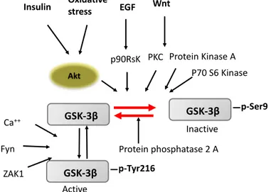
Neuronal cell survival and synaptic plasticity by promoting Tau phosphorylation is one of the activated GSK-3β dependent event and has widely reported in many studies (Mandelkow, E. M., 1992; Hong, J. G., 2012). GSK-3β is ubiquitous serine/threonine kinase under physiological condition and inactivated by phosphorylation through upstream kinases. Activated GSK-3β negatively regulates many cells signaling pathways (i.e., Wnt signaling and insulin signaling pathway) and also promotes cell death (Grimes, C. A., 2001). GSK-3β requires phosphorylation at Tyr216 in the activation loop for its activity (Hughes, K., 1993; Lochhead, P. A., 2006) although this Tyr phosphorylation is thought to be autophosphorylation (Cole, A., 2004), however, phosphorylation by Src family tyrosine kinases has also been reported (Goc, A., 2014). Inhibition of GSK-3β activity regulates by phosphorylation at the N-terminal Ser9 by several protein kinases, such as PKA, PKB, p90RSK and Akt (V 2001; Doble, B. W., 2003; Jope, R. S., 2004, Sutherland, C., 1993; Jensen, J., 2007). Phosphorylated Ser9 binds to a pocket for priming phosphorylation in the substrate-binding
11
region and to reduce the binding affinity for substrates (Dajani, R., 2001; Ter Haar, E., 2001). However, regulation at the molecular level is not yet fully understood, for example, it isnot known what amount of GSK-3β remains inactive without Tyr216 phosphorylation. Therefore, it is unclear whether all GSK-3β molecules are automatically activated by autophosphorylation after de novo synthesis or if they require any signal for activation. For instance, insulin and other growth factors activate Akt, which in turn phosphorylates GSK-3β at Ser9, rendering the kinase inactive and resulting in decreased phosphorylation of downstream substrates, such as glycogen synthase, Tau, β-catenin, etc. (Liu, S. J.,2004; Zhang, Y., 2018).
In general GSK-3β is able to phosphorylate Tau at multiple sites although the most prominent phosphorylation sites are, Ser-198, Ser-199, and/or Ser-202 (Tau-1 site) and Ser-396 and/or Ser-404 (PHF-1 site) (Godemann, R., 1999; Agarwal-Mawal, A., 2003; Lovestone, S., 1996; Sun, W., et al 2002). It has been seen that phosphorylation at Ser396 and Ser404 seems to precede phosphorylation at the other sites (Godemann, R., 1999). GSK-3β preferentially phosphorylates many of its substrates after they have been pre-phosphorylated by other kinases, whereas the stretch of amino acids in Tau that includes the phosphorylatable residues, Ser396, Ser400, and Ser404, can be directly phosphorylated by GSK-3β without the prior activity of other kinases (Leroy, A., 2010; Hanger, D. P., 2011) and thus, these sites apparently have become the potential target to be studied in detail in the AD brain. However, phosphorylation at Ser404 is critical to this process, and substitution of this residue by alanine ablates phosphorylation of both Ser396 and Ser400. It appears, therefore, the primary phosphorylation of Ser404 by GSK-3β can itself serve as a primed residue for the subsequent sequential
12
phosphorylation of Tau at Ser400 and Ser396 by GSK-3β (Leroy, A., 2010; Hanger, D. P., 2011).Nonetheless, a recent study strongly supports and confirms that phosphorylation at Tau Ser 396-404 (PHF-1) is one of the earliest events in the AD and thus this study highlights the chronological order of tau phosphorylation sites along with the AD progression levels (Mondragón‐ Rodríguez, S., 2014). Aiming to identify early events and modification at phosphorylation level, many studies are currently focused on understanding the role of GSK-3β for Tau phosphorylation in early-onset AD-brain.
Since GSK-3β phosphorylates Tau at many sites seen in PHFs, it is suggested that activation of GSK-3β strongly promotes NFTs in the AD-brain but many studies which are focused on understanding the biochemical regulation of GSK-3β in AD brain are in contradiction, and many of them have demonstrated no up-regulation of this kinase in AD-brain. Thus, one of the possibilities is that abnormal hyperphosphorylation might be because some other modifications occur on Tau which makes it a better substrate for phosphorylation (Liu, F., 2002). While another study from Pei and Jin-Jing et al. has investigated the regional and intracellular distributions of active and inactive forms of GSK-3β in brain staged for neurofibrillary changes and demonstrated increased active GSK-3β, but no changes in inactive GSK-3β status were found in AD-brain (Pei, J. J., 1999; Leroy, K., 2002). Further to broaden our understandings, one of the recent epigenetic-based studies suggested the hyperactivity of GSK-3β along with NFT formation could be just targeted in the early stage of AD brain (Braak stage I-II) whereas turn off in a later stage, i.e. braak stage IV-VI (Nicolia, V., 2017). Therefore, the exact mechanism of GSK-3β activation/inhibition in Tau hyperphosphorylation is a misconception and not fully known.
13
Although further work is needed to clarify these controversial findings.
1.2.3 GSK-3β and oxidative stress
Biochemical analysis of different regions (i.e., pre-cortex, cortex, hippocampus, inferior parietal lobule) in postmortem AD-brain at various stages (i.e. pre-clinical AD (PCAD), mid impaired cognition (MCI) and AD) demonstrated that AD brain is highly sensitive to increased oxidative stress (Butterfield, D. A., 2006; Butterfield, D. A.,2007; Sultana, R., 2007 Butterfield DA 2008). Despite of mild OS, increased OS is being addressed in many mediating regulatory pathways, i.e. Akt activation, down-stream inhibited Gsk-3β (Grimes, C. A., 2001) and insulin resistance (Yu, T., 2006). Although contrary to this finding, one noticeable effect of oxidative stress has demonstrated in a recent study mentioning GSK-3β activation and so Tau phosphorylation at the specific site could be mediated by oxidative stress. This study suggested a novel mechanism of GSK-3β regulation in which N-terminal cleavage of GSK-3β is catalyzed by calpain; a calcium-dependent non-lysosomal cysteine protease expressed ubiquitously in mammals. This cleavage results in loss of the inhibitory domain and producing two fragments with kinase activity.
Moreover, it is very well known that oxidative stress usually induces calcium overloads through mitochondria ROS overproductions and results in cellular calcium sparks (Hamilton, D. J., 2013) and calpain activation (Feng, Y.,2013). Based on such studies, activation of GSK-3β is not only restricted to upstream kinases in insulin cascade, i.e. Akt, but also can be achieved through other parallel mechanisms, i.e., inhibited protein phosphatase 2A (PP2A) and activated calpain proteases. These studies widely open the
14
possibilities to further enquire the complexity of GSK-3β activation/inhibition-mediated signaling in AD brain in various aspects.
Figure 3: Multiple mechanism for GSK- 3β activation/inhibition regulation through various
upstream signaling.
1.2.4 GSK-3β and Insulin Resistance (IR): A double sided player in AD and IR
Lately, it has been attributed in few studies that impaired cognition in the AD-brain can be recovered by insulin/glucose administration (Barone, E., 2018). Such evidence directly indicates a crucial link between AD-brain and insulin resistance (impaired glucose utilization). Glucose utilization regulated by insulin and insulin-like growth factor (IGF) but reduced insulin signaling (clinically known as insulin resistance) resulted in inhibited downstream insulin cascade signaling; PI3K/Akt kinase and ultimately led to increased GSK3-β activity (Talbot, K., 2012; Rivera, E. J., 2005; Steen, E.,2005) Tau phosphorylation. (van der Harg, J. M., 2017; 56 Bomfim, T. R., 2012, Zhang, X., 2016). In addition, autopsies were performed on patients with DM and/or
1
Insulin Oxidative stress EGF Wnt
Akt GSK-3β GSK-3β p-Ser9 GSK-3β p-Tyr216 Inactive Active PKC p90RsK Protein Kinase A P70 S6 Kinase Protein phosphatase 2 A Ca++ Fyn ZAK1
15
AD, which resulted in reducing levels and activity of PI3K/Akt signaling pathway in their frontal cortex, with a large amount of phosphorylated GSK-3β and abnormal tau hyperphosphorylated proteins (Steen, E.,2005; Nicolia, V., 2017; Liu, Y., 2011) . Interestingly, the defects of this signaling pathway are more severe in individuals who suffer both DM and AD (T2DM-AD). Excessive activation of GSK-3β through inactivated Akt is closely related to Tau hyperphosphorylation. However, this is not the case always followed in AD progression as other studies reported increased GSK-3 β activation without decreased Akt activation levels in the postmortem AD brain (Griffin, R. J., 2005; Yarchoan, M., 2014; Pei, J. J., 2003; Tramutola, A., 2015). So, it is remained to reconcile all the evidence that can provide a precise mechanism at the specific time point of aging (i.e., early onset to late onset).
Although besides human data, new insights in AD and insulin resistance signaling have been intervened by Eugenio et al., using 3xTg-AD mice model This study demonstrated increased insulin resistance mainly in old age of mice (12 months) which further led to reduced Akt activation and increased GSK-3 β, however intranasal insulin treatment recovered the Akt activation (Barone, E., 2018; Barone, E., 2016). From all the observations, one convincible explanation could be that insulin resistance mediated GSK-3β activation is one of the early events in AD-brain which no longer activated along with the aging. In the agreement of this hypothesis, one another study explained that the mechanism responsible for the increase in Tau phosphorylation in response to insulin deficiency in the late-onset AD is unlikely to be an elevated GSK-3β activity (in fact, a decrease in the active form of the kinase was observed); instead, the specific inactivation of protein phosphatase 2A (PP2A) may be responsible (Liu, C., 2010)
16
1.3 Brain and Oxidative Stress: A Staircase to AD Development
1.3.1. Oxidative Stress
“Reactive oxygen species” are one of the major byproducts of any aerobic metabolism known as free radicals. Principally, a free radical is any chemical species that contains one or more free unpaired electrons in the outer orbital of their atomic structure which makes them highly reactive species that reacts with any other species to achieve the stability (Halliwell, B., 1995). Endogenously ROS production governed by multiple cellular metabolisms: i) NADPH oxidase (NOX) complex in cell membranes, mitochondria, peroxisomes, and endoplasmic reticulum (Muller, F., 2000; Derick, H. A. N., 2001) ii) oxidative phosphorylation of electron chain reaction in mitochondria, iii) in inflammatory responses through neutrophils and macrophages. Exogenously ROS can be generated by several pollutants, smoke, UV radiation, Drugs, hormone, X-Rays, Xenobiotics, and gamma rays through direct or indirect methods (Valko, M., 2006).
The cellular system can tolerate modest and mild oxidative stress and potentially respond to the cellular damage by upregulating stress-responsive gene (e.g., HO-1/2) expression, upregulating antioxidant defense enzyme (e.g., Superoxide dismutase: SOD) and some other protective protein system (Chaperone). Nevertheless, severe OS may be responsible for reversible or non-reversible damage to the DNA, Proteins, and lipids that causes the structural and functional alterations (Nakamura, H., 1997) an imbalance between this two-counteract activity consequences the causes of oxidative stress (OS) (Sies, H., 1989).
17
OS participates in the biological system at various levels. At low concentration, ROS could be responsible for increased free intracellular Ca+2 (Orrenius, S., 1989; McConkey, D. J., 1996) as a second messenger, increased nitric oxide in phagocytosis as a second messenger (Kogishi, J. I.,2000), free iron (Halliwell,1992) to catalyze OH• generation, and as a part of signal transduction (Finkel, T. 2000). ROS has been suggested to act as the second messenger to regulate the activity of protein kinases and phosphatase (Hensley, K., 2000). Contrary, at high concentration (as the result of imbalanced homeostasis) ROS can react with biological macromolecules, i.e. proteins, DNA, Lipid, carbohydrate, RNA, etc. This reaction leads the oxidative damage to these molecules and cellular dysfunction that causes the cell death by apoptosis (Stadtman, E. R., 1998; Nordberg, J., 2001)
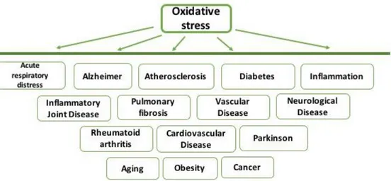
OS is involved in a wide spectrum of diseases and linked with several pathologies (Figure 4).
Figure 4: ROS involvement in different disease pathologies.
In 1956, Denham Harman suggested that free radicals produced during aerobic respiration cause cumulative oxidative damage, resulting in aging and
18
death. He noted parallels between the effects of aging and of ionizing radiation, including mutagenesis, cancer, and gross cellular damage (Harman D.1956; Hempelmann L. H.,1953). As per free radical theory of aging, it has been observed that species with high energy consumption by mitochondria and high metabolic rate ultimately results in unlimited O2•−production that linked to a faster rate of respiration, associated with an enormous generation of oxygen radicals and hastens aging which gives rise a shorter maximum lifespan potential (MLSP) (Beckman, K. B., 1998).
However, oxidative damages in such diseases are balanced by the antioxidant system in both enzymatic and non-enzymatic manner. The enzymatic oxidation system includes superoxide dismutase (SOD), catalase (CAT) and glutathione peroxidase (Gpx). Nonenzymatic antioxidants are derived from the plants, fruits and vegetables, i.e. lipid soluble α-tocopherol, carotenoids and many flavonoids and water-soluble glutathione, uric acid and ascorbic acids (Sies, H., 1997; Beckman, K. B., 1998). Besides, uric acids, β-carotene, and bilirubin are majorly considered as endogenous antioxidants produced in cells in-vivo (Halliwell, B., 1989).
1.3.2 Oxidative stress in AD brain
Many hypotheses were proposed to be involved in AD pathology which includes the amyloid cascade, excitotoxicity, oxidative stress, and inflammation hypotheses and all are centered on the role of Aβ, which ultimately cause the oxidative stress in the brain (Drake, J., 2003; Hardy, J., 2002). Therefore, studies suggested that oxidatively modified proteins are considered as a significant factor to contribute in etiology or pathogenesis of
19
many neurodegenerative diseases, including Alzheimer’s disease (AD), and Parkinson’s disease (PD) as well (Butterfield, D. A., 2007a; Butterfield, D. A., 2002; Cross, C. E., 1987; Markesbery, W. R.,1997). For many years it has been noticed that increased protein oxidation, protein nitration, and lipid peroxidation appear in neurofibrillary tangles and neuritic plaques of AD patients, and simultaneously the levels of oxidation products are also increased in cerebrospinal fluid of AD patients (Hensley, K., 1998).
Figure 5: Aging and AD associated characteristics.
A brain is exclusively prone to secondary carbonylation via lipid peroxidation because it contains a large concentration of polyunsaturated fatty acids (PUFA) in the brain tissue. Lipid peroxidation process considered as a toxic due to the allylic hydrogen abstraction and occurrence of subsequent radical chain reactions and propagation on acyl chain of polyphenols. Such processes may spread between the membrane system and even adjacent cells (Butterfield, D. A., 2002; Butterfield, D. A., 2010a;
Castegna, A., 2004). Status of lipid peroxidation in AD brain can be measured by the detection of elevated levels of free or protein-bound acrolein,
HNE(4-AD
Aβ
p-Tau
Oxidative stress
20
hydroxynonenal), isoprostane 8, 12-iso-iPF2α-Ⅵ, F2-isoprostane (F2-IsoP), and F4-neuroprostane (F4-NP) (Markesbery, W. R., 2005; Praticò, D., 2004; Butterfield, D. A., 2002; Butterfield, D. A., 2010a; Castegna, A., 2004).
Following the HNE abundance, many proteins/enzymes were further identified with protein nitration (3-nitrotyrosine) modification from early stage in AD brain (Butterfield, D. A., 2008). In particular for example BVR-A; one of the reductase enzyme and component of heme degradation pathway (HO/BVR-A) and kinase in insulin signaling pathway, has found with 3-NT (3-nitrotyrosine) modifications, resulted in reduced phosphorylated BVR-A which is necessary for its kinase activity in insulin signaling cascade (Barone, E., 2011a). Thus, oxidation derived changes to a protein could reduce the propensity of other PTMs and their functions as well. Lately, redox proteomics has become an advancement in the field to measure the oxidative stress-mediated total protein modification in AD brain (Butterfield, D. A., 2003) to provide an overview upon AD pathology.
Interestingly, few studies from the past revealed that increased OS has the significant impact on several signaling pathways; that not only causing damage because of the action of free radicals, but deranges signaling pathways leading to tau hyperphosphorylation, a hallmark of the disease and could involve in AD pathology. Evidently, both oxidative stress and reduced insulin signaling activation in the brain (known as brain insulin resistance), were proposed to favor Tau hyperphosphorylation in AD although the molecular mechanisms are still unclear ( Talbot, K., 2012; Steen, E., 2005; Rivera, E. J., 2005; Zhang, X., 2016; Morales-Corraliza, J., 2016; Sajan, M. P., 2016; Yarchoan, M., 2014; Bomfim, T. R., 2012; Butterfield, D. A.,2014). Tau phosphorylation due to increased OS has been highlighted by Zhu et al. in their study where they demonstrated the link between p38(one of
21
the tau kinase), tau phosphorylation, oxidative stress, and cell cycle-related events in Alzheimer disease (Zhu, X., 2000) and Lloret, A., et al. showed in their study that Aβ and tau toxicities in Alzheimer’s are linked via oxidative stress-induced p38 activation (Giraldo, E., 2014). At the consequences, both aging and AD brain encountered with oxidative stress which further i) oxidizes the protein through PTMs mechanism and ii) alters signaling pathways including kinases led to ultimate Tau phosphorylation.
1.3.3 BVR-A/HO-1 System
Increased quantity of pro-oxidants (ROS) in brain up regulates many anti-oxidant responses including; stress responses gene expression and enzyme activities. Biliverdin reductase-A and heme-oxygenase (BVR-A/HO) is evolutionary conserved and one of the major anti-oxidant systems to maintain the cellular redox homeostasis. It’s up-regulation provides the earliest defense response to the stress (Poon, H. F.,2004). HO-1 is the very first rate-limiting enzyme which is responsible for the breakdown of heme moiety into equimolar molecules biliverdin, carbon monoxide (CO), and ferrous iron (Maines, M. D., 1997), this reaction latter catalyzes the biliverdin into bilirubin by biliverdin reductase (BVR). Exposing to ROS, this enzyme elevates the availability of physiological bilirubin, CO and ferritin compelling antioxidant, anti-apoptotic and anti-inflammatory properties, therefore HO-1 known as a key player in maintaining cellular homeostasis (Ryter, S. W.,2005). Typically two main isoforms of heme oxygenase, i.e. HO-1 and HO-2 has been recognized and categorized on the basis of expression of their genes and distribution in the distinct tissue (Ryter, S. W., J 2006; Abraham, N. G., 2008).HO-1 (about 32 kD) is encoded by gene
22
HMOX1 and HO-2 (36 kD ) encoded by HOMX2 respectively in the tissue (Gozzelino, R., 2010). HO-2 is considered to be a constitutive isoenzyme most highly expressed in neuronal tissues (Brain) and testis contributing to cell homeostasis, whereas HO-1 is thought to be an inducible form with relatively low expression in most tissues (Zhu, X., 2011; Ryter, S. W., J 2006; Maines, M. D., 1997), though HO-1 mainly regulates protection against several stress conditions; ischemia (Burger, D., 2009), hypoxia (Chang, A. Y., 2009) and ROS (Cooper, K. L., 2009) and also known as heat shock protein (Hsp32).
Expression of HO-1 at the HOMX1 gene level is regulated by transcription repressor Bach-1 under basal condition but under pro-oxidant state its upregulation at both gene and protein level controlled by two upstream enhancer regions, E1 and E2 (Sun, J., 2002), which contains multiple stress- responsive elements (SREs), also known as antioxidant-responsive elements (AREs). These emerging evidences suggest its up-regulation is highly dependent on ROS stimuli and represent an oxidative inducible nature of this protein (Barone, E., 2014). AREs share a consensus sequence (GCnnnGTA) with the Maf recognition element (MARE) (Stewart, D.,2003). The interactions between MAREs and heterodimers formed by a Maf protein (MafK, MafF, or MafG) and an NF-E2-related factor 2 (Nrf2), or activator protein 1 (AP-1) play a direct role in HO-1 induction (Maines, M. D., 2005a; Sun, J.,2002).
Up-regulation of HO-1 upon ROS induction includes double mechanisms; i) by inducing conformational modification of Bach1 structure, which leads to its translocation from the nucleus to the cytoplasm where
23
Bach1 is ubiquitinated and degraded thereby, releasing transcriptional repression; and ii) by promoting the ubiquitination and consequent degradation of Keap1, which under normal conditions sequesters Nrf-2 into the cytoplasm, avoiding its transcriptional activity (He, X., 2007; Zenke-Kawasaki, Y., 2007).
Figure 6: HO-1/BVR system and regulation.
1.3.4 Role of BVR-A in the regulation of cells stress response
Similar to heme oxygenase, BVR possesses two isoforms as well; BVR-A and BVR-B (Kapitulnik, J., 2009; Maines, M. D., 2005b; Pereira, P. J. B., 2001). At functional level both these enzymes generate BR, but only BVR-A reduces BV-alpha into the powerful antioxidant and anti-nitrosative molecule BR-IX-alpha (thereafter BR) (Barone, E., 2009; Stocker, R., 2004),
24
whereas BVR-B prefers the other BV isoforms, such as BV-β, BV-γ, and BV-δ (Kapitulnik, J., 2009; Maines, M. D., 2005b; Pereira, P. J. B., 2001). Both BVR-A and BVR-B were identified in humans, with age-dependent characteristics. Biliverdin reductase-A is the primary form detected in the adult (95–97% BR is found in the bile), whereas BVR-B is predominant (~ 87%) in the fetus (Cunningham, O., 2000). Under normal condition, Biliverdin reductase co-expresses with HO-1/HO-2 as heat shock inducible enzymes (Ewing, J. F., 1993).
BVR is not only a reductase enzyme but also has been identified as serine/threonine/tyrosine kinase involved in various cellular function (Kapitulnik, J., 2009; Maines, M. D., 2005b). However, activation of BVR-A through phosphorylation at Ser/Tyr/Thr sites are essential for its dual characteristic function (reductase and kinase activity) (Kapitulnik, J., 2009; Lerner-Marmarosh, N., 2005). From the studies, it is demonstrated that BVR-A is capable of autophosphorylation which is required for its so-called reductase activity (Lerner-Marmarosh, N., 2005) whether phosphorylation through other kinases, i.e. insulin receptor kinase (IRK) is necessary to activate its own kinase activity (Kapitulnik, J., 2009; Lerner-Marmarosh, N., 2008; Maines, M. D. 2007; Tudor, C., 2008). Highlighting BVR-A’s activity, it does regulate many down-stream pathways like;
1. Catalyze the last step in the heme-degradation pathway by reducing the γ-meso (methylene) bridge of BV to BR (Kapitulnik, J., 2009). 2. Modulates the activity of members of conventional and atypical
groups of PKC isozymes (PKC-βII and PKC-ζ, respectively) (Kapitulnik, J., 2009; Lerner-Marmarosh, N., 2007; Maines, M. D. 2007; Gibbs, P. E., 2012b).
25
3. Functions as a scaffold protein for the formation a ternary complex with MEK1 and ERK1/2, placing ERK in a position that enables its activation by MEK (Kapitulnik, J., 2009; Lerner-Marmarosh, N., 2008).
4. Regulate the expression of stress-responsive genes such as HO-1 (Tudor, C.,2008) and iNOS (Di Domenico, F., 2013; Gibbs, P. E., 2012a). (Demonstrated in the figure below).
Figure 7: multifaceted Role of BVR-A.
Activated BVR-A (phosphorylated form) further regulates two central signaling pathways in insulin cascade; MAPK and phosphatidylinositol-3-kinase (PI3K) (Kapitulnik, J., 2009). Both the pathways synergistically play a significant role in neuronal development, neuronal plasticity and learning/memory consolidation (Horwood, J. M., 2006; Akter, K., 2011).
26
In recent years, published work from Maines group directs new insight on BVR-A’s propensity as a scaffold protein in signaling pathways.They discovered that the phosphotyrosine binding motif Y198MKM in BVR-A after being phosphorylated by IRK, interacts with NPXY motif of IRK. YMXM binding motif of any protein including IRS1/IRS2 and PI3K after phosphorylated by IRK, become docking site for src-homology domain-containing proteins and help in the assembly of a multiprotein complex (Miralem, T., 2016). A significant BVR fragment was found to activate insulin/IGF-1 signaling by its ability to interact with the intracellular kinase domain of the insulin receptor and to stimulate glucose uptake (Gibbs, P. E., 2014). As BVR and Akt both are downstream target in insulin signaling pathway and activated by IRK, they also share similarity at i) functional level: both targeted by ROS stimuli, IGF-1/2 insulin and hypoxia (Datta, S. R.,1999; Miralem, T.,2005; Salim, M., 2001; Gibbs, P. E., 2010), and ii) structural level: Akt isoforms and hBVR share certain similarities in their protein-protein interaction domain. The N-terminal pleckstrin homology (PH) domain of the Akt isoforms (aa1–113) (Coffer, P. J., 1998; Agamasu, C., 2015), and the C-terminal half of hBVR, which folds as a large 6-stranded β sheet (Kikuchi, A., 2001; Whitby, F. G., 2002), are the protein-protein interaction domains (Gibbs, P. E., 2012a).
Sequence identified in the β sheet of BVR is 225RNRYLSF which is identical to the canonical Akt interaction motif RxRxxS and this motif is also found in all Akt substrate, non-substrate binding proteins and Akt co-activator i.e members of proto-oncogenes TCL1 family (TCL1, MTCP1, TCL1b) (Laine, J.,2000; Brazil, D. P., 2002), noticeably the underlined amino acids are prominently required for proposed interactions between proteins.
27
Figure 8: Schematic overview of BVR as a scaffold protein
Overall, activated BVR is recognized as a key player compelling its pleiotropic properties (Reductase, kinase and scaffold protein) in the cellular system under the various signal inductions.
Since Alzheimer disease has also partial involvement of increased OS within aging and imbalanced antioxidant system, many recent studies are focused on understanding the pivotal role of BVR-A in AD pathology because of its pleotropic functions and abundance in ROS signaling.
28 1.4 Glucose Metabolism in the Brain
Nobody realizes that some people expend tremendous energy merely to be normal.
“Albert Camus, Notebooks 1942-1951
Human brain depends on glucose as its primary source of energy. In the adult brain, neurons have the highest energy demand (Howarth, C., 2012), as per requiring continuous delivery of glucose from the blood. Humans brain accounts for ~2% of the body weight, but it consumes ~20% of glucose-derived energy making it the primary consumer of glucose (~5.6 mg glucose per 100 g human brain tissue per minute) in the entire body (Erbsloh, F., 1958). Glucose metabolism provides the fuel for physiological brain function through the generation of ATP, generates several neurotransmitters, and foundation for critical for brain physiology and disturbed glucose metabolism in the brain underlies several diseases affecting both the brain itself as well as the entire organism.
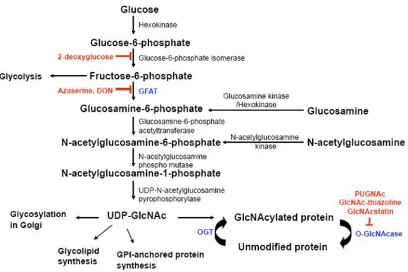
One of the prioritized function of glucose metabolism is to provide the substantial energy source, i.e. ATPs to the neurons to possess information, balance the action potential flow, postsynaptic potential, maintenance of ion gradients, the neuronal resting potential (Howarth, C., 2012; Erbsloh, F., 1958; Harris, J. J., 2012; Ivannikov, M. V., 2010).However, approximately 2–5% of total glucose is also utilized in the hexosamine biosynthetic pathway (HBP) to produce glucosamine-6-phosphate and, ultimately, UDP-N-acetylglucosamine (UDP-GlcNAc) (Liu, F., 2009; Copeland, R. J., 2013). UDP-GlcNAc is the donor substrate for O-linked-ß-N-acetylglucosamine transferase (OGT), which catalyzes protein O-GlcNAcylation, a post-translation process transferring a single moiety of N-acetyl-D-glucosamine residues from UDP-GlcNAc to the hydroxyl side chains of serine/threonine
29
residues of proteins and similar to phosphorylation. The reversible attachment of O-linked GlcNAc to proteins is catalyzed by O-GlcNAcase (OGA), which regulates its removal. The activation of OGT is sensitive to UDP-GlcNAc substrate and, therefore, to altered intracellular glucose metabolism (F. liu 2009, C.X. Gong 2016, B.D. Lazarus 2009, K. Vaidyanathan 2014, Y. Yu, 2012).
Figure 9: Hexosamine biosynthetic pathway (HBSP).
The synthesis of UDP-GlcNAc from glucose and enzymes involved in the process are shown and inhibitors of HBP and GlcNAcylation are shown in red (Figure 9). Interestingly, both the post-translation modification process; O-GlcNAcylation and phosphorylation are mutually related. Indeed, several studies reported that O-GlcNAc could either occur reciprocally to
30
serine (Ser) and threonine (Thr) phosphorylation or can interact with adjacent phosphorylated sites influencing their modification (Yu, Y., 2012, Deng, Y., 2008; Li, X., 2006; Trinidad, J. C., 2012). Interestingly altered glucose metabolism has been involved in several pathophysiological conditions, i.e. insulin resistance, which is one of the commons among the older people. Development of Insulin resistance in the peripheral system including pancreas, liver, white adipose tissue and brain generally thought to be primary etiological evident for glucose metabolism dysfunction and a significant decline in insulin signaling response in those tissues were observed with age (Salmon, A. B. 2012).
In the line, it has also shown that increased oxidative stress in the brain can directly promote insulin resistance (Barone, E., 2011a) and alter the glucose metabolism that promotes severe cognitive decline. Thus, impairment in glucose metabolism and insulin resistance in peripheral organs including brain in elder age could be a part of AD-like dementia. However, how this oxidative stress and impaired glucose metabolism participates at the synergistic level in AD-like dementia, have discussed in the next subtitle.
1.4.1 Impaired glucose metabolism, oxidative stress (OS) and Alzheimer
Symptom severity in Alzheimer progression was only limited to the NFTs and Aβ depositions until the series of studies provided some intriguing evidence that has confirmed the involvement of early metabolism dysfunction in the pathology. NFTs and Aβ depositions are profoundly has been seen in healthy aging and non-demented patients (mentioned before) as well which is not distinguishable at pathological status. PET analysis of AD patients has revealed the reduced cerebral metabolic rate for glucose
31
(CMRglc) (Mosconi, L., 2008). As per the required energy for functional synapses and axonal transportation are usually provided through glucose consumption and ATP production along with balanced ROS homeostasis and clearance pathways in normal aging but disturbed ROS homeostasis results in increased oxidative stress and strongly associated with stress-mediated abnormalities in the brain (Mattson, M. P., 2006). Nevertheless, reduced glucose uptake principally minimizes the generation of superoxide anions which in turn results in lower the level of H2O2 and OH• and lessen the
damage to proteins and other biomolecules. (Mattson, M. P., 2006). Contrary to the reduction in glucose uptake which promotes the lower OS in the brain, aging deliberately causes ROS generation by other means i.e. perturbation of ER, peroxisome and mitochondria (Zorov, D. B., 2006, Circu, M. L., 2010) and abnormal NFTs/ Aβ depositions which elevate the risk of higher ROS in the brain (Lambert, M. P., 1998). Further many studies in cell culture and ex-vivo rat muscle showed that this increased OS reduces insulin-stimulated glucose uptake, reduction of the downstream insulin signaling mediators proteins, i.e. Akt, IRS-1(insulin receptor), GSK-3β (summarized mechanism in figure 3) and reduced glucose transporter translocation to the cellular membrane (Salmon, A. B., 2012). Therefore, it’s very well predicted that age-associated increased OS and metabolic dysfunction (impaired glucose uptake) in the brain is a significant contributor to the pathophysiological status of AD patients.
Positron emission tomography (PET) imaging with 2-[18F] fluoro-2-deoxy-D-glucose (FDG) as the tracer has long been used to track AD-related brain changes by providing qualitative and quantitative estimates of the cerebral metabolic rate of glucose (CMRglc). This study brought the significant contribution of glucose metabolism forefront in the AD pathology
32
when a reduction in glucose uptake has confirmed in AD patients compared to the age-matched healthy patients (Mosconi, L., 2008).
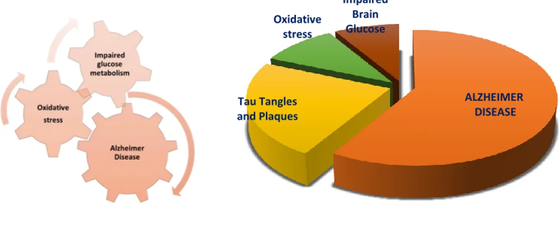
Besides these observations as mentioned above, many other studies are focused on identifying the other alternative mechanism that could further be a leap in AD pathology. To shed light on this concern, lately, it has been discovered in a few studies that reduced glucose availability in the brain could raise the probability of occurrence of protein phosphorylation than glycosylation simultaneously. The balanced interplay between these two post-translation modifications could be one of the key mechanisms to rescue the NFTs formation and tangles in AD brain. All these studies taken together provide us an overview that reveals an important link between oxidative stress, impaired glucose metabolism and AD pathology underlining to a different stage of aging.
Figure 10: Relationship among the oxidative stress, Alzheimer disease and impaired glucose
metabolism. ALZHEIMER DISEASE Tau Tangles and Plaques Oxidative stress Impaired Brain Glucose
33
1.4.2 Cross talk between O- GlcNAcylation and phosphorylation in Alzheimer disease: A link between AD pathology and impaired glucose uptake
These two PTM (phosphorylation and glycosylation) mechanism for any protein, shares reciprocal manner of regulation in which inhibition of protein kinase (Protein Kinase A, Protein C) and inhibition of O-GlcNAcase increase the global O-GlcNAc levels while shows the decreased phosphorylation for many events involved in cell development signaling or homeostasis (Griffith, L. S., 1999; Wang, Z., 2007). Interestingly, the site-specific target for O-GlcNAc and phosphorylation could occur on A) same site (Ser/Thr) B) competitive
34
proximal site of Ser/Thr C) Occupied both proximal sites (Chutikarn Butkinaree 2010). This interplay between O-GlcNAclytion and phosphorylation can be more complicated than just a competitive mechanism for any cell signaling and the balance between these two PTM at the same time on the same protein could achieve the cell fate in cell survival decision. Interestingly, AD brain identified with aberrant glycosylated Tau other than being hyperhosphorylated (Liu, F., 2002) and this study further supports the idea that impaired glucose uptake in AD brain favors the reduced glycosylation modification on Tau protein but coincides with increased abnormal phosphorylation.
1.4.3 O-GlcNAcylation of Tau and APP in Alzheimer disease
Brain Amyloid deposition and Tau aggregates are the clear evidence for the AD, but impaired glucose metabolism coincides in AD brain and responsible for synaptic dysfunctionality as a result of Aβ neurotoxicity. However, impaired glucose metabolism (Hypometabolism) becomes clinically evident in a significant manner for many different disorders. Alterations of brain glucose metabolism seem to occur also in subjects affected by mild cognitive impairment (MCI), before the appearance of clinical signs of dementia. Therefore, it is suggested this event is a cause, rather than a consequence of AD (Gong, C. X., 2016; Yu, Y., 2015). In past three decades PET (positron emission topography) analysis of AD brain has revealed its importance as an early onset biomarker in AD pathology (Blennow, K.,2006), validating the coexisted AD and insulin resistance together. The new hypothesis suggested the limited glucose uptake gives rise reduced GLUT1, GLUT 3 in neuronal cells supporting the increased insulin resistance in the AD (Calvo‐Ochoa, E.,
35
2015; Correia, S. C., 2012; de la Monte, S. M., 2009). The occurrence of O-GlcNAcylation to any protein is UDP-GlcNAc (a substrate for OGT) dependent via Hexosamine biosynthetic pathway (HBP), but the failure of O-GlcNAcylation is now considered as a sensor for limited nutrition supply to the brain. Therefore, glucose hypometabolism within diseases, i.e., diabetes and AD favors the reduced O-GlcNAclytion and reciprocally increased phosphorylation. Post-translation glycosylation of several nuclear and cytoplasmic proteins has reported by Torres and Hart including cytoskeleton protein Tau and membrane-associated protein APP (Hart, G. W., 2007).
Figure 12: Impaired brain glucose metabolism contributes to neurofibrillary degeneration in
Alzheimer's disease
Mass spectrometry results of Tau aggregates in AD patients show many sites occupied for O-GlcNAcylation and less for phosphorylation. In the Study
36
from Lefebvre group mention the Inhibiting phosphatase activity results in increased phosphorylation and vice versa O-GlcNAcylation (Lefebvre, T., 2003). Data from fasting mice demonstrate decreased O-GlcNAcylation and increased phosphorylation (Li, X., 2006).
Figure 13: Potential protective role of O-GlcNAcylation within the healthy brain, which
may become deficient in the AD brain.
Impairments in glucose utilization (may perhaps by decreased cell-surface glucose transporters) as indicated above in the figure would result in reduced flux down the hexosamine biosynthetic pathway (HBSP) and ultimately lead to reduced O-GlcNAc on Tau, APP, and nicastrin (a non-catalytic component of the -secretase complex), which could result in the increased production of NFTs and amyloid plaques, the hallmark features of the AD brain.
37
Intriguingly, during AD progression brain O-GlcNAc levels appear to decrease while phosphorylation increases. Indeed, samples of frontal cortex from AD patients displayed a significant reduction in global O-GlcNAc levels but increased Tau hyperphosphorylation as compared to controls (Gatta, E., 2016; Yuzwa, S. A., 2014; Liu, F., 2004). However, the reduction of total protein O-GlcNAcylation in the AD is still a matter of controversy since other studies reported an inverse trend (Förster, S., 2014; Frenkel-Pinter, M., 2017; Griffith, L. S., 1995). The abnormal phosphorylation/O-GlcNAcylation ratio of Tau is critical to neurodegeneration, as increased phosphorylation promotes Tau aggregation into tangles, while higher levels of Tau O-GlcNAcylation are protective against tangles formation (Gong, C. X., 2016; Li, X., 2006; Liu, Y., 2009; Zheng, B. W., 2016). Immunofluorescent studies on human brain samples revealed an inverse relationship between Tau O-GlcNAcylation and phosphorylation (Liu, F., 2009, Liu, Y., 2009). Further for the reason that reciprocity sustains the O-glycosylation and phosphorylation, many studies have confirmed the increased O-GlcNAclytions and decreased phosphorylation with selective OGA inhibitors in-vitro. Treatments using inhibitors of OGA has been tested on different mouse models of AD or tauopathy showing in general that increasing the levels of O-GlcNAcylation can reduce disease hallmark and lower cognitive decline (Jacobsen, K. T., 2011; Kim, C., 2013; Yuzwa, S.A., 2012; Graham, M. E., 2011).
The APP, a membrane-bound protein that results in Aβ deposition during AD development (as already mentioned in 1.3.1 in the beginning) has been observed with decreased O-GlcNAcylation modification comparatively phosphorylation. When T2DM has found being associated with Aβ toxicity
38
in AD model, its considered that impaired glucose metabolism can contribute in the development of AD through drop off in a total O-GlcNAcylated APP (Griffith, L. S.,1995). Griffith et al. first discovered Post-translation modification (O-GlcNAcylation) of APP and APP associated proteins became the attention for its role in APP processing. Following this observation, it has been found that increased O-GlcNAcylation of APP leads to decreased production of Aβ (40) in cultured SH-SY5Y cells with non-selective OGA inhibitors (Kim 2013). Surprisingly study in 5xFAD Aβ mice model (conceive amyloid pathology), shows a decreased level of Aβ (40) and Aβ (42), limited plaques, less neuroinflammation and recovered cognition ability in the brain while exposing to long-term selective OGA inhibitors (Macauley, M. S., 2010). In the case of site-specific O-GlcNAcylation, both APP and APP processing enzyme (nicastrin-non-catalytic components of secretase complex) were spotted on Ser-708 being O-GlcNAcylated. (Kim, C., 2013). Regardless of the known pathway of γ-secretase for Aβ generation, it is believed that decreased O-GlcNAcylation on nicastrin induces APP processing and produces Aβ fragments. Reconciling the glucose hypometabolism with increased APP processing refer to the hypothesis of overlapping T2DM/insulin resistance with hyperphosphorylated Tau and excess Aβ depositions in AD.
39
Chapter 2
:
Background and Aim
In a past decades neurofibrillary tangles (NFTs) and Aβ plaques has spotted in brain specific region as a hallmark of AD, that gradually causes cognitive impairment. PET analysis together with biochemical analysis has confirmed the significance of NFTs and plaques in the AD pathology but we further need to strength on the causes that lead to cognitive impairment before it distinguishes in severe AD symptoms.
Interestingly, Insulin resistance together with impaired glucose metabolism and increased ROS production in the brain has been linked with AD development (Barone, E., 2011a; 2011 b; 2014, and 2015; Butterfield, D. A., 2014; Hart, G. W., 2011). These alterations in the signaling pathways have seen very often in a large population of old people with increasing in numbers every year. Thus, in the current situation the biggest challenge is not only to discover the biomarkers but also to understand the molecular mechanism of pathology at early-onset AD and late-onset as well.
For the first-time, the link between oxidative stress and impaired glucose transport has been evidenced in cultured neurons, which is followed by a decrease in cellular ATP levels due to a dysfunctional respiratory chain reaction in mitochondria (Keller, J. N.,1997; Luo, Y., 1997). Further studies suggested that altered glucose metabolism is a very early change in the AD (De Leon, M. J., 2001; Mosconi, L., 2008; Small, Gary W., 1995; Reiman, E. M., 1996; Mosconi, L., 2006) and legitimately correlates the clinical disabilities with dementia (Blass, J. P., 2002). Occurrence of insulin resistance in early AD due to impaired glucose metabolism concomitantly stimulates the oxidative stress in the AD brain and have recognized as early
40
symptoms. This alteration can be an indication for the transition of pre-clinical AD (PCAD) to the MCI stage. Although one study suggests that OS occurs before the onset of AD symptoms and that oxidative damage is found before robust Aβ plaque formation (Butterfield, D. A., 2014). Thus, insulin resistance, oxidative stress and impaired glucose metabolism promotes a feedback mechanism and involved as a vicious cycle in early phase of AD development.
It is known that OS and impaired brain metabolism affects downstream targets; i) GSK-3β and, ii) BVR-A/HO-1, which plays a significant role in several signaling and among all OS mediated Akt activation and GSK-3β inhibition is a part of neuroprotective mechanism in the AD brain (Talbot, K., 2012; Steen, E., 2005; Rivera, E. J., 2005; Zhang, X., 2016; Morales-Corraliza, J., 2016; Sajan, M. P., 2016; Yarchoan, M., 2014; Bomfim, T. R., 2012; Butterfield, D. A.,2014). When it comes to understand the role of HO-1/BVR-A system in the AD, one of the studies from Butterfield and the group has been reported an astonishing observation which revealed that oxidative/nitrosative post-translation modification of HO/BVR system associated with its decreased phosphorylation forms (BVR-A activity) in MCI and AD brain (Barone E 2011a and 2011b). On the other hand, based on its scaffold properties, another study from Maines group has reported the BVR-A/Akt mediated GSK-3β activation (Miralem, T., 2016) and also data from our group showed that BVR-A plays a significant role as a scaffold protein in CK1 mediated BACE-1 phosphorylation and resulting in Aβ depositions in aging model (Triani, F., 2018). So further BVR-A is a central attraction in my study because previously our group has shown that reduced total BVR-A level and its activity proportionally associated with the increased levels of OS in AD and MCI brain (Barone, E., 2011a; Barone, E.,