Enhanced adherence of human uremic erythrocytes to vascular
endothelium: Role of phosphatidylserine exposure
M
ARIOB
ONOMINI, V
ITTORIOS
IROLLI, F
EDERICOG
IZZI, S
ILVIOD
IS
TANTE, A
LFREDOG
RILLI,
and M
ARIOF
ELACOInstitute of Nephrology, Department of Medicine, and Biomorphology Department, G. d’Annunzio University, Chieti, Italy
Enhanced adherence of human uremic erythrocytes to vascular the cell [2]. Thus, loss of membrane asymmetry and in endothelium: Role of phosphatidylserine exposure. particular the appearance of PS at the outer membrane
Background. The exposure of phosphatidylserine (PS) on
surface is associated with many physiological and patho-the outer leaflet of erythrocyte membrane may have several
logical phenomena including enhancement of coagulative pathophysiological consequences including increased
erythro-reactions and cell recognition by phagocytes [reviewed in cyte adherence to endothelial cells, a finding that seems relevant
in pathologies with reported vascular injury. 2 and 3]. Apoptosis also is accompanied by PS exposure
Methods. Because PS externalization increases in erythro- at the cell surface [4]. In addition, it has been demonstrated cytes from patients suffering from chronic uremia, which is that PS externalization participates in RBC adherence frequently associated with vascular damage, the adherence of
to the endothelium [5–9], a process that may be relevant uremic erythrocytes to human umbilical vein endothelial cell
in pathologies with reported vascular damage [8]. (HUVEC) monolayers and the role of PS exposure on such
cell-cell interaction were studied. There is a high prevalence of vascular disease in patients
Results. The number of uremic erythrocytes adhering to suffering from chronic renal failure (CRF). CRF is also HUVEC was markedly greater than with normal erythrocytes
characterized by a number of structural and functional and significantly correlated (r⫽ 0.88) with the percentage of
anomalies of the RBC membrane [10–13]. Recently, we PS-exposing erythrocytes in the population. Adhesion to the
found a significant increase in PS-exposing RBC in chronic monolayers was significantly decreased when uremic
erythro-cytes were preincubated with either annexin V or PS-containing uremic patients compared with healthy controls [14]. The liposomes, and was strongly greater for PS-positive than PS- abnormality seems related to the uremic state regardless negative fluorescence-activated cell sorter (FACS)-sorted
ure-of the dialysis treatment, and is most likely caused by mic erythrocytes. Binding occurred preferentially in the gaps
inhibition of PS transport from the outer to the inner of HUVEC monolayers and was enhanced by matrix exposure.
leaflet of RBC plasma membrane. We also showed that Uremic erythrocytes adhered to immobilized thrombospondin,
and binding to endothelial cells was significantly reduced when surface-exposed PS promotes the susceptibility of uremic monolayers were incubated with antibodies to thrombospondin. RBC to phagocytosis [15] and thus may be involved in
Conclusions. These findings suggest that PS externalization
the shortened RBC lifespan of chronic uremia [16]. may promote increased uremic erythrocyte adhesion to
endo-On the basis of these observations, it is possible to thelium, possibly via a direct interaction with matrix
thrombo-hypothesize that abnormal exposure of outer-leaflet PS spondin.
in uremic RBC may significantly affect their propensity for adhesion to endothelial cells. The present study was undertaken to examine the adherence of human uremic The phospholipids of the normal human erythrocyte
RBC to human umbilical vein endothelial cells (HUVEC) (RBC) are distributed asymmetrically in the bilayer of
and to test the role of RBC PS externalization on such the red cell membrane, with the aminophospholipid
phos-cell-cell interaction. phatidylserine (PS) located exclusively in the inner leaflet
[1]. The maintenance of this asymmetry is an
energy-requiring process of major physiological importance for METHODS
Study population
Key words: thrombospondin, endothelial cells, uremia, vascular injury, Twenty healthy control subjects (10 women, 10 men;
apoptosis, coagulation, chronic renal failure.
mean age 57⫾ 2 years; range 31 to 72) and 20 stable uremic patients on chronic maintenance hemodialysis Received for publication February 28, 2002
Accepted for publication May 8, 2002 (10 women, 10 men; mean age 59 ⫾ 2 years; range 30 to 70) were included in this study after giving informed 2002 by the International Society of Nephrology
consent. Patients were not diabetics [17] nor did they Preparation of phospholipid vesicles
receive recombinant human erythropoietin. Other exclu- Liposomes (small unilamellar vesicles) were prepared sion criteria were: blood transfusion within the past six by sonication as previously reported [18]. Phospholipids months; iron, folic acid and vitamin B12deficiency; uncon- were suspended in chloroform/methanol (90/10), dried
trolled hypertension; active infection; use of drugs that under nitrogen and resuspended in PBS by vortexing. might interfere with erythropoiesis (such as theophylline The mixtures were then sonicated at 4⬚C for five minutes and angiotensin-converting enzyme inhibitors); and ma- at 30 W using a Fisher Sonic Dismembrator model 300. lignant or systemic disease. All hemodialysis patients Liposomes contained either 70 molar percent phosphati-were being dialyzed three times a week with a four-hour dylcholine (PC) and 30 molar percent PS (PC/PS lipo-dialysis session using bicarbonate dialysate, and none somes) or only phosphatidylcholine (PC liposomes). reused synthetic membranes.
Endothelial adherence assay Materials
Confluent EC monolayers were washed twice with PBS Fluorescein isothiocyanate-labeled annexin V (FITC- to remove traces of serum, covered with RBC suspen-AnV), phosphate-buffered saline (PBS), Hanks’ buffered sions, and incubated for 45 minutes at 37⬚C while being saline solution (HBSS), HEPES buffer, M199 medium,
mechanically agitated. Nonadherent erythrocytes were fetal calf serum (FCS), glutamine, heparin, endothelial
removed by washing five times with PBS. The RBC that cell (EC) growth factor, trypsin-edathamil, thrombin,
ethy-had adhered to the EC were videotaped via an inverted lenaglycol-bis (-aminoethyl ether)-N,N⬘-tetra-acetic acid
phase-contrast microscope (Leica DM IRB, Leitz, Ger-(EGTA), phosphatidylcholine (PC) from egg yolk, and
many) equipped with a charge-coupled device (CCD) l-␣-phosphatidylserine from bovine brain were obtained
video camera (Model CoolSnap; RS Photometric, Tuc-from Sigma Chemical Co. (St. Louis, MO, USA).
Dul-son, Arizona, USA). Adherent RBC were counted in 25 becco’s calcium/magnesium-free PBS was obtained from
random microscope fields ranging over the entire endo-Mascia Brunelli (Milan, Italy). Anti-thrombospondin
thelial cell monolayer and reported as the number of mouse monoclonal antibody (clone A4.1) and control
adherent cells per 1000 HUVEC. monoclonal antibody (trpE) were from Oncogene
Re-For studies on the influence of subendothelial matrix search Products (Boston, MA, USA). Endothelial cells
exposure on RBC adherence, HUVEC were pretreated (HUVEC) were from Clonetics Corp (San Diego, CA,
for five minutes at 37⬚C with 0.1 U/mL thrombin in HBSS, USA). Thrombospondin from human platelets and
fi-1% bovine serum albumin (BSA), 50 mmol HEPES ph bronectin were purchased from Calbiochem (La Jolla,
7.4 (HAH) or with HAH alone as control. These working CA, USA).
conditions of thrombin exposure have been shown to be associated with maximal adhesogenic effect [19].
Preparation of cells
To examine the effect of different components on the
Erythrocytes. Blood was drawn by venipuncture into
binding of uremic RBC to endothelial cells, RBC suspen-evacuated tubes containing ethylenediaminetetraacetic
sions from uremic patients were incubated for five min-acid (EDTA). After centrifugation at 700⫻ g for five
utes with FITC-AnV (100 nmol/L) to mask RBC surface-minutes at 4⬚C, the plasma and buffy coat were removed,
exposed PS [15] or with liposomes (final concentration the RBC pellet was washed three times with PBS, and
5 mmol/L phospholipids) composed of either PC or of then resuspended to 1% hematocrit in the same medium
a mixture PC/PS, before being layered over HUVEC. for flow cytometry assay or in M199 medium for
adher-In another set of experiments, confluent HUVEC mono-ence assay.
layers were washed in HAH and incubated for three hours
Endothelial cells. Endothelial cells (HUVEC) were
at 37⬚C with a monoclonal antibody recognizing throm-cultured at 37⬚C in 5% CO2in 75-cm2flasks (Nunc,
Naper-bospondin (TSP) [20] at the concentration of 50g/mL ville, IL, USA). The culture medium was M199
supple-or with control antibody at an equivalent concentration. mented with 20% (vol/vol) heat-inactivated FCS, 2 mmol/L
An adherence assay was then performed as described l-glutamine, penicillin/streptomycin (100 U/mL and 100
above. g/mL), 100 g/mL heparin and 100 g/mL EC growth
factor. The medium was changed under sterile conditions
Adherence to immobilized proteins
every two days until the cells reached confluence (3 to
Multiwell slides were coated with 50g/mL of purified 4 days). Cultures grown to confluence, after two washes
protein (TSP, FN, or BSA) and incubated at 37⬚C for with calcium/magnesium-free PBS, were harvested using
one hour in 95% humidity. The slides were then blocked 0.01 trypsin edathamil, split 1:3, and further cultured in
with 1% BSA in HBSS for one hour under the same new flasks. Confluent monolayers from passage 2 up to
conditions. RBC suspensions of 1% Hmt in HBSS were passage 6 were used in adherence assays, within two days
Fig. 1. Adherence of erythrocytes from uremic patients on hemodialy-sis (N⫽ 20) and normal subjects (N ⫽ 20) to endothelial cell monolayers.
Adherence was by a static assay. Error bars depict the standard error. *Significant difference between normal and uremic erythrocytes
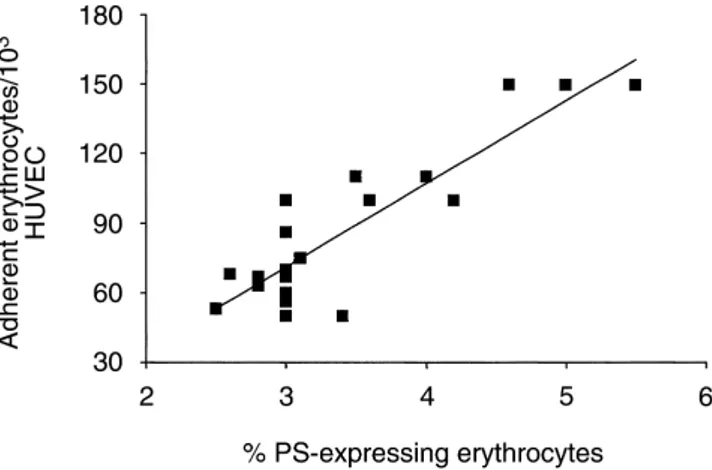
adher-Fig. 2. Correlation in chronic uremic patients on hemodialysis therapy
ent to HUVEC.
(N⫽ 20) between phosphatidylserine (PS)-exposing normal human erythrocyte (RBC) and static RBC adherence to endothelial cell mono-layers. The FITC-annexin V positive RBC population was measured
by FACS analysis (r⫽ 0.88; P ⬍ 0.0001). cover the entire slide. After incubation for 45 minutes
at 37⬚C, the slides were washed three times with PBS and the adherent RBC were counted in a minimum of
by using the unpaired or paired t test, for unpaired and 20 fields via an inverted phase contrast microscope
paired values respectively. Pearson’s correlation coeffi-equipped with a CCD video camera, as described above.
cient (r ) was used to test the association between pairs
Measurement of PS exposure in the RBC population of variables. All results are expressed as a mean⫾ SEM,
and P⬍ 0.05 is considered statistically significant. The percentage of PS-exposing cells in erythrocyte
samples was measured by a flow cytometric assay based on FITC-AnV labeling [14]. Briefly, isolated RBCs were
RESULTS
diluted 1:100 (3 to 5⫻ 107RBC/mL) to a final volume of
In recent studies we had observed greater PS exposure 0.25 mL in a binding buffer consisting of 10 mmol/L of
in RBC from uremic patients than in healthy controls HEPES-Na (pH 7.4), 136 mmol/L of NaCl, 2.7 mmol/L
[14, 15]. Because PS exposure might play its part in RBC of KCl, 2 mmol/L of MgCl2, 1 mmol/L of NaH2PO4, 5
adherence to endothelium [5–9], the present study was mmol/L of glucose, 5 mg/mL of BSA, and 2.5 mmol/L
designed to investigate whether the abnormal PS expo-of CaCl2. After addition of 100 nmol/L of FITC-AnV
sure in uremic RBC might influence their binding to and incubation for 15 minutes at room temperature in
endothelial cells (HUVEC). The number of PS-exposing the dark, an aliquot of the sample was directly aspirated
red cells was determined by annexin V labeling and flow into the flow cytometer (Epics Elite; Coulter Electronics,
cytometry, and the adherence to confluent HUVEC mono-Hialeah, FL, USA). The RBC population was defined by
layers by a static adherence assay. Adhesion to non-size in forward and side scatter plots: gated cells were
activated HUVEC by RBC from chronic uremic patients counted as annexin positive if they had a mean
fluores-on maintenance hemodialysis was significantly greater cence of at least 1.0. The flow cytometer software was used
(P⬍ 0.001) than adhesion by normal RBC (Fig. 1). The to calculate the percentage of annexin V-positive cells.
percentage of PS-exposing RBC was also significantly For some experiments, FITC-AnV RBC from uremic
greater in uremic patients than in healthy subjects (3.43⫾ patients were FACS-sorted to obtain either a PS-positive
0.18 vs. 0.65⫾ 0.03% annexin V-positive RBC, P ⬍ 0.001). or PS-negative fraction. Sorted cells were collected into
A strong correlation (r⫽ 0.88, P ⬍ 0.0001) was found tubes containing 1 mL of PBS and washed once with saline
between the number of uremic RBC adherent to HUVEC solution. PS-expressing RBC were next incubated with
and the percentage of PS-exposing RBC in the suspen-EGTA (2.5 mmol/L) for 30 minutes to remove annexin
sion (Fig. 2). and washed once with saline solution. Cells (either
PS-The latter results suggested that RBC adherence might expressing or non-PS-expressing RBC) were then
incu-depend on the percentage of PS-exposing cells in the bated with HUVEC monolayers for static adherence assay.
population. To clarify this possibility, we first
FACS-Data analysis sorted annexin V-positive uremic RBC to obtain either
PS-expressing or non-PS–expressing red cells. PS-posi-Data were analyzed using the statistical software
tive RBC showed a markedly greater propensity for ad-Sigma-Stat 2.0 for Windows (Jandel Scientific Software,
Fig. 3. Effect of PS competitors on uremic erythrocyte adherence to endothelial cell monolayers. Erythrocyte suspensions from hemodialysis
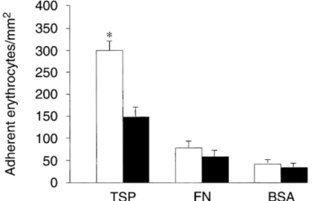
Fig. 4. Uremic (䊐) and normal (䊏) RBC adherence to immobilized
patients (N⫽ 10) were incubated for five minutes with one of the
proteins. Multiwell slides were coated with 50g/mL of indicated
pro-following before the static adherence assay with HUVEC: buffer
(Con-teins and then blocked with 1% BSA for 60 minutes. A washed suspen-trol), annexin V, PS vesicles (30% PS, 70% PC, 5 mmol/L lipid), PC
sion of RBC (1% Hmt in HBSS) was placed on the immobilized proteins vesicles (100% PC, 5 mmol/L lipid). Error bars depict the standard
for 45 minutes, followed by three washes with PBS. *Significant differ-error. *Significantly different from the control.
ence between normal and uremic RBC adherence to TSP (P⬍ 0.001, N⫽ 10).
did PS-negative uremic RBC (25⫾ 12 RBC/103HUVEC;
P⬍ 0.02, N ⫽ 6). Next, RBC from hemodialysis patients
were incubated with FITC-annexin V to mask surface-exposed PS before they were added to HUVEC mono-layers. Preincubation with annexin V significantly re-duced RBC adherence to HUVEC (P⬍ 0.001; Fig. 3), indicating that annexin V shields PS on the RBC from interacting with endothelial cells. Uremic RBC adher-ence was also significantly inhibited (by 76%) by the presence of phospholipid vesicles containing PS, whereas the presence of vesicles containing PC alone had no inhibi-tory effect on adherence (Fig. 3). In the aggregate, these
Fig. 5. Effect of monoclonal antibody to thrombospondin (TSP) on
data indicate a major role for a PS-rich domain in the uremic RBC adherence to endothelial cell monolayers. HUVEC mono-layers were incubated with monoclonal antibody to TSP or a control outer hemileaflet of the uremic RBC membrane in
caus-monoclonal antibody (trpE) before being layered in a static assay with ing RBC adherence to endothelial cells. uremic RBC suspensions. Results are expressed as percentage of the Ultrastructural observation of RBC-HUVEC binding buffer-treated control adherence, which is set to 100%. Error bars represent SEM calculated from the absolute values from 8 experiments showed that the adherence of uremic RBC to endothelial
and normalized relative to the adherence of the buffer-treated control. cells occurred preferentially to the edges of cells and the *Significant differences from the control.
gap between cells (data not shown), as previously reported for calcium ionophore-treated PS-exposing RBC [8]. Since such a finding would indicate the importance of
(Fig. 4). There was markedly greater adherence by ure-subendothelial matrix in uremic RBC-HUVEC binding,
mic erythrocytes to immobilized TSP than to FN or to we decided to evaluate whether uremic RBC endothelial
BSA (non-specific control). In comparison with normal adhesion could be further enhanced by increasing matrix
RBC, RBC from uremic patients showed a significantly exposure. To generate exposure of the matrix, HUVEC
increased adherence to TSP, while exhibiting no statisti-were pretreated with agents such as thrombin, which cal difference in their adherence to either FN or BSA cause the cells to retract [19]. Treatment of endothelial (Fig. 4). The ability of uremic RBC to adhere to immobi-cells with thrombin significantly increased uremic RBC lized TSP was inhibited by 72% when red cells were adherence. Expressed as an adherence ratio (RBC ad- preincubated with annexin V (76⫾ 12 RBC/103HUVEC
herence with thrombin pre-treatment divided by RBC vs. 300⫾ 22 RBC/103HUVEC for untreated RBC, P⬍
adherence without thrombin pretreatment), this adher- 0.001, N⫽ 10).
ence-promoting effect was 2.11⫾ 0.25 with a range of In additional experiments, HUVEC monolayers were 1.20 to 3.50 (N⫽ 8; P ⬍ 0.005). incubated with a monoclonal antibody recognizing TSP We next examined the adherence of erythrocytes to [20] or a control monoclonal antibody, before static ad-herence assay. As shown in Figure 5, uremic RBC adher-immobilized adhesive proteins of the endothelial matrix
ence proved significantly decreased (P ⬍ 0.001) when RBC to HUVEC monolayers may occur via a direct interaction between PS and TSP in the subendothelial HUVEC had been pretreated with anti-TSP antibody,
whereas irrelevant antibody had no effect on adherence. matrix [8]. TSP is a “matricellular” protein [23], capable of functional interactions with a variety of cell types and matrix proteins [24–26]. It is synthesized and secreted
DISCUSSION
by endothelial cells and some other cell types in culture The exposure of PS on the external leaflet of the RBC and is incorporated into the extracellular matrix of these plasma membrane can have several pathophysiological cells [27–29]. TSP is trimeric and multifunctional, making consequences with particular regard to the processes of it suitable for participation in various and complex adhe-hemostasis, cell phagocytosis and cell-cell interaction sive events.
[2, 3]. The present study found not only increased RBC Our results show that uremic RBC adhered preferen-PS exposure, confirming our previous findings [14, 15], tially to immobilized TSP as distinct from FN or BSA. but also an enhanced adherence of uremic RBC to endo- Adherence to TSP by uremic RBC was significantly greater thelial cell monolayers, which represents a new observa- than adherence by normal RBC and was markedly inhib-tion in chronic renal failure. Our results suggest that PS ited by preincubation of red cells with annexin V, which on the uremic RBC surface may serve as a recognition further suggests a significant adhesogenic effect for PS site for the HUVEC monolayers. These results also sug- exposed on the RBC membrane. Furthermore, adherence gest a role for subendothelial matrix factors, possibly to endothelial cells of uremic RBC significantly de-TSP, in promoting the abnormal adherence of uremic creased when HUVEC monolayers were pretreated with RBC to endothelial cells. antibodies directed against TSP. Taken together, these This study is consistent with the observation pre- results indicate a possible role for a TSP-mediated mech-viously made in other conditions that loss of membrane anism in the abnormal adherence of uremic RBC to phospholipid asymmetry in RBC may lead to RBC-endo- vascular endothelium. This issue, however, deserves fur-thelium binding [5, 6, 8, 21, 22]. Several observations ther investigation, since an inability to completely block argue in favor of a role for surface-exposed PS in the adherence under conditions that decrease the availability adherence of uremic RBC to endothelial cells. A linear of TSP suggest the existence of other adherence-promot-correlation between adherence and the number of ing factors.
exposing PS RBC was found. In addition, a remarkable The RBC-endothelial interaction seems especially difference was observed between HUVEC adhesion of pronounced in pathologies characterized by vascular PS-exposing or PS-negative uremic RBC. Strong evi- damage and a subpopulation of PS-exposing RBC, such dence for the involvement of PS exposed on the uremic as sickle cell anemia, diabetes, and thalassaemia [6, 21, RBC surface in binding to endothelial cell monolayers 22]. It has been shown that adhesion of sickle erythro-is provided by results of experiments with PS competi- cytes to the vascular endothelium may cause inhibition tors. A significant reduction in adherence was observed of endothelial DNA synthesis, up-regulation of endo-when uremic RBC were preincubated with annexin V. thelin-1 gene expression, decrease in the levels of nitric This has a propensity for binding to the PS domain, which oxide synthase mRNA and protein, and inhibition of nitric may render it unavailable for other potential receptor- oxide activity [abstract; Phelan et al: Blood 86(Suppl 1): mediated processes [15]. Similarly, PS-containing lipid 418a, 1995] [30–32].
vesicles decreased uremic RBC binding by competing Many studies point to endothelial dysfunction and re-with PS-exposing RBC for the apparent binding sites on duced nitric oxide bioactivity in chronic uremic patients the HUVEC monolayer. Specificity for a PS pathway is [33–40], a finding that may play a role in the pathogenesis demonstrated by the absence of any inhibitory effect by of vascular disease in these patients. Our present data PC only liposomes. Although our data do not exclude indicate that increased PS exposure on the surface of the possibility that other factors also may be important, uremic erythrocytes causes adhesion of these cells to endo-they indicate a major role for externalized PS in the thelial cell monolayers. Whether abnormal RBC adhe-adherence of uremic RBC to HUVEC monolayers. siveness contributes to endothelial dysfunction in uremia
Regarding the endothelial cell’s participation in uremic is a concept that still requires exploration. RBC-HUVEC binding, though we have not attempted to
Reprint requests to Mario Bonomini, M.D., Institute of Nephrology, fully delineate this aspect, both the localized interaction
“SS. Annunziata” Hospital, Via dei Vestini, 66100 Chieti, Italy. between uremic erythrocytes and HUVEC monolayers E-mail: [email protected]
and the doubly increased RBC adhesiveness found when
matrix exposure had been generated, suggest that an REFERENCES adherence-promoting factor(s) is (are) involved in the
1. Bretscher MS: Asymmetric lipid bilayer structure for biological subendothelial matrix. Recent studies have shown that membranes. Nature 236:11–12, 1972
2. Devaux PF, Zachowski A: Maintenance and consequences of the adherence of calcium ionophore-treated PS-exposing
membrane phospholipid asymmetry. Chem Phys Lipids 73:107– 21. Kuypers FA, Yuan J, Lewis RA, et al: Membrane phospholipid asymmetry in human thalassemia. Blood 91:3044–3051, 1998 120, 1994
22. Wood BL, Gibson DF, Tait JF: Increased erythrocyte phosphati-3. Zwaal RFA, Schroit AJ: Pathophysiologic implications of
mem-dylserine exposure in sickle cell disease: Flow-cytometric measure-brane phospholipid asymmetry in blood cells. Blood 89:1121–1132,
ment and clinical associations. Blood 88:1873–1880, 1996 1997
23. Bornstein P: Diversity of function is inherent in matricellular 4. van Engeland M, Kuijpers HJH, Ronsevers FCS, et al: Plasma
proteins: An appraisal of thrombospondin 1. J Cell Biol 130:503– membrane alterations and cytoskeletal changes in apoptosis. Exp
506, 1995 Cell Res 235:421–430, 1997
24. Asch AS, Tepler J, Silbiger S, Nachman RL: Cellular attachment 5. Schlegel RA, Prendergast TW, Williamson P: Membrane
phos-to thrombospondin. Cooperative interaction between recepphos-tor sys-pholipid asymmetry as a factor in erythrocyte-endothelial cell
inter-tems. J Biol Chem 266:1740–1745, 1991 actions. J Cell Physiol 123:215–218, 1985
25. Adams JC, Lawler J: Diverse mechanisms for cell attachment to 6. Wali RK, Jaffe S, Kumar VK, Kalra VK: Alterations in
organiza-platelet thrombospondin. J Cell Sci 104:1061–1076, 1993 tion of phospholipids in erythrocytes as factor in adherence to
26. Asch AS, Nachman RL: Thrombospondin: Phenomenology to endothelial cells in diabetes mellitus. Diabetes 37:104–111, 1988
function. Prog Hemost Thromb 89:157–176, 1989 7. Closse C, Dachary-Prigent J, Boisseau MR:
Phosphatidylserine-27. Mosher DF, Doyle MJ, Jaffe EA: Synthesis and secretion of related adhesion of human erythrocytes to vascular endothelium.
thrombospondin by cultured human endothelial cells. J Cell Biol Br J Haemat 107:300–302, 1999
93:343–348, 1982 8. Manodori AB, Barabino GA, Lubin BH, Kuypers FA:
Adher-28. Reinders JH, DeGroot PG, Dawes J, et al: Comparison of secre-ence of phosphatidylserine-exposing erythrocytes to endothelial
tion and subcellular localization of von Willebrand protein with matrix thrombospondin. Blood 95:1293–1300, 2000
that of thrombospondin and fibronectin in cultured human vascular 9. Kalra VK, Banerjee R, Sorgente N: Heterotypic and homotypic
endothelial cells. Biochim Biophys Acta 844:306–313, 1985 cell-cell adhesion molecules in endothelial cells. Biotechnol Appl
29. Lawler J, Hynes RO: The structure of human thrombospondin, Biochem 12:579–585, 1990
an adhesive glycoprotein with multiple calcium-binding sites and 10. Inauen W, Staublli M, Descoeudre C, et al: Erythrocyte deform- homologies with several different proteins. J Cell Biol 103:1635–
ability in dialysed and non-dialysed uraemic patients. Eur J Clin 1648, 1986
Invest 12:173–176, 1982 30. Hebbel RP, Mohandas N: Sickle cell adherence, in Sickle Cell 11. Kelly RA, Canessa ML, Steinman TI, Mitch WE: Hemodialysis Disease: Basic Principles and Clinical Practice, edited by Embyry and red cell cation transport in uremia: Role of membrane free SH, Hebbel RP, Mohandas N, Steinberg MH, New York, Raven fatty acids. Kidney Int 35:595–603, 1989 Press Ltd, 1994, pp 217–230
12. Lindner A, Gagne E-R, Zingraff J, et al: A circulating inhibitor 31. Mosseri M, Bartlett-Pandite AN, Wonc K, et al: Inhibition of of the RBC membrane calcium pump in chronic renal failure. endothelium-dependent vasorelaxation by sickle erythrocytes. Am Kidney Int 42:1328–1335, 1992 Heart J 126:338–346, 1993
13. Perna AF, Ingrosso D, Zappia V, et al: Enzymatic methyl esterifi- 32. Evans HG, Ryley HC, Hellet J, Lewis MJ: Human red blood cation of erythrocyte membrane proteins is impaired in chronic cells inhibit endothelium-derived relaxing factor (EDRF) activity.
Eur J Pharmacol 163:361–364, 1989 renal failure. J Clin Invest 91:2497–2503, 1993
33. Morris ST, Jardine AG: The vascular endothelium in chronic 14. Bonomini M, Sirolli V, Settefrati N, et al: Increased erythrocyte
renal failure. J Nephrol 13:96–105, 2000 phosphatidylserine exposure in chronic renal failure. J Am Soc
34. Nakayama M, Yamada K, Yamamoto Y, et al: Vascular endothelial Nephrol 10:1982–1990, 1999
dysfunction in patients on regular dialysis treatment. Clin Nephrol 15. Bonomini M, Sirolli V, Reale M, Arduini A: Involvement of
42: 117–120, 1994 phosphatidylserine exposure in the recognition and phagocytosis
35. Gris J-C, Branger B, Velina F, et al: Increased cardiovascular of uremic erythrocytes. Am J Kidney Dis 37:807–814, 2001
risk factors and features of endothelial activation and dysfunction 16. Kaye M: The anemia associated with renal disease. J Lab Clin
in dialyzed uremic patients. Kidney Int 46:807–813, 1994 Med 52:83–100, 1958
36. Kari JA, Donald AE, Vallance DT, et al: Physiology and bio-17. Wilson MJ, Richter-Lowney K, Daleke DL: Hyperglycemia
in-chemistry of endothelial function in children with chronic renal duces a loss of phospholipid asymmetry in human erythrocytes.
failure. Kidney Int 52:468–472, 1997 Biochemistry 32:11302–11310, 1993
37. Bonomini M, Reale M, Santarelli P, et al: Serum levels of soluble 18. Fadok VA, Voelker DR, Campbell PA, et al: Exposure of phas- adhesion molecules in chronic renal failure and dialysis patients.
phatidylserine on the surface of apoptotic lymphocytes triggers Nephron 79:399–407, 1998
specific recognition and removal by macrophages. J Immunol 38. Noris M, Remuzzi G: Physiology and pathophysiology of nitric 148:2207–2216, 1992 oxide in chronic renal disease. Proc Assoc Am Phys 111:602–610, 19. Manodori AB, Matsui NM, Chen JY, Embury SH: Enhanced 1999
adherence of sickle erythrocytes to thrombin-treated endothelial 39. Vallance P, Leone A, Calver A, et al: Accumulation of an endog-cells involves interendothelial cell gap formation. Blood 92:3445– enous inhibitor of nitric oxide synthesis in chronic renal failure.
3454, 1998 Lancet 339:572–575, 1992
20. Prater CA, Plotkin J, Jaye D, Frazier WA: The properdin-like 40. Blum M, Yachnin T, Wollman Y, et al: Low nitric oxide produc-type I repeats of human thrombospondin contain a cell attachment tion in patients with chronic renal failure. Nephron 79:265–268,
1998 site. J Cell Biol 112:1031–1040, 1991