Scuola Normale Superiore Pisa
The Wiskott-Aldrich syndrome protein in dendritic cells is required for polarization of the microtubules cytoskeleton, synapse formation and
activation of T cells
Thesis submitted for the Degree of Doctor Philosophiae
Academic year 2008-2009
Candidate: Julian Pulecio
Supervisors: Dr. Oscar R. Burrone Dr. Federica Benvenuti
1. INTRODUCTION... 4
1.1. Wasp Disease, general traits ... 4
1.1.1. Role of WASp in DCs ... 7
1.2. Dendritic cells ... 8
1.2.1. Subsets of dendritic cells ... 8
1.2.1.1. DCs in non-lymphoid tissues... 9
1.2.1.2. DCs in lymphoid tissues...10
1.2.1.3. Plasmacytoid DCs in lymphoid and non-lymphoid tissues...11
1.2.1.4. In vitro generated DCs ...12
1.2.2. DC specific functions ...12
1.2.2.1. Antigen uptake and pathogen recognition...12
1.2.2.1.1. Receptor mediated endocytosis...13
1.2.2.1.2. Phagocytosis...13
1.2.2.1.3. Macropinocytosis ...14
1.2.2.2. Antigen presentation by DCs ...14
1.2.2.2.1. Dendritic cell maturation ...16
1.2.2.2.2. DC trafficking...18
1.2.3. T-cell stimulation by DCs ...21
1.2.3.1. Immune synapse...22
1.2.3.1.1. IS and T cell activation...23
1.2.3.1.2. T cell polarization...24
1.2.3.1.3. T cell activation in vivo...26
1.2.3.1.4. DCs in synapse formation...28
1.2.3.1.5. Information exchange at the IS...30
1.2.4. Interleukin 12, an example of the three signals integration...32
2. MATERIALS AND METHODS ...35
3. RESULTS...42
Maturation of WASp- DCs ...43
Phagocytic defect in WASp- DCs ...43
Role of WASp on DC displacement and migration ...48
DC trajectories in vivo and in vitro ...48
Naïve T cell priming ...53
In vitro DC-T cell interaction ...55
Role of the MTOC in DC signaling...62
Cytokine polarization at the immune synapse...68
T-cell activation after specific polarized signaling ...73
4. DISCUSSION ...77
5. CONCLUSIONS ...89
6. BIBLIOGRAPHY ...90
The Wiskott-Aldrich syndrome (WAS) is an X-linked primary immunodeficiency characterized by recurrent infections, thrombocytopenia and a predisposition to develop autoimmune phenomena and lymphoreticular malignancies. The disease arises form mutations in the gene that codes for the WAS protein (WASp), a key regulator of actin dynamics in hematopoietic cells. Extensive analysis of WASp activation, regulation and function in T lymphocytes, have contributed to the understanding of the molecular basis of the immunodeficiency in WAS patients. However, it is increasingly evident that a general impairment of hematopoietic cell functions contributes to the pathogenesis of the disease
Dendritic cells (DCs) are professional antigen-presenting cells (APCs) pivotal in the initiation of primary immune responses against pathogens and in the maintenance of peripheral T-cell tolerance against self-antigens. Despite clear indications of a role of WASp in the cytoarchitecture and migration of immature DCs, little is known about the effect of WASp deficiency on the ability of DCs to handle antigens and to interact with and activate naïve T-cells
The aim of this project was to extend the present knowledge on the biology of DCs by studying the role of WASp in several aspects of their activity and to increase our understanding of the role of DCs in WAS pathogenesis. Using a murine model of WASp deficiency (WASp-) we focused on four main topics in DCs. First, we evaluated the ability of WASp- DCs to internalize and process physiologically relevant forms of antigens. Second, we measured the ability to migrate and encounter naïve T cells in vivo and in vitro. Third, we studied the ability to physically interact, present antigens and form stable synapses with T cells. Finally, we evaluated the efficacy of WASp- DCs to support T-cell activation in vivo and in vitro. In this work we demonstrate that WASp is a key protein in DCs, required to properly activate T cells. We also reveal a new mechanism of polarization in DCs that supports the synaptic delivery of cytokines and enhances T-cell activation.
1. INTRODUCTION
1.1.
Wasp Disease, general traits
The Wiskott-Aldrich syndrome (WAS) is an X-linked recessive disorder characterized by thrombocytopenia, small platelet volume, eczema, recurrent bacterial and viral infections, autoimmune diseases, increased risk of malignancies and abnormal B- and T-cell function.
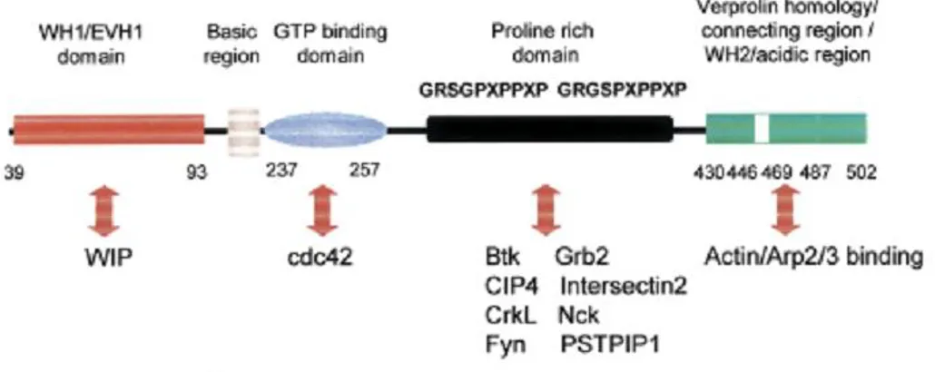
The disease was linked to a single gene composed of 1823 bp and 12 exons, which is present in the X chromosome and only transcribed in the hematopoietic cells. The product is a 502 amino acid called the Wiskott-Aldrich protein (WASp). The protein contains an N-terminal domain called EVH1 or WH1, a basic region followed by a 14 residue stretch (GTPase binding domain or cdc42/Rac-interacting binding motif) a proline-rich region containing multiple consensus motifs for Scr3 homology 3 (SH3) domain binding and an acidic C-terminal region (VCA domain).
The C-terminus of WASp is responsible of binding and activating the Arp2/3 complex, a potent nucleator of actin polymerization1. This domain is made up of a WASp homology 2 (WH2) sequence followed by a short central (C) sequence and a terminal acidic (A) sequence. WH2 domains bind monomeric actin 2, while the C and A sequences bind to the Arp2/3 complex.
The N-terminal domain binds to the WASp interacting protein (WIP) that regulates WASp
activity3 . WASP also possesses a binding domain (CRIB, also called GBD) for the GTP-bound (activated) form of the small GTPase cdc42, as well as a basic sequence that binds to phosphatidylinositol 4,5-bisphosphate (PIP2) 4.
The native form of WASp suppresses its capacity to trigger Arp 2/3 complex-mediated actin nucleation. This autoinhibitory behavior is obtained by folding the GBD and the EVH1 domains to residues within the VCA domain. WASp activity is
evoked following cell stimulation by three different ways: disruption of the blockage by interactions between cdc42 and the GBD domain, interaction between SH3 domain-containing proteins and the proline-rich region, or by phosphorylation of tyrosine residues (Figure1).
Figure 1. Functional domains of WASp and their interacting proteins. In the
inactive state, WASP has an autoinhibitory conformation, with the molecule folded in such a way as to enable a stable interaction between the CRIB and C domains. This structural constraint is disrupted by interactions between activated cdc42 and the WASp GBD or, alternatively, between SH3 domain-containing proteins, such as PSTPIP1 and Nck, and the WASp proline rich domain.
WASp is a member of a family of proteins that participate in the transduction of signals from the cell surface to the actin cytoskeleton 5. Other members of this proteins family are the three isoforms of SCAR/WAVE 1-3 and the ubiquitously expressed N-WASp.
The mechanisms responsible for the pathophysiology of WAS are directly linked to deficient actin organization in haematopoietic cells. The aberrant cell migration in hematopoietic stem lineage precursors has been proposed as the basis of the profound disorders of the immune cell development, functioning, and homeostasis in WAS patients6. However, the biological defects that have the most profound effects on the functioning of the immune system as a whole have not yet been clearly determined.
Actin remodeling represents an integral component of cell activation. Regulating the temporal and spatial distribution of actin polymerization is therefore essential for eliciting appropriate cell responses to environmental signals7.
Several studies performed with lymphocytes from WAS patients have revealed both signaling and cytoskeletal defects, including aborted mitosis, abnormal pattern of actin filaments, impaired macrophage phagocytosis8, NK cell cytotoxicity9, reduced B-cell adhesion and migration10 and T-cell activation11 7. The human WAS gene has 86% of similarity with the murine WAS gene. Thus, to better understand the role of WASp in the immune system, Snapper’s group generated a WASp-deficient mouse containing a targeted disruption of the GBD/CRIB motif. This murine model share most of the pathology features reported in humans5. A summary of the cellular defects that have been reported7 for the WASp-deficient mouse model is listed in Table 1.
Table 1. Functional defects in WASp-deficient cells
Cell type Functional defects
Haematopoietic progenitors12 Decreased homing efficiency
Monocytes/macrophages7; 13 Abnormal morphology Absent podosomes
Chemotactic/migratory defects Adhesion defects
Phagocytic defects T cells11; 1214 Abnormal morphology
Impaired CD3-mediated proliferation Impaired thymocyte development
Impaired capping of actin and T-cell receptors B cells15 Abnormal morphology with shortened microvilli Platelets16 Reduced number and volume
Lack of WASp leads to defective development and maturation of T-cell and B-cell lineage cells in mice. However, it is not clear whether a functional redundancy of WASp and N-WASp is present. Cotta de Almeida et al. addressed this problem
generating a conditional double-knockout strain and suggested that T-cell development depends on the combined activity of WASp and N-WASp17. Nonetheless, WASp serves a unique role for peripheral T cell function. In peripheral T cells WASp plays a critical role in the reorganization of the actin cytoskeleton. WASp-deficient T cells show a reduced capacity to form an asymmetric patch of adhesion molecules and surface receptor following TCR engagement that leads to an impaired migration and proliferation14; 16.
WASp plays a crucial role in the cytoskeletal regulation of B lymphocytes. WASp-deficient B cells are unable to respond to chemotactic gradients and have an impaired motility, caused by their inability to polarize the cell body10. Moreover, actin polymerization mediated by cdc42/Rac1, has been shown to be affected in WASp deficient B cells, causing an aberrant pattern of membrane protrusions upon stimulation with IL-415. These defects have been proposed as an explanation to the aberrant peripheral B-cell maturation and homeostasis in WASp-deficient B cells (Westerberg 2008).
1.1.1. Role of WASp in DCs
Several defects in the cytoskeletal architecture of DCs such as aberrant podosome formation and altered membrane adjustments required for migration, have been reported18 in human and mouse cells. To examine the functional role of WASp in DCs, most of the studies have been carried out using the murine deficient model. WASp- DCs fail to assembly podosomes during the response to chemotactic gradients, in particular, it has been shown that formation of podosomes and recruitment of the integrin Beta-2 is strongly compromised in the absence of WASp, causing a defect on the ability of adhesion to the Intracellular adhesion marker ICAM-1. ICAM-1 is a relevant molecule in stabilizing cell-cell interactions and facilitating endothelial migration. In vitro experiments resulted in abnormal DC
ligands CCL19, CCL3 and CCL2119. In vivo, WASp- DCs have a defective migration from skin and periphery towards the secondary lymph organs, which correlates with a reduced priming of naïve T cells20.
1.2.
Dendritic cells
DCs can be catalogued as a heterogeneous family of leukocytes that integrate information and transmit it to lymphocytes. Their main role is to recognize specific molecules upon infection of invading pathogens that trigger their differentiation into immunogenic Antigen Presenting Cells (APC), capable of priming and sustaining the expansion of naïve T cells.
Known as professional APC, DCs have a notable role in the initiation and modulation of the immune response due to their distribution and capacity to reach the lymphoid organs, areas where the CD4+ and CD8+ T cells accumulate. They have a unique capacity to translate the environmental signals into specific classes of adaptive immune responses by polarizing T-cell development. DCs have an inherent high efficiency for antigen presentation that allows them to induce strong T cell responses in small number and low levels of antigen21. DCs play also an essential role in the generation of both central and peripheral T-cell tolerance by inducing deletion, anergy or regulation of T lymphocytes22.
1.2.1. Subsets of dendritic cells
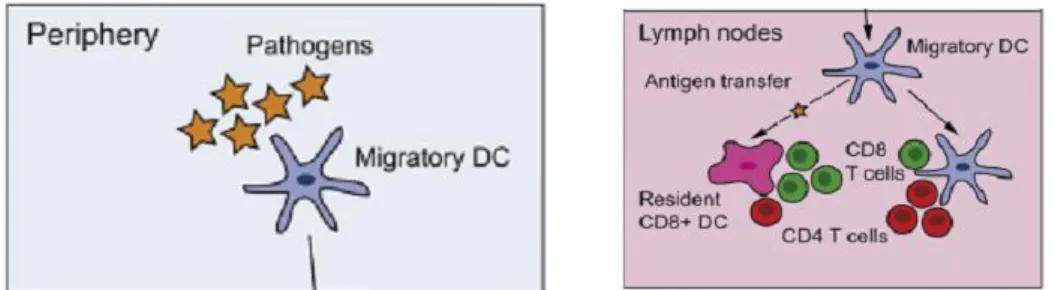
The phenotypic and functional analyses of DCs found in thymus, spleen and lymph nodes have revealed that DCs are a heterogeneous population of cells that can be classified in several ways that are still debated. In general, they can be divided into 2 major populations: (1) non-lymphoid tissue migratory and lymphoid tissue– resident DCs and (2) plasmacytoid DCs (pDCs, also called natural interferon-producing cells)21.
Migratory and tissue-resident DCs have two main functions: the maintenance of self-tolerance and the induction of specific immune responses against invading pathogens (figure 2)23 , while the major function of pDCs is to secrete interferon-alpha in response to viral infections and to prime T cells against viral antigens.
1.2.1.1. DCs in non-lymphoid tissues
Non-lymphoid tissue DCs are present in the pancreas and the heart, at filtering sites such as the liver and the kidney, and at environmental interfaces as lung, gut, and skin. Among interface DCs, epidermal DCs also called Langerhans cells (LC) are the most studied. LC constitutively express major histocompatibility complex (MHC) class II and high levels of the lectin langerin24. These interstitial DCs express low levels of the integrin CD11b and coexpress alpha-Eβ7 (CD103), a ligand of the cell adhesion molecule E-cadherin expressed by most epithelial cells. In addition to CD103+ DCs, tissues also contain another major DC population that is characterized as MHC class II+ CD11c+ CD11bhi CD103–langerin.
Langerhans cells were first described by Steinman in 197825, who proposed a model where the DCs can be found in the epidermis or in the spleen. In this model, DCs can exist in two functional states: immature and mature. Immature DCs are located in the periphery and efficiently uptake self and non-self antigens but are quite inefficient for T cell activation. Only upon encounter of pathogens and mediators of inflammation, DCs enter to a development program called maturation. Maturation causes downregulation of their endocytic capacity, activation of the antigen processing machinery that generates complex of MHC molecules with peptides derived from the internalized antigens and an increase of T-cell costimulatory molecules. In parallel, changes in the pattern of chemokine receptors and adhesion molecules and modifications in the cytoskeleton structure induce the migration from the periphery to secondary lymphoid organs, where the antigens are efficiently presented to T cells, thus initiating adaptive immune
In summary, tissue DCs uptake antigens and migrate continuously through afferent lymphatics vessels to the T-cell areas of lymph nodes (LN), a process that increases in response to inflammatory signals. The constant DC efflux from tissues to the tissue-draining LN requires constant replacement with new cells in order to keep the tissue-DC homeostasis26.
1.2.1.2. DCs in lymphoid tissues
Lymphoid tissue–resident DCs are the most studied DC population in mice, but little information is available on their human counterparts. Lymph node DCs are a heterogeneous population as they include blood-derived lymphoid tissue–resident CD8+, CD4+, double-negative spleen equivalent DCs, and migratory DCs entering via the afferent lymphatics that vary according to the LN draining site26.
In mice, splenic DCs constitutively express MHC class II and the integrin CD11c. They are further classified into two major subsets that include CD4+CD8– CD11b+ DCs that localize mostly in the marginal zone and CD8+CD4–CD11b– DCs localized mostly in the T-cell zone27. CD4- CD8- CD11b+ DCs have also been identified and are called double-negative DCs. CD8+ DCs are specialized in MHC class I presentation, whereas CD4+ DC subset is specialized in MHC class II presentation. CD8+ DCs have also been shown to cross-present cell-associated antigens, whereas CD4+ DCs are unable to do so28. The DC population present in the mucosa-associated lymphoid tissues, is equivalent to the one found in the spleen.
DCs that reside in the LN and in the spleen play a major role in the induction of immune response against blood-borne pathogens, since their function is to monitor the blood for the presence of infectious agents that spread through circulation. Intravenous inoculation of inflammatory compounds such as lipopolysaccharide (LPS) or CpG induce the activation of the LN DCs, their migration to the T-cell areas of spleen and LNs, the acquisition of a mature
phenotype and the secretion of large amount of IL-12, confirming their major role in immunosurveillance.
Figure 2. Role of the two main DC populations. Migratory DCs scan the periphery
looking for pathogens, they drift away towards the lymphoid organs and encounter resident DCs, where they present those antigens to CD4 and CD8 T cells.
1.2.1.3. Plasmacytoid DCs in lymphoid and non-lymphoid tissues
Plasmacytoid DCs (pDCs) are a subset of DCs in both humans and mice with the ability to sense and respond to viral infections mainly by secreting large amounts of type I interferons. pDCs constitutively express MHC class II molecules and lack most lineage markers29. Murine pDCs lack CD11b and express low levels of the integrin CD11c and the lineage markers CD45RA/B220+ and ly6C /GR-1+, express PDCA1 and siglec-H, identified as a specific surface markers for mouse pDCs30. Human pDCs express very low to no level of CD11c, they express CD4 and CD45RA antigens, the c-type lectin receptor BDCA2, and the molecule BDCA4, a neuronal receptor often used to isolate pDCs, and high levels of the interleukin-3 receptor (CD123). pDCs constitutively express the IFN regulatory factor (IRF-7) that allows the rapid secretion of vast amounts of IFN- in a signaling pathway that starts by engagement of Toll-like receptors 7 and 931. pDCs circulate in blood and are found in steady-state at the spleen, thymus, LN, and the liver. Human and mice pDCs enter the LN through the high endothelial venules and accumulate in the paracortical T-cell rich areas. In contrast to the other DCs subtypes, pDCs do not efficiently migrate to peripheral tissue in the steady state with the exception of the liver29.
1.2.1.4. In vitro generated DCs
The establishment of defined cell culture systems to generate DCs in vitro has been useful for functional and intracellular studies. The first method reported to differentiate mouse DCs in vitro involves cultures of bone-marrow or spleen precursors in medium that is supplemented with granulocyte/macrophage colony stimulating factor (GM-CSF), with or without interleukin-4 (IL-4). Surface marker examinations showed that the resulting DCs are partially similar to some of the lymphoid-organ-resident DC subsets found in vivo32.
A more accurate system to generate lymphoid organ-like DCs emerged with the use of the FMS-like tyrosine kinase 3 ligand (FLT3L). Bone marrow precursors cultured with FLT3L differentiate into DCs with similar expression patterns of CD11b, CD24, CD172, Toll-like receptors, chemokine receptors and the ability to secrete IL-12 and the chemokine ligand 5 (CCL5)33.
1.2.2. DC specific functions
The main function of DCs is the internalization of pathogens, followed by processing and presentation of antigen peptides to naïve T cells.
1.2.2.1.
Antigen uptake and pathogen recognition
One of the properties that define DCs as potent antigen-presenting cells, is their ability to efficiently uptake particles and pathogens by endocytosis. Endocytosis is highly active in immature DCs and downregulated upon cell maturation, assuring the efficient sampling of the environment in the periphery and simultaneously limits the range of antigens that the cells will be able to present after leaving the marginal tissues34. There are three main endocytosis pathways: receptor mediated endocytosis, phagocytosis and macropinocytosis35.
1.2.2.1.1. Receptor mediated endocytosis
The efficiency of endocytosis is increased by non-specific binding of solutes to the cell membrane and even more by the capture of soluble antigens through specific high affinity receptors that are concentrated into specialized endocytic transport vesicles. DCs express a wide range of endocytic receptors that are grouped into two main families. The first encloses receptors for the Fc portion of immunoglobulins (FcRs) and complement receptors (CRs) which are involved in the uptake of particles that are opsonized by immungloublins or complement factors. A second class of endocytic receptors comprises the scavenger receptors (SRs) and C-type lectin family receptors that directly recognize specific structures on both self-antigens and pathogens36.
1.2.2.1.2. Phagocytosis
The uptake of large particulate antigens by phagocytosis is the prevalent form of antigen uptake in vivo for both pathogen-derived and endogenous antigens. The process starts with the engagement of specific cell surface receptors, that trigger a signaling cascade mediated by the Rho-family GTPases (Rho, Rac and Cdc42) that ends up with the extensive reorganization of the actin cytoskeleton, forming cell-surface extensions that zip up around the pathogen and engulf it37.
The membrane protrusion and the activation of signaling pathways depend on the nature of the particle to be ingested and the receptors that recognize it. Phagocytosis might take place by engagement of specific receptors. In the FcR-mediated phagocytosis, the cells extend pseudopods that engulf the particle and subsequently fuse to form a phagosome, a process that requires the activation of cdc42 for the pseudopod extension and Rac for pseudopod fusion and phagosome closure. On the other hand, the CR-mediated phagocytosis does not induce pseudopod formation. The coordinated action of chemokines and integrin ligation controlled by Rho induces the formation of the phagocytic cup38.
Phagocytosis by DCs is essential in host defense against infections. Immature DCs are able to phagocytose Gram positive and Gram negative bacteria, mycobacteria, yeast cells and parasites39. Additionally DCs have a main role in the clearance of apoptotic cells by the recognition of molecules that are absent on live cells, as calreticulin, phosphatidylserine and lysophospholipin.
1.2.2.1.3. Macropinocytosis
Macropinocytosis contributes to bulk fluid-phase uptake via the formation of membrane protrusions that collapse and fuse with the plasma membrane generating large endocytic vesicles that allow the sampling of large volumes of extracellular milieu. Macropinocytosis is constitutively active in immature DCs. This process is essential for the uptake of soluble antigens released by pathogens or externally provided upon intradermal or intravenous injection.
1.2.2.2. Antigen presentation by DCs
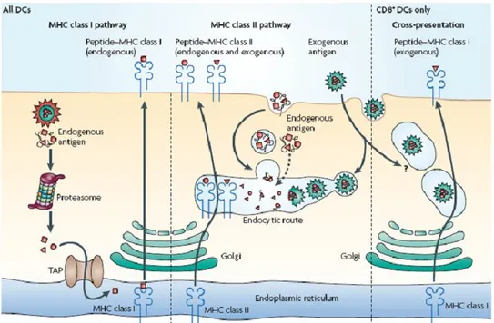
Upon internalization, antigens are degraded into small immunogenic epitopes that associate with the MHC and are transported to the plasma membrane where they trigger the activation of naïve T lymphocytes. In particular, the activation of CD8+ and CD4+ T lymphocytes requires recognition by the T cell receptor (TCR) of epitopes associated with MHC class I and MHC class II, respectively. The antigen processing pathways that lead to the formation of peptide-MHC complexes rely on proteolysis occurring in the proteasomes and lysosomes35.
It is accepted that MHC class II molecules encounters exogenous antigens in the endocytic pathway and MHC class I is loaded with endogenous antigens in the endoplasmic reticulum (ER). However, it has been shown that MHC-II complexes can present intracellular antigens from intracellular proteins and MHC-I can present peptides derived from exogenous antigens, a process called cross-presentation40; 41.
DCs perform antigen processing and presentation via MHC class I in proteins coming from alternative translation products, proteins found in the cytosol (either endogenous or viral) and proteins retrotranslocated to the cytosol and imported into the ER. Regardless the route of entry, most of the peptides loaded on the MHC-I complex are produced by the proteasome and further trimmed by cytosolic or ER resident peptidases. Proteins are trimmed in their N-terminal region by the proteasome and peptide products are shuttled in the ER where the final MHC-I peptide complex is formed and eventually presented on the cell surface42.
DCs have the highest cross-presentation efficiency, allowing the entry of exogenous antigens into the MHC class I pathway of antigen presentation43. Soluble proteins, immune complexes, pathogens and cellular antigens have been reported to be cross-presented. However, the mechanisms by which the APCs transfer internalized antigens to the MHC class I loading pathway are not well understood. In most of the cases a limited endocytic degradation and transport into ER seem to be required, being a mechanism to allow the release of the antigen from the endocytic structures to the cytosol44.
DC cross-presentation plays a key role in priming of CD8+ T cells in response to exogenous agents such as bacterial or viral infection, as well as in the maintenance of both, central and peripheral tolerance to self antigens by the deletion or anergy of self-reacting cytotoxic lymphocytes45.
Antigens loaded on the MHC-II come from exogenous proteins that are internalized by DCs through different mechanisms of endocytosis or endogenous proteins that reside in the secretory system. The MHC-II is assembled in the ER and transported to early endosomes and further on to late endosomes and lysosomes, along this path it can bind polypeptide precursors that are trimmed by various proteases to end up reaching the plasma membrane46 (Figure 3).
Figure 3. Antigen presentation pathways by DCs. All DCs have functional MHC class
I and MHC class II presentation pathways. MHC class I molecules present peptides that are derived from proteins degraded mainly in the cytosol, which in most DC types comprise almost exclusively endogenous proteins. MHC class II molecules acquire peptide cargo that is generated by proteolytic degradation in endosomal compartments. The precursor proteins of these peptides include exogenous material that is endocytosed from the extracellular environment and also endogenous components. CD8+ DCs have a unique ability to deliver exogenous antigens to the MHC class I (cross-presentation) pathway, although the mechanisms involved in this pathway are still poorly understood. TAP, transporter associated with antigen processing47.
1.2.2.2.1. Dendritic cell maturation
Concomitantly to the uptake and processing of the antigens, DCs recognize pathogen associated patterns (PAMPs) and sense inflammatory signals by different classes of membrane and intracellular receptors. Receptor engagement triggers signal cascades that lead to the production of inflammatory cytokines, upregulation of co-stimulatory molecules and to the altered expression of chemokine receptor profiles, providing them the ability to stimulate naïve T lymphocytes48.
One of the best characterized classes of receptors that directly contribute to the inflammatory responses to pathogens is the Toll-like receptor family (TLR). Mammalian TLRs are a family of at least 12 transmembrane proteins that collectively recognize lipids, carbohydrate, peptide and nucleic acid molecules
expressed by different microorganisms, and differ from each other in ligand specificities, expression patterns and the inducible target genes49.
Expression of TRLs 1, 2, 4, 5 and 6 is confined to the cell surface and appears to be specialized mainly in the recognition of bacterial products, where TLR4 plays an essential role in the recognition of LPS, a major component of Gram- bacteria. TLRs 3, 7, 8 and 9 are expressed on the membrane of endocytic vesicles or other intracellular organelles and are specialized in the detection of viral nucleic acids. This family of TLRs has an essential role in the antiviral immune responses mediated by the secretion of type I interferons. TLRs 9 and 7 are involved in the recognition of the 2’-deoxyribo(cytodine-phosphato-guanosine) (CpG) DNA motifs found in bacteria and viral DNA, while TLR3 is engaged by double-stranded RNA ('mimicked' by poly(I:C))50.
Upon activation of their ligands, TLRs transduce signals through pathways involving diverse adaptor proteins containing Toll/IL-1R (TIR) domains. TIR activation triggers signaling cascades that end up with expression of host defense genes including inflammatory cytokines, type I interferon cytokines, the up-regulation of co-stimulatory molecules (CD40, CD80 and CD86), downup-regulation of chemokine receptors such as CCR1, CCR5 and CCR6 and upregulation of CCR2, 5, 7 and 19, which drives DCs into the afferent lymphatic vessels and the LN51.
1.2.2.2.2. DC trafficking
After antigen uptake and processing, DCs sense and integrate different environmental signals and eventually migrate to T-cell rich areas of secondary lymphoid organs. There, antigen-specific immune responses are initiated by engagement of the TCR with the cognate peptide-MHC complex presented by the DC.
Antigen-loaded tissue DCs migrate to draining LNs through the lymph. These DCs penetrate the endocortex and reach the high endothelial venules (HEV). Thus, T and B cells that home to LNs by route of HEVs are first exposed to antigen loaded tissue DCs than lymphoid resident DCs. In this way, T cells are preferentially updated with information of antigens in the periphery52.
The entry of DCs from peripheral tissues into the draining lymphatic vessels as well as their migration from the lymph into the LN cortex depends on the chemokine receptor CCR7 and its ligands CCL19 and CCL21. The prevailing model to explain how CCR7+ DCs arrive at peripheral lymphatic vessels is that they respond to a chemotactic gradient of CCR7 ligands, which originates from the lymphatic vessel. In mice, there are two known functional genes that encode CCL21. One form of CCL21, the CCL21-Leu, is expressed in the periphery by initial lymphatic vessels. The other form of CCL21, CCL21-Ser, is only expressed in the terminal lymphatic vessels and LN. The gradient caused by this divergent chemokine expression pattern might be the cause of DC migration53.
DC migration from the periphery to the T-cell zone of lymphoid organs is a process that requires reorganization of the cytoskeleton and the plasma membrane. It has been shown that RAC1 and RAC2 are required for migration from the skin to the lymph nodes. Absence of both RAC1 and RAC2 strongly impairs the extension of dendrites by DCs and the mobilization of DCs to LN54; 55.
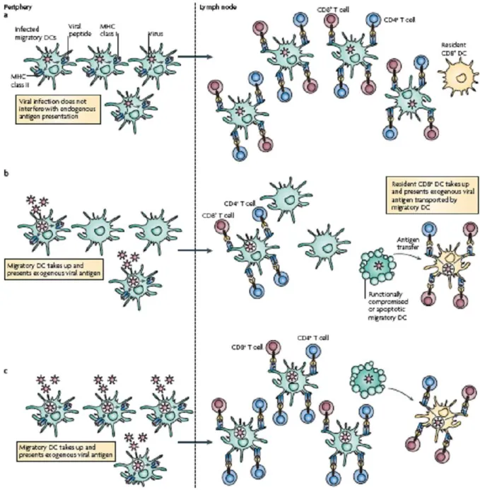
Is widely accepted that migratory DCs have a major role in carrying outer antigens from peripheral tissues to the lymph nodes and these can be somehow presented
to the CD8+ DCs. Instead, self antigens are presented in the steady state on MHC class I and II molecules by both immature resident in lymphoid organs CD8+ DCs and mature migratory DCs, with the former being biased towards MHC class I cross-presentation and the latter towards MHC class II presentation.
Three different scenarios can be hypothesized for antigen-presenting functions of migratory and lymphoid resident DCs (Figure 6). A first situation is one in which the migratory DCs are themselves infected by a virus that has no deleterious effect on the DCs. In this situation, the migratory DCs are the main subset that presents endogenously produced antigens on MHC class I molecules 56and MHC class II molecules, in the draining LN.
The second situation is one in which the migratory DCs are not infected, but the migratory DCs are still required to carry viral antigens to the lymph nodes, whether endogenously expressed or captured from infected cells, and transfer the antigens to the resident DCs in the form of endosomal vesicles or apoptotic bodies57. The main resident DC population that acquires the antigen would be the CD8+ DCs, due to their special ability to capture dead cells or cell fragments. The migratory DCs that survive and have not been inactivated by viral immunoevasins, as well as the resident CD8+ DCs, present the antigens according to their intrinsic abilities 58; 59.
The third scenario occurs in large scale infection of tissues that are drained by the lymph node. In this case the relative antigen presentation efficiency of the lymphoid and non-lymphoid DCs is almost the same on a per-cell basis, but the comparative number of migratory DCs that display antigen is much higher and their relative contribution to cross-presentation within the lymph node increases59. This hypothetical model implies the recruitment of monocytes to sites of inflammation, followed by their conversion into DCs. In this way, the monocyte-derived DCs may share some of the antigen-presenting functions that are initially carried out by the migratory and lymphoid-organ-resident DC subsets60.
Figure 4. DC migration and antigen presentation. An scheme with three
hypothetical scenarios showing how different populations of DCs may migrate and act in a synergetic way to counteract tissue infection47.
1.2.3. T-cell stimulation by DCs
DCs play a crucial role in the induction of adaptive immunity and in the maintenance of peripheral tolerance to self and non-pathogenic environmental antigens. They can determine the fate of naïve T cells by three signals. The first occurs with engagement of the TCR with the cognate peptide-MHC complex, which triggers a TCR cascade that determine the antigen-specificity of the response. The second consists in the engagement of the CD28 and CTLA-4 co-stimulating receptors, to respectively influence the T cell response in a positively or negative way, controlling in this manner the initiation of the protective immunity. The last signal is delivered by cytokines and chemokines produced by DCs, which are sensed and integrated by the T cells. The combined effect of diverse released cytokines determine the proliferation, survival and ability to differentiate into effector cytotoxic T cells, Th1, Th2, Th17 or T regulatory cells47.
It is widely accepted that DC maturation state is the critical switch that provides the signals for effector and memory T cell development, diverting T cells from anergy or deletion to protective immunity. On the other hand, immature DCs have been reported to have a inefficient antigen-presenting capacity, inducing peripheral T cell tolerance by antigen-specific T cell deletion, functional inactivation or by generation of regulatory T cells61. DCs capacity to efficiently prime naïve T cells resides in the increased expression of MHC complexes, adhesion and co-stimulatory molecules, which controls the stability and the duration of the DC-T cell contact62. In addition, mature DCs secrete enhancing cytokines like the interleukin-12 (IL-12) and interferons type I/II that support the survival and the differentiation into effector T cells.
1.2.3.1. Immune synapse
In order to deliver the three signals required to prime naïve T cells, a physical contact must be established with the antigen presenting cell (APC). The organized structure that takes place in the region of contact is called the immune synapse (IS). The formation of the IS is driven by different molecular mechanisms such as polarized recycling of receptors, passive lateral diffusion and cytoskeletal-mediated movement of molecules.
Immune synapses can have different morphological patterns with specific arrangements of membrane proteins and receptors. The simplest type of synapse experimentally observed has simple enrichment of receptors at the contact site. A more complex arrangement with central accumulation of both TCRs and adhesion molecules at the contact site is known as mature synapse63.
The mature IS is composed of two concentric regions; first, the central supramolecular activation cluster (cSMAC), where the TCR, the co-stimulatory molecules (mainly CD40-CD40L and CD28-B7) and the intracellular signaling molecules PKCθ (105), lck, fyn, and ZAP-70 are concentrated in the inner side of the T cell membrane63; 64. Although it is accepted that the initiation of TCR signals occurs in peripheral microclusters that begin to form prior to IS formation, the cSMAC has been proposed as a place for TCR signal enhancement and TCR degradation65 or as a site for enhanced receptor engagement and prolonged signaling 66. Surrounding the cSMAC, a peripheral integrin-rich ring (pSMAC) is found, where LFA-1 interacts with ICAM-1 and which concentrates intracellular talin in the T cell provides an adhesive anchoring64 (Figure 5).
Figure 5. Structure of the Immune Synapse from the T cell side. TCR interaction
with peptide–MHC class II molecules recruits tyrosine kinases such as ζ-chain-associated protein 70 kDa (ZAP70) and FYN, adaptor proteins and actin polymerization-regulatory molecules such as the small GTPases cdc42 and RhoA, WASP, inducing localized actin polymerization at the cSMAC through the ARP2/3 complex. The pSMAC is composed of leukocyte function-associated antigen 1 (LFA1) molecules that interact with ICAM1 expressed by the DC and also regulates the actin cytoskeleton through LFA1 interaction with talin67.
1.2.3.1.1. IS and T cell activation
After formation of the IS, engagement of the TCR initiates a signaling cascade involving Lck, ZAP70, Itk and Vav, which result in the accumulation of Cdc42. Cdc42 in turns induces WASp activation and triggering of cytoskeletal rearrangements that results in polarization and activation of the T cell67.
As the TCR is engaged, cortical actin concentrates at the contact region following its clearance towards the edges of the contact sites, in order to form a peripheral ring to the pSMAC68. These changes are thought to depend on increased membrane fluidity, caused by a transient dephosphorylation of ezrin, radixin and moesin (ERM), which blocks the crosslink of the actin cytoskeleton with the plasma membrane and provokes a decreased cellular motility. This in turn is associated with Ca2+ dependent phosphorylation and deactivation of the motor
Accumulation of F-actin at the T cell-DC interface is the result of induced localized activation of multiple actin regulatory pathways, in particular the actin related proteins 2/3 (Arp2/3) complex. Activation of Arp2/3 is mediated by the interaction with WASp, WAVE2 and HS1. WASp is recruited to the site of TCR activation through its interaction with the SLP-76 associated adapter protein Nck, where it is activated via Vav-1 dependent stimulation of cdc4269. Vav-1 also activates Rac1, which results in the activation of WAVE2. However, experiments indicate that WASp deficient T cells still polymerize F-actin at the T cell–DC contact region, implying that WASp might control T cell activation in an alternative way, affecting the endocytosis and the exocytosis70.
Inhibition of actin polymerization in T cells by cytochalasin D prevents synapse formation, and interferes with actin-myosin functions preventing movement of surface proteins to the contact zone71; 72.
1.2.3.1.2. T cell polarization
Formation of the IS results in the polarization of the T cell, which is orchestrated by a protein network that includes four complexes, the Scribbled (Scrib), partitioning defect (PAR), Crumbs and a core planar cell polarity (PCP).
The Scrib complex consists of scrib, Lgl and Dlgh. In particular, Dlg has been found to be translocated to the IS, and it is thought to be responsible of reorganization of the cytoplasm, rearrangements of surface proteins and redirection in the transport of RNA and proteins73. Other proteins such as scrib, Crumbs 3 and Par3 rapidly relocalize to the IS, and their absence causes a reduction in the cell motility, conjugate formation and lytic activity74.
Polarization of the T cell also provokes that the centrosome, which is the main microtubules organizing center (MTOC), dissociates from its position near the nuclear envelope and moves towards the contact site T cell-DC. MTOC movement
reorients the microtubule network and the whole cell, bringing the MTOC-associated organelles, such as Golgi complex and the endocytic recycling compartment 75 (Figure 6).
The centrosome (MTOC) is found in the center of the microtubule cytoskeleton and contains the centrioles (barrel-shaped cylinders composed of microtubule triples) and the pericentriolar material (PCM) which is mainly composed by -tubulin. The -tubulin is a key player in the polymerization of microtubules from and - tubulin subunits. Microtubules have a minus end that is proximal to the MTOC and a more dynamic plus end that lengthens away. Microtubule stability given by acetylation of the alfa and beta tubules was shown to be important for TCR-mediated polarization76. MTOC reorientation is a hallmark of cell polarity in various cellular processes like asymmetric cell division and directional migration.
A study by Combs77, shows that MTOC polarization is integrated into the TCR signaling through interaction between the dynein and the adhesion-and-degranulation-adaptor protein (ADAP), which might provide a link between the microtubule cytoskeleton, microtubule motor proteins and the actin cytoskeleton through ADAP/VASP actin binding. This hypothetical linkage between plus-end microtubules and the cortical actin cytoskeleton is supported by data where inhibition of microtubule polarization by colchicine, induced an early retraction of the actin-based protrusions in T cells before IS formation68 .
Another event that makes part of T cell polarization is the formation of the distal-pole complex. An actin-rich structure which is thought to have a role for pulling away and sequestering negative regulators from the TCR activation complex, and might be required for distinguishing the fate of recently activated T cells into memory or effector79.
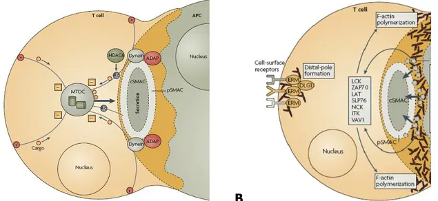
A B
Figure 6. T cell polarization upon IS formation. A) Upon TCR recognition, the T cell
reorient the MTOC beneath the contact region with the DC. MTOC polarization is commanded by signaling coming from the IS. A complex that consists of adhesion- and degranulation-adaptor protein (ADAP) and the microtubule motor dynein is thought to “grab” the MTOC. Microtubules have been shown to anchor to the pSMAC during TCR engagement. Moreover, microtubule plus-end complexes are proposed to link the microtubule cytoskeleton to the cortical T-cell actin cytoskeleton, thereby providing additional force for MTOC movement through an unknown mechanism. Histone deacetylase-6 (HDAC6), which is important for microtubule stability, is required for TCR-mediated MTOC polarization. Once the MTOC is polarized, cargo travels along microtubules using minus-end directed movement to the MTOC, which directs secretion towards the DC surface78. B) Reorganization resulting from T-cell activation leads to the
formation of the distal pole complex which is formed by the ERM cytolinkers and interacts with DLG1 in order to recruit away from the SMACs molecules such as CD43 or the P-selectin glycoprotein ligand 1 (PSGL1)67; 78.
1.2.3.1.3. T cell activation in vivo
IS formation has been mainly studied using in vitro models which do not consider the gradients, factors and external signals that might affect the contacts between T cells and APCs. In 2002, the first in vivo studies showed that in intact LN, T cells are highly motile and DCs are able to scan a high number of them over time. Nevertheless, when DCs are loaded with specific antigen the interactions became stable, with an average duration in the order of hours80; 81.
Von Andrian and colleagues recently proposed a three-phase model for T-cell–DC interaction in vivo. According to this model, antigen-specific interactions of T lymphocytes with DCs are transient between 2 and 8 h following the encounters,
stable between 8 and 24 h, and again transient by 24–36 h. The stable interaction in the 8–24 h period probably corresponds to the organization and maintenance of the IS, and is required for complete T-cell activation82.
It is likely that in vivo, T-cell behavior is strongly influenced by chemoattractant forces in the presence of lymph and blood flow. Since T cell activation is characterized by both transient and stable interactions, it has been proposed that chemokine-mediated signals compete with TCR-mediated stop signals and that the combination of the two types of signals determines the duration of T cell−APC interactions. Studies have demonstrated that the accumulation of chemokine receptors at the T cell−APC contact site requires formation of a productive IS and chemokine (CXCL12 and 5) directed secretion by APCs. In this way, T cell responsiveness to other chemoattractant sources is reduced and stability of T cell−APC interactions is increased83 (Figure 7).
Figure 7. Chemokine receptors in T-DC synapses. Chemokine receptors recruitment
to the IS in T cells is mediated by recruitment of chemokines 5 and CXCL12. In DCs represents a mechanism used to reinforce the synapse and avoid early splitting due to external chemoattractant sources, thus, enhancing T cell activation83.
1.2.3.1.4. DCs in synapse formation
Molecular events that contribute to synapse formation and maintenance in T cells have been characterized quite extensively using artificial APC, whereas little is known about the mechanisms controlling the coordinated transfer of different signals from DCs to T cells.
The first evidences of the active role of the APC in the IS were given by Al-alwan et al, who demonstrated that DCs actively polarize F-actin and fascin, during formation of IS with CD4 T cells84; 85. Later, it was assessed that DCs rearrange their actin cytoskeleton towards naive CD4+ T cells only in the presence of specific MHC-peptide complexes69. Concomitantly, Kondo et al 86 showed that IS could take place in the absence of antigen, inducing TCR signaling and T cell proliferation, probably caused by the density of HLA class II molecules on the surface of DCs in conjunction with the pool of displayed self-peptides. Benvenuti et al. described for the first time the functional role of the maturation state of the DC together with the presence of antigens on IS. Immature DCs were able to establish multiple transient contacts of low stability and with no mature immune synapses taking place. However, they observed that in the absence of antigen, DC maturation induced a minor increase in CD3, LFA-1, and LAT clustering at the immune synapse, but effective clustering, TCR signaling and T cell activation required both DC maturation and antigen recognition 62; 87.
Bloom has documented the role of spinophilin in formation of IS by DCs. This adaptor protein initially found in the dendritic spines of neurons in nervous central system, shows dramatic changes in its distribution accompanying the formation of the immunological synapse. Spinophilin contains a PDZ domain, which is often found in scaffold proteins that bind the cytoskeleton as in T cells, and controls polarity as SCRIB88(figure 8). The spinophilin null phenotype has a great impact on the triggering of a highly effective immune response.
Figure 8. DCs polarization in Immune synapses. In immature DCs, spinophilin is
located throughout the cytoplasm but redistributes to the plasma membrane upon stimulus-induced maturation. In DCs interacting with T cells, spinophilin is polarized dynamically towards the IS in an antigen-dependent manner and induce the polarization of adaptors proteins such as the plexins88.
Another adaptor protein with a shared role in neuronal synapses is plexin-A1. Plexin-A1 belongs to a family of cell surface proteins that are known to act as receptors for semaphorins. In DCs, plexin-A1 appears to be retained in an intracellular compartment, making its way to the cell surface after TNF- stimulation, where it clusters in a multifocal pattern localizing to the T cell synapse89.
A recent report revealed an unexpected function of synapse formation in DCs. Riol-blanco et al. demonstrated that CD40 signaling upon IS formation induce ATK1 activation, which inhibits the apoptosis of DCs in stable conjugates with T cells. In parallel, they claimed that soluble factors secreted by both T cell and DC are not enough to increase DC survival90.
1.2.3.1.5.Information exchange at the IS
Factors secreted by lymphocytes are often released into an environment that is densely populated with many cell types, which brings the problem of specificity of intercellular communication. In particular, CD8+ and CD4+ T cells must operate their secretory responses in a targeted way as to avoid activating or killing the wrong cell. First studies of the MTOC, secretory organelles and Golgi in T cells suggested the existence of a mechanism for targeted secretion towards the APC.
Currently, it is established that many hematopoietic cells are able to perform directed secretion. Mast cells and granulocytes polarize their degranulation in response to FcR cross-linking91, while natural killer cells and cytotoxic lymphocytes direct the content of their secretory lysosomes towards a specific target92.
Imaging studies have shown that lytic granules are released by cytotoxic T lymphocytes (CTL) at a defined point within the synapse75. In particular, it has been shown that CTL first reorient the MTOC upon TCR signaling towards the IS, next the MTOC docks at the cellular membrane and finally the lytic granules are secreted. Recently, it has been shown that MTOC and lytic granule polarization are independently regulated in response to the strength of TCR signaling93.
In vitro and in vivo experiments have shown that several important cytokines, such as IL-2, accumulate beneath the IS in T helper cells after stimulation by an APC94. Huse et al have shown that T helper cells use 2 directionally distinct pathways for secretion of cytokines and chemokines. The first one, release directly towards the IS in an antigen specific way molecules such as IL-2, interferon- and IL-10. The second is multidirectional and includes the secretion of TNF, IL-4 and CCL3, which seems to be involved in inflammation responses and the mobilization of bystander cells95.
Ii is very likely that the polarized and the multidirectional secretion pathways are regulated by specific vesicle markers that control their fate after being produced in the Golgi and anchored to the microtubules. This phenomenon was observed by Stow in macrophages, tracing the trafficking markers of different cytokine-containing vesicles96; 97.
Signals are continuously delivered to the T cell during prolonged interactions with DCs. MTOC reorientation controls the directed secretion of cytokines and chemokines, which might travel along the microtubules using their slow-growing end and then being released towards the engaged DC. Alternatively, stores of IL-2, IL 4 and IL-5 are directed towards the DC after MTOC reorientation98(Figure 6).
There have been some reports suggesting that also DCs might make use of the polarized secretion. Semino et al showed that in conjugates with NK cells, immature DC increase their free Ca2+ concentration, rearrange their cytoskeleton and co-ordinately secret IL-18 towards the interacting NK cell99. Borg et al. showed that the formation of specific DC/NK conjugates induces the polarization of the IL-12 toward the synapse and provoke NK cell activation100. There is only one report that studies the directed secretion in T cell-DC conjugates, suggesting that IL-1B and cathepsin D are released toward the IS. However, the mechanism and the molecular complexes that support polarization in DCs are not well understood101.
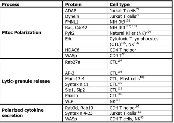
MTOC polarization has been functionally linked to polarized secretion of cytokines and lytic in immune cells. A list of the proteins linked to these processes and the cell type where they have been studies is summarized in the Table 2.
Table 2 Proteins identified in polarization and secretion pathways in several cell types.
Process Protein Cell type
ADAP Jurkat T cells77
Dynein Jurkat T cells77
FMNL1 NIH 3t3102
Rac, Cdc42 NIH 3t3102; 103
Pyk2 Natural Killer (NK)104
Erk Cytotoxic T lymphocytes
(CTL)105, NK106 HDAC6 CD4 T helper Mtoc Polarization WASp CD4 T95 Rab27a CTL107 AP-3 CTL108
Munc13-4 CTL, Mast cells109 Syntaxin 11 CTL110
Slp1, Slp2 CTL111
Paxilin CTL105
Lytic-granule release
WIP NK112
Rab3d, Rab19 CD4 T helper95 Syntaxin 4-23 Jurkat T cells113 Polarized cytokine
secretion
WASp CD4 T cells, NK95
1.2.4. Interleukin 12, an example of the three signals integration IL-12 is one of the most important cytokines produces by DCs upon TLR engagement. IL-12 is produced by several DC subsets after challenging with different bacterial strains, that stimulates TLRs 3, 4 or 9114. IL-12 is a covalenty linked heterodimer formed by a 35 kDa light chain, known as p35, and a 40 kDa heavy chain known as p40. The p35 protein is homologous to other single-chain four-alpha helical cytokines like IL-6 and granulocyte colony-stimulating factor (G-CSF), whereas p40 is homologous to the extracellular domain of members of the hematopoietic cytokine-receptor family (Figure 9)115.
IL-12 positively regulates its own production via the induction of IFN-gamma, which primes monocytes and neutrophils for further IL-12 production. Conversely, IL-12 production is inhibited by other cytokines including IL-10, IL-11, IL-13 and type I interferons117; 118.
Figure 9. IL-12 structure. IL-12 is an heterodimer composed by a light chain (p35)
and a heavy chain (p40). The bioactive form known as p70 binds the IL-12 receptor expressed by T cells116.
Studies with deficient mice for both IL-12 subunits or for the IL-12 receptor have revealed that IL-12 has an important role in favoring T helper 1 (Th1) response119. Both in vivo and in vitro, IL-12 is required for the optimal differentiation of CD4+ T cells into high-level IFN--producing Th1 cells120.
Though initially it was believed that IL-12 was sufficient to induce Th-1 cell differentiation, it has been shown that it may be more important for amplifying and fixing the phenotype of already committed Th1 cells than for directly priming naive CD4+ T cells for Th1-cell differentiation121.
IL-12 is synergistic with CD28 stimulation, and facilitates the T-cell proliferation and IFN-gamma production. In particular, IL-12 enhances the generation and cytotoxicity of T lymphocytes, inducing the transcription of genes that encode cytotoxic granule-associated molecules and upregulating the expression of adhesion molecules122; 123.
The Il-12 receptor is composed of two chains, 1 and 2 123and is mainly expressed by activated T cells, NK, DCs and in low levels by resting T cells. The affinity of IL-12 for either subunit alone is low, but coexpression of both 1 and 2
subunits generates IL-12 high-affinity binding sites. IL-12p40 interacts predominantly with the 1 subunit, whereas p35 interacts largely with the 2 subunit.
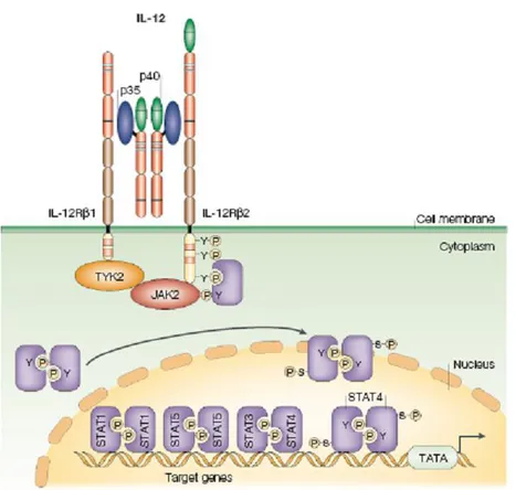
The specific effects of IL-12 are caused by ligand-induced autophosphorylation and transphosphorylation of receptor-associated Janus kinases (JAK). JAK activation induces tyrosine phosphorylation of the receptor subunits located in the intracellular domain. These phosphorylated tyrosines serve as docking sites for STATs (signal transducers and activators of transcription) and potentially other signaling molecules. IL- 12 specifically induces the tyrosine phosphorylation and DNA binding of two STAT family members, STAT3 and STAT4 (Figure 10). Their activation has been shown to be necessary but not sufficient to explain the ability of IL-12 to induce Th1 differentiation124.
Figure 10. IL-12 receptor signaling pathway The interleukin-12 (IL-12) receptor is
composed of two chains, IL-12Rβ1 and IL-12Rβ2. Signal transduction through IL-12R induces tyrosine phosphorylation, primarily of the Janus family kinases JAK2 and TYK2, which, in turn, phosphorylate and activate signal transducer and activator of transcription 1 (STAT1), STAT3, STAT4 and STAT5. The specific cellular effects of IL-12 are due mainly to its ability to induce activation of STAT4116.
2. MATERIALS AND METHODS
MiceSix to eight weeks old C57BL/6 females were purchased from Harlan (Milano, Italy). WASp- mice on a C57BL/6 (CD45.2) genetic background were a gift from S. Snapper (Massachussets General Hospital, Boston). GFP-centrin mice were generated from a construct given by Michel Bornens (Institut Curie, Paris) and were a gift from Chantal Desdouets (Institut Cochin, Paris). OVA-specific, MHC class-I OT-I and MHC class-II OT-II, TCR transgenic mice were purchased from the Jackson Laboratories. CD45.1 congenic C57BL/6 (kind gift from Pierre Guermonprez, Institut Curie, Paris) were bred to OT-I mice to obtain OT-I/CD45.1.
Mice were bred and maintained in sterile isolators. Animal care and treatment were conducted in conformity with institutional guidelines in compliance with national and international laws and policies (European Economic Community [EEC] Council Directive 86/609; OJL 358; December 12, 1987).
Cells
Bone marrow-derived DCs were differentiated in vitro from the bone marrow of the different mouse genotypes using culture medium containing Fms-like tyrosine kinase 3 ligand (Flt3L). DCs were used for experiments between day 7 and 8 when expression of Cd11c was higher than 80%.
For experiments with endogenous DCs, spleens from mice were extracted, homogenized and digested with collagenase D (1,6 mg/ml, Roche) and DNase I (0,1 mg/ml, Roche). Enrichment of DCs was performed by density gradient in an Optiprep solution (Sigma) 1,068 g/cm3 .The very low density fraction mainly composed by DCs was recovered and subjected to purification using CD11c microbeads (Miltenyi Biotec). OT-I and OT-II cells were isolated from total lymph
Antibodies and FACS reagents
The following antibodies for FACS analysis were purchased from BD Pharmingen: FITC and PE-conjugated anti-CD11c, FITC and PE-conjugated anti I-Ab, PE-conjugated anti-CD86, PE-PE-conjugated anti-CD11b, PE-Cy5-PE-conjugated anti-CD8, PE-conjugated anti CD45.1, biotinylated anti-CD69, biotinylated anti-CD3. CFSE (5-(6)-carboxyfluorescein diacetate succinimidyl diester), SNARF and CMTMR were purchased from Molecular Probes.
Bacterial Infection
Salmonella typhymurium strain ATK-GFP was kindly gifted by M. Rescigno (IEO, Milan). Bacteria were grown in LB medium (kanamicin 25 mg/ml + ampicilin 50 mg/ml) until it reached an O.D of 0.6. HEK293 or BM-DCs were infected at different infection ratios in IMDM medium during 1 hour, gentamicin (50g/ml) was added for an additional hour. Medium was washed away thoroughly and cells were lysed with Triton X-100 (0.5%). The bacteria internalized were released and plated on agar petri dishes with kanamicin + ampicilin. The day after, the number of grown colonies was counted and correlated with the initial number of bacteria. For FACS reading, cells were not lysed after treatment with gentamicin but collected and the intensity of the GFP signal was correlated to the number of bacteria phagocytosed for each DC.
Time-lapse video microscopy (Trajectories)
For the dynamic analysis of DCs trajectories, 3x10⁵ immature or LPS-pulsed DCs (overnight, 10 g/ml) were plated on fibronectin-coated coverslips and placed into a chamber on a Zeiss LSM510 META Axiovert 200M reverse microscope at 37°C in a 5% CO2 atmosphere. Transmitted light images were taken with a 63X objective and a 3CCD camera every 30 seconds during 40 minutes. Recording of the trajectories, displacement analysis, and velocity measurements were made using the Image J software. For analysis of the conjugate formation, mature DCs were incubated for one hour with the MHC class-I restricted peptide of ovalbumin
257-264(SIINFEKL) (0,1 nM) before plating. 1 x10⁵ OT-I cells were added to the dish and images were taken starting 5 min after landing on the same plane of DCs. Each DC was analyzed along the length of the movie and the number and duration of contacts established with T cells was scored.
In vivo migration assay
WASp- and wild-type (WT) BM-DCs were harvested at day 7 and labeled with 5-(6)-carboxyfluorescein diacetate succinimidyl diester(CFSE, molecular probes) 2M, according to manufacturer instructions. After labeling, 5x10⁵ or 2x10⁶ cells, depending on the experiments, were injected into the footpad of C57BL/6 mice. To quantify the number of migrating DCs single cell suspensions from the draining popliteal lymph node were obtained by digestion in collagenase D at day 1, 2 and 3 post-injection. The absolute numbers of CFSE+/CD11c+ cells were quantified by FACS by acquiring all cells in each sample.
Immunostaining on lymph node sections
For localization of DCs within lymph nodes C57BL/6 WT mice were injected with 5x10⁵ WT or 1x10⁶ WASp- CFSE-labeled DCs. Lymph nodes were harvested at day 2 and fixed in paraformaldehyde. Tissues were snap frozen in Tissue-Tek. Frozen sections were fixed in cold acetone and incubated with biotinylated anti-mouse CD3 followed by Alexa-647-conjugated streptavidin. Images were acquired using a LSM 510 Meta using 40 /0.40 NA oil objectives and MetaView 4.6 software (Molecular Devices, Downingtown, PA).
Adoptive transfer and T cell activation
1x10⁶ OT-I/CD45.1 cells were purified as described above and injected intra venously into recipient host. For priming with BM-DCs, cells were pulsed with graded dose of the MHC class-I restricted peptide of ovalbumin (SIINFEKL) for 3
after DC injection, popliteal draining LNs were collected, digested in collagenase and the percentage of I/CD45.1 cells was evaluated by gating on OT-I/CD45.1. For comparison of the priming ability of DCs in LN, we quantified the number of CFSE+ DCs in each sample (by gating on CFSE+ cells). To analyze the CFSE dilution profile of transferred OT-I cells, T cells were labeled with CFSE and the dilution profile was analyzed by gating on CD8+/CD45.1+ cells.
Time-lapse video microscopy (MTOC reorientation)
For the analysis of MTOC dynamics of reorientation, 2x10⁵ centrin-GFP DCs pulsed for 5 hours with CpG (1 g/ml), LPS (1 ng/ml) and SIINFEKL peptide (10 nM). DCs were plated on fibronectin-coated coverslips, placed into a chamber with IMDM medium on a Zeiss LSM510 META Axiovert 200M reverse microscope at 37°C in a 5% CO2 atmosphere. OT-I cells labeled with the vital dye SNARF (Molecular Probes) were added few minutes before starting the record. Transmitted light and fluorescence images were taken with a 63X objective and a 3CCD camera every 30 seconds for at least 40 minutes. The dynamics of centrin-GFP spots corresponding to the MTOC were tracked frame by frame in every single cell, choosing the plane with the brightest GFP spot. Number of cells that reoriented the MTOC, elapsed time between the establishment of the contact and reorientiation, and duration of the polarized condition were analyzed using the Image J software.
DC-T conjugates formation
The formation of DC-T conjugates was assessed by FACS analysis. 5 x10⁵ DCs activated by TLR agonist for time periods from 0 to 12 hours. DCs were pulsed with SINFEKL peptide, stained with SNARF and mixed with CFSE-labeled T cells (1:1 ratio). Green/red doublets were quantified by FACS after 20 min of interaction at 37°C. Data were expressed as percentage of T cells engaged in doublets over the total number of T cells.
Immunocytochemistry
DCs were stimulated at different time points with CpG (1 g/ml), LPS (1 ng/ml), pulsed with graded doses of SIINFEKL peptide and transferred to slides coated with fibronectin (Sigma-Aldrych, 10 g/ml). For synapse formation, OT-I or OT-II cells were added to DCs in a 1:1 ratio and incubated at 37 for 30 minutes. In some experiments, OT-I cells were labeled with CFSE (2 M). After fixation with 4% paraformaldehyde/PBS, primary and secondary antibody staining was done in PBS/BSA 0,1% /saponin 0,05%. The dilutions for the primary antibodies were: rat -tubulin (1:400), IL-12 p40/p70 (1:100), Vb5.1/5.2 (1:100), TNF (1:100), VAMP-7(1:500), cd11c (1:100). Anti VAMP-7 antibodies were a kind gift of Thierry Gally (Institut Jaques Monod, Paris), anti-tubulin antibody was purchased from AbD Serotec, all the other antibodies were purchased from BD-Pharmingen. The secondary antibodies were mouse Alexa-647, rat Alexa-488, rat Alexa-555, rabbit Alexa-555 from Molecular Probes. Phalloidin-Texas red (Sigma) was used to detect polymerized F-actin. Confocal images were acquired in a LSM510 META Axiovert 200M reverse microscope with a 63x objective. Z-projection of slices, 3D and image analysis were performed using Zeiss LSM image examiner and image J. At least 30 conjugates for slide were analyzed in at least three independent experiments.
Analysis of polarization
The analysis of polarization was performed on individual DCs in contact with a single T cell. This was the most represented condition in our experiments. To score conjugates with polarized MTOC, we calculated the ratio between the DC diameter and the distance of the MTOC to the synapse region. Conjugates in which such value was lower than 0,3 were considered as “polarized”. We defined cytokine-containing vesicles using a standardized threshold calculated with Image J on Z-projections of confocal sections. The distance between the MTOC and all vesicles was measured on individual cells and plotted as average distance in at