A
LMA
M
ATER
S
TUDIORUM ·
U
NIVERSITÀ DI
B
OLOGNA
Scuola di Scienze
Corso di Laurea Magistrale in Fisica
Characterization of X-ray Detectors based on
Organic Semiconducting Single Crystals
Relatore:
Presentata da:
Prof.ssa Beatrice Fraboni
Enrico Zanazzi
Correlatore:
Dott.ssa Laura Basiricò
Sessione I
i
Abstract
Negli anni recenti, lo sviluppo dell’elettronica organica ha condotto all’impiego di materiali organici alla base di numerosi dispositivi elettronici, quali i diodi ad emissione di luce, i transistor ad effetto di campo, le celle solari e i rivelatori di radiazione. Riguardo quest’ultimi, gli studi riportati in letteratura si riferiscono per la maggiore a dispositivi basati su materiali organici a film sottile, che tuttavia presentano problemi relativi ad instabilità e degradazione. Come verrà illustrato, l’impiego di singoli cristalli organici come materiali alla base di questi dispositivi permette il superamento delle principali limitazioni che caratterizzano i rivelatori basati su film sottili. In questa attività sperimentale, dispositivi basati su cristalli organici semiconduttori verranno caratterizzati in base alle principali figure di merito dei rivelatori. Tra i campioni testati, alcuni dispositivi basati su singoli cristalli di 6,13-bis (triisopropylsilylethynyl)-pentacene (TIPS-Pentacene) e 5,6,11,12-tetraphenyltetracene (Rubrene) hanno mostrato interessanti proprietà e sono stati quindi maggiormente studiati.
iii
Abstract
In recent years, the development of the organic electronics has led to the employment of organic materials as basic materials for the operation of many electronic devices, such as organic light emitting diodes, organic field effect transistors, organic solar cells and radiation sensors. As regards radiation sensors, the studies reported in literature mainly refer to devices based on thin film organic semiconductors, which however present problems due to instability and degradation. As it will be illustrated, organic single crystals overcame most of the major limitations inherent to thin film-based detectors. In this experimental work, OSSCs-film-based detectors will be characterized in terms of the principal detectors’ figures of merit. Among the samples tested, devices based on 6,13-bis(triisopropylsilylethynyl)-pentacene (TIPS-Pentacene) and 5,6,11,12-tetraphenyltetracene (Rubrene) have shown interesting properties and therefore they have been more deeply investigated.
v
Contents
Introduction ...1
1 Organic electronics ...3
1.1 Introduction to organic materials ...3
1.2 Organic semiconductors ...6
1.3 Charge carrier transport in organic semiconductors ...9
1.4 Charge carrier injection into organic semiconductors ... 10
1.5 Single crystal growth of organic semiconductors ... 11
2 Radiation detection ... 15
2.1 Radiation interactions with matter ... 15
2.2 Radiation detectors ... 18
2.3 Solid state detectors ... 19
2.4 Solid state detectors based on organic thin film materials ... 21
2.5 Solid state detectors based on organic single crystals ... 22
3 Materials and methods... 25
3.1 Sample preparation ... 25
3.2 Measurement setups ... 26
3.2.1 Measurement setup (X-rays) ... 26
3.2.2 Measurement setup (LED) ... 29
3.3 Measurement procedure ... 31
3.4 Data analysis ... 34
3.5 Characterization of the reference devices ... 36
4 Characterization of TIPS-based detectors ... 41
vi
4.2 TIPSSC_02 – Planar Au electrodes (Solvent DCB 1%) ... 44
4.3 TIPSSC_07 – Planar Ag electrodes (Solvent Tetralin 5.75 mg/mL) ... 45
4.4 TIPSSC_09 – Planar Au electrodes (Solvent Tetralin 4.9 mg/mL) ... 54
4.5 Summary ... 56
5 Characterization of Rubrene-based detectors ... 59
5.1 Rux_03 – Rux_04 – Rux_05 ... 60
5.2 Rux_08 ... 61
5.3 Rux ... 63
5.4 Summary ... 70
6 Characterization of other OSSCs-based detectors ... 71
6.1 DMTPDS... 72 6.1.1 DMTPDS_01 ... 72 6.1.2 DMTPDS SC3 MS - DMTPDS 4 MS - DMTPDS poly 4 MS ... 74 6.2 Naphthalene ... 75 6.3 NTHI_01 – NTHI_02 ... 76 6.4 Summary ... 78 7 Final Discussion ... 79 8 Conclusions ... 85
Appendix A – List of figures ... 87
Appendix B – List of tables ... 93
Acknowledgments ... 95
1
Introduction
With the invention of the transistor around the middle of the last century, inorganic semiconductors such as Si or Ge began to take over the role as the dominant material in electronics from the previously dominant metals. At the same time, the replacement of vacuum tube based electronics by solid state devices initiated a development which by the end of the 20th century has led to the omnipresence of semiconductor microelectronics in our everyday life. Now at the beginning of the 21st century we are facing a new electronics revolution that has become possible due to the development and understanding of a new class of materials, commonly known as organic
semiconductors. Unlike the inorganic counterpart, the use of organic semiconductors opens the
possibility for large-area fabrication using low-cost, wet processing techniques, such as spin-casting, spray casting and inkjet printing. Moreover, mechanical flexibility of these materials allows the fabrication of flexible devices, opening the field known as flexible electronics.
In recent years, numerous electronic devices, including field effect transistors, light-emitting diodes, photovoltaic cells and radiation sensors, have employed organic materials as the active component. As regards radiation detectors, organic materials could be viable candidates for the detection of higher energy photons (X- and gamma-rays), and they were first suggested for radiation detection in the early 1980s. However, the interest in these materials was mostly focused on their scintillating properties, possibly because the material requirements are particularly stringent in the case of direct detectors, as evidenced by the few reports present in the literature on radiation detection based on organic thin films, where stability, reproducibility and the attainment of good sensitivity are still open issues. In fact, it must be pointed out that the direct conversion of ionizing radiation into an electrical signal within the same device is a more effective process than indirect conversion, since it potentially improves the signal-to-noise ratio and it reduces the device response time. Thanks to their particular properties, Organic Semiconducting Single Crystals (OSSCs) are ideal candidates to directly detect X-ray radiation, since they overcome most of the major limitations inherent to thin film-based detectors, as will be discussed in detail. Moreover, being based on carbon, their low effective atomic number is similar to the average human tissue-equivalent Z value and makes them ideal candidates for radiotherapy and medical applications, which would benefit greatly from the improved accuracy of tissue-equivalent dosimeters. In fact, there are currently no low-cost, large-area detectors with tissue-equivalence response available.
2
Within the i-flexis1 European project, this experimental work has been dedicated to the study and
characterization of direct X-ray detectors based on OSSCs. Sixteen samples belonging to different organic molecules have been tested and characterized as direct X-ray detectors in terms of the most important detectors’ figures of merit (sensitivity, dark current, signal-to-noise ratio). Samples based on single crystals of the following organic molecules will be the object of this study:
6,13-bis(triisopropylsilylethynyl)pentacene (TIPS-Pentacene);
5,6,11,12-tetraphenyltetracene (Rubrene);
1,2-dimethyl-1,1,2,2-tetraphenyldisilane (DMTPDS);
5,6,11,12-tetraphenyltetracene (Naphthalene);
Methylphenylnaphtaleneimide (NTHI)
Among the previous molecules, detectors based on both TIPS-Pentacene and Rubrene have shown interesting properties and a significant response under X-rays, although lower than that of other OSSCs-based X-ray detectors reported in literature. Therefore, these samples have been more deeply investigated.
The first chapter of this thesis will illustrate the general properties of organic materials, discussing the charge transport mechanism of organic semiconductors and the principles related to the metal/semiconductor interfaces. In closing, after introducing the organic single crystals, the principal methods for their growing will be described.
In the second chapter, after an introduction on the radiation interaction with matter, an overview on radiation detectors will be illustrated. Among the radiation detectors, a focus on solid state detectors and on their most important figures of merit will be reported. Moreover, an overview on organic semiconductor X-ray detectors based on thin films and single crystals will be illustrated in the last two subsections.
The third chapter will describe the general procedure adopted throughout the experimental work, including the description of the sample preparation method and the measurement setup. Subsequently, the measurement procedure and the data analysis method will be illustrated. In closing, the last subsection will report the characterization results of the reference devices.
The fourth and fifth chapter will report the characterization results of the TIPS- and Rubrene-based detectors, respectively.
Although the other OSSCs-based samples (DMTPDS-, Naphthalene- and NTHI-based devices) have not shown a significant response under X-rays, the characterization results of the related samples will be shown in the sixth chapter.
The seventh chapter will be a discussion of the results obtained through the entire activity and in the eighth and last chapter we will report the conclusions.
1
3
1 Organic electronics
In this chapter, an introduction on the fundamental properties of organic materials will be treated in subsection 1.1. Among the organic materials, organic semiconductors are the basis of the organic electronics and their most important properties will be discussed in subsection 1.2. Subsequently, in subsection 1.3, the charge transport mechanism of these materials will be illustrated. Since metal/semiconductors interfaces are very important in electronic devices, a summary on this topic will be reported in subsection 1.4. After introducing the organic single crystals, in subsection 1.5 the principal methods for their growing will be described.
1.1 Introduction to organic materials
The adjective organic used in the expression organic electronics refers to the fact that, in this branch of electronics, the active materials used for the fabrication of devices are organic compounds. Although the distinction between “organic” and “inorganic” compounds is not always straightforward, an organic molecule is usually defined as a chemical compound containing carbon. Consequently, in order to explain and understand the electrical properties of organic compounds it is necessary to describe the electronic configuration of the carbon atom and the way it forms chemical bonds with other atoms (of the same type or belonging to different chemical species). Carbon is an element belonging to the group 14 of the periodic table. The members of this group are characterized by the fact that they have four electrons in the outer energy level. Since there are three naturally occurring isotopes of carbon (12, 13 and 14), but the carbon-12 is the most stable and the most abundant, we will refer to the carbon-carbon-12 whenever the name “carbon” will be used throughout this thesis. [1] [2]
In order to understand the electronic properties of organic compounds it is essential to describe the way carbon electrons are distributed in space and the way they are bonded to the nuclei. In other words, it is necessary to introduce the concepts of atomic orbital and orbital hybridization.
According to quantum mechanics, the wave-like behavior of an electron may be described by a complex wavefunction depending on both position and time ; the square modulus is equal to a probability density: the integral of the square modulus over a certain volume gives the probability of finding, at a certain instant , the electron in that volume. We can therefore define as atomic orbital the region of space in which the probability of finding an electron is at least 90%. Each orbital is defined by a different set of quantum numbers and, due to the Pauli’s
4
exclusion principle, it contains a maximum of two electrons. The shape of each orbital depends on the mathematical definition of the wavefunction and therefore on the value assumed by the l quantum number. As shown in Figure 1.1, for we obtain the orbitals 1s and 2s, which are shaped as spheres centered in correspondence with the atom nucleus. The three orbitals on the bottom are the 2p orbitals and are obtained for . In this case each orbital is shaped as a couple of ellipsoids with a point of tangency in correspondence with the atom nucleus. The three p orbitals are reciprocally orthogonal: if we consider a cartesian coordinate system centered in the nucleus, these orbitals appear aligned along the three axes and are therefore called 2px, 2py and 2pz orbitals.
Since the carbon atom has , the ground-state electron configuration of carbon may be expressed, according to the IUPAC standard rules, by the following notation: 1s22s22p2. [2]
Figure 1.1: Shape of the orbitals 1s, 2s and 2p. [2]
In order to understand the charge transport mechanisms within the organic materials, it is necessary to describe the concept of hybrid orbital, introduced by Linus Pauling in 1931. Let us take n different orbitals, each one described by its own wavefunction . When hybridization occurs, these orbitals are linearly combined in order to form n new hybridized orbitals, each one corresponding to its own wavefunction . From a qualitative point of view, hybridization may be thought of as a “mix” of atomic orbitals which results in the formation of new, isoenergetic orbitals more suitable for the description of specific molecule structure, as that of the methane, shown in Figure 1.2. As can be seen, methane is characterized by a tetrahedral geometry, in which the carbon atom occupies the tetrahedron center while the four hydrogen atoms are placed in correspondence with the four tetrahedron corners. [1] [3]
5
The carbon atom can give rise to three different hybridization (sp3, sp2 and sp), depending on the bonds that it arranges with other atoms. [4]
In the case of methane, the structure may be explained by considering the hybridization of carbon outer orbitals (the 2s and the three 2p orbitals) which results in the formation of four sp3 orbitals (Figure 1.3). The four sp3 orbitals of carbon partially overlap with the 1s orbitals of hydrogen atoms, giving rise to four covalent bonds which are usually indicated as σ-bonds.
Figure 1.3: Sp3 hybridization. [2]
The hybridization sp2 takes place, for instance, in the ethylene molecule CH2=CH2 (Figure 1.4).
In this case, the 2s orbital and two 2p orbitals (let us assume 2px and 2py) hybridize and form a set
of three sp2 orbitals which lie on the XY plane and are located in correspondence with the corners of an equilateral triangle (Figure 1.5). The fourth, unhybridized 2pz orbital lies along a direction
which is perpendicular to the plane containing the hybridized sp2 orbitals. [1]
6
Figure 1.5: Sp2 hybridization. [2]
When two sp2-hybridised carbon atoms come into close contact in order to form a chemical bond, the orbitals overlap occurs at two different levels. On one hand, one can notice the formation of a covalent σ -bond resulting from the intersection between two sp2 orbitals along the line joining the two carbon atoms' nuclei. The other two sp2 orbitals overlap with the hydrogen atoms' 1s orbitals and form two other covalent σ -bonds. When the two carbon atoms come into contact, a partial overlap between the two unhybridized 2pz orbitals occurs. This overlap is responsible for the
formation of a second covalent bond between the carbon atoms, called π-bond. These two types of covalent bond are shown in Figure 1.6. [1] [2]
Figure 1.6: formation of π and σ bonds. [2]
Since the σ bond is characterized by a much larger overlap volume, the σ bonds are more energetic than the π bonds. This phenomenon has very important consequences for the electrical behavior of the molecule: while σ electrons are strictly confined into the small volume between the two carbon atoms' nuclei, π electrons are able to move into a larger volume and their interaction with the nuclei is relatively weak. [1] [2]
1.2 Organic semiconductors
In order to understand how organic devices work it is essential to have a clear picture of the conduction mechanisms in organic conductors and semiconductors.
Let us define a polymer as a molecule composed of repeating structural units called monomers, which are characterized by a low relative molecular mass and are interconnected typically by means
7
of covalent bonds. More specifically, conductive polymers are usually defined as organic polymers able to conduct electricity, exhibiting a conductive or a semiconductive behavior. Conductive polymers can be roughly grouped into three different categories:
conjugated polymers;
polymers containing aromatic cycles;
conjugated polymers containing aromatic cycles.
All the molecules belonging to the previous categories have in common the alternation of single and multiple bonds (usually, double bond) in their structure. This is shown for example in Figure 1.7a for the simplest conjugated molecule, polyacetylene. [2]
a) b)
Figure 1.7: a) polyacetylene and b) overlap of p-orbitals in conjugated polymers. [2]
In these molecules, the p-orbitals of the π-electrons overlap (Figure 1.7b), thus, the arrangement
of the electrons is reconfigured concerning the energy levels. We can separate the molecular energy levels into two categories: π and π*, bonding and anti-bonding respectively, forming a
band-like structure (Figure 1.8). The occupied π-levels are the equivalent of the valence band in
inorganic semiconductors. [3]
Figure 1.8: bonding and anti-bonding π molecular orbitals. [3]
The electrically active level is the highest one and it is called Highest Occupied Molecular Orbital (HOMO). The unoccupied π*-levels are equivalent to the conduction band. In this case, the electrically active level is the lowest one, called Lowest Unoccupied Molecular Orbital (LUMO). The resulting band gap is given by the difference of the energy between HOMO and LUMO. If we
8
consider a polymer chain with N atoms using the quantum mechanical model for a free electron in a one dimensional box, the wave functions for the electrons of the polymer chain is given by:
Equation 1.1
with n positive integer and where h is the Plank constant, m the electron mass and L the conjugation length, which, if we consider N atoms separated by a distance d within the polymer chain, is equal to . Therefore, if the π-electrons from the p-orbitals of the N atoms occupy these molecular orbits, with 2 electrons per orbit, then the HOMO should have an energy given by:
( )
Equation 1.2
whereas, the LUMO will have an energy of:
( )
Equation 1.3
All energies are supposed to be measured with respect to vacuum energy level as reference. Thus, the energy required to excite an electron from the HOMO to the LUMO is given by their energies difference:
Equation 1.4
It is evident that the band gap is inversely proportional to the conjugation length , and, as a
consequence, to the number of atoms in the polymer chain. If the band gap is high the
material is an insulator, if it is low the material is a conductor. Usually the most of the organic semiconductors have a band gap between 1.5 to 3 eV. [3]
In organic electronics, semiconductors based both on single crystals and polycrystalline materials can be employed. Single crystals of conjugated organic molecules are the materials with the highest degree of order and purity among the variety of different forms of semiconductors. Electronic devices comprising these materials are by far the best performers in terms of the fundamental parameters such as charge-carrier mobility, exciton diffusivity, concentration of defects and operational stability. [5]
9
Figure 1.9 shows, for example, the optical microscopy image of two different types of organic single crystals reported in literature: 4-hydroxycyanobenzene (4HCB) and 1,5-dinitronaphthalene (DNN). [6]
Figure 1.9: Optical microscopy images of (a) 4-hydroxycyanobenzene (4HCB) and (b) 1,5- dinitronaphthalene (DNN) organic single crystals. [6]
1.3 Charge carrier transport in organic semiconductors
In subsection 1.2 we have shown that if we hypothesize an ordered, periodic structure, then the electronic properties of a single polymer chain may be illustrated through a band diagram showing a band gap in which a conduction and valence bands may be clearly identified.
However, when polymer molecules aggregate, the resulting polymeric solids exhibit different crystallinity degree, varying from almost perfect crystals to amorphous solids. As a consequence, charge carrier transport in such solids varies in a range delimited by two extreme cases: band transport and hopping. [1] [7]
Band transport is normally observed only in pure, single organic crystals. In such materials, charge mobility depends on temperature according to the following power law:
Equation 1.5
In highly disordered polymeric solids, such as amorphous solids, transport usually proceeds via hopping and is thermally activated. In amorphous solids, molecules are arranged in a random, disordered way. Therefore, energy states are not organized in continuous bands separated by an energy gap but instead localized energy states (i.e. existing only for discrete values of the wave number k) occur. The density of these states is usually described using a couple of Gaussian distributions, being centered in correspondence with the energy level where the majority of levels appear. The functions peaks may be interpreted as analogous to conduction and valence bands in crystalline semiconductors (Figure 1.10). [1] [2]
10
Figure 1.10: energy diagram in different type of organic semiconductors. [1]
In amorphous solids, charge flow takes place when electrons start moving (hopping) from lower energy levels to higher energy levels. This flow is due to an increment in electrons energy which may be caused both by temperature or the application of an external electric field. A simple model frequently used in order to express mathematically the mobility dependence on the factors cited above is the following:
(
) ( √
) Equation 1.6
where is a parameter called activation energy (i.e. the minimum amount of energy to be provided in order to start conduction) and represents the applied electric field.
The case of semicrystalline polymeric solids is perhaps the most complicated, from an analytical point of view. These solids usually assume a polycrystalline structure, in other words they may be thought of as many crystalline grains immersed into an amorphous matrix. While within the grains charges move thanks to band transport, the problem arises in correspondence with the grain boundaries. Here, mobile charges are temporarily immobilized (trapping) thus creating a potential barrier which electrostatically repels same sign charges; as a consequence, charge mobility is greatly decreased. A simple expression utilized in order to express mobility in polycrystalline semiconductors is given by the following:
(
) Equation 1.7
In Equation 1.7, µ0 is the mobility in crystalline grains and Eb is the height of potential barrier. [1]
1.4 Charge carrier injection into organic semiconductors
In electronic devices based on organic semiconductors, metal/organic semiconductor interfaces are very important and can strongly influence both the type and the amount of charge carrier injected into the channel. In principle, all organic semiconductors should be able to allow both
11
kinds of charge carriers transport. Therefore, achieving n-type or p-type conduction should only depend on the metal employed for the electrodes that should be able to efficiently inject one type of charge carriers into the semiconductor layer. Indeed, charge injection strongly depends on the energy level matching between the Fermi level of the metal electrodes and organic semiconductors energy levels, namely, lowest unoccupied molecular orbital (LUMO) and highest occupied molecular orbital (HOMO). One of the fundamental aspects of the metal/semiconductor interface is the Fermi level alignment, described by the Mott-Schottky model. When a neutral metal and a neutral semiconductor are brought in contact, the Mott-Schottky model predicts that their bulk Fermi levels will align, causing band bending in the semiconductor (Figure 1.11). [3]
Figure 1.11: Energy-band diagrams under thermal equilibrium for a) a metal (left) and an intrinsic semiconductor (right) that are not in contact; b) a metal/semiconductor contact, with band bending region in the semiconductor, close to the interface with the metal. Ec and Ev indicate the edge of the conduction band and the edge of the valence
band; EF indicates the position of the Fermi level. [3]
Due to the band bending, a non-Ohmic Schottky barrier can be formed at the interfaces between metal and semiconductor. As a consequence, charge transport can be limited by injection through the Schottky barrier and is characterized by thermal excitation of charge carriers over the barrier, resulting in thermally excited temperature dependence. Mott-Schottky model is generally used as a guideline for choosing the contact metal. The height of the injection barrier will be given by the difference between the metal Fermi level and the HOMO or LUMO levels of the organic semiconductor for holes and electrons respectively.
There are several aspects that can modify the Mott-Schottky-type of band bending. One of these is the formation of surface dipoles at the interface between the metal and the organic semiconductor. Another aspect that can have a strong influence in charge injection is the presence of traps at the metal/organic interface that are mostly produced during contact fabrication. [3]
1.5 Single crystal growth of organic semiconductors
As already mentioned, organic semiconductors can be employed for the fabrication of electronic devices either as polycrystalline thin film or single crystals. Organic semiconducting thin films are used in numerous applications such as organic field-effect transistors (OFETs), organic light-emitting diodes (OLEDs), and organic solar cells because of their light weight, flexibility, solubility, low-temperature processability, large-scale yields, and low cost. Spin coating, drop
12
casting, or printing techniques can be applied in the production of prototype electronic devices. However, grain boundaries, defects, dislocations, and impurities make polycrystalline organic films not suitable for the investigation of the intrinsic properties of organic semiconductors. Instead, organic single crystals that can be prepared with high purity, low density of defects and high performances are ideal model compounds. Moreover, they provide an ideal platform for the studies of the intrinsic physical properties of organic semiconductors. [8]
As reported in literature, the study of organic single crystal has led to their employment in electronic devices as for example OFETs and X-ray detectors. [9] [10] [11]
A large number of organic crystal growth methods have been developed (solution, gas-phase and melt-growth method) and most have been based on modified inorganic crystal growth method. The best-performing OSSCs are usually grown by Chemical Vapor Deposition (CVD), which involves the use of multi-zone heated tubes, in which a vapor of molecules is carried by a convenient inert gas onto a cold wall. However, due to the nature of this growth process, it is difficult to grow very large crystals. Crystal growth from the melt could represent an interesting alternative to CVD; nonetheless, although in some cases crystallization from the melt of OSSCs proved to be successful, stability problems (especially due to enhanced photo-oxidation rates at temperatures approaching the melting temperature) often limit the possibility of taking advantage of this crystallization technique for organic materials. These problems may be overcome using growth from solution. This approach has a high versatility, with the capability to deliver very large (up to several cm3) and pure crystals, and with low energy requirements (i.e. no need for dramatic heating, cooling or vacuum) and ease of implementation. This latter point is worthy of particular attention, since it implies an extremely low cost, especially when large-area detectors are envisaged. [6] [9] In this subsection we report three single crystal growth methods: the solvent evaporation method, the physical vapor transport (PVT) method and the Bridgman growth method. We remand to [8] and [6] for further information.
The solvent evaporation method is the simplest and most effective method to grow single crystals, and most organic crystals used for crystal structure analysis are grown by this method. Using an organic solvent, if a beaker containing a saturated solution is not covered too tightly, the solvent can slowly evaporate forming supersaturated solution. Then nuclei (seeds) spontaneously form, growing into larger crystals (Figure 1.12). [8]
Figure 1.12: solvent evaporation method. [8]
The physical vapor transport (PVT) is a method that combines crystal growth with material purification. There are different PVT methods frequently employed: open systems, closed systems, and semi-closed system. In an open system, an inert gas controls the speed of sublimation, deposition, and crystal growth of organic molecules. Considering Figure 1.13, the material is heated
13
in zone 1 and sublimed in a flow of carrier gas under pressures ranging from a few Torr to atmospheric pressure. The molecular vapor crystallizes downstream at a lower temperature in zone 2, with pure crystals separated from impurities due to the temperature gradient and the flow of the carrier gas. [8]
Figure 1.13: PVT in an open system. [8] .
The Bridgman growth method is used for the growth of large single crystals inside sealed ampoules. In this method, a quartz ampoule is filled with powdered material. The ampoule is sealed under vacuum or with an inert gas, and then moves through a temperature gradient. At a certain temperature, crystal nucleation is induced at the tip of the ampoule, and the crystallization front propagates through the melted material. The ampoule moves slowly across the temperature gradient, and as the solubility of many impurities in the melt is different from the crystal solubility, the deposited impurities are separated from the crystals. However, if the solubility of the impurities in the melt is almost the same as that in the crystals, impurities cannot be removed by this method. Therefore, purification needs to be carried out in a separate process before crystal growth. [8]
15
2 Radiation detection
In order to understand the general properties of radiation detectors, an introduction on the radiation interaction with matter will be reported in subsection 2.1. Subsequently, in subsection 2.2 an overview on radiation detectors including the different operation modes will be illustrated. Solid state detectors based on inorganic semiconductor materials will be treated in subsection 2.3. Moreover, an overview on organic semiconductor detectors based on thin films and single crystals will be illustrated in subsections 2.4 and 2.5, respectively.
2.1 Radiation interactions with matter
To organize the discussion that follow, it is convenient to arrange the four major categories of radiation into the following matrix:
Figure 2.1: the four major categories of radiation. [12]
The entries in the left column represent the charged particles that, because of the electric charge carried by the particle, continuously interact by means of the Coulomb force with the electrons present in any medium through which they pass. The radiations in the right column are uncharged and therefore are not subject to the Coulomb force. Instead, these radiations must first undergo an interaction that radically alters the properties of the incident radiation in a single encounter. In all cases of practical interest, the interaction results in the full or partial transfer of energy of the incident radiation to electrons or nuclei of the constituent atoms, or to charged particle products of nuclear reactions. If the interaction does not occur within the detector, these uncharged radiations (e.g., neutrons or gamma rays) can pass completely through the detector volume without revealing the slightest hint that they were ever there. The horizontal arrows shown in the diagram illustrate the results of such interactions. An X- or gamma ray, through the processes described below, can transfer all or part of its energy to electrons within the medium.
16
Although a large number of possible interaction mechanism are known for gamma rays in matter, only three major types play an important role in radiation measurements: photoelectric
absorption, Compton scattering, and pair production. All of these processes lead to the partial or
complete transfer of the gamma-ray photon energy to electron energy. [12]
Photoelectric absorption: in this process, a photon undergoes an interaction with an absorber
atom in which the photon completely disappears. In its place, an energetic photoelectron is ejected by the atom from one of its bound shells. The interaction is with the atom as a whole and cannot take place with free electrons. The photoelectron appears with an energy given by
where hν is the energy of the original photon and Eb represents the binding energy of the
photoelectron in its original shell. For gamma-ray energies of more than a few hundred keV, the photoelectron carries off the majority of the original photon energy. In addition to the photoelectron, the interaction also creates an ionized absorber atom with vacancy in one of its bound shell. This vacancy is quickly filled through capture of a free electron from the medium and/or rearrangement of electrons from other shells of the atom. Therefore, one or more characteristic X-ray photons may also be generated.
The photoelectric process is the predominant mode of interaction for X-rays of relatively low energy. The process is also enhanced for absorber materials of high atomic number Z. No single analytic expression is valid for the probability of photoelectric absorption per atom over all ranges of Eγ (photon’s energy)and Z, but a rough approximation is
where the exponent n varies between 4 and 5 over the gamma-ray energy region of interest. [12]
Compton scattering: this interaction takes place between the incident gamma-ray and an
electron in the absorbing material. The photon transfers a portion of its energy to the electron (assumed to be initially at rest), which is then known as a recoil electron.
Figure 2.2: Compton scattering. [12]
Equation 2.1
γ
17
The incoming gamma-ray is than deflected by an angle θ with respect to its original direction. Because all angles of scattering are possible, the energy transferred to the electron can vary from zero to a large fraction of the gamma-ray energy. The expression that relates the energy transfer and the scattering angle for any given interaction can simply be derived by writing simultaneous equations for the conservation of energy and momentum. Using the symbols defined in Figure 2.2 we can show that:
where m0c 2
is the rest-mass energy of the electron (0.511 MeV). The probability of Compton scattering per atom of the absorber depends on the number of electrons available as scattering targets and therefore increases linearly with Z. [12]
Pair production: if the gamma ray exceeds twice the rest-mass energy of an electron (1.02
MeV), the process of pair production is energetically possible. As a practical matter, the probability of this interaction remains very low until the gamma-ray energy approaches several MeV and therefore pair production is confined to high-energy gamma rays. In the interaction (which must take place in the coulomb field of a nucleus), the gamma-ray photon disappears and is replaced by an electron-positron pair. All the excess energy carried in by the photon above the 1.02 MeV required to create the pair goes into kinetic energy shared by the positron and the electron. No simple expression exists for the probability of pair production per nucleus, but its magnitude varies approximately as the square of the absorber atomic number. [12]
The relative importance of the three processes described above for different absorber materials and gamma-ray energies is conveniently illustrated in Figure 2.3.
Figure 2.3: The relative importance of the three major types of gamma-ray interaction. The lines show the values of Z and hν for which the two neighboring effects have the same probability. [12]
18
The line at the left represents the absorber atomic number at which photoelectric absorption and Compton scattering are equally probable as a function of the photon’s energy. The line at the right represents the absorber atomic number at which Compton scattering and pair production are equally probable. Three areas are thus defined on the Z, energy parameter space where photoelectric absorption, Compton scattering, and pair production each predominate. [12]
2.2 Radiation detectors
Before discussing specifically the features of the radiation detectors based on organic materials, we first outline some general properties of a radiation detector. We begin with a hypothetical detector that is subject to a quantum of radiation, which could be an individual X- or gamma-ray photon. In order for the detector to respond at all, radiation must undergo interaction through one of the mechanism discussed in subsection 2.1.
The net result of the radiation interaction in a wide category of detectors is the appearance of a given amount of electric charge within the detector active volume. Our simplified detector model thus assume that a charge Q appears within the detector at time t = 0 resulting from the interaction of a single quantum of radiation. Next, this charge must be collected to form the basic electrical signal. Typically, collection of the charge is accomplished through the imposition of an electric field within the detector, which causes the positive and negative charges created by the radiation to flow in opposite direction. The time required to fully collect the charge varies greatly from one detector to another: this time reflects both the mobility of the charge carriers within the detector active volume and the average distance that must be traveled before arrival at the collection electrodes. We therefore begin with a model of a prototypal detector whose response to a quantum of radiation will be a current that flows for a time equal to the charge collection time. The sketch in illustrate one example for the time dependence the detector current might assume, where tc
represents the charge collection time.
∫ Equation 2.4
Figure 2.4: current vs time in a radiation detector. [12]
As described by Equation 2.4, the time integral over the duration of the current must simply be equal to Q, the total amount of charge generated in that specific interaction.
We can now introduce a fundamental distinction between three general modes of operation of radiation detectors. The three modes are called pulse mode, current mode and mean square voltage
19
In pulse mode operation, the measurement instrumentation is designed to record each individual quantum of radiation that interacts in the detector. In most common applications, the time integral of each burst of current, or the total charge Q, is recorded since the energy deposited in the detector is directly related to Q. At very high event rates, pulse mode operation becomes impractical or even impossible.
In current mode operation, we assume that the measuring device has a fixed response time T, then the recorded signal from a sequence of events will be a time-dependent current given by
∫
Equation 2.5
Because the response time T is typically long compared with the average time between individual current pulses from the detector, the effect is to average out many of the fluctuations in the intervals between individual radiation interactions and to record an average current that depends on the product of the interaction rate and the charge per interaction.
The mean square voltage mode of operation is most useful when making measurements in mixed radiation environments when the charge produced by one type of radiation is much different than that from the second type. [12]
2.3 Solid state detectors
With respect to gas radiation detectors, the use of solid state detectors is of great advantage in many radiation detection applications. For example, detectors dimension can be kept much smaller than the equivalent gas-filled detectors because solid densities are some 1000 times greater than that for a gas. [12]
Considering solid state detectors, high energy photons (X- and gamma-rays) can be detected with two different categories of functional materials: scintillators and semiconductors. In both cases, the interaction with a high energy photon first induces primary excitations and ionization processes (ions and electrons) which, at a second stage, interact within the volume of the detection material and produce a majority of secondary excitations (electron–hole pairs), within a picosecond timeframe. The by-products of both the primary and secondary excitations are electron–hole pairs (excitons) that can be transduced into an output signal following different pathways in semiconductor detectors and in scintillators, described in more detail in the following.
In a scintillator, the excitons transfer their energy to luminescent centers which are often intentionally introduced. These centers release the energy radiatively, and the resulting photons, typically in the visible wavelength range, escape the scintillator and are collected by a coupled photo-multiplier tube (PMT) or a photodiode to obtain an electrical signal associated to the incident radiation beam. [6]
In a semiconductor detector (e.g. CdTe, SiC), an electric field is applied to dissociate the electron– hole pairs and to sweep the electrons and holes to the positive and negative electrodes, respectively. The resulting photocurrent is directly recorded as the output electrical signal associated to the high energy radiation particles. The direct conversion of ionizing radiation into an electrical signal within the same material, and thus within one single device, is a more effective process than indirect
20
conversion, since it potentially improves the signal-to-noise ratio and it reduces the device response time. The material requirements for the two different detection mechanisms share some similarities: high stopping power to maximize the absorption efficiency of the incident radiation, high purity to minimize exciton trapping, and good uniformity to reduce scattering and good transparency, possibly coupled to the ability to grow the material into a large size to increase the interaction volume. For semiconductors, a high and balanced carrier mobility and a low intrinsic carrier density are essential to obtain a high sensitivity and a low background current. On the other hand, scintillators must have an efficient cascade energy transition series to achieve a high light emission yield. [6]
High purity silicon and germanium were the first materials to be used as solid state detectors, and are still widely employed thanks to their extremely good energy resolution which, however, can only be achieved at cryogenic temperatures. This prompted the development of novel compound semiconductors such as CdTe, SiC and CdZnTe which can offer excellent performance at room temperature, superior in a few aspects to Ge.
Nonetheless, the difficulty to grow large-size, high-quality crystals of these II–VI compound materials at a low cost is limiting their application in very high-tech and specific detectors, e.g. in satellites and as pioneering medical diagnostic tools. A non-negligible further drawback of these materials is their limited availability, and often their toxicity. These limitations have prompted the need to find alternative novel semiconducting materials.
The main requirements for a good solid state semiconductor detector are common to all semiconductors and are briefly detailed below:
High sensitivity, which is defined as the detector’s capability of producing a usable signal for a given type of radiation and energy. [13]
High resistivity ( >109 Ω cm) and low leakage current. Low leakage currents when an electric field is applied during operation are critical for low noise operation. The necessary high resistivity is achieved by using larger band gap materials ( > 1.5 eV) with low intrinsic carrier concentrations.
A small enough band gap so that the electron–hole ionization energy is small (< 5 eV). This ensures that the number of electron–hole pairs created is reasonably large and results in a higher signal to noise ratio.
High atomic number (Z) and/or a large interaction volume for efficient radiation–atomic interactions. The cross-section for photoelectric absorption in a material of atomic number Z varies as Zn, where 4 < n < 5. For high-sensitivity and efficiency, large detector volumes are required to ensure that as many incident photons as possible have the opportunity to interact in the detector volume. A further related requirement is that the detector material must have a high density, although this is essentially guaranteed simply by the fact that a solid material is employed for the detector material in contrast to gas-based detectors.
High intrinsic µ product. The carrier drift length is given by µ E, where µ is the carrier mobility, is the carrier lifetime and E is the applied electric field. Charge collection is determined by which fraction of photo-generated electrons and holes effectively traverses the detector and reaches the electrodes.
21
High-purity, homogeneous, defect-free materials, to ensure good charge transport properties, low leakage currents, and no conductive short circuits between the detector contacts.
Electrodes which produce no defects, impurities or barriers to the charge collection process and which can be used effectively to apply a uniform electric field across the device. This requirement is also related to the need to avoid material polarization effects which may affect the time response of the detector.
Surfaces should be highly resistive and stable over time to prevent increases in the surface leakage currents over the lifetime of the detector.
It is obvious that not all of the above requirements can be easily met by a single material, but the dramatic advancements in the organic semiconductor research field recently have stimulated studies on the potential application of organic semiconductors as solid-state detectors. [6]
2.4 Solid state detectors based on organic thin film materials
Let us now consider the organic materials and their applications in solid state detectors. The first studies performed featured the neutron irradiation of polyacetylene and polythiophene films, which induced an increase in the film conductivity, linear with the irradiation dose but irreversible. [6] To date, only a few examples of simple, direct detectors based on organic semiconductors have been reported, and they all refer to thin films based on organic semiconducting polymers and small molecules. When compared to silicon-based detectors, conjugated polymers, despite their lower carrier mobility and inferior radiation tolerance, exhibit the advantage that large areas can be covered and possibly nanostructured via wet-processing, thus increasing the detector’s active surface at a much lower cost. Moreover, radiation detectors made from polymers exhibit greater mechanical flexibility in comparison to inorganic solids. However, the issue of the degradation of semiconducting polymers represents a practical problem for the fabrication of effective semiconducting polymer-based intrinsic, direct detectors. In fact, these detectors base their operability on the measurement of the resistivity (conductivity) of the polymeric semiconductor, which increases (decreases) upon device exposure to the ionizing radiation, due to material degradation. This means that the above described devices are not able to perform for prolonged periods, neither to be repeatedly used with reproducible performance, hence resulting in detectors with a very short operative lifetime and, in the best possible case, as disposable devices. [6]
A first step towards the solution of these problems was made recently, when semiconducting polymer-based drop-cast films with a thickness of approximately 10–20 mm showed linearity up to dose rates of 60 mGy/s. The sensitivity of these devices reached values of 100–400 nC mGy-1cm3, which are comparable to silicon devices. [14] The time response of the X-ray photocurrent measured for the thin film devices was less than 150 ms, and no sign of radiation damage was observed for doses in excess of 10 Gy, although no information on the reproducibility of the results was reported. Though the authors of these reports do not always mention it explicitly, it has to be noted that in these devices the polymeric semiconductor thin films are always coupled to metallic electrodes or substrates, which are exposed to the ionizing radiation together with the organic layer. The importance and the role of the metallic electrodes and/or the substrate in the performance of these devices has been stressed in several dedicated works, since they act as the primary X-ray
22
photo-conversion layers, producing secondary electrons which are injected into the organic thin film and thus produce the electrical signal output. [6] [14] [15]
A study that regards semiconducting thin film-based detectors and is worth mentioning, is reported in the article written by Intaniwet et al. [16]. In this article the authors performed a study on poly(triarylamine) (PTAA)-based X-ray detectors. In these devices a single layer of PTAA is deposited on indium tin oxide (ITO) substrates, with top electrodes selected from Al, Au and Ni. Results show that the choice of electrode contact material has a large effect on device performance. A high rectification Schottky diode can be achieved using a metal with a work function lower than the HOMO level of the polymer. The resulting higher barrier height metal-polymer contact produces a fast time-independent response with very stable photocurrent output and a high signal-to-noise ratio. When using PTAA, it was found that Al is very suitable for the metal contact. In contrast, diodes with lower barrier heights, fabricated with either Au or Ni contacts, show a long-lived, slow transient response to X-ray irradiation, because of X-ray-induced charge injection and the build-up of space charge close to the metal-polymer interface (Figure 2.5). Therefore, when selecting the material for the contacts on a polymeric sensor, the metal’s work function should lie between the HOMO and LUMO levels of the chosen polymer. [16]
Figure 2.5: Response of the ITO/PTAA/metal sensors, with 30 μm thick PTAA layers, upon exposure to 17.5 keV X-rays through (a) Al and (b) Au top contacts with dose rates increasing over time (6, 13, 20, 27, 33, 40, 47, 54, 60, and 67 mGy/s). The devices are exposed to X-radiation for 90 s for the Al contact and for 180 s for the Au contact. Operational voltages: (c) 10, (d) 20, (e) 60, (f) 100, (g) 150, and (h) 300 V. Insets: magnified plot of a single response
when exposed to an X-ray dose rate of 47 mGy/s and operated at 300 V. [16]
2.5 Solid state detectors based on organic single crystals
Although the studies reported in literature are still few, OSSCs are very promising materials to directly detect X-ray radiation, as their particular properties overcome most of the major limitations inherent to organic materials discussed previously. For example, their long range molecular packing order and lack of grain boundaries impart unique transport properties to all OSSCs such as high carrier mobilities (up to 40 cm2V-1s-1), transport anisotropy and long exciton diffusion lengths (up to 8 mm, as opposed to the few nanometers in organic thin films and blends used in photovoltaic and photodetecting devices). These features, coupled with their high resistivity and low dark currents
23
due to the relatively large band gap, are enhanced, in the case of solution-grown OSSCs, by the possibility of tuning the crystal dimensions up to mm3. Moreover, a notable unique property of OSSCs is that they can efficiently and intrinsically photo-convert X-ray photons into an electrical signal, without the need for the intervention of extra metal/substrate layers. [6]
Among the articles reported in literature, Fraboni et al. [11] reported devices based on solution-grown OSSCs (from two different molecules: 4-hydroxycyanobenzene (4HCB, Figure 2.6 a,b) and 1,8-naphthaleneimide (NTI ,Figure 2.6 c,d). These samples have been fabricated and operated in air, under ambient light and at room temperature, at voltages as low as few volts, delivering well reproducible performances and a stable linear response to the X-ray dose rate.
Figure 2.6: optical microscopy images (a,c) and molecular structure (d,b) of single crystals of 4HCB and NTI. [11] Moreover, in the same article the authors show that the detectors’ response may be influenced by the emission of secondary electrons from high- Z electrodes or substrates. Considering Figure 2.7, it is evident that the collected electrical signal is larger when the electrodes are exposed, possibly due to an extra contribution of secondary electrons released by the interaction of the X-rays with the metal electrodes. The same behavior has been observed for various combinations of substrates (quartz, SiO2, Cu), metal electrodes (Ag, Au, Cu), and geometries, thus assessing that high- Z
substrates and/or electrodes give a non-negligible contribution to the collected electrical signal. However, the fabrication of all-organic devices with the aim to remove the possible contribution from higher-atomic-number device components, demonstrated that the signal is comparable to that obtained from devices with shielded metallic electrodes. This confirms that organic single crystals exposed to X-rays can directly convert the incoming radiation into an electrical signal with no need for additional high-Z components in the device. [11]
24
Figure 2.7: Left: sketched of the two different measurement configurations used to probe the detectors, that is, with unshielded (upper panel) and shielded (lower panel) metallic electrodes. Right: comparison of the ∆I=Ion-Ioff
response vs the dose rate for a device with shielded (solid red circles) and unshielded (solid black squares) Ag electrodes, compared with all-organic identical geometry device (solid blue stars). [11]
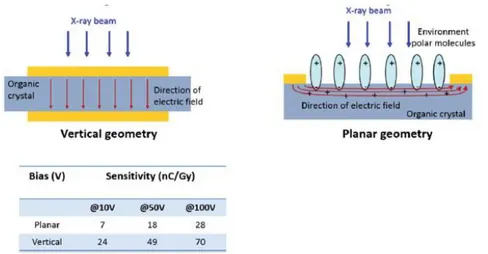
Another study that is reported in [6] and is worth mentioning, regards the investigation of the role and the effects of the molecular anisotropic packing in the X-ray photo-response of OSSCs. In this article the authors report two samples: one based on 4HCB and one based on 1,5-dinitronaphthalene (DNN) molecules, which have been chosen because they generate crystals with different geometries (platelets in the case of 4HCB and needles for DNN). Although the results show that the crystal shape and geometry does not affect the detector performance, the anisotropic packing of the molecule affects the electronic transport properties of the crystal, inducing a carrier mobility which can vary up to 4 orders of magnitude along the three crystal axes in the 4HCB. However, it is noteworthy that all axes can be used for the effective detection of ionizing radiation. Interestingly, in 4HCB crystals, the largest sensitivity is obtained in the vertical geometry, even if in this configuration the electrodes do not connect the crystal axis with the best molecular π-stacking and carrier mobility, as shown in Figure 2.8. In the same figure a sketch of the planar geometry, which is the electrodes geometry of most of the samples under test in this experimental work, is also reported. [6]
Figure 2.8: Sketch of the electric field distribution in the vertical and planar geometry in 4HCB samples (upper panel). Sensitivity values in both configurations for three bias voltages. [6]
25
3 Materials and methods
In this chapter the general procedure adopted throughout the experimental work will be described. The samples preparation method, including description of materials and tools employed, will be treated in subsection 3.1. Subsequently, in subsection 3.2, the measurement setup will be described. The measurement and data analysis procedures will be illustrated respectively in subsections 3.3 and 3.4. In closing, the characterization results of the reference devices will be shown in subsection 3.5.
3.1 Sample preparation
Initially placed on a glass substrate, the organic single crystal is fixed by its edges using Ag paste. Subsequently, a 50 µm diameter Au wire is placed crosswise on the crystal as a shadow mask, in order to create a channel between the metal electrodes fabricated by means of vacuum evaporation. The metal deposition process is performed in a high vacuum chamber (10-5 torr), using either gold or silver as deposition material (150 mg). Initially, the metal is properly placed on a filament inside the chamber. At the pressure of 10-2 torr, achieved with a rotary pump, metal drops are created by heating the filament. Subsequently, the chamber is opened and the samples are placed on their support. At the pressure of 10-5 torr, achieved with a turbomolecular pump, the deposition is performed. Figure 3.1 shows an example of a crystal after the deposition process.
Figure 3.1: Image taken with the optical microscope showing a single crystal after the deposition process. As can be seen, it is possible to identify the channel.
26
After the deposition process, the organic crystal is electrically contacted using gold wires (Figure 3.2) and connected inside an aluminum case. The sample is therefore ready to be characterized under X-rays.
a) b)
Figure 3.2: a) Image of a complete sample and b) enlargement of the crystal area, where it is possible to identify the gold wires.
3.2 Measurement setups
The most of the measurements will be performed with the samples exposed to X-rays. However, in the case of two samples (TIPSSC_07 and Rux), we will perform measurements using a LED of 375 nm wavelength. This will be done in order to compare both responses, with the aim to reach a better interpretation of the photoconversion process occurring in X-ray detectors. In fact, although controversial, the dynamic of photoconversion of visible photons in organic materials has been already treated in literature [17] [18]. On the other hand, the interaction mechanism between organic materials and X-rays is less known.
In this subsection the measurement setups employed in the case of both X-ray irradiation and LED illumination will be described.
3.2.1 Measurement setup (X-rays)
A schematic of the measurement setup is reported in Figure 3.3.
27
The detector is placed in a shielded area at the distance of 21 cm from a molybdenum X-ray tube (PANalytical2, PW 2285/20), which operates at a voltage of 35 kV and at the current defined by the user (5 mA : 30 mA). The electrometer (Keithley3, mod. 6517A) is connected to both the detector and a PC, allowing the user to interface with the system through the LabView software.
Figure 3.4 shows an image of the shielded area, where it is possible to identify the X-ray tube and the aluminum case containing the sample under test.
Figure 3.4: shielded area.
Samples are therefore subjected to a dose rate that depends on the X-ray tube’s operation current. Figure 3.5 shows the dose rate-current calibration graph and Table 3.1 shows numerically the relation between the current and the dose rate at the distance of 21 cm from the beam source.
5 10 15 20 25 30 35 0 20 40 60 80 100 120 140
160 Dose rate (mGy/s) Linear Fit of Sheet1 D
Do se rate (mG y/s) I (mA) Equation y = a + b*x Weight Instrument Residual Sum of Squares 0,55522 Adj. R-Squar 0,99935
Value Standard Err
D Intercept -0,0978 0,43778
D Slope 3,9203 0,04079
L=21cm from the beam source V=35kV
Calibration Mo-tube Bologna
10/01/2014 dosimetro PMX-III
Figure 3.5: Calibration of the Mo-tube. Dose rate vs current.
2
www.panalytical.com
3
28 Current (mA) Dose rate (mGy/s) 5 19.4 10 40.0 15 57.3 20 77.7 25 98.6 30 117.0 35 138.7
Table 3.1: Relation between current and dose rate at the distance of 21 cm from the beam source.
The sample under test will be subjected to a given incident photon flux, that can be calculated by means of a proper X-ray spectra simulator4 developed by Siemens. By setting the anode material, the X-ray tube operation voltage and the dose rate it is possible to obtain the photon flux, as reported in Table 3.2.
Dose rate Photon Flux Ф (mGy/s) (cm-2s-1) 19.4 2.98·1010 40.0 6.16·1010 57.3 8.82·1010 77.7 1.20·1011 98.6 1.52·1011 117.0 1.79·1011 138.7 2.14·1011
Table 3.2: Relation between dose rate and photon flux.
For a given incident photon flux Ф (expressed in cm-2s-1), the photocollection efficiency of the sample is given by:
Equation 3.1
where is the difference between the current under irradiation and the dark current, is the channel area of the crystal, is the electron charge and is the ratio between the photon energy and the energy of pair creation (2.5 times the bandgap). [11] The previous quantities and the photocollection efficiencies of the TIPSSC_07 and the RUX samples exposed to X-rays at the dose rate of 98.6 mGy/s are reported in Table 3.3.
29 Sample Photon energy (ev) Dose rate (eV gr-1 s-1) Photon flux (cm-2s-1) Channel area (cm2) ∆I (A) Energy gap (ev) β f (%) TIPSSC_07 1.70·104 6.16·1014 1.52·1011 5.0·10 -5 4.10·10-10 2 3400 4.96 Rux 6.0·10-4 1.10·10-9 2.2 3090 1.22 Table 3.3: Photocollection efficiencies for the TIPSSC_07 and Rux samples at the X-ray dose rate of 98.6 mGy/s.
3.2.2 Measurement setup (LED)
The LED employed for the measurements (HWA/WYS ultrafire5 mod. wf-501B) is characterized by a wavelength of 375 nm. A picture of the measurement setup is reported in Figure 3.6.
a) b)
Figure 3.6: a) Measurement setup for the LED measurements; b) sample under illumination.
The photon flux is calculated by using a silicon-based UV photodiode of 2.5 mm diameter supplied by EOS6 (Figure 3.7).
Figure 3.7: measurement set up for the LED photon flux calculation.
5
www.ultrafire.net
6
30
When the LED is switched on, the ∆V induced by the LED to the photodiode can be appreciated by means of the oscilloscope (GW Instek7 mod. GDS-2204A). At the distance of 5: 20 cm between the LED and the photodiode, the ∆V does not change significantly and is equal to 14V (Figure 3.8).
Figure 3.8: The ∆V induced by the LED to the photodiode can be appreciated on the oscilloscope’s screen. Since the responsivity of the photodiode at 375 nm is equal to 204·105 V/W, the incident power is given by:
Equation 3.2
Since the area of the photodiode is equal to 0.049 cm2, the irradiance (incident power per unit area) is given by:
Equation 3.3
The energy of the incident photon is equal to:
Equation 3.4
Therefore the photon flux is given by:
Equation 3.5 7 www.gwinstek.com
