UNIVERSITÀ DEGLI STUDI DELLA TUSCIA DI VITERBO DIPARTIMENTO DI ECOLOGIA E BIOLOGIA
Corso di Dottorato di Ricerca in Genetica e Biologia Cellulare - XXVIII Ciclo
BO/11
TITOLO TESI DI DOTTORATO DI RICERCA
NF-Y AND LAMIN A IN CELL PROLIFERATION AND CANCER
Tesi di dottorato di: Dott.ssa Cicchillitti Lucia Firma
Coordinatore del corso Tutore
Prof. Giorgio Prantera Dott.ssa Giulia Piaggio
Firma Firma
.
Data della discussione 06/05/2016
ABSTRACT
Nuclear transcription factor (NF-Y) is a sequence-specific transcription factor that binds DNA on a sequence spanning the CCAAT a common element present on several gene promoters. It has long been considered an activator of genes involved in growth promotion including cell cycle regulatory genes. NF-Y is composed of three subunits (-YA –YB –YC) all required for the DNA binding and transactivation activity. NF-YA is the regulatory subunit of the NF-Y protein. Numerous findings highlight that NF-Y is involved in cancer.
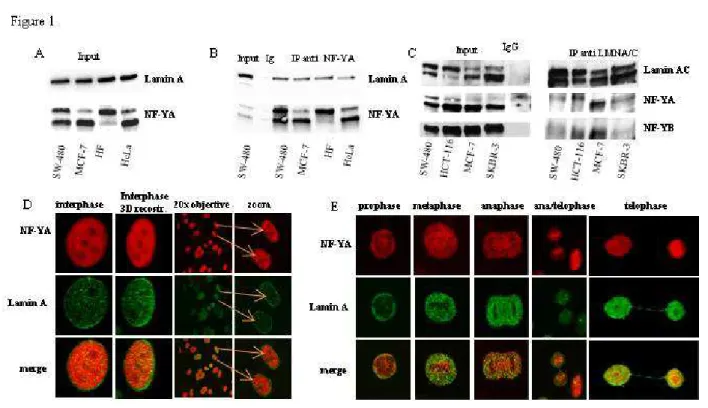
In manuscript I, to get clues on NF-Y function(s) in cancer cells, we performed a mass spectrometry screening of a pool of proteins that co-precipitate with the NF-YA subunit. By this screening we identified lamin A as a novel putative NF-Y interactor. This result was validated by co-IP experiments using different cell lines. Confocal analysis confirmed this interaction in different phases of mitosis. In order to investigate the possible involvement of lamin A/NF-Y complex in gene regulation, we generated the chromatin fractions. Co-IP experiments confirmed the occurrence of lamin A /NF-Y complex in the chromatin. We further isolated euchromatin from proliferating cells in order to evaluate the possible involvement of lamin A in actively transcribed genes involved in cell cycle progression where NF-Y plays a key role as transcription factor. Our data demonstrated that NF-Y and lamin A co-localize in transcriptionally active region. We performed ChIP experiments to explore the possible recruitment of lamin A on promoters of NF-Y target genes demonstrating that lamin A physically interacts with several promoter regions carrying CCAAT-boxes, such as CCNB2, DHFR, CCNA2, CDK1, CCNB1, CDC25C, TOPO2a and PCNA. Gain and loss of function experiments revealed that lamin A counteracts NF-Y transcriptional activity impacting on cell cycle progression. We performed luciferase assays using the CCNB2 promoter driven luciferase reporter construct as a sensor of NF-Y activity in cellular and mouse models. These experiments revealed that lamin A counteracts NF-Y activity on CCNB2 promoter. Moreover, data obtained from experiments performed under serum deprivation or oxidative stress conditions emphasized the importance of lamin A activity in cell proliferation in the tumour microenviroment characterized by low nutrient supply and excessive reactive oxygen species (ROS) production.
In manuscript II, we analysed the possible involvement of NF-Y and lamin A in stratifying endometrial cancer (EC). EC is a major cause of mortality for patients worldwide. EC is classified as type I or type II based on histologic properties. Type I, also called the endometrioid type (EEC) because of its histologic similarity to the endometrium, accounts approximately 70–80% of sporadic
EC. Most type I tumours occur in the setting of unopposed estrogen stimulation, leading to endometrial hyperplasia. According to FIGO definition, type I ECs include lower grade EECs (grade 1 and 2 EEC). Unlike type I tumors, type II lesions are not related to estrogen exposure or endometrial hyperplasia and include high risk malignancies, as poorly differentiated high-grade EEC (G3), and non endometrioid endometrial carcinomas (NEM) such as serous papillary and clear cell carcinomas. In general, patients with EC have a good prognosis since early diagnosis is frequent and the disease has usually not spread beyond the uterus. However, the prognosis for recurrent or metastatic EC remains poor. Although most cases of low grade ECs do not behave aggressively, in rare instances, even low-grade, well-differentiated ECs can progress in a highly aggressive manner. In this study, we analysed several EC tissues to find novel clinical and biological features to help the diagnosis and consequently the treatment of early EEC. A retrospective cohort of several formalin-fixed, paraffin-embedded (FFPE) specimens from patients with EC were analysed. Total RNA and proteins were extracted and analyzed, respectively, by quantitative PCR and western blotting. Our correlation studies identify NF-YAs, a splicing isoform of NF-YA, , and lamin A as two novel potential biomarkers in ECs. It has been recently demonstrated that NF-YAs belongs to the embryonic stem cell transcription factor circuitry. Lamin A has been shown to be involved in cancer development and tumor aggressiveness. We observed that NF-YAs is exclusively expressed in EC tissues, while lamin A is strongly down-modulated in EC compared with benign tissues and its loss of expression correlates with higher histologic grade and aggressiveness. Results obtained in low grade EEC (grade 1) tissues demonstrated that NF-YAs expression is heterogeneous, with 55% of samples expressing the short isoform compared to 100% in G2 and G3 EEC and NEM. Interestingly, the presence of NF-YAs was related with lower lamin A protein and mRNA levels. It is worth to note that the presence on NF-YAs and loss of lamin A expression was consistently associated with lower estrogen receptor (ERs) expression and related with miR-200 family upregulation and ZEBs decreased expression, indicators of EC aggressiveness, thus supporting the potential role on NF-YAs and lamin A as novel prognostic correlation biomarkers with a potential for a more systematic integration in clinical practice for individualized therapy in EC, in particular in low grade malignancy.
Our studies help to promote our understanding of the mechanisms of NF-Y activity providing a molecular evidence for the direct transcriptional modulation of cell cycle related genes by lamin A/NF-Y nuclear protein complex. Moreover, they open up a possibility to use lamin A and NF-YAs expression, in combination with ERs status, in the diagnosis and treatment of early EC .
CONTENTS
1 INTRODUCTION 5
1.1 NF-Y 5
1.2 Lamin A 8
1.3 Endometrial cancer (EC) 11
1.4 Epithelial-mesenchymal transition (EMT) in EC. 12
2 THESIS AIMS AND RESULTS 17
2.1 Manuscript I 17 2.2 Manuscript II 19 3 CONCLUDING REMARKS 21 4 AKNOWLEDGEMENTS 25 5 REFERENCES 26 6 MANUSCRIPT I 32 6.1 Abstract 32 6.2 Introduction 33
6.3 Material and methods 34
6.4 Results 39 6.5 Discussion 44 6.6 References 45 6.7 Figures 50 6.8 Supplementary figures 55 7 MANUSCRIPT II 59 7.1 Abstract 59 7.2 Background 60
7.3 Material and methods 63
7.4 Results 64 7.5 Discussion 67 7.6 Conclusions 69 7.7 References 70 7.8 Tables 76 7.9 Figures 78
1 INTRODUCTION
1.1 NF-Y
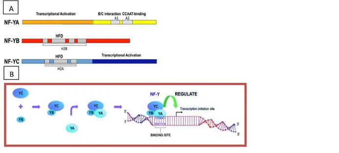
NFY (also called CBF, aCP1 and CP1 ) is a ubiquitous protein, composed of 3 subunits, NFYA, -YB, and -YC, whose genes are highly conserved from yeast to mammals. All 3 subunits are required for NF-Y binding to the consensus sequence, the CCAAT-box. The CCAAT box is one of the most common cis-acting elements found in the promoter and enhancer regions of a large number of genes in eukaryote. The CCAAT box location within promoters is fixed at −60/−100 nucleotides from the transcriptional start site (TSS) and, whenever tested, has been shown to be crucial for promoter activity. The structure and the DNA-binding mode of the NF-YB/NF-YC dimer are highly reminiscent of that of other histone-fold domains (HFDs), and an activation domain is present in the NF-YA subunit (1). NF-YA is the regulatory subunit of the trimer. The crystal structure of NF-Y bound to a 25 bp CCAAT oligonucleotide shows that the HFD dimer binds to the DNA sugar-phosphate backbone, mimicking the nucleosome H2A/H2B-DNA assembly. First, YB and YC interact to form heterodimers through their HFDs. The NF-YB/NF-YC heterodimer then interacts with NF-YA to form the heterotrimeric NF-Y transcription factor. The absence of any of the NF-Y subunits results in loss of binding of the NF-Y complex to DNA and NF-Y-directed transcription (Figure 1).
A bioinformatic analysis of promoters of cell-cycle regulatory genes shows an abundance of CCAAT boxes in promoters regulated during the G2/M transition (2). Consistent with this, the NF-Y complex supports basal transcription of a class of regulatory genes responsible for cell-cycle A A
Figure 1: (A) Scheme of NF-Y subunits. (B) NF-Y complex formation. NF-Y consists of three different subunits, NF-YA, NF-YB and NF-YC, which are all necessary for formation of NF-Y complexes and binding to CCAAT boxes to activate transcription. The arrow with bar indicates transcription initiation site
A
A bioinformatic analysis of promoters of cell
CCAAT boxes in promoters regulated during the G2/M transition which are mitotic cyclin complexes (3
binding of NF-Y to cellular promoters is essential for cell prolifer
It has been reported the presence of two major NF aa), the short isoform lacking a 28
are present at different levels in various cellular context an exon encoding the majority of a glutamine
(NF-YAs) lacks this. NFYAl
preferentially expressed in epithelial cells and the latter in lymphoid cells (
The level of NF-YAl increases through mouse and human embryonic stem (ES) cell differentiation, whereas NF-YAs is significantly downregulated (
isoforms remains to be elucidated, but hematopoietic stem cells (15).
Although mutations in NF-Y subunits have never been specifically identified in tumours, systematic examination of expression profiles indicates that NF
cancer. Expression of NF-YA in normal cells is modulated during the cell cycle (4 Figure 3: Description of the two isofroms of NF
exon 3.
Figure 2: The NF-Y complex support basal transcription of a class of responsible for cell cycle progression, among which are mitotic cyclin complexes
bioinformatic analysis of promoters of cell-cycle regulatory genes shows an abundance of CCAAT boxes in promoters regulated during the G2/M transition progression
which are mitotic cyclin complexes (3-10). Taken together, these studies demonstrate that the Y to cellular promoters is essential for cell proliferation.
It has been reported the presence of two major NF-YA isoforms, “long” (347 aa)
, the short isoform lacking a 28-amino acid within the NF-YA amino-terminal domain (11), that are present at different levels in various cellular context (Figure 3). NFYA-long (
an exon encoding the majority of a glutamine-rich transactivation domain, whereas NFYA and NFYAs show distinct expression patterns; the former is preferentially expressed in epithelial cells and the latter in lymphoid cells (12,
increases through mouse and human embryonic stem (ES) cell differentiation, is significantly downregulated (14). Functional significance of the two NF ns to be elucidated, but recent data indicate that NF-YAs promotes self
Y subunits have never been specifically identified in tumours, systematic on profiles indicates that NF-Y targets are upregulated in different types of
YA in normal cells is modulated during the cell cycle (4 Description of the two isofroms of NF-YA originating from alternative splicing of
Y complex support basal transcription of a class of responsible for cell cycle progression, among which are mitotic cyclin complexes
cycle regulatory genes shows an abundance of progression (Figure 2), among Taken together, these studies demonstrate that the
(347 aa) and “short” (318 terminal domain (11), that long (NF-YAl) contains rich transactivation domain, whereas NFYA-short show distinct expression patterns; the former is
,13).
increases through mouse and human embryonic stem (ES) cell differentiation, significance of the two NF-YA promotes self-renewal of
Y subunits have never been specifically identified in tumours, systematic upregulated in different types of YA in normal cells is modulated during the cell cycle (4) and its
YA originating from alternative splicing of Y complex support basal transcription of a class of regulatory genes responsible for cell cycle progression, among which are mitotic cyclin complexes.
abrogation plays an important role in downregulating several cell-cycle control genes in differentiated cells (5,7,9,10). Previous studies aimed at understanding the biological role of NF-Y took advantage of a loss of function approach, such as expression of dominant-negative NF-YA mutants and conditional deletion of the mouse NF-YA gene. When a dominant-negative NF-YA mutant that interacts with -YB/YC but does not bind DNA is expressed in mouse fibroblasts, retardation of cell growth is observed (16). The knock out of the NF-YA subunit in mice leads to embryo lethality; moreover, inactivation of the NF-YA gene in mouse embryonic fibroblasts results in inhibition of cell proliferation and growth arrest at various phases of the cell cycle (17-18). It has been demonstrated that NF-Y modulates the promoter activity of several genes in response to DNA-damaging agents (19,20), and NF-Y overexpression increased the proliferation rate of cancer cells harbouring endogenous mutant p53 (Figure 4).
Next, it has been shown that NF-Y interacts in vivo with mutant p53 and increases DNA synthesis, which is impaired upon abrogation of NF-YA expression (8,21). Clinical studies have indicated that patients with upregulated expression of NF-Y target genes have poor prognosis in multiple cancers (22). Using global gene expression profiles, the involvement of NF-Y in cancer-associated pathways has been recently reported across human cancers (23). In agreement with its wide involvement on human cancers, previous studies described that NF-Y interacts with different partners. Indeed, in normal cells NF-YA binds to deacetylase enzymes (HDACs) while in transformed cells the acetylase p300 is preferentially recruited (8,9). Although some NF-Y interactors are already known, several partners through which NF-Y exerts its role still need to be characterized.
Figure 4: Model proposing the molecular mechanism underlying the transcriptional control of cell cycle-related genes by mut-p53/NF-Y or wtp53/NF-Y protein complexes.
1.2 Lamin A
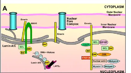
Lamins are type V intermediate filament (IF) proteins and the major components of the nuclear lamina that scaffold adjacent to the inner nuclear membrane (Figure 5).
As IF proteins, lamins exhibit a typical tripartite structure consisting of an α-helical rod domain flanked by globular N-terminal head and C-terminal tail domains. The C terminus contains a nuclear localization signal (NLS) and a structural motif similar to a type s immunoglobulin fold (Ig-fold), likely involved in protein–protein interactions (Figure 6).
A-type lamins, whose most represented isoforms are lamin A and C, are alternatively spliced products of the same gene, LMNA, and are found in roughly similar amounts in most tissues. The lamin A gene is 57.6 kb long and consists of 12 exons, encoding two globular domains and a central-helical coiled-coil rod domain. Lamin C is encoded by exons 1 to 9 and a portion of exon 10. Lamin A results from alternative splicing, which adds exons 11 and 12 and removes the lamin-C-specific portion of exon 10. Diseases caused by mutations in genes encoding nuclear lamins are generally termed laminopathies (24).
Lamins play important roles in nuclear architecture, mechanosignaling (25) and chromatin dynamics (26) and impact on stem cell proliferation and differentiation (27,28). Disruption of one or more of these functions due to lamin mutations cause a group of inherited diseases affecting various tissues and organs or causing accelerated ageing (26).
Figure 5: Scheme of interactions of A-type lamins and NE-associated proteins with DNA, chromatin complexes, and related transcription factors.
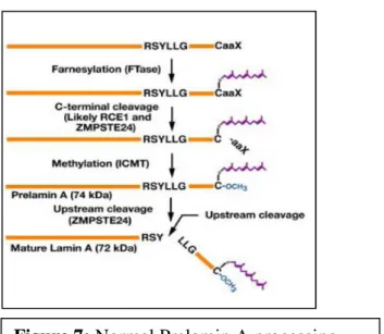
Unlike lamin C, lamin A is translated as prelamin A and undergoes posttranslational processing steps at the C-terminal CaaX motif (Figure 7). Farnesylation of prelamin A occurs at a key aminoacid, cysteine 661, within the C-terminal CaaX box. Cysteine 661 is farnesylated by the dimeric protein farnesyl transferase (29). The modification is necessary for further processing of the lamin A precursor, consisting of methylation of the same residue by the enzyme Icmt, and double cleavage leading to production of mature lamin A. Mature lamin A and lamin C are solubilized in mitosis and can also localize throughout the nucleoplasm in interphase cells.
Figure 5: Scheme of the LMNA gene. Examples of the main mutations that cause laminopathies are shown above the gene; not all mutations are listed. In the case of HGPS, MAD, FPLD, AR-EDMD, RD and CMT2B1, the most common causative LMNA mutation (or only mutation) is shown. In the cases of AD-EDMD, AWS, LGMD1B, GLD and DCM1A, a representative mutation among multiple causative mutations is included. AD-EDMD, autosomal dominant Emery–Dreifuss muscular dystrophy; AR-EDMD, autosomal recessive Emery–Dreifuss muscular dystrophy; AWS, atypical Werner syndrome; CMT2B1, Charcot–Marie–Tooth disorder, type 2B1; DCM1A, dilated cardiomyopathy, type 1A; FPLD, Dunnigan familial partial lipodystrophy; GLD, generalized lipodystrophy; HGPS, Hutchinson–Gilford progeria syndrome; LGMD1B, limb girdle muscular dystrophy, type 1B; MAD, mandibuloacral dysplasia; RD, restrictive dermopathy.
It has been shown that lamin A stabilizes the nuclear lamina and chromatin, with implications for epigenetic stabilization and limiting of DNA breaks. Interactions of lamins with chromatin occur through domains termed lamina-associated domains (LADs) with the implication that lamins associate with chromatin at the nuclear lamina at the nuclear periphery. LADs are often located in repressive chromatin structures with an enrichment of this compartment at the nuclear periphery (24 30). Although most lamins are found near the nuclear membranes, nucleoplasmic populations also exist, which may have distinct roles (31-34). Lamins are often aberrantly expressed or localized in tumours. With respect to its multiple functions, it is convincible to presume that change of lamin A protein levels may contribute to tumourigenesis and progression.
The expression of the A-type lamins is often reduced or absent in cells that are highly proliferative, including various human malignancies such as colon cancer, cervical cancer, lung cancer, prostate cancer, gastric cancer, ovarian cancer and leukemia and lymphoma (35-39). Several studies reported that miR-9 is able to target and reduce lamin A expression (40,41). miR-9 has been identified as both an oncogene and a tumor suppressor depending on different cancer types. In gastric cancer(42), endometrial cancer (43), brain cancer (44) and leukemia (45) miR-9 is observed upregulated and oncogenic, whereas in cervical cancer (46.), colorectal cancer (47) and ovarian cancer (48) it is observed downregulated and anti-tumorigenic. miR-9 overexpression was also correlated with cancer progression, metastasis and poor prognosis (43). Moreover, cells lacking lamin A proliferate faster and display inefficient cell cycle arrest upon contact inhibition (48). Recent data highlight the specific functions of a small pool of lamina-independent A-type lamins, located throughout the nucleoplasm, in the regulation of early tissue progenitor cell proliferation and commitment (31,50,51).
1.3 Endometrial cancer
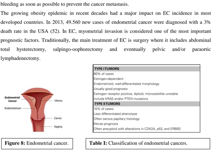
Endometrial cancer (EC) is the most common genital tract malignancy and occurs in reproductive and postmenopausal women. EC develops in the inner lining of the uterus, also called the endometrium (Figure 8). The human endometrium is stratified into two functional layers: the transient superficial stratum functionalis and the permanent deeper stratum basalis adjacent to the myometrium. The superficial stratum functionalis is lined by luminal epithelium, contains superficial glandular epithelium and stroma and is completely shed and regenerated during the monthly menstrual cycle and after childbirth.
The development of EC is most prevalent in postmenopausal women. For populations within this category, it is highly recommended to have a pelvic exam every year and to report any vaginal bleeding as soon as possible to prevent the cancer metastasis.
The growing obesity epidemic in recent decades had a major impact on EC incidence in most developed countries. In 2013, 49.560 new cases of endometrial cancer were diagnosed with a 3% death rate in the USA (52). In EC, myometrial invasion is considered one of the most important prognostic factors. Traditionally, the main treatment of EC is surgery where it includes abdominal total hysterectomy, salpingo-oophorectomy and eventually pelvic and/or paraortic lymphadenectomy.
Most EC cases are sporadic, with only 10% considered familiar (53,54). In general, patients with EC have a good prognosis since early diagnosis is frequent and the disease has usually not spread beyond the uterus. However, women with recurrent and/or metastatic EC of either type have a poor prognosis, with a median survival of 7–12 months (55). These patients require more effective systemic therapy than is presently available. Currently, adjuvant and systemic treatment of recurrent and metastatic EC are based on conventional chemotherapy and anti-hormonal treatment. In order to improve therapy it is important to understand the processes which inhibit and stimulate cancer
progression. EC is classified as type I or type II based on histologic parameters, clinical behavior and epidemiology (Table I). Type I, also called the endometrioid type (EEC) because of its histologic similarity to the endometrium, accounts approximately 70–80% of sporadic EC. Most type I tumors occur in the setting of unopposed estrogen stimulation, leading to endometrial hyperplasia. The cellular action of estrogens is mediated trough the estrogen receptors (ERs) that belong to the nuclear steroid receptor superfamily. Two distinct ERs, defined as ER-α and ER-β, have been identified. In the human uterus, ER-α is the predominant subtype. Unlike type I tumors, type II lesions are not related to estrogen exposure or endometrial hyperplasia and include high risk malignancies, as high grade EECs, serous papillary and clear cell carcinoma. Some biological molecules have been identified as prognostic markers in EC, such as KRAS, PTEN, EGFR, FGFR, P53, HER2, and ERs. (56). Expression of ERs has been correlated with stage, histologic grade and survival. Loss of ERs has been significantly associated with aggressive phenotype and poor survival in EC patients (57). In particular, early stage, well differentiated ECs usually retain ERs expression, whereas advanced stage, poorly differentiated tumours often lack one or both receptors. Recently, it has also been observed an association between lack of ER-α and epithelial-mesenchymal transition (EMT) (58,59).
1.4 Epithelial-mesenchymal transition (EMT) in EC.
EMT enables epithelial cells to acquire a like mesenchymal potential with increase motility and ability to extravasate and circulate. A classification into three types of EMT has been proposed (60)(Figure 9).
Type 1 EMT is used during development to generate cells with mesenchymal features out of epithelial cells. The primitive epithelium, specifically the epiblast, gives rise to primary mesenchyme via an EMT. This primary mesenchyme can be re-induced to form secondary epithelia by a MET. It is speculated that such secondary epithelia may further differentiate to form other types of epithelial tissues and undergo subsequent EMT to generate the cells of connective tissue, including astrocytes, adipocytes, chondrocytes, osteoblasts, and muscle cells. It is a “clean” and entirely physiological process and not associated with inflammation, fibrosis, or an invasive phenotype. Type 2 EMT, in contrast, occurs during tissue repair in response to traumatic or inflammatory injury. Under normal circumstances, type 2 EMT is limited to an acute repair process (e.g., wound healing) and can be beneficial, as it provides tissue replacement. Unlike the type 1 EMT, the type 2 EMT is expressed over extended periods of time and can eventually destroy an affected organ if the primary inflammatory insult is not removed or attenuated. Finally, type 3 EMT is associated with migratory and invasive features of tumor cells. A characteristic of type 3 EMT is that it originates from cells that have already undergone malignant transformation. Thus, the genetic and epigenetic changes typical for cancer cells, such as the activation of oncogenes and the inactivation of tumor suppressors, can act in concert with the EMT program. The composition of the basement membrane also changes, altering cell-ECM interactions and signaling networks. The next step involves EMT and an angiogenic switch, facilitating the malignant phase of tumor growth. Progression from this stage to metastatic cancer also involves EMTs, enabling cancer cells to enter the circulation and exit the blood stream at a remote site, where they may form micro- and macro-metastases, which may involve METs and thus a reversion to an epithelial phenotype. After the transition to a mesenchymal state, cells can also change back to an epithelial state in a process known as mesenchymal-epithelial transition (MET) (Figure 10).
In EC, alteration of EMT markers have been identified in metastatic disease and associated with reduced survival (Table II). Importantly, loss of epithelial markers such as E-cadherin is associated with adverse prognosis in both EEC and NEM tumor types (61-63). Increased expression of the E-cadherin transcription repressors Twist, Snail, and Slug has been demonstrated in EC cell lines and in tumor samples, and down-regulation of E-cadherin immunoreactivity has been described in both EEC and nonendometrioid malignancies (64). It has been shown that E-cadherin repressors Slug, ZEB1, and HMGA2 were expressed preferentially at the myoinvasive front of EEC and that EMT-like changes could be induced in vitro through extracellular signal-regulated kinases ERK) 1/2 phosphorylation (65).
miRNAs are small non-coding RNA elements that control cellular function by modulating the stability and translation of multiple target mRNAs at the post-transcriptional level (Figure 11). They play important roles in development, cellular differentiation, proliferation, cell-cycle control, and cell death.
It is recognized that, in order for tumors to develop, cells must acquire cellular characteristics that are very different from those of healthy cells. Since individual miRNAs have potentially hundreds of target genes, miRNA dysregulation will have a profound effect on the regulation of the cellular machinery and contribute to enabling cancer hallmarks. Several studies have identified miRNAs, which are differentially expressed in EC compared to healthy endometrial tissues (66). For example, miR-205 (67-70), and miR-96 cluster (hsa-miR-96, hsa-miR-182 and hsa-miR-183) (70,71) were found upregulated in EC compared to benign tissues. Other studies identified three miRNAs (499, 135b and 205) as upregulated and five (10b, 195, miR-30a-5p, miR-30a-3p and miR-21) as downregulated (72). In particular, the miR-200 family members have been extensively studied with respect their role in EMT in various tissues, where they target the expression of many genes, such as the transcription factors ZEBs (73,74). It has been already shown that elevated levels of all miR-200 family, in all stages of EC, inversely correlates with the expression of ZEBs. miR-200s upregulation has been demonstrated in type I EEC compared to normal endometrial tissues (75,76) in keeping with observations in other tumours, such Figure 11: The miRNA processing pathway has long been viewed as linear and universal to all mammalian miRNAs. This canonical maturation includes the production of the primary miRNA transcript (pri-miRNA) by RNA polymerase II or III and cleavage of the pri-miRNA by the microprocessor complex Drosha–DGCR8 (Pasha) in the nucleus. The resulting precursor hairpin, the pre-miRNA, is exported from the nucleus by Exportin-5–Ran-GTP. In the cytoplasm, the RNase Dicer in complex with the double-stranded RNA-binding protein TRBP cleaves the pre-miRNA hairpin to its mature length. The functional strand of the mature miRNA is loaded together with Argonaute (Ago2) proteins into the RNA-induced silencing complex (RISC), where it guides RISC to silence target mRNAs through mRNA cleavage, translational repression or deadenylation, whereas the passenger strand (black) is degraded. In this review we discuss the many branches, crossroads and detours in miRNA processing that lead to the conclusion that many different ways exist to generate a mature miRNA.
as melanoma (77,78), ovarian cancer (79), and colorectal carcinoma (80). Recently, it has also been suggested that miR-200 family, under influence of estrogen, maintains an epithelial phenotype in lower grade EEC (81). However, based on hormone status, miR 200a upregulation has been linked with outcome of EC patients. A recent study correlated miR-200a with prolonged survival in ERs positive subgroup, whereas an inverse trend was observed in the ERs negative soubgroup (82).
2 THESIS AIMS AND RESULTS
2.1 MANUSCRIPT I
The lamin A/NF-Y protein complex reveals an unknown transcriptional mechanism of cell cycle regulation.
Lucia Cicchillitti, Isabella Manni, Carmine Mancone, Toni Alonzi, Fabrizio Carlomosti, Lucia dell’Anna, Giulia Dell’Omo, Mauro Picardo, Paolo Ciana, Maurizio Capogrossi, Marco Tripodi, Alessandra Magenta, Maria Giulia Rizzo, Aymone Gurtner, and Giulia Piaggio
The analysis of global regulatory perturbations across human cancers pointed at NF-Y as one of the transcription factors responsible for oncogenic transcriptional changes. Although mutations in NF-Y subunits have never been specifically identified in tumours, systematic examination of protein expression profiles indicates that NF-YA transcriptional activity is upregulated in different types of cancer. Thus, identification of NF-Y protein partners can help to the characterization of mechanisms associated with its tumorigenic potential. In the present study, starting with a mass-spectrometry screening, we identified a novel nuclear protein complex formed by lamin A and NF-Y involved in chromatin binding and cell proliferation. Using a combination of biochemical, cell biology and molecular imaging techniques, we demonstrated that NF-Y, physically interacting with lamin A, strongly impacts on cancer cell proliferation. Changes in lamin A expression have been reported in a variety of cancers, correlating with tumorigenic potential and more aggressive phenotype. In our study, ChIP experiments demonstrate that lamin A physically interacts with several promoter regions of cell cycle genes in a NF-Y dependent manner. In particular, we detected lamin A binding to promoter regions encompassing CCAAT boxes of actively transcribed NF-Y target genes, such as CCNB2, DHFR, CCNA2, CDK1, CCNB1, CDC25C, TOPO2a and PCNA, as demonstrated by histone methylation marks and pol II recruitment. Moreover, we showed that lamin A has a role in transcriptional regulation of several NF-Y target genes impairing its transcriptional activity. Numerous studies showed that lamin A can modulate cell signaling through several mechanisms, for example, by sequestering transcription factors in inactive complexes, modulating post-translational modifications and degradation, and regulating transcriptional complexes. We hypothesizes that lamin A hinders the targeting of NF-Y to its consensus sites and highlight a dose-dependent effect of lamin A binding. In fact, we observed an increased NF-Y transcriptional activity in LMNA silenced cells and a basal CCNB2 promoter activity inversely correlated to lamin A expression. Moreover, gain and loss of function experiments revealed that LMNA counteracts NF-Y transcriptional activity impacting on cell cycle progression. It has already been observed that lamin A interactions often appear to be confined to promoter subregions rather than to entire promoter regions. Our data support a view of lamin A as modulator of NF-Y transcriptional activity
by its interaction with NF-YA, and are consistent with a locus-specific regulation of lamin A interactions with promoters important for cell cycle regulation and tumor progression. To demonstrate in vivo the impact of LMNA on NF-Y transcriptional activity we took advantage of MITO-Luc mouse model, that we recently developed, harbours a strictly NF-Y dependent promoter in front of a luciferase reporter allowing us to monitor the NF-Y activity in a spatiotemporal manner within the entire living organism. Data obtained strongly support the physiological impact of lamin A expression in cell proliferation. We suggest that changes in lamin A expression could modulate NF-Y activity and, consequently, its oncogenic transcriptional potential. To investigate the role of lamin A in cell cycle progression we compared the ability of SW-480 and SW-480 LMNA-KD cells to grow under low nutrient or oxidative stress conditions Our data indicate that the lamin A/NF-Y complex strongly impacts on cancer cell proliferation under cellular stress conditions, Further exploration to uncover the molecular mechanism(s) by which lamin A/NF-Y complex acts as crucial regulator in diverse cellular processes and, in particular, in cancer could be important to improve and potentially provide new clues into new therapeutic approaches for cancer treatment.
2.2 MANUSCRIPT II
Prognostic role of NF-YA splicing isoforms and lamin A status in low grade endometrial cancer.
Lucia Cicchillitti, Giacomo Corrado, Mariantonia Carosi, Malgorzata Ewa Dabrowska, Rossella Loria, Rita Falcioni, Giuseppe Cutillo, Giulia Piaggio, and Enrico Vizza.
Endometrial cancer (EC) is a major cause of mortality for patients worldwide. Although most cases of low grade ECs do not behave aggressively, in rare instances, even low-grade, well-differentiated ECs can progress in a highly aggressive manner. Current clinical approaches in the treatment of EC mainly relies on surgical FIGO classification, histologic subtype, and histologic grade. Identification of novel molecular markers may be helpful to avoid risk of over-and under treatment of EC patients and to overcome recurrence. In this study we analyzed several EC tissues to find novel clinical and biological features to help the diagnosis and treatment of early ECs.
In this study, a retrospective cohort of FFPE specimens from patients with EC and benign (NE) specimens from patients who underwent a hysterectomy to treat other benign disease (n = 13) were collected. According with the histologic grade, we analysed 29 low grade (G1), 49 high grade endometrioid (G2-G3) and 10 non endometrioid EC tissues (NEM). Biopsies were sampled for primary tumors in hysterectomy specimens.
A recent study based on both informatics analysis and microarray expression profile of the motifs of known transcription regulators and experimental evidence from ENCODE, identified NF-Y as one of the key components the key transcription factors involved in gynecological cancers. NF-Y is composed of three different subunits: YA, YB and YC. The association between YB and NF-YC provides a docking site for NF-YA, and NF-YA is the regulatory subunit of the complex responsible for sequence-specific DNA binding. Subunit YA has two different isoforms, NF-YAl (long) and NF-YAs (short), resulting from alternative splicing. It has recently demonstrated that NF-YAs belongs to the embryonic stem cell transcription factor circuitry. We analysed the expression of NF-Y in several FFPE specimens by comparing the protein expression level of two subunits of NF-Y complex, NF-YA and NF-YB, in EC and benign endometrial tissues. We found that the exclusive expression of NF-YAl characterizes benign endometrial tissues, whereas the appearance of the NF-YAs is specifically associated with a tumour phenotype. In fact, the short form was detectable only in EC tissues. Very interestingly, NF-YAs was expressed in all high grade EEC and NEM tissues, whereas it was detected only in 55% of our low grade G1 EEC samples. This result suggests that NF-YAs could represent a diagnostic marker in early EC. To explore the potential role of NF-YAs in EC aggressiveness, we stratified G1 EEC tissues in two subgroups: one expressing only NF-YAl (NF-YAs-) and another expressing both isoforms (NF-YAs+).
Concomitantly, we analysed the ERs status, whose loss of expression has been well documented in advantage stages and poorly differentiated tumours, in all our cohort of FFPE EC samples. A massive down-modulation of ESR1 mRNA in 61,2% high grade EC and in NEM tissues was observed, in agreement with literature data indicating a strong correlation between the downmodulation of this gene with advanced stage of EC. Focusing on our G1 subgroups (NF-YAs- and NF-YAs+), very interestingly we observed a correlation with the loss of ESR1 and the presence of NF-YAs. In fact, expression of ESR1 was lower in NF-YAs+ compared with NF-YAs- tissues. These evidences indicate a possible involvement of NF-YAs expression in EC aggressiveness. Lack of ESR1 has been recently associated with epithelial to mesenchymal transition (EMT). We analysed mRNA levels of several EMT markers, such as E-Cadherin, N-Cadherin, miR-200 family and its direct targets, ZEB1 and ZEB2. Analysis of the qRT-PCR data showed that an augmented percentage of EC tissues exhibits a low E-cadherin/N-cadherin (E/N) ratio, an index of differentiated phenotype, together with an increase of E-Cadherin mRNA (CDH2) expression compared with benign tissues, and this modulation correlated with a more aggressive clinophatologic phenotype. The same analysis, performed in our G1 subgroups, revealed that both E/N ratio and CDH2 mRNA levels are not related with NF-YAs expression, since both groups showed the same expression pattern. miR-200s has already been demonstrated to be differentially expressed in EC compared to healthy endometrial tissues and associated with EMT in EC. Our analysis confirmed that all members of the family (miR-200a, miR- 200b, miR-200c, and miR-141) were up-regulated in all stages of EC compared to benign tissues and their expression inversely correlates with ZEB1 and ZEB2 mRNA expression. In our G1 subsgroups, we observed a consistent increase of miR-200 family expression inversely related to ZEB1 mRNA levels in G1 EEC NF-YAs+ compared with NF-YAs-, thus indicating a possible involvement of NF-YAs in miR-200 family regulation. Several studies identified A-type lamins as an indicator of differentiated tumour cells and demonstrated to represent a potential biomarker for various types of cancer. We observed a significant correlation of loss of lamin A expression with stage and histologic grade in EC. Interestingly, clustering of NF-YA isoforms in G1 EEC indicated that NF-YAs+ samples consistently exhibited lower lamin A expression compared with YA-. Our findings indicate NF-YAs and lamin A as molecules with a potential for a more systematic stratification of low grade EC malignancy.
3 CONCLUDING REMARKS
One of the issues of our laboratory is to address the role of NF-Y in cancer. NF-Y is one of the transcription factors responsible for aberrant oncogenic transcription occurring in several cancers. We firstly focused our study on the identification of novel NF-Y protein partners in order to better characterize the mechanism associated with its tumorigenic potential. Starting with a mass-spectrometry screening, we identified a nuclear protein complex formed by lamin A and NF-Y involved in chromatin binding, cell proliferation and cancer progression. Besides its localization to the nucler lamina, we observed that a small fraction of lamin A is also present in the nucleoplasm. We focused our study on the occurrence of lamin A/NF-Y association in the nucleoplasm compartment and, in particular, on chromatin where NF-Y exerts its role as transcription factor. We demonstrated that lamin A binds to promoter regions encompassing CCAAT boxes of NF-Y target genes, such as CCNB2, DHFR, CCNA2, CDK1, CCNB1, CDC25C, TOPO2a and PCNA, and this binding is mediated by NF-Y. We clearly demonstrate that lamin A physically interacts with NF-Y target genes actively transcribed, as demonstrated by histone methylation marks and pol II recruitment, and that lamin A inhibits NF-Y transcriptional activity modulating transcription in a manner dependent on local chromatin marks. Our results are consistent with previous evidences demonstrating that down-regulation of lamin A/C leads to dissociation of lamin A/C from promoters by enhancing transcriptional permissiveness (83). It has been observed that lamin A interactions often appear to be confined to promoter subregions rather than to entire promoter regions. Our data support a view in which lamin A, through its ability to bind NF-Y, exerts a locus-specific interaction with promoters important for cell cycle regulation and tumor progression. In our study, an inverse correlation between lamin A and several NF-Y target genes expression level was observed, thus supporting the role of lamin A as regulator of NF-Y transcriptional function. We validated these evidences by in vivo imaging involving the use of a genetically engineered mouse model called MITO-Luc (for mitosis-luciferase), in which an NF-Y–dependent promoter controls luciferase expression. Data obtained strongly support the physiological impact of lamin A expression in cell proliferation. Interestingly, our data obtained treating cancer cells under low nutrient and oxidative stress conditions(Figure 10) indicate that loss of lamin A in cancer cells may confer the ability to grow under low nutrient supply and oxidative stress and to adjust to a changed environment in vivo by inducing gene expression so that the tumor continues to grow.
Figure 10
Cancer cells from solid tumors become metabolically stressed, when nutrients are insufficient within poorly vascularized regions. Metabolic stress results from
glutamine and glucose, partly through excessive reactive oxygen species (ROS) production. Oxygen radicals may augment tumor invasion and metastasis by increasing the rates of cell migration. During transformation into invasive
morphology and adhesive mode, resulting in a loss of normal epithelial polarization and differentiation, and a switch to a more motile, invasive phenotype
adaptations might reveal cancer cell liabilities that can be exploited for therapeutic benefit. Further exploration to uncover the molecular mechanism(s) by which NF
crucial regulator in diverse cellular processes and, in particular, in
improve and potentially provide new clues into new therapeutic approaches for cancer treatment. Loss of lamin A expression have been reported in a variety of
potential and more aggressive
expression could modulate NF-Y activity and, its oncogenic transcriptional potential
To understand the clinical impact of lamin A expression NF-Y expression, we translated
Figure 10: To investigate the role of
experiments and compared the ability of SW
serum or after hydrogen peroxide treatment (200µM H
SW-480 cells was partially reduced upon growth factor deprivation (A) or upon oxidative stress conditions (B) compared with control cells, whereas the growth of SW
always not impaired. This results
observed both under serum deprivation and oxidative stress conditions in LMNA
with control cells, even if at low extent (C), suggest a rate proliferation gain of function related to th increase NF-Y translational acitvity for the cells with reduced lamin A levels.
Cancer cells from solid tumors become metabolically stressed, when nutrients are insufficient within poorly vascularized regions. Metabolic stress results from severe deprivation of oxygen, glutamine and glucose, partly through excessive reactive oxygen species (ROS) production. Oxygen radicals may augment tumor invasion and metastasis by increasing the rates of cell migration. During transformation into invasive carcinoma, epithelial cells undergo profound alterations in morphology and adhesive mode, resulting in a loss of normal epithelial polarization and differentiation, and a switch to a more motile, invasive phenotype. A better understanding of these ions might reveal cancer cell liabilities that can be exploited for therapeutic benefit. Further exploration to uncover the molecular mechanism(s) by which NF-Y/ lamin A complex acts as crucial regulator in diverse cellular processes and, in particular, in cancer could be important to improve and potentially provide new clues into new therapeutic approaches for cancer treatment.
oss of lamin A expression have been reported in a variety of cancers, correlating with tumori potential and more aggressive phenotype (35-39). Our data suggest that changes in lamin A
Y activity and, in particular, lamin A downmodulation may increase genic transcriptional potential.
To understand the clinical impact of lamin A expression in cancer and its possible correlation with our findings in a clinical study focused on the identification of To investigate the role of LMNA in cell cycle progression, we performed time course experiments and compared the ability of SW-480 and SW-480 LMNA-KD cells to grow in 0,1% serum or after hydrogen peroxide treatment (200µM H2O2). As shown in this
480 cells was partially reduced upon growth factor deprivation (A) or upon oxidative stress conditions (B) compared with control cells, whereas the growth of SW-480 LMNA
always not impaired. This results, together with the higher basal CCNB2 promoter luciferase activity observed both under serum deprivation and oxidative stress conditions in LMNA
with control cells, even if at low extent (C), suggest a rate proliferation gain of function related to th Y translational acitvity for the cells with reduced lamin A levels.
Cancer cells from solid tumors become metabolically stressed, when nutrients are insufficient severe deprivation of oxygen, glutamine and glucose, partly through excessive reactive oxygen species (ROS) production. Oxygen radicals may augment tumor invasion and metastasis by increasing the rates of cell migration. carcinoma, epithelial cells undergo profound alterations in morphology and adhesive mode, resulting in a loss of normal epithelial polarization and A better understanding of these ions might reveal cancer cell liabilities that can be exploited for therapeutic benefit. Further Y/ lamin A complex acts as cancer could be important to improve and potentially provide new clues into new therapeutic approaches for cancer treatment.
cancers, correlating with tumorigenic ur data suggest that changes in lamin A in particular, lamin A downmodulation may increase
and its possible correlation with on the identification of LMNA in cell cycle progression, we performed time course
KD cells to grow in 0,1% ). As shown in this figure, the growth of 480 cells was partially reduced upon growth factor deprivation (A) or upon oxidative stress 480 LMNA-KD cells was promoter luciferase activity observed both under serum deprivation and oxidative stress conditions in LMNA-KD cells compared with control cells, even if at low extent (C), suggest a rate proliferation gain of function related to the
novel molecular biomarkers in EC. Current clinical approaches in the treatment of EC mainly relies on surgical FIGO classification, histologic subtype, and histologic grade. Identification of novel molecular markers may be helpful to avoid risk of over-and under treatment of EC patients and to overcome recurrence. A recent study based on both informatics analysis of the motifs of known transcription regulators and experimental evidence from ENCODE, identified NF-Y as one of the key components of the transcription regulation factories of gynecological cancer (84). We analyzed NF-Y expression levels in a cohort of formalin-fixed, paraffin-embedded (FFPE) EC tissues. In our study we identified a specific splicing isoform of the regulatory subunit of NF-Y, NF-YAs, as a new potential indicator of aggressiveness in G1 endometrial endometrioid adenocarcinoma (EEC). We observed that NF-YAs protein was undetectable in benign tissues, whereas it was consistently expressed in high grade EEC and in NEM subtypes. Interestingly, only in G1 EEC a heterogeneous expression of NF-YA isoforms was observed with some samples expressing exclusively the long form (NF-YAl) and others samples expressing both isoforms. This results prompted us to stratify G1 EEC in two subgroups: one expressing only NF-YAl (NF-YAs-), and another, including 40 % of G1 EEC tissues analysed, expressing both isoforms (NF-YAs+). It is worth to note that patients with G1 tumors involving only endometrium and no evidence of intraperitoneal disease have a low risk (<5%) of nodal involvement. Although most cases of G1 EEC do not behave aggressively, in rare instances, even low-grade, well-differentiated endometrial adenocarcinomas can progress in a highly aggressive manner. We hypothesize that the molecular feature related to NF-YA isoforms expression could be a relevant biomarker to predict the outcome of these cancers. The exclusive presence of NF-YAl form in benign tissues suggests that it may represent a marker of differentiation and that the presence of NF-YAs may be linked with an increase of a pool of poorly differentiated cells in tumors tissues. Lamin A has been demonstrated to play a key role in sensing tissue elasticity in differentiation and the reduction in its expression frequently correlates with cancer subtypes and cancer aggressiveness, proliferative capacity and differentiation state (85). To evaluate the possible involvement of lamin A also in EC, we analyzed its protein and mRNA expression levels. Interestingly, lamin A was consistently down-modulated in EC both at mRNA and protein level. Lamin A loss was further increased in high grade EEC and non-endometrioid endometrial adenocarcinoma (NEM), thus indicating lamin A as a novel potential marker of EC aggressiveness. It is worthwhile to note that decreased lamin A levels were observed in our subgroup of NF-YAs expressing G1 EECs, thus further supporting the hypothesis of a possible involvement of NF-YAs in tumor differentiation and aggressiveness. We confirmed this correlation by evaluating several indicators of EC aggressiveness, such as estrogen receptor status, miR-200 family expression and EMT markers, miR-200 family members and ZEBs.
Our studies presented in this thesis indicate NF-Y/lamin A complex as an important regulator of cancer cells proliferation. In particular, we identified NF-YAs, a specific isoform of NF-YA, and lamin A as two novel potential targets and predictive markers for new therapeutic approaches in EC, in particular in low grade EEC, which may contribute in determining a patient's prognosis and in tailoring adjuvant therapies.
4 AKNOWLEDGEMENTS
The work included in this thesis was performed at department of Research, Advanced Diagnostics and Technological Innovation, Regina Elena National Cancer Institute.
During the three years’ study, I received help from many people. Thus, I’d like to express my gratitude to:
My supervisor, Dr. Giulia Piaggio, for giving me the opportunity to achieve the PhD academic qualification and for her support and help with my projects.
My collegues: Emmanuela Falcone, Luise de Latouliere, Francesca Garibaldi, Aymone Gurtner and Isabella Manni for their support and help during these years.
My collaborators: Giacomo Corrado, Enrico Vizza, Alessandra Magenta, Mariantonia Carosi, Malgorzata Ewa Dabrowska, Rossella Loria, Rita Falcioni, Giuseppe Cutillo, Fabrizio Carlomosti, Lucia dell’Anna, Giulia Dell’Omo, Mauro Picardo, Paolo Ciana, Maurizio Capogrossi, Marco Tripodi, Alessandra Magenta, Maria Giulia Rizzo for their contribution with my projects.
5 REFERENCES
1 Mantovani R. The molecular biology of the CCAAT-binding factor NF-Y. Gene. 1999; 239: 15-27.
2 Linhart C, Elkon R, Shiloh Y. Deciphering transcriptional regulatory elements that encode specific cell cycle phasing by comparative genomics analysis. Cell Cycle. 2005; 4: 1788-97.
3 Zwicher J, Lucibello FC, Wolfraim LA, Gross C, Truss M, Engeland, K, et al. Cell cycle regulation of the cyclin A, cdc25C and cdc2 genes is based on a common mechanism of transcriptional repression. EMBO J. 1995; 14: 4514-22.
4 Bolognese F, Wasner M, Dohna CL, Gurtner A, Ronchi A, Muller H et al. The cyclin B2 promoter depends on NF-Y, a trimer whose CCAAT-binding activity is cell cycle regulated. Oncogene. 1999; 18: 1845-53.
5 Farina A, Manni I, Fontemaggi G, Tiainen M, Cenciarelli C, Bellorini M, et al. Down-regulation of cyclin B1 gene transcription in terminally differentiated skeletal muscle cells is associated with loss of functional CCAAT-binding NF-Y complex. Oncogene. 1999; 18: 2818-27.
6 Korner K, Jerom V, Schmidt T, Muller T. Cell cycle regulation of the murine cdc25B promoter- essential role for NF-Y and a proximal repressor element. J Biol Chem. 2001; 276: 9662-69.
7 Gurtner A, Manni I, Fuschi P, Mantovani R, Guadagni F, Sacchi A, et al. Requirement for down-regulation of the CCAAT-binding activity of the NF-Y transcription factor during skeletal muscle differentiation. Mol Biol Cell. 2001; 14: 2706-15.
8 Di Agostino S, Strano S, Emiliozzi V, Zerbini V, Mottolese M, Sacchi A, et al. Gain of function of mutant p53- the mutant p53/NF-Y protein complex reveals an aberrant transcriptional mechanism of cell cycle regulation. Cancer Cell. 2006; 10: 191-202. 9 Gurtner A, Fuschi P, Magi F, Colussi C, Gaetano C, Dobbelstein M, et al. NF-Y
dependent epigenetic modifications discriminate between proliferating and postmitotic tissue. PLoS One. 2008; 3(4): e2047.
10 Manni I, Caretti G, Artuso S, Gurtner A, Emiliozzi V, Sacchi A, et al. Posttranslational regulation of NF-YA modulates NF-Y transcriptional activity. Mol Biol Cell. 2008; 19: 5203-13.
11 Li XY, Hooft van Huijsduijnen R, Mantovani R, Benoist C, Mathis D. Intron-exon organization of the NF-Y genes. Tissue-specific splicing modifies an activation domain. Biol Chem. 1992; 267: 8984-90.
12 Ishimaru F, Mari B, Shipp MA. The type 2 CD10/neutral endopeptidase 24.11 promoter: functional characterization and tissue-specific regulation by CBF/NF-Y isoforms. Blood. 1997;89:4136-45
13 Ceribelli M, Benatti P, Imbriano C, Mantovani R. NF-YC complexity is generated by dual promoters and alternative splicing. J Biol Chem. 2009 Dec 4;284(49):34189-200. 14 Grskovic M, Chaivorapol C, Gaspar-Maia A, Ramalho-Santos M. Systematic
identification of cic-regulatory sequences active in mouse and human embryonic stem cells. PLos Genet. 2007;3:e145.
15 Zhu J, Zhang Y, Joe GJ, Pompetti R, Emerson SG NF-Ya activates multiple hematopoietic stem cell (HSC) regulatory genes and promotes HSC self-renewal. Proc Natl Acad Sci U S A. 2005;102:11728-33.
16 Hu Q, Maity SN. Stable expression of a dominant negative mutant of CCAAT binding factor/NF-Y in mouse fibroblast cells resulting in retardation of cell growth and inhibition of transcription of various cellular genes. J Biol Chem. 2000; 275: 4435-44. 17 Bhattacharya A, Deng JM, Zhang Z, Behringer R, de Crombrugghe B, Maity SN. The B
subunit of the CCAAT box binding transcription factor complex (CBF/NF-Y) is essential for early mouse development and cell proliferation. Cancer Res. 2003; 63: 8167-72.
18 Benatti P, Dolfini D, Viganò A, Ravo M, Weisz A, Imbriano C. Specific inhibition of NF-Y subunits triggers different cell proliferation defects. Nucleic Acids Res. 2011; 39: 5356-68.
19 Manni I, Mazzaro G, Gurtner A, Mantovani R, Haugwitz U, Krause K, et al. NF-Y mediates the transcriptional inhibition of the cyclin B1, cyclin B2, and cdc25C promoters upon induced G2 arrest. J Biol Chem. 2001; 276: 5570-76.
20 Imbriano C, Gurtner A, Cocchiarella F, Di Agostino S, Basile V, Gostissa M , et al. Direct p53 transcriptional repression- in vivo analysis of CCAAT-containing G2/M promoters. Mol Cell Biol. 2005; 25: 3737-51.
21 Gurtner A, Fuschi P, Martelli F, Manni I, Artuso S, Simonte G , et al. Transcription factor NF-Y induces apoptosis in cells expressing wild-type p53 through E2F1 upregulation and p53 activation. Cancer Res. 2010; 70: 9711-20.
22 Yamanaka K, Mizuarai S, Eguchi T, Itadani H, Hirai H, Kotani H. Expression levels of NF-Y target genes changed by CDKN1B correlate with clinical prognosis in multiple cancers. Genomics. 2009; 94: 219-27.
23 Goodarzi, H.L., Elemento, O., Tavazoie, S. Revealing global regulatory perturbations across human cancers. Mol Cell. 2009; 36: 900-11.
24 Worman HJ. Nuclear lamins and laminopathies. J Pathol. 2012 Jan;226(2):316-25. 25 Maraldi NM , Capanni C , Del Coco R , Squarzoni S , Columbaro M , Mattioli E ,et al.
Muscular laminopathies- role of preLMNA in early steps of muscle differentiation. Adv Enzyme Regul. 2011;.51: 246-56.
26 Camozzi D, Capanni C, Cenni V, Mattioli E, Columbaro M, Squarzoni S, et al. Diverse lamin-dependent mechanisms interact to control chromatin dynamics. Focus on laminopathies. Nucleus. 2014; 5: 427-40.
27 Mattioli E, Columbaro M, Capanni C, Maraldi NM, Cenni V, Scotlandi K, et al. PreLMNA-mediated recruitment of SUN1 to the nuclear envelope directs nuclear positioning in human muscle. Cell Death Differ. 2011; 18: 1305-15.
28 Worman HJ, Schirmer EC. Nuclear membrane diversity- underlying tissue-specific pathologies in disease? Curr Opin Cell Biol. 2015; 34: 101-12.
29 Barrowman J, Hamblet C, George CM, Michaelis S. Mol Biol Cell. Analysis of prelamin A biogenesis reveals the nucleus to be a CaaX processing compartment. Mol Biol Cell. 2008 Dec;19(12):5398-408.
30 Kind J, van Steensel B. Genome-nuclear lamina interactions and gene regulation. Curr Opin Cell Biol. 2010; 22: 320-525.
31 Naetar N, Korbei B, Kozlov S, Kerenyi MA, Dorner D, Kral R, et al. Loss of nucleoplasmic LAP2alpha-LMNA complexes causes erythroid and epidermal progenitor hyperproliferation. Nat Cell Biol. 2008; 11: 1341-8.
32 Gesson K, Vidak S, Foisner R. Lamina-associated polypeptide (LAP)2α and nucleoplasmic lamins in adult stem cell regulation and disease. Semin. Cell. Dev. Biol. 2014; 29: 116-24.
33 Kubben N, Adriaens M, Meuleman W, Voncken JW, van Steensel B, Misteli T. Mapping of LMNA- and progerin-interacting genome regions. Chromosoma, 2012; 121: 447-64.
34 Collas P, Lund EG, Oldenburg AR. Closing the (nuclear) envelope on the genome- how nuclear lamins interact with promoters and modulate gene expression. Bioessays. 2014; 36: 75-83.
35 Prokocimer M, Davidovich M, Nissim-Rafinia M, Wiesel-Motiuk N, Bar DZ, Barkan R, et al Nuclear lamins- key regulators of nuclear structure and activities, J Cell Mol Med. 2009; 13: 1059–1085.
36 Capo-chichi CD, Cai KQ, Simpkins F, Ganjei-Azar P, Godwin AK, Xu XX. Nuclear envelope structural defects cause chromosomal numerical instability and aneuploidy in ovarian cancer. BMC Med. 2011; 9-28.
37 Capo-chichi CD, Cai KQ, Smedberg J, Ganjei-Azar P, Godwin AK, Xu XX. Loss of A-type lamin expression compromises nuclear envelope integrity in breast cancer. Chin J Cancer. 2011; 30: 415–25.
38 Belt EJ, Fijneman RJ, van den Berg EG, Bril H, Delis-van Diemen PM, Tijssen M, et al. Loss of LMNA/C expression in stage II and III colon cancer is associated with disease recurrence. Eur J Cancer. 2011; 47: 1837–45.
39 Wu Z, Wu L, Weng D, Xu D, Geng J, Zhao F. Reduced expression of LMNA/C correlates with poor histological differentiation and prognosis in primary gastric carcinoma. J Exp Clin Cancer Res. 2009; 28-8.
40 Jung HJ, Coffinier C, Choe Y, Beigneux AP, Davies BS, Yang SH, et al. Regulation of prelamin A but not lamin C by miR-9, a brain-specific microRNA. Proc Natl Acad Sci U S A. 2012 Feb 14;109(7):E423-31.
41 Yang SH, Procaccia S, Jung HJ, Nobumori C, Tatar A, Tu Y et al. Mice that express farnesylated versions of prelamin A in neurons develop achalasia. Hum Mol Genet. 2015 May 15;24(10):2826-40.
42 Rotkrua P, Akiyama Y, Hashimoto Y, Otsubo T, YuasaY et al.. MiR-9 downregulates CDX2 expression in gastric cancer cells. Int J Cancer. 2011 Dec 1;129(11):2611-20. 43 Myatt SS, Wang J, Monteiro LJ, Christian M, Ho KK, Fusi L, Dina RE, et al. Definition
of microRNAs that repress expression of the tumor suppressor gene FOXO1 in endometrial cancer. Cancer Res. 2010 Jan 1;70(1):367-77.
44 Nass D, Rosenwald S, Meiri E, Gilad S, Tabibian-Keissar H, Schlosberg A et al. MiR-92b and miR-9/9* are specifically expressed in brain primary tumors and can be used to differentiate primary from metastatic brain tumors. Brain Pathol. 2009 Jul;19(3):375-83.
45 Senyuk V, Zhang Y, Liu Y, Ming M, Premanand K, Zhou L et al. Critical role of miR-9 in myelopoiesis and EVI1-induced leukemogenesis. Proc Natl Acad Sci U S A. 2013 Apr 2;110(14):5594-9.
46 Hu X, Schwarz JK, Lewis JS Jr, Huettner PC, Rader JS, Deasy JO et al. A microRNA expression signature for cervical cancer prognosis. Cancer Res. 2010 Feb 15;70(4):1441-8.
47 Bandres E, Agirre X, Bitarte N, Ramirez N, Zarate R, Roman-Gomez J et al. Epigenetic regulation of microRNA expression in colorectal cancer. Int J Cancer. 2009 Dec 1;125(11):2737-43
48 Maresca G, Natoli M, Nardella M, Arisi I, Trisciuoglio D, Desideri M, et al. LMNA knock-down affects differentiation and progression of human neuroblastoma cells. PLoS One. 2012; 7 (9)- e45513
49 Guo LM, Pu Y, Han Z, Liu T, Li YX, Liu M et al. MicroRNA-9 inhibits ovarian cancer cell growth through regulation of NF-kappaB1. FEBS J. 2009 Oct;276(19):5537-46. 50 35 Lund E, Oldenburg AR, Delbarre E, Freberg CT, Duband-Goulet I, Eskeland R, et al.
LMNA/C-promoter interactions specify chromatin state-dependent transcription outcomes. Genome Res. 2013; 23: 1580-9.
51 36 Lund E, Duband-Goulet I, Oldenburg A, Buendia B, Collas P. Distinct features of LMNA-interacting chromatin domains mapped by ChIP-sequencing from sonicated or micrococcal nuclease-digested chromatin. Nucleus. 2015; 6: 30-9.
52 R. Siegel, D. Naishadham, A. Jemal. Cancer statistics, 2013CA Cancer J Clin, 63 (2013), pp. 11–30.
53 Ryan AJ, Susil B, Jobling TW, Oehler MK. Endometrial cancer. Cell Tissue Res. 2005 Oct;322(1):53-61.
54 Amant F, Moerman P, Neven P, Timmerman D, Van Limbergen E, Vergote I. Endometrial cancer.Lancet. 2005 Aug 6-12;366(9484):491-505.
55 Oza AM, Elit L, Tsao MS, Kamel-Reid S, Biagi J, Provencher DM, et al. Phase II study of temsirolimus in women with recurrent or metastatic endometrial cancer: a trial of the NCIC Clinical Trials Group. J Clin Oncol (2011) 29:3278–85.
56 Zhang Y, Zhao D, Gong C, Zhang F, He J, Zhang W, et al. Prognostic role of hormone receptors in endometrial cancer: a systematic review and meta-analysis. World J Surg Oncol. 2015 Jun 25;13:208.
57 Backes FJ, Walker CJ, Goodfellow PJ, Hade EM, Agarwal G, Mutch D, et al. Estrogen receptor-alpha as a predictive biomarker in endometrioid endometrial cancer. Gynecol Oncol. 2016 Mar 10. pii: S0090-8258(16)30062-2.
58 Wik E, Ræder MB, Krakstad C, Trovik J, Birkeland E, Hoivik EA, et al. Lack of estrogen receptor-α is associated with epithelial-mesenchymal transition and PI3K alterations in endometrial carcinoma. Clin Cancer Res. 2013 Mar 1;19(5):1094-105. 59 Kent CN, Guttilla Reed IK. Regulation of epithelial-mesenchymal transition in
endometrial cancer: connecting PI3K, estrogen signaling, and microRNAs. Clin Transl Oncol. 2016 Feb 8.
60 Kalluri, R.; Weinberg, R.A. The basics of epithelial-mesenchymal transition. J. Clin. Invest. 2009, 119, 1420–1428.
61 K. Holcomb, R. Delatorre, B. Pedemonte, C. McLeod, L. Anderson, J. Chambers E-cadherin expression in endometrioid, papillary serous, and clear cell carcinoma of the endometrium Obstet Gynecol, 100 (2002), pp. 1290–1295.
62 T. Saito, M. Nishimura, H. Yamasaki, R. Kudo Hypermethylation in promoter region of E-cadherin gene is associated with tumor dedifferention and myometrial invasion in endometrial carcinoma Cancer, 97 (2003), pp. 1002–1009.
63 N. Sakuragi, M. Nishiya, K. Ikeda, et al. Decreased E-cadherin expression in endometrial carcinoma is associated with tumor dedifferentiation and deep myometrial invasion Gynecol Oncol, 53 (1994), pp. 183–189.
64 E. Colas, N. Pedrola, L. Devis, et al. The EMT signaling pathways in endometrial carcinoma Clin Transl Oncol, 14 (2012), pp. 715–720.
65 N. Montserrat, A. Mozos, D. Llobet, et al. Epithelial to mesenchymal transition in early stage endometrioid endometrial carcinoma Hum Pathol, 43 (2012), pp. 632–643.
66 Ulfenborg B, Jurcevic S, Lindlöf A, Klinga-Levan K, Olsson B miREC: a database of miRNAs involved in the development of endometrial cancer. BMC Res Notes. 2015 Mar 28;8:104. doi: 10.1186/s13104-015-1052-
67 67 Chung TKH, Cheung T-H, Huen N-Y, Wong KWY, Lo KWK, Yim S-F, et al. Dysregulated microRNAs and their predicted targets associated with endometrioid endometrial adenocarcinoma in Hong Kong women. Int J Cancer. 2009;124:1358
68 Cohn D, Fabbri M, Valeri N, Alder H, Ivanov I, Liu C, et al. Comprehensive miRNA profiling of surgically staged endometrial cancer. Am J Obstet Gynecol. 2010;202:656. e-656.e658
69 Wu W, Lin Z, Zhuang Z, Liang X. Expression profile of mammalian microRNAs in endometrioid adenocarcinoma. Eur J Cancer Prev. 2009;18:50
70 Ratner ES, Tuck D, Richter C, Nallur S, Patel RM, Schultz V, et al. MicroRNA signatures differentiate uterine cancer tumor subtypes. Gynecol Oncol. 2010;118:251 71 Jurcevic S, Olsson B, Klinga-Levan K. MicroRNA expression in human endometrial
adenocarcinoma. Cancer Cell Int. 2014;14:88
72 Tsukamoto O, Miura K, Mishima H, Abe S, Kaneuchi M, Higashijima A, et al. Identification of endometrioid endometrial carcinoma-associated microRNAs in tissue and plasma. Gynecol Oncol. 2014;132:715
73 Feng X, Wang Z, Fillmore R, Xi Y. MiR-200, a new star miRNA in human cancer. Cancer Lett. 2014 Mar 28;344(2):166-73.
74 Zaravinos A. The Regulatory Role of MicroRNAs in EMT and Cancer. J Oncol. 2015;2015:865816.
75 Snowdon J, Zhang X, Childs T, Tron VA, Feilotter H. The microRNA-200 family is upregulated in endometrial carcinoma. PLoS One. 2011;6(8):e22828.
76 Panda H, Pelakh L, Chuang TD, Luo X, Bukulmez O, Chegini N. Endometrial miR-200c is altered during transformation into cancerous states and targets the expression of ZEBs, VEGFA, FLT1, IKKβ, KLF9, and FBLN5. Reprod Sci. 2012 Aug;19(8):786-96.
77 Mueller DW, Rehli M, Bosserhoff AK. miRNA expression profiling in melanocytes and melanoma cell lines reveals miRNAs associated with formation and progression of malignant melanoma. J Invest Dermatol. 2009 129: 1740–1751.
78 Elson-Schwab I, Lorentzen A, Marshall CJ. MicroRNA-200 family members differentially regulate morphological plasticity and mode of melanoma cell invasion. PLoS One 2010; 5(10): e13176.
79 Iorio MV, Visone R, Di Leva G, Donati V, Petrocca F, et al. MicroRNA signatures in human ovarian cancer. Cancer Res. 2007: 67: 8699–8707.
80 Xi Y, Formentini A, Chien M, Weir DB, Russo JJ, et al. Prognostic values of microRNAs in colorectal cancer. Biomark Insights 2006; 2: 113–121.
81 Krasner C. Aromatase inhibitors in gynecologic cancers. J Steroid Biochem Mol Biol. 2007: 106: 76–80.
82 Dong Y, Si JW, Li WT, Liang L, Zhao J, Zhou M, et al. 200a/141 and miR-205 upregulation might be associated with hormone receptor status and prognosis in endometrial carcinomas. Int J Clin Exp Pathol. 2015 Mar 1;8(3):2864-75.
83 Guelen L, Pagie L, Brasset E, Meuleman W, Faza MB, Talhout W, et al. Domain organization of human chromosomes revealed by mapping of nuclear lamina interactions. Nature. 2008 ; 12: 453(7197):948-51.
84 Pappa KI, Polyzos A, Jacob-Hirsch J, Amariglio N, Vlachos GD, et al. Profiling of Discrete Gynecological Cancers Reveals Novel Transcriptional Modules and Common Features Shared by Other Cancer Types and Embryonic Stem Cells. PLoS One. 2015 Nov 11;10(11):e0142229.