International PhD
PLANT HEALTH TECHNOLOGIES AND PROTECTION OF AGROECOSYSTEMS
XXVII CYCLE 2012-2014
Molecular characterization of Rhizoctonia spp. isolates and sustainable approaches to control Rhizoctonia diseases in ornamental nursery
This thesis is presented for the degree of Doctor of Philosophy by
PIETRO TINDARO FORMICA
COORDINATOR TUTOR
I
Contents
1. The genus Rhizoctonia ... 1
1.1. Introduction ... 1
1.2. Anastomosis group (AG) of Rhizoctonia solani and BNR and molecular analysis . 3 1.3. Systematics of Rhizoctonia species ... 7
Genus Thanatephorus [Rhizoctonia s. str.] ... 7
Genus Ceratobasidium [binucleate Rhizoctonia (BNR)] ... 9
1.4. Biology ... 12
1.5. Pathogenicity and hosts... 15
1.5.1. Seed decay ... 15
1.5.2. Damping-off of seedling ... 15
1.5.3. Crown and root rot ... 16
1.5.4. Hypocotyl and stem cankers ... 18
1.5.5. Bud rot... 18
1.5.6. Aerial (web, leaf, thread) blight ... 19
1.5.7. Turf grasses patch ... 19
2. Rhizoctonia diseases on ornamental plants in Europe and Italy ... 27
2.1. Rhizoctonia diseases management ... 29
2.1.1. Chemical control ... 29
2.1.2. Integrated pest management ... 30
2.1.3. Biological control ... 32
2.1.3.1. Trichoderma spp. ... 35
2.2. Soil disinfestation (fumigation) ... 38
2.2.1. Metham sodium and dazomet ... 39
2.2.1.1. Metham sodium, dazomet and Trichoderma spp. ... 40
2.2.2. New experimental fumigant: dimethyl disulfide (DMDS) ... 42
3. Thesis aim ... 44
4. Occurrence and characterization of Rhizoctonia species causing diseases on ornamental nurseries in southern Italy ... 48
II
4.2. Results... 49
4.3. Discussion ... 61
5. In vitro antagonism of BCAs against Rhizoctonia spp. ... 63
5.1. Materials and methods ... 63
5.2. Results... 65
5.3. Discussion ... 68
6. Efficacy of different fungicides and BCAs in controlling of Rhizoctonia root rot on Dodonaea viscosa in growth chamber assays ... 69
6.1. Introduction ... 69
6.2. Materials and methods ... 69
6.3. Results... 72
6.4. Discussion ... 73
7. Effects of fumigation on survival of Rhizoctonia spp. and Trichoderma spp... 75
7.1. Introduction ... 75
7.2. Materials and methods ... 75
7.3. Results... 78
7.4. Discussion ... 88
8. Effect of a new fumigant against binucleate and multinucleate Rhizoctonia isolates .... 89
8.1. Materials and methods ... 89
8.2. Results... 92
8.3. Discussion ... 95
9. Conclusion ... 96
1 1. The genus Rhizoctonia
1.1. Introduction
Rhizoctonia species are soil-borne pathogens causing root and foliar diseases on a
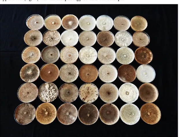
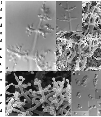
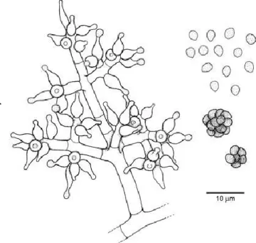
wide range of agronomic crops, turf grasses, ornamental plants, and fruit and forest trees worldwide (Adams, 1988; Sneh et al., 1991; Couch, 1995). Rhizoctonia solani is the most widespread species, with a host range that includes over 500 plant species (Farr et al., 1995). The genus concept of Rhizoctonia spp. was established by de Candolle (1815) (Sneh et al., 1998). However, the lack of specific characters led to the classification of a mixture of unrelated fungi as Rhizoctonia spp. (Parmeter and Whitney, 1970; Moore, 1987). Ogoshi (1975) enhanced the specificity of the genus concept for Rhizoctonia by elevating the following characteristics of R. solani to the genus level. Based on this revised genus concept, species of Rhizoctonia can be differentiated by mycelia colour (Fig. 1), number of nuclei per young vegetative hyphal cell (fig. 2) and the morphology of their teleomorph.
2
Figure 2 – Nuclei in Rhizoctonia solani (a) and Rhizoctonia BNR (b) hyphae
The teleomorph of Rhizoctonia spp. belongs to the sub-division Basidiomycota, class
Hymenomycetes.
The anamorphs of Rhizoctonia are heterogeneous. Moore (1987) placed the anamorphs of Thanatephorus spp. in Moniliopsis and reserved the genus Rhizoctonia for anamorph of ustomycetous fungi which have septa with simple pores.
Moniliopsis species have smooth, broad hyphae with brown walls, multinucleate
cells, dolipore septa with perforate parenthesomas and teleomorphs in the genera
Thanatephorus and Waitea. Related on the binucleate Rhizoctonia spp., the
anamorphs of the R. repens group (teleomorph Tulasnella) were assigned to the new genus Epulorhiza. Anamorph of Ceratobasidium was assigned to the new genus Ceratorhiza (Moore, 1987). Moore’s system is taxonomically correct and justified. At present, the concept of genus Rhizoctonia has become clear from these taxonomical studies at the molecular level (Gonzalez et al., 2001). However, many researchers (Sneh et al., 1998) in the world still retain the name Rhizoctonia for
3 1.2. Anastomosis group (AG) of Rhizoctonia solani and BNR and molecular
analysis
Genetic diversity is broad in Rhizoctonia genus, to the extent that the species is segregated into anastomosis groups (AGs) based on hyphal reactions of individuals. In 1969, J. R. Parmeter and his colleagues reintroduced the concept of "hyphal anastomosis" which implies that isolates of Rhizoctonia spp. that have the ability to recognize and fuse (i.e. "anastomose") with each other are genetically related, whereas isolates of Rhizoctonia spp. that do not have this ability are genetically unrelated. Anderson (1982) defined the anastomosis as a manifestation of somatic, or vegetative, incompatibility.
Anastomosis reactions between hyphae of paired isolates of R. solani consist of several types; such as perfect fusion (C3), imperfect fusion (C2), contact fusion (C1) and no reaction (C0) (Matsumoto et al., 1932; Carling et al. 1996). These classes have been accepted by many researchers and are useful for a better understanding of the genetic diversity of R. solani populations, because of the background genetically supported by vegetative or somatic compatibility (VC or SC) of confronted isolates (MacNish et al, 1997). The four classes of reactions can be distinguished according to the degree of interaction between hyphae from interacting isolates.
The category that represents the total fusion is the C3. It occurs for the same anastomosis group, same vegetative compatibility population (VCP) and the same isolate. The merger is realized by the fusion of walls and membranes accompanied with protoplasm connection. The anastomosis point frequently is not obvious with a diameter equal or nearly equal hyphal diameter. Anastomosing cells and adjacent cells may die, but generally do not.
The C2 category occurs in same AG, but not between different VCPs. Evidences are a wall connection and an uncertain membrane contact, besides the death of anastomosing and adjacent cells.
C0 and C1 are characterized by a little or no hyphal fusion between isolates. An apparent wall contact between hyphae without a wall penetration and membrane-membrane contact is typical for the C1 category; occasionally one or both
4 anastomosing cells and adjacent cells die. These categories occur between different AGs or in the same AG.
Affinity for hyphal fusion (anastomosis) (Parmeter et al., 1969; Parmeter and Whitney, 1970; Ogoshi et al., 1983a, b; Burpee et al., 1980) has been used to characterize isolates among R. solani, R. zeae, R. oryzae, R. repens and binucleate
Rhizoctonia spp. with Ceratobasidium teleomorphs.
To date, isolates of multinucleate R. solani have been assigned to 13 anastomosis groups (AG-1 to AG-13), which may possess similar characteristics, such as host preference, pathogenicity, and type of disease symptom caused (Carling et al. 2002a). Isolates of R. zeae and R. oryzae have been assigned to Z and WAG-O, respectively (Sneh et al., 1998; Carling et al., 1999, 2002a). Isolates of binucleate
Rhizoctonia spp. with Ceratobasidium teleomorphs have been reported. A system
developed in Japan (Ogoshi and Uti,1979, Ogoshi et al., 1983 a,b; Sneh et al., 1998; Hyakumachi et al., 2005) includes 21 anastomosis groups designated A to AG-U, in which at present AG-J and AG-M still are in question as members of binucleate
Rhizoctonia. Another system developed in the USA (Burpee et al., 1980) includes 7
anastomosis groups designed as CAG-1 to CAG -7. CAG-1 corresponds to AG-D, CAG-2 to AG-A, CAG-3 and CAG-6 to AG-E, CAG-4 to AG-F, CAG-5 to AG-R, and CAG-7 to AG-S (Sneh et al., 1998; Ogoshi et al., 1983a, b). At present, the anastomosis system based on AG-A through AG-U is widely accepted by many researchers.
Although the anastomosis method is accurate, valid, and largely used, it is sometimes impossible to determine to which AG an isolate belongs by anastomosis, because certain isolates do not anastomose with representatives of any known AG while some isolates have lost their capability to self-anastomose (Hyakumachi and Ui 1988). On the other hand, isolates of certain AGs anastomose also with isolates of more than one AG (Sneh et al. 1991; Carling 1996). In addition, determination of AGs by hyphal anastomosis requires meticulous microscopic experience, and it is a time-consuming procedure.
As suggested by Sharon et al. (2006, 2008) the introduction of various molecular and biochemical tools have confirmed the genetic relatedness validity of the AGs and
5 greatly advanced the accuracy of its classification. On the other hand the variety of these methods has been used to develop rapid PCR-based diagnostic tools for accurate identification of the isolates to AGs and their subgroups.
Sharon et al. in 2006 described the advances in various molecular techniques for classification of multinucleate Rhizoctonia (MNR); later, in 2008, continued the studies on binucleate and uninucleate Rhizoctonia (BNR and UNR). All the various molecular methods used for classification of Rhizoctonia spp., including isozyme analysis, total cellular fatty acids analysis, electrophoretic karyotyping, DNA–DNA hybridization, RAPD, AFLP, repetitive probe, AT-rich DNA RFLP, single-copy nuclear RFLP, rDNA RFLP, and rDNA sequence analysis have been explored (Tab. 1).
It was assessed that DNA sequences encoding ribosomal RNA genes, especially the internal transcribed spacer regions (ITS1 and ITS2) flanking the 5.8S subunit (rDNA-ITS sequence analysis), among the various molecular classification methods used for classification of Rhizoctonia spp., seems to be the most appropriate one. This technique is based on rDNA-ITS sequence alignment analysis (by which the genetic relatedness of the isolates is exhibited by clustering of isolate sequences in a tree), complemented with detailed percent sequence similarity within and among AGs and subgroups; these are compared with the anastomosis grouping method (Sharon et al. 2006, 2008).
Despite the fact that new methods and techniques have been developed in fungal systematics, classification of Rhizoctonia species still is considered to be in developmental stage. In general terms, systematic approaches for this group of fungi need both the study either the determination of the characterization of anastomosis groups, either the rDNA-ITS sequence analysis. MacNish et al. (1996) deemed appropriate for study of population biology, that molecular techniques might be complemented with hyphal anastomosis behaviour. For the characterization of AG8 isolates, a pool of methods, including hyphal anastomosis, pectin isozymes, RAPD and DNA fingerprinting, was used.
6
Table 1 - Relative efficacies of the various molecular methods used for classification of Rhizoctonia spp. (Sharon et al. 2006)
Method Different AG Same AG Subgroups within AG Individuals
Nucleic acids
a. DNA–DNA hybridization +++ +++
b. RFLP (restriction fragment length polymorphism)
- 18S, 28S rDNA +++ + +
- ITS (internal transcribed spacer) rDNA +++ +
- AT-rich DNA + + +++
- Single-copy nuclear DNA + + +++
c. DNA fingerprinting
- RAPD (random amplified polymorphic DNA) +++
- AFLP (amplified fragment length
polymorphism) +++
d. DNA sequencing
- 18S, 28S rDNA +++ +++ +
- ITS (internal transcribed spacer) rDNA +++ +++ +++
e. Electrophoretic karyotyping + + +
Protein
a. Isozymes
b. Zymograms + + +++
Cellular fatty acids +++ + +
7 1.3. Systematics of Rhizoctonia species
Members of the form genus Rhizoctonia D.C. are considered as a complex mixture of filamentous fungi that differ in many significant features, including their sexual stages (teleomorph), asexual stages (anamorph), having in common the possession of a non-spored imperfect state, usually referred to as the Rhizoctonia anamorph (Sneh
et al. 1991, Talbot, 1970, Tu and Kimbrough 1978).
The group includes several of the most devastating crop pathogens like
Thanatephorus cucumeris (Frank) Donk (anamorph = Rhizoctonia solani Kühn), the
majority of orchid mycorrhizal symbionts (mainly belonging to genus
Ceratobasidium D.P. Rogers) and a collection of saprotrophic organisms of different
systematic placement. The Rhizoctonia anamorph is characterized by several common features present among members of the entire Rhizoctonia species complex. Taxa from the group have been rearranged into several groups of higher fungi, including both Ascomycota and Basidiomycota, and split into several genera, employing criteria such as the analysis and ultrastructural comparison of septal apparatus. Until very recently, classification for some of the groups within the complex has been exclusively based on criteria such as hyphal anastomosis, since other types of diagnostic features are usually scarce in these fungi. Phytopathological studies in the complex have represented the major contingent of contributions in the group, especially in the case of R. solani. Some members of the complex have been reported to be protective isolates against pathogenic members of Rhizoctonia and some other fungal pathogens. (Gonzales Garcia et al., 2006)
Genus Thanatephorus [Rhizoctonia s. str.]
The genus Thanatephorus (Ceratobasidiaceae, Ceratobasidiales, Basidiomycota), was initially proposed by Donk (1956) to designate teleomorphic phases of the
Rhizoctonia solani multinucleate anamorph. It is commonly accepted that Thanatephorus applies to most parasitic fungi (as in the case of R. solani) which are
characterized by hypochnoid and cymose hyphae just above basal hyphae (Roberts, 1999). Somatic hyphae in Thanatephorus are constantly wider (more than 10 μm in diameter) (Roberts, 1999) than in Ceratobasidium, a closely related genus in the family Ceratobasidiaceae.
8 The morphology of Botryobasidium Donk (Donk, 1956) differs from Thanatephorus owing to the presence of short-sterigmate basidia, no repetitive basidiospores and the absence in culture of monilioid cells or sclerotia. Furthermore, Langer (1994) has provided evidence in Botryobasidium of septal pores with continuous parenthesomas, in contrast with discontinuous parenthesomas in both Thanatephorus and Ceratobasidium. Donk (1956) simultaneously established the genera
Thanatephorus and Uthatobasidium, reserving the later epithet for taxa similar to
Thanatephorus, but saprophytic and not producing sclerotia. Furthermore, Talbot and Keane (1971) proposed the name Oncobasidium P.H.B. Talbot and Keane for plant pathogenic taxa similar to Thanatephorus but not producing sclerotia. Finally, authors like Roberts (1999), considered these two genera to be synonyms of
Thanatephorus, assuming that parasitism in this genus is considered to be facultative
(T. cucumeris isolates are commonly reported to occur as saprotrophs), and generic distinction could be considered as weak for these three names. Other genera close to
Thanatephorus have been recently synonymized by Roberts (1999), including Ypsilonidium Donk, Cejpomyces Svrcek and Pouzr., Aquathanatephorus C.C. Tu and
Kimbr. (=R. solani AG1) and Tofispora G. Langer. Evolutive relationships between
Thanatephorus and its close relative Ceratobasidium remain controversial.
Employing classical taxonomic approaches, some authors (Stalpers and Andersen, 1996; Roberts, 1999) have considered both genera to be part of a generic complex, where delimitation among them presents some difficulties, and differences in morphometrical features and ecological behaviour are gradual along the several taxa within both genera. Roberts (1999) considered, after studying the type material for all accepted taxa of both genera, in combination with a preliminary ITS-based molecular phylogeny of selected species, that the two genera should be considered as synonyms. Recently, González et al. (unpublished), in an ITS-based phylogeny of family Ceratobasidiaceae (including taxa from Ceratobasidium, most accepted R.
solani anastomosis groups and sequences from genus Waitea), suggested that both
genera, although closely related, must be retained as independent entities within
Ceratobasidiaceae. The phylogenetic reconstruction carried out also demonstrated
9 1998; González et al., 2001), of Rhizoctonia solani (Thanatephorus cucumeris) into at least four different biological species. Thus, molecular analyses suggested that
Thanatephorus praticola Kotila and Flentje could be assigned to define, at a specific
level, AG 4 isolates (and their subgroups); T. sasakii (Shirai) C.C. Tu and Kimbr. could represent the valid epithet to name isolates from AG 1-IA and AG 1-IC; T.
microsclerotium (G.F. Weber) Boidin could represent AG1-IB strains, while the rest
of AGs actually defined, should be confined to Thanatephorus cucumeris s. str.
Genus Ceratobasidium [binucleate Rhizoctonia (BNR)]
The genus Ceratobasidium (Ceratobasidiaceae, Ceratobasidiales, and
Basidiomycota) was initially proposed by Rogers (1935) to accommodate four taxa
(C. calosporum Rogers, the type species of the genus designated by him, C.
cornigerum (Bourd.) Rogers, C. sterigmaticum (Bourd.) Rogers and C. obscurum
Rogers), some of them usually included as part of a complex mixture of genera and species arranged in the several groups and sections recognized for wide ancient genera like Corticium or Hypochnum. Thus, two of the above mentioned taxa, C. cornigerum and C. sterigmaticum formed part of section Botryoidea of Corticium. Donk (1931) was the first author to segregate part of the Ceratobasidiales, erecting the genus Botryobasidium Donk within the family Tulasnellaceae, due to the presence in members of the new genus of basidiospores capable of germinating by repetition, a diagnostic feature in phragmobasidiate fungi. Later, Rogers (1935) suggested such a taxonomical concept by adding Ceratobasidium to this group of fungi. Martin (1948) equally defined the family Ceratobasidiaceae to accommodate the genus. Donk (1956), being conscious that Botryobasidium still contained certain heterogeneity of “holo-” and “heterobasidiate” taxa, segregated these last elements (species with autoreplicative spores and large sterigmata) into two genera,
Thanatephorus and Uthatobasidium Donk, which were subsequently included by
Jülich (1981) within his concept of Ceratobasidiales. Both C. sterigmaticus and C.
obscurum are considered to belong to Thanatephorus in the modern concept of the
genus, and are excluded from Ceratobasidium (Roberts, 1999). Donk (1958) erected the genus Koleroga to accommodate K. noxia Donk, a species morphologically close to Ceratobasidium, except for the absence of autoreplicative spores. Talbot (1965)
10 demonstrated the presence of this type of spore in some collections of K. noxia. Subsequently, some authors (Roberts, 1999) considered the genus Koleroga as a nomenclatural synonym of Ceratobasidium (C. noxium). Currently, there are between 10 (Roberts, 1999) and 11 (Kirk et al., 2001) species accepted for the genus. Species of Ceratobasidium are characterized for the presence of a non-sporulating,
Rhizoctonia-like (genus Ceratorhiza) anamorphic phase, binucleate somatic hyphae
(uninucleate in Ceratobasidium bicorne) and saprophytic, mycorrhizal or parasitic teleomorphic phases. Ceratobasidium taxa produce effuse fruitbodies of ceraceous consistency, with globose to sphaeropedunculate basidia, produced directly from basal hyphae or in raceme-like groups, usually showing a division between hypo- and epibasidium, producing basidiospores with high rates of repetitive germination (Rogers, 1935). This set of diagnostic characters (mostly referred to hyphal cytology and nutritional behaviour), differentiate Ceratobasidium from Thanatephorus. Concerning the nomenclature of anamorphic stages of the genus, only the above mentioned genus Ceratorhiza is considered to date, to be the correct name to designate anamorphs with Ceratobasidium teleomorph (Roberts, 1999). From an ecological point of view, the genus includes saprophytic, symbiotic and even parasitic taxa. Thus, most species have been described as saprophytic on soil or plant debris (wood and leaf litter from both angiosperm and gymnosperm hosts), or as forming part of the fungal component of orchid mycorrhizae. A small number of taxa have been reported as parasites of herbaceous plants (some of them of economic interest) or bryophytes. Regarding its evolutive relationships, the genus could represent for several authors a transitional evolutive line to modern basidiomycetes, from typically heterobasidiate (with clearly segmented basidia, possessing hipo- and epibasidia), resembling Tulasnella-like forms (probably the closest heterobasidious relative to Ceratobasidium), towards typically holobasidiate forms with non-segmented basidia and true sterigmata. In this sense and in accordance with its current systematic position (Roberts, 1999; Kirk et al., 2001), the family
Ceratobasidiaceae could represent the most primitive group of holobasidiomycetes,
with hymenial structures showing morphologies where segmentation of the basidia is still easily observed, and partition on sterigmata occurs. Studies on the ultrastructure
11 of septal apparatus in Ceratobasidium and Thanatephorus have revealed their affinities with the remaining holobasidiate basidiomycetes. In this sense, Binder et
al. (2005) have recently reported phylogenetic evidence of the relationships of the
12 1.4. Biology
Rhizoctonia solani causes different types of diseases to a wide variety of plants, all
over the world and under different environmental conditions (Adams, 1988; Sneh et
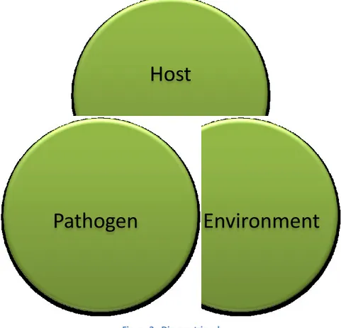
al., 1991; Couch, 1995). In order to understand the Rhizoctonia disease occurrences
should be considered the host, the pathogen and the environment (fig. 3)
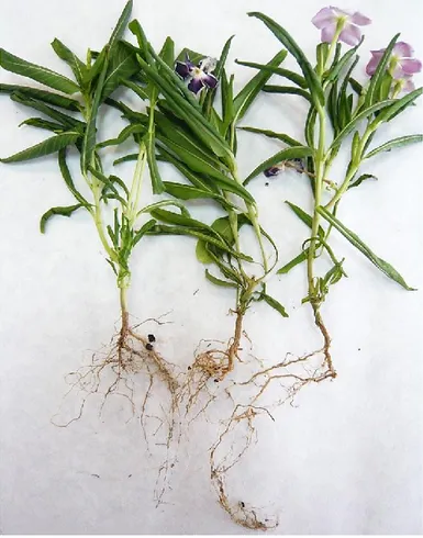
Rhizoctonia spp. attack its hosts during their juvenile stage of development such as
seed, seedlings and cuttings (figure 4) and diseases can occur in cases of severe epidemics under specific conditions. Rhizoctonia species are responsible of root and stem rot, leaf spot, seedlings damping-off and foliar web blight (Chase, 1991; Benson and Cartwright, 1996; Hyakumachi et al., 2005; Rinehart et al., 2007). Besides the type of germination is the most critical phase, because it influences the length of exposure of the seedling to the invasion by pathogen (Singh, 1955; Ruppel
et al. 1964). In hypogeal germination the cotyledons remain underground and
susceptible to attack whereas in epigeal germination the cotyledons are carried above
Host
Environment
Pathogen
13 the soil, and may thereby escape decay. Factors that delay the germination may increase seed decay and preemergence damping-off. The susceptibility of the host to the fungus attack declines with maturation and lignification of tissues; thus the pathogen attacks the aerial parts, under very moist conditions (Baker, 1970).
Rhizoctonia species remain for all the year in more or less continuous vegetative
growth even if it forms resting structures that allows to avoid unfavourable condition. Once the environmental conditions come back closely to the optimum range, the pathogen starts again to differentiate fresh mycelium to infect hosts newly. The resting structure, sclerotia, not only are necessary to face the unfavourable conditions (i.e. dry periods) but are also a means of dissemination by wind or water (Echandi, 1965; Baker, 1970).
Environmental factors are very important in influencing the development of Rhizoctonia diseases. The temperature at which infection occurs, the ability to
develop in lower soil levels, the ability to form sclerotia, the growth rate and the survival in a certain area may or may not show up given the environment and host. Due to the phylogenetic heterogeneity, these environmental conditions differ among
= Host high susceptibility seed germination seedling growth mature plant flowering fruit
14 taxa, and even among isolates belonging to the same species. The optimal relative humidity, essential for the production of teleomorphs, could range between 40-100%. For sexual sporulation the pathogen needs the intake of O2 and an efficient removal
of CO2. The optimum temperature range for sexual fruiting is from 20 to 30°C.
Moreover, variations in day/night temperatures also seem to play an important role in fruiting. Uchida et al. (1986) reported in Rhizoctonia species that light stimulates hymenial formation but inhibits the ripening of basidia. This intuition is confirmed by the fact the higher rates of sporulation are usually found by night, followed by a drop in diurnal temperatures. Plants are more prone to attack by pathogens when stressed by an inhospitable environment and usually caused by a lack of nutrients or related to the existence of a strong hydrophobicity gradient in the colonizing substrates (Adams and Butler, 1983; Kotila, 1947; Flentje, 1956).
15 1.5. Pathogenicity and hosts
Rhizoctonia diseases of ornamental plants can occur in cases of severe epidemics. A wide range of disease symptoms have been recorded, including root and stem rot, leaf spot, seedlings damping-off and foliar web blight (Chase, 1991; Benson and Cartwright, 1996; Hyakumachi et al., 2005; Rinehart et al., 2007).
BNR typically are weakly virulent, less destructive pathogens or not pathogenic considered as mycorrhizal or biocontrol agents (Harris et al., 1994; Andersen and Rasmussen, 1996; Hwang and Benson, 2002; Burns and Benson 2000). However, several studies reported BNR as pathogenic on economically important agricultural and horticultural crops (Priyatmojo et al., 2001; Kuramae et al., 2007; Aiello et al., 2012).
1.5.1. Seed decay
Rhizoctonia may invade the seed while still in the fruit, decaying in there or merely
infecting it. The decay process is then resumed after the seed is planted and before germination (Baker, 1947; Neergaard, 1958).
The seed may also be invaded by growth of the fungus from infested soil in which it is planted; the grater the amount of inoculum in the soil, the more certainly and the more rapidly this occurs. In either case, the invaded seed serves as a food base, enabling the pathogen to reach adjacent seedling. It is thus in common to find seedlings with pre-emergence damping-off in the vicinity of a rotted seed. The greater the distance seed are separated, the less the probability of such spread (Singh and Singh, 1955). Thus, losses are usually greater in seed flats and nursery beds than in the wild.
1.5.2. Damping-off of seedling
Rhizoctonia may attack newly emerged plants or cuttings at the base of stem causing
extensive water-soaked lesions. As a consequence, the attack may followed by wilt and collapse of the plant. The damping-off can occur in pre- and post-emergence phase. The first one is an extension of seed decay; the two together are often designated as “poor stand”. The damping-off may be considered as a delayed attack,
16 due to unfavourable environment (temperature or moisture) or insufficient inoculum for faster action, of the seed decay. The longer that seedling emergence in delayed, the greater the opportunity for invasion and the greater is the preemergence damping-off. The emergence is tightly related on the seed depth, seed vitality, type of germination (hypogeal or epigeal) and the physical soil conditions (temperature, moisture or pH) (Singh, 1955; Hartley et al., 1918; Germ, 1960; Sinclair, 1965; Leach, 1947; Beach, 1949; Peace, 1962; Baker, 1970). The post-emergence damping-off represents still a further delay in attack and/or expression of symptoms. Symptoms may develop any time after emergence through the soil surface, until the seedling is still in the juvenile stage. The susceptibility of the seedling declines with maturation and lignification of tissues. This increased resistance may be due to conversion of pectin to calcium pectate, rendering the tissues resistant to the polygalacturonase of the fungus (Bateman and Lumsden, 1965).
Symptoms appear on the stem near soil level, but may later advance downward into the roots. Small bits of soil or organic matter dangle from the coarse mycelium of
Rhizoctonia attached to the infected seedlings when removed from the soil (Duggar,
1916). Thus by examination, with a hand lens, it is possible to distinguish the coarse, hyaline to brown mycelium of Rhizoctonia.
The disease may spread into two directions: in circular or irregular patches, when seed is randomly sown and in turf grass; in linear strips if it is sown in rows. These represent either a dispersion ways of inoculum either a via for the pathogen introduction. In disinfested soil, commonly used in nursery, the introduction of the pathogen will success with seed or transplants, soil fragments or infected tissues splashed in by water, carried in by wind or workers, or survival on the pots. The more nearly sterile the soil the more rapid will be the spread, and the larger the area of spread (Baker, 1957, 1962)
1.5.3. Crown and root rot
Crown rot is typified by extensive water-soaked, dark brown lesions at the crown level that girdled entire stem and an internal brown discoloration of cortical tissue (Fig. 6). Crown rot of sugar beet is one of the historic diseases caused by R. solani.
17 Infection apparently occurs in the young leaflets or in leaf bases, and causes petiole decay. As the crown leaves die out, lateral young ones appear. The crown of the plant may eventually be killed and the fungus advance into the top of the fleshy root, causing a dry brown decay (Edson, 1915; Walker, 1957).
Root rot is not generally the most important type of disease caused by this fungus, but severe losses can be produced on some crops. Dark, circular to oblong, sunken cankers with brown borders develop at the point of origin of secondary roots (Fig. 5). Secondary roots may be invaded directly, or may be cut off by the basal lesions. Underground fleshy roots are often affected by cankers, as well as being attacked at the crown. Plants died a few days later due to the disruption of translocation of water and nutrients. As reported by Durbin (1957) there are some subterranean type of R. solani (such as the alfalfa isolates) that are more tolerant of CO2
than the surface or aerial strains. Among all the species susceptible to root rot there are several agronomical crops including sweet pea, alfalfa, cereals, sugar beet and ornamentals as well (Weber and Foster, 1928; Samuel and Garrett, 1932; Blair, 1942; Moore, 1959). On turnips concentric light and dark bands are produced on the side of the root in early season; later, the lesions may be large, rough, dry and pithy. Attack may occur in the field, as well as in storage (Dana, 1925; Lauritzen, 1929). Sclerotia may develop on the surface of the cankers on either host. Tulip bulbs may be russetted by R. solani
18 (Moore, 1959).
R. solani has been shown to attack the roots of conifers (Hartley, 1921) and coffee
trees (Crandall and Arillaga, 1955), but the importance of this trouble under field condition has not been demonstrated.
1.5.4. Hypocotyl and stem cankers
Stem lesions may develop in fully mature plants with well-developed secondary tissue. The stem rot of carnation is very destructive where this plant is grown in soil infested by R. solani or where infected cutting are used. A soft moist rot starts at the soil surface and advances into the stem; the decayed cortex easily rubs off, leaving the stele beneath. Strands of mycelium, and sometimes sclerotia, appear on the surface. Roots remain intact until late in the disease, but the tops wilt, turn brown, and die (Baker and Sciaroni, 1952). The fungus causes a destructive “neck rot” of gladiolus, infecting at soil level through the leaf bases, and producing brown shredded lesions from decay of parenchymatous tissue. Cankers may occur on mature tomato plants at the base of stem branches; these often have alternate light and dark bands. Infection may occur through a leaf in contact with the soil and spread to the cortex and stele (Conover, 1949). Basal cankers or “foot rot” may also be produced on tomato plants in glasshouses (Small, 1927).
A more unusual type of stem rot was produced on aquatic plants in the coastal waters of Virginia and North Carolina following the opening of a canal. Dark lesions occurred at the soil surface, regardless of the depth and concentration of sea water, producing great destruction to four genera of plants. This strain of R. solani was able to attack potatoes and that from potatoes to attack the aquatic plants (Bourn and Jenkins, 1928).
1.5.5. Bud rot
The flower buds, which are the first to arise from the crown, are killed. Lateral dormant buds may later grow out sometimes giving a witches’-broom effect (Wilhelm, 1957). A preplanting soak of the plant in gibberellin (10 ppm) accelerates the rate of emergence of shoots from the soil and thus decreases Rhizoctonia bud rot.
19 1.5.6. Aerial (web, leaf, thread) blight
Under condition of high relative humidity, warm temperature and reduced sunlight, aerial blight may develop on the canopy of bedding plants and other plants in the greenhouse and landscape (Weber and Roberts, 1951; Wehlburg and Cox, 1966; Frisina and Benson, 1987). R. solani, that lead an aerial existence independent of the soil, may spread through the tops of plants.
Foliar blight initially develops as water-soaked lesions on stem and leaves. In succulent plants it rapidly develops in complete plant collapse; while in less succulent ones foliage concentric rings may result from diurnal expansion of lesion (Chase, 1987).
Web blight symptoms occur in the interior portion of the canopy, while the outer canopy leaves remain healthy, typically (Frisina and Benson, 1989).
Stems also were infected and turned dark, reddish, brown or black as disease progressed. If new shoots come out in dry weather they remain healthy and presumably resistant. The small brown spots are concentrically brown-ringed.
1.5.7. Turf grasses patch
The strains that cause brown patch of turf grasses appear to act in every way as the truly aerial forms just discussed, though growing so close to the ground that soil-surface types might be expected. Leaves are infected through stomata and through mowing wounds. Initial leaf symptoms observed are small, tan lesions that enlarged and become surrounded by reddish brown margins over time. Eventually grass leaves become necrotic and brown in colour. The diseased areas of grass are usually 30-90 cm in diameter, but may reach 6-15 m. A dark purplish-green advancing margin 1,3-5,0 cm wide, in which the mycelium is webbed, is visible in the mornings, but soon dries, and the central leaves die and turn light brown.
The crown and the roots are only rarely invaded. Sclerotia are formed near the base of the plant. Because grass is close to the moist soil, there may be a good deal of dew and guttation fluid on the leaves at night; the environment is therefore similar to those trees in the humid tropics.
20
Table 2 - Binucleate Rhizoctonia groups and hosts
Group AG-A: (Mazzola, 1997; Sneh et al., 1998; Yang et al., 2007; Polizzi et al., 2009c, 2010b; Wang and Wu, 2012; Miles et al. 2013; Yang and Wu 2013, Li et al. 2014)
Symptoms root rot, damping-off, browning, tortoise shell, crown rot, stem rot and stem canker Host
strawberry, sugar beet, bean, pea, sunflower (Helianthus annuus Linn.), tomato, melon, cucumbear (Cucumis sativas Linn.), leaf lettuce, spinach, peanut, potato, Solanum tuberosum, apple, swiss chard, Dodonea viscosa, Thryptomene saxicola and foxtail millet (Setaria italica) Note Some isolates in this group form mycorrhizal associations with orchids
Group AG-Ba (Sneh et al., 1998)
Symptoms grey sclerotium disease, sclerotium disease, gray southern blight Host rice, Echinochloa crugalli subsp. submitica var. typica, and foxtail millet
Note ---
Group AG-Bb (Sneh et al., 1998)
Symptoms brown sclerotium disease, grey sclerotium disease, and sheath spot Host fox tail, millet, and rice
Note ---
Group AG-C (Sneh et al., 1998; Hayakawa et al., 1999)
Symptoms symbiosis (orchids)
Host orchids, sugar beet seedlings, subterranean clover, and wheat Note No important pathogens have been reported
Group AG-D: I, II, III (Sneh et al., 1998; Toda et al.,1999; Hayakawa et al., 2006)
Symptoms sharp eye spot, yellow patch, foot rot, Sclerotium disease, snow mold, root rot, damping-off, lesions on stems, winter stem rot and sheath rot
Host cereals, turf grass, wheat, barley, sugar beet, clove, pea, onions (Allium cepa Linn.), potato, cotton, bean, soybean, mat rush, foxtail millet, subterranean clover and Zoysia japonica Note Recently this group is classified into subgroup AG-D (I) that causes Rhizoctonia patch and
winter patch diseases. AG-D (II) causes elephant footprint disease Group AG-E (Sneh et al., 1998)
Symptoms web-blight, damping-off, seedlings, and symbiosis (orchids) Host
bean, pea, radish, onion, leaf lettuce, tomato lima bean, snap bean, soybean, peanut, cowpea (Vigna savi), flax, sugar beet, Rhododendron L., long leaf pine (Pinus palustris Mill.), slash, lobolly pine (Pinus taeda L.), and rye (Secale cereale L.)
Note ---
Group AG-F (Sneh et al., 1998; Eken and Demirci, 2004, Yin et al., 2011, Aiello et al., 2012; Saroj et al. 2013; Meza-Moller et al. 2014; Harveson and Bolton 2013)
Symptoms roor rot, watermelon vine decline, damping-off and dry rot canker Host
bean, pea, radish, onion, peanut, leaf lettuce, tomato, subterranean clover radish, tomato, cotton, taro, strawberry (source: DDJB), Fragaria x ananassa, Musa spp., watermelon (Citrullus lanatus), Tagetes erecta and sugar beet
Note ---
21
2003; Fenille et al., 2005; Polizzi et al., 2009°; Tuncer and Eken 2013) Symptoms damping-off, root rot, and browning
Host strawberry, sugar beet, bean, pea, tomato, melon, sunflower, peanut, yacoon, apple, Rhododendron Linn., Fragaria x ananassa, Viburnum tinus and pepper (Capsicum annuum)
Note
Non-pathogenic binucleate Rhizoctonia spp. provide effective protection to young bean seedlings against root rot caused by R. solani AG-4 (Leclerc et al., 1999). On pepper is non-pathogenic
Group AG-H (Hayakawa et al., 1999)
Symptoms symbiosis (orchids)
Host Dactylorhiza aristata (Orchidaceae)
Note ---
Group AG-I (Mazzola, 1997; Sneh et al., 1998; Ravanlou and Banihashemi, 2002)
Symptoms root rot and symbiosis (orchids)
Host strawberry, sugar beet, wheat, apple, orchids, and Fragaria x ananassa
Note ---
Group AG-J: (Sneh et al., 1998)
Symptoms none
Host apple
Note ---
Group AG-K (Demirci, 1998; Li et al., 1998; Sneh et al., 1998; Ravanlou and Banihashemi, 2002; Tuncer and Eken 2013)
Symptoms Root rot
Host sugar beet, radish, tomato, carrot, onion, wheat, maize, Allium cepa (source: DDJB), Pyrus communis (pear) (source: DDJB), Fragaria x ananassa and pepper (Capsicum annuum) Note On pepper is non-pathogenic
Group AG-L: No special diseases have been reported (Sneh et al., 1991)
Symptoms ---
Host ---
Note ---
Group AG-N: No special diseases have been reported (Sneh et al., 1991)
Symptoms ---
Host ---
Note ---
Group AG-O: No special diseases have been reported (Sneh et al., 1991)
Symptoms ---
Host ---
Note ---
Group AG-P: (Sneh et al., 1998; Yang et al., 2006)
Symptoms black rot and wirestem Host tea (Camellia Linn.), red birch
22
Group AG-Q: (Sneh et al., 1998)
Symptoms none
Host Bentgrass
Note ---
Group AG-R: (Sneh et al., 1998;Yang et al., 2006, 2008)
Symptoms wire stem and rhizome blight
Host bean, pea, radish, onion, leaf lettuce, tomato, lima bean, snap bean, soybean, cowpea, peanuts, red birch, azalea and ginger
Note ---
Group AG-S (Demirci, 1998; Sneh et al., 1998)
Symptoms no specific diseases
Host azalea, wheat, barley, and azalea
Note ---
Group AG-T: (Hyakumachi et al., 2005)
Symptoms stem rot and root rot
Host miniature roses
Note ---
Group AG-U: (Hyakumachi et al., 2005)
Symptoms stem rot and root rot
Host miniature roses (Rosa rugosa Thunb.)
23
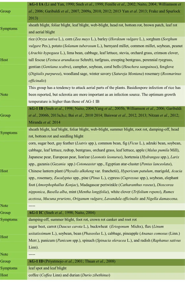
Table 3 - Multinucleate Rhizoctonia isolates and hosts
Group
AG-1 IA (Li and Yan, 1990; Sneh et al., 1998; Fenille et al., 2002; Naito, 2004; Williamson et
al., 2006; Garibaldi et al., 2007, 2009a, 2010, 2012; 2013 Yan et al. 2013; Frake and Spurlock 2013)
Symptoms sheath blight, foliar blight, leaf blight, web-blight, head rot, bottom rot, brown patch, leaf rot and aerial blight
Host
rice (Oryza sativa L.), corn (Zea mays L.), barley (Hordeum vulgare L.), sorghum (Sorghum vulgare Pes.), potato (Solanum tuberosum L.), barnyard millet, common millet, soybean, peanut (Arachis hypogaea L.), lima bean, cabbage, leaf lettuce, stevia, orchard grass, crimson clover, tall fescue (Festuca arundiacea Schreb), turfgrass, creeping bentgrass, perennial ryegrass, gentian (Gentiana scabra), camphor, soybean, coral bells (Heuchera sanguinea), foxglove (Digitalis purpurea), woodland sage, winter savory (Satureja Montana) rosemary (Rosmarinus officinalis)
Note
This group has a tendency to attack aerial parts of the plants. Basidiospore infection of rice has been reported, but sclerotia are more important as an infection source. The optimum growth temperature is higher than those of AG-1 IB
Group
AG-1 IB (Sneh et al., 1998; Naito, 2004;Yang et al., 2005b, Williamson et al., 2006; Garibaldi
et al., 2006b, 2013a,b,c; Bai et al., 2010 2014; Baiswar et al., 2012, 2013; Nitzan et al., 2012; Miranda et al. 2014
Symptoms sheath blight, leaf blight, foliar blight, web-blight, summer blight, root rot, damping-off, head rot, bottom rot and seedling blight
Host
corn, sugar beet, gay feather (Liatris spp.), common bean, fig (Ficus L.), adzuki bean, soybean, cabbage, leaf lettuce, redtop, bentgrass, orchard grass, leaf lettuce, apple (Malus pumila Mill), Japanese pear, European pear, lion'ear (Leonotis leonurus), hortensia (Hydrangea spp.), Larix spp., gazania (Gazania spp.) Cotoneaster spp., Egyptian atar-cluster (Pentas lanceolata), Chinese lantern plant (Physalis alkekeng var. franchetii), Hypericum patulum, marigold, Acacia spp., rosemary, Eucalyptus spp., pine (Pinus L.), cypress (Cupressus spp.), soybean, elephant foot (Amorphophallus Konjac), Madagascar periwinkle (Catharanthus roseus), Dioscorea nipponica, Basella alba, mint (Mentha longifolia), white clover (Trifolium repens), Rumex acetosa, Mucuna pruriens, Origanum vulgare, Lavandula officinalis and Nigella damascena.
Note ---
Group AG-1 IC (Sneh et al., 1998; Naito, 2004)
Symptoms damping-off, summer blight, foot rot, crown rot canker and root rot
Host
sugar beet, carrot (Daucus carota L.), buckwheat (Eriogonum Michx), flax (Linum
usitatissimum L.), soybean, bean (Phaseolus L.), cabbage, pineapple (Ananas comosus (Linn.) Merr.), panicum (Panicum spp.), spinach (Spinacia oleracea L.), and radish (Raphanus sativus Linn).
Note ---
Group AG-1 ID (Priyatmojo et al., 2001; Thuan et al., 2008)
Symptoms leaf spot and leaf blight
24
Note this subgroup was recently reported in the Philippines (Priyatmojo et al., 2001) Undetermined subgroup: buckwheat, flax, spinach, and radish
Group
AG-2-1 (Satoh et al., 1997; Camporota and Perrin, 1998; Sneh et al., 1998; Rollins et al., 1999;
Khan and Kolte, 2000; Naito, 2004; Paulitz et al., 2006; Zhang et al., 2009; Baiswar et al., 2010; Bai et al., 2010; Mercado Càrdenas et al., 2012; Caesar et al. 2014; Misawa and Kuningana 2013)
Symptoms damping-off, leaf rot, leaf blight, root rot, foot rot, bottom rot, bud rot, head rot, seedling blight and leaf margin necrosis
Host
sugar beet, wheat (Triticum aestivum Linn.), potato, cowpea (Vigna unguiculata (Linn.) Walp), canola, rape (Brassica napus Linn.), cauliflower (Brassica oleracea var. botrytis Linn.), mustard (Sinapis Linn.), turnip (Brassica rapa Linn.), pepper (Piper Linn.), Silene armeria, spinach, leaf lettuce, strawberry (Fragaria ananassa Duchesne), tulip (Tulipa gesneriana Linn.), tobacco (Nicotiana Linn.), clover (Medicago Linn.), table beet, canola, cabbage, Ctenanthe oppenheimiana, Dioscorea nipponica, tobacco, Lepidium draba and chinese chives (Allium tuberosum Rottler ex Sprengel)
Note This group includes the AG-2-1 tulip strain (former AG-2t) and the AG-2-1 tobacco strain (former homogenous Nt-isolates) (Kuninaga et al., 2000)
Group AG-2-2 III B (Sneh et al., 1998; Priyatmojo et al., 2001; Naito, 2004; Caesar et al., 2009; Woodhall et al., 2012a; Garibaldi et al. 2014; Zhao and Wu 2014)
Symptoms brown sheath blight, dry root rot, root rot, brown patch, large patch, black scurf, stem rot, stem blight, web blight, Rhizoctonia rot, damping-off, stem canker, collar rot, and crown brace rot
Host
rice, soybean, corn, sugar beet, edible burdock (Arctium lappa), taro (Colocasia esculenta), Dryopteris spp., elephant foot, crocus, saffron (Crocus sativus Linn.), redtop, bentgrass, St. Augustine grass, turf, balloon flower (Platycodon grandiflorum), Christmas-bells (Sandersonia aurantiaca), Hedera rhombea, mat rash, gladiolus, ginger, Iris Linn, Centaurea stoebe, potato and Rebutia perplexa
Note ---
Group AG-2-2 IV: (Sneh et al., 1998; Naito, 2004)
Symptoms leaf blight, foliage rot, root rot, and stem rot
Host sugar beet, carrot, eggplant (Solanum Linn), pepper, spinach, stevenia (Stevenia Adams et Fisch), and turfgrass
Note ---
Group AG-2-2 LP: (Aoyagi et al., 1998)
Symptoms large patch
Host Zoysia grass
Note ---
Group AG 2-3: (Naito and Kanematsu, 1994; Sumner et al., 2003)
Symptoms leaf blight and root rot
Host soybean
Note ---
25
Symptoms crown rot, brace rot, and damping-off
Host corn and carrot
Note ---
Group AG-2-BI: (Carling et al., 2002b)
Symptoms nonpathogenic
Host isolates, obtained only from soils and plants in forests
Note
former name is AG-BI; Undetermined subgroup: sesame (Sesamum Linn.), white mustard (Sinapsis alba), primrose (Primula spp.), white lace flower (Ammi majus), carnation, baby’s-breath (Gypsophila paniculata), rusell prairie gentian (Eustoma grandiflorum), snap bean, lima bean, and Chinese radish
Group AG 3: PT, TB (Sneh et al., 1998; Kuninaga et al., 2000; LaMondia and Vossbrinck, 2011, 2012; Wu et al., 2012; Muzhinji et al. 2014a; McCormack et al. 2013; Tuncer and Eken 2013) Symptoms black scurf, leaf spot, target leaf spot, damping-off and root rot
Host
PT: potato with black scurf symptoms; maize with stem bases and roots rot TB: tobacco with target leaf spot symptoms
Pepper (Capsicum annuum)
Note Undetermined subgroup: eggplant, sugar beet, tomato, and wheat. Their pathological and ecological information is less
Group
AG-4: HG-I, HG-II, HG-III (Baird, 1996; Holtz et al., 1996; Sneh et al., 1998; Fenille et
al.,2002; Ravanlou and Banihashemi, 2002; EI Hussieni, 2003; Kuramae et al., 2002, 2003; Naito, 2004; Yang et al., 2005c, Garibaldi et al., 2006a; Yang et al., 2007; O’ Brien et al., 2008; Aiello et al., 2008a, b; Hsiao et al., 2008; Aiello et al., 2009; Garibaldi et al., 2009b, c, d, Polizzi et al., 2009b; Polizzi et al., 2010a, c; Srinivasan and Visalakchi, 2010; Polizzi et al., 2011a, b; Bai et al., 2012; Liao et al., 2012; Mathew et al., 2012; Nitzan et al., 2012; Woodhall et al., 2012b; Muzhinji et al. 2014b; Yang and Wu 2013)
Symptoms damping-off, root rot, stem canker, black scurf, fruit rot, stem rot, leaf blight, web blight, crown rot, basal rot, wilt and leaf yellowing and wilting
Host
pea, sugar beet, melon, soybean, adzuki bean, common bean, snap bean, lima bean, carrot, spinach, taro, tomato (Lycopersicon esculentum Mill.), potato, alfalfa (Medicago sativa Linn.), elephant foot, arrowleaf clover, beans, barley, buckwheat, cabbage, canola, turnip, carnation, cauliflower, Chinese chive, chrysanthemum, corn, cotton (Gossypium Linn.), table beet, tobacco, turfgrass, wheat, white lupine, parsley (Petroselinum Hill), Cineraria Linn., stock, poinsettia, primrose, hybrid bouvardia, Citrus Linn., cauliflower, Euphorbia spp., geranium (Pelargonium spp.), Russel prairie gentian, statice (Limonium spp.), baby’s-breath, Astragalus membranaceus, lamb’s lettuce, swiss chard, mung bean (Vigna radiata), Lagunaria patersonii, redwood, african daisy (Osteospermum), orange jessamine (Murraya paniculata), Washington lupine (Lupinus polyphyllus), Hosta fortune, fan columbine (Aquilegia flabellata), Coprosma repens and C. lucida, Chamaerops humilis, Streptosolen jamesonii, sunflower, Passiflora mollissima, Tabebuia impetiginosa, Rhodiola secholiensis, Besella rubra, Pisum sativum, mint (Mentha longifolia)
26
Group AG-5 (Li, et al., 1998; Demirci, 1998; Sneh et al., 1998; Ravanlou and Banihashemi, 2002; Eken and Demirci, 2004; Naito, 2004; Mattew et al.,2012; Yang and Wu, 2012)
Symptoms root rot, damping-off, black scurf, brown patch, stem cacker and symbiosis (orchids) Host soybean, adzuki bean, apple, barley, chickpea, common bean, lima bean, potato, strawberry,
sugar beet, table beet, tobacco, turfgrass, wheat, white lupine and Pisum sativum
Note ---
Group AG-6: HG-I, GV (Mazzola, 1997; Meyer et al., 1998; Sneh et al., 1998; Carling et al., 1999; Pope and Carter, 2001; Naito, 2004; Tuncer and Eken 2013)
Symptoms root rot, crater rot, and symbiosis (orchids)
Host apple, wheat, carrot, carnation and pepper (Capsicum annuum) Note all isolates from forests are non-pathogenic
Group AG-7: (Naito, et al., 1993; Baird and Carling, 1995; Carling, 1997, 2000; Carling et al.,1998; Rani et al. 2013; Kamel et al. 2010)
Symptoms damping-off, root rot, and black scurf
Host carnation, cotton, soybean, watermelon (Citrullus lanatus (Thunb.) Mansfeld), Raphanus Linn., and potato
Note ---
Group AG-8: (Sneh et al., 1998; Naito, 2004)
Symptoms bare patch
Host barley, cereals, green pepper, potato, and wheat
Note ---
Group AG-9: (Sneh et al., 1998; Naito, 2004).
Symptoms black scurf
Host potato, crucifers, wheat, and barley.
Note ---
Group AG-10: (Sneh et al., 1998; Schroeder and Paulitz, 2012)
Symptoms (weak pathogenic) root rot Host barley, wheat and canola
Note ---
Group AG-11: (Kumar et al., 2002).
Symptoms damping-off and hypocotyls rot Host barley, lupine, soybean, and wheat
Note ---
Group AG-12: (Kumar et al., 2002)
Symptoms symbiosis (orchids)
Host Dactylorhiza aristata (Orchidaceae)
Note ---
Group AG-13: (Carling et al., 2002a).
Symptoms none
Host cotton
27 2. Rhizoctonia diseases on ornamental plants in Europe and Italy
Several studies of last twenty years reported a broad plant-host group susceptible to
Rhizoctonia diseases. Different symptoms such as root and stem rot, leaf spot,
seedling damping-off and foliar leaf blight are caused by Rhizoctonia spp. in different countries of Mediterranean basin.
In Europe 7 different species of Rhizoctonia are reported as pathogen on ornamental plants. In detail: R. crocorum is reported to be pathogen on Dianthus sp., Humulus
lupulus and Morus sp., in Bulgaria (Bobev, 2009). Rhizoctonia endophytica var. endophytica and Rhizoctonia endophytica var. filicata are reported on Picea abies in
Norwey (Roll-Hansen and Roll-Hansen, 1968). Roberts (1999) have found out
Rhizoctonia rubiae on Rubia sp. in France. Rhizoctonia tuliparum and Rhizoctonia violacea are pathogen on Tulipa sp. in Poland and Citrus sp. in Italy, respectively
(Mulenko, 2008; Greuter, 1991). The main specie that has more than 500 host and has been reported in 8 European country (Bulgaria, Denmark, Germany, Greece, Italy, Madeira Islands and Spain) is R. solani (Aiello 2008 a, b; Aiello 2009; Bobev 2009; Braun 1930; Crous et al. 2004; Garibaldi 2006a, b; Garibaldi 2009 a, b, c, d; Garibaldi 2007; Garibaldi 2003 a, b; Gonzalez et al. 2011; Holevas et al. 2000; Kowalski and Andruch, 2012; Mulenko et al. 2004; Mulenko et al. 2008; Polizzi et
al. 2009 a; Polizzi 2010 et al. a, b; Polizzi 2011 et al. a, b; Richardson 1990,
Garibaldi et al. 2013 a, b, c, d, Garibaldi et al. 2010, Garibaldi et al. 2012, Garibaldi
et al. 2014). In Italy intensive researches have been conducted on vegetable crops
and ornamental plants.
These studies allowed to identify R. solani as causal agent of aerial blights on
Anubias heterophylla, Aquilegia flabellata, Catharanthus roseus, Digitalis purpurea, Heuchera sanguinea, Hosta fortunei, Lantana camara (Garibaldi et al. 2003a;
Garibaldi et al. 2009a; Garibaldi et al. 2006a; Garibaldi et al. 2009b,c,d; Garibaldi et
al. 2007; Garibaldi et al. 2003a,b). Most of these reports concern on surveys
conducted in Sicilian nursery, especially located in the Eastern side of the Island. Damping-off caused by R. solani has been reported on Chamaerops humilis,
28 2010a; Aiello et al. 2008a, b; Polizzi et al. 2011a). Polizzi et al. 2009a; Aiello et al. 2009; Polizzi et al. 2011b reported crown and root rot on Coprosma lucida and C.
repens, Murraya paniculata, Passiflora mollissima. Greuter et al. (1991) wrote a
checklist of Sicilian fungi in which is reported R. violacea as causal agent on Citrus sp.
29 2.1. Rhizoctonia diseases management
Since the earliest days of agriculture, humans have had to protect their crops against yield loss from weeds, insect pests and diseases (Lamberth et al., 2013).
Understanding the life history of pathogenic fungi is critical for developing appropriate strategies for disease management (Vilgalys and Cubeta, 1994).
Plants are protected from infections by a “skin”, a waxy cuticular layer atop the cell wall. Would-be pathogens breaching this barrier encounter an active plant immune system that specifically recognize pathogen and altered-self molecules generated during infection. Consequent regulation of a network of inducible defences can halt pathogen proliferation and signal distal plant organs to become nonspecifically primed against further infection (Dangl et al., 2013).
Although R. solani and binucleate Rhizoctonia spp. attack numerous ornamentals, control strategies are similar for both (Chase, 1987, 1991; Frisina and Benson, 1988). The most important consideration is the judicious use of preventative measures, regardless of venue.
An effective program can encompass primarily, or in combination, cultural practices, fungicides, biological control and eradicants (Benson and Cartwright, 1996).
2.1.1. Chemical control
Ever since the destructive potential of Rhizoctonia pathogens on plants was realized, application of synthetic fungicides has been the most used disease control measure employed by the farmers. Even today thus disease-prone and high-yielding varieties have been developed, fungicides treatments are needed to maintain crop health. Chemical fungicides have been largely successful used against Rhizoctonia species pathogens on many different crops and in different areas.
Chemical control of Rhizoctonia has been the subject of hundreds of studies conducted since 1913 when stem-formalin treatment was first recommended to control potato black scurf (Winston, 1913).
Rhizoctonia diseases are controlled by seed, soil treatments or foliar applications using fungicides of diverse chemical groups. From 1935 to 1965 quintozene was the
30 most popular chemical fungicide and had had monopoly for Rhizoctonia control. Later, in 1966, the first systemic fungicide, carboxin, was commercially launched and provided highly effective against R. solani damping-off and root rot (Kataria and Gisi, 1996). Nowadays two chemicals are constantly used for managing Rhizoctonia disease: tolclofos-methyl, included in AH-fungicides (Aromatic Hydrocarbons) and pencycuron, a phenylureas following the FRAC Code List© 2014. Tolclofos-methyl
has been used for its excellence in controlling almost all types of Rhizoctonia diseases on a high number of crop species under diverse environmental conditions. Whereas, pencycuron has been developed specifically to control rice sheath blight and potato scurf (Kataria and Gisi, 1996).
In 2013, Haralson et al. conducted two trials for managing Cylindrocladium and Rhizoctonia root rot on blueberry. In each trial were tested fludioxonil, azoxystrobin and flutolanil. These fungicides significantly reduced lesion incidence and lesion length in both trials except flutolanil that reduced lesion length only in the second trial. For controlling Rhizoctonia root rot azoxystrobin may be used in a rotation, because it may be subject to resistance development. Haralson et al. (2013) concluded that azoxystrobin may provide control in the short to medium term, but other fungicides are needed for use as rotation partners. Azoxystrobin, flutolanil and fludioxonil should be considered as part of a rotation for the control of Rhizoctonia root rot.
2.1.2. Integrated pest management
The concept of disease control has evolved over the years. In the early days, the goal was to eradicate the pathogens as effectively as possible, using drastic chemicals while ignoring the consequences to the environment. It was only later realized that in addition to the pathogen, there are other components in disease development, such as, the host and the biotic and abiotic components of the environment, which can be manipulated to suppress the disease. Consequently, the integrated-management approach, as detailed below, became a major pillar in disease suppression, and the term “disease management” replaced that of “disease control”. IPM gradually replaced pest, pathogen and weed control, not only in terminology but also in
31 concept. The concept of IPM was first developed and disseminated by entomologists in the 1950s; it was only later adopted by plant pathologists, initially for foliar diseases. The further adoption of IPM for soil-borne diseases, which is more complex understanding, occurred later still. In 1998, Kogan defined the IPM such a “decision support system for the selection and use of pest control tactics, singly or harmoniously coordinated in a management strategy, based on cost/benefit analyses that take into account the interest of and the impact on producers, society and the environment”. Later, in 2010 Chellemi stated that the IPM involves the coordinated use of multiple tactics to maintain damage from specific pests below an economic threshold and to conserve beneficial organism. Thus, the integration of management tools is a basic principle of IPM, regardless of its many definitions (Kendrick, 1988; Kogan, 1998; Katan, 2006; Gray et al., 2009; Chellemi, 2010; Katan 2012, 2014). Katan (2006, 2012) has described the basic principles of IPM related to soil disinfestation as follows:
1. Combining and integrating methods for pest management, or alternating them, should be at the heart of IPM. Integration means the harmonious use of multiple methods (Gray et al., 2009)
2. Any methods for disease suppression, even if it is only partially effective, should be examined and considered since it might be effective when combined with other methods
3. As with any group of pests, IPM for soil-borne pathogens has to be holistic. All sources of inoculum, at all sites, have to be managed, during the entire life cycle of the pathogen, especially those inocula that reinfest the soil after disinfestation 4. The impact of disease management on the crop, the agricultural and
non-agricultural environments, natural resources and human health should be considered
5. The tool selected to manage one pest should be compatible with those used to manage others pests of particular crop
Disease monitoring integrated with economic injury thresholds and a detailed knowledge of the pathosystem should be used to plan the rational application of
32 management strategies. Papaviza and Lewis (1979) commented that the literature on integrated management of Rhizoctonia in the field was practically non-existent. Fidanza and Dernoeden (1996) reported success in controlling Rhizoctonia blight in ryegrass with nitrogen source and fungicide applications. In 2001, the combination of azoxystrobin and the Bacillus isolate MSU-127 was showed to be effective in Rhizoctonia crown and root rot of sugar beet reduction and greatest root and sucrose yield increase (Kiewnick et al. 2001).
2.1.3. Biological control
Since 80 years, biological control of soil-borne pathogens by introduced microorganism has been studied, but only since last decades researches have focused their attentions on biological control and the companies have developed programs for the production of organism, the biological control agent (BCA), as commercial products.
In plant pathology, the term biological control, or its abbreviated synonymous “biocontrol”, has been applied to the use of microbial antagonists to control diseases (Pal and McSpadden Gardener, 2006).
Biological control, in general, is defined as the reduction of pest populations by natural enemies and typically involves an active human role. Thus, could be considered as the suppression of damaging activities of one organism by more other organisms. With regards to plant disease, biological control includes the cultural practices such as the rotation and planting of disease resistant cultivars. Moreover, the inducing host resistance could be considered a form of biological control. However, biological control sensu strictu refers to the utilization of microbial inoculants in order to suppress a single pathogen by a single antagonist, or by a complex of antagonists, and a single type or several classes of plant disease (Pal and McSpadden Gardener 2006).
Garrett (1965) described the biological control as a set of conditions or practices useful to reduce survival or activity of a pathogen through the activity of any other living organism. This means that the biocontrol agents (BCAs) were evaluated as a substitute of chemicals. Today the definition of biological control is replaced by the
33 “biological management” of plant disease, so to cover all aspects referring to suppression rather that real control of plant diseases accomplished by BCAs (Tjamos
et al. 2010). So the BCAs are organisms that interact with the components of disease
triangle to manage the disease (host, pathogen and environment). BCAs may contrast pathogen by physical contact and/or a high-degree of selectivity for the pathogen (direct antagonism) and/or by deplete the food base and by physical occupation of site, or by producing compounds that have a direct effect on the pathogen (indirect antagonism). All the mechanisms are probably never mutually exclusive.
A typical example of direct antagonism is represented by the parasitism, a state in which one organism, the parasite, lives on or inside another organism, the host, and derives its nourishment and other needs from it. Microorganisms that are parasitic on other fungi are usually referred to as mycoparasites (Baker and Cook, 1974). Many mycoparasites occur on a wide range of fungi and some of them have been proposed to play an important role in disease control (Adams, 1990; Lo, 1997; Maloy, 1993). Mycoparasitism is a potential mode of action and relies on the production of fungal cell-wall-degrading enzymes (Elad 1995). In literature there are several examples of mycoparasites. For example, Weindling (1932) suggested the inoculation of soil with
Trichoderma spores to control damping off of citrus seedling caused by Rhizoctonia solani (92). Besides, T. viride and other Trichoderma species were observed to
parasitize also Rhizoctonia bataticola (Baker and Cook, 1974). More recently T.
harzianum and T. hamatum have been marketed as wound dressings for ornamental
and forest trees and decay inhibitors for utility poles (Adams, 1990; Maloy, 1993). Mycoparasitism is performed in a four-step process (Chet, 1987; Handelsman and Parke, 1989; Harman and Nelson, 1994; Lam and Gaffney, 1993; Lo, 1997;Lorito et
al. 1994;Tunlid et al. 1992):
1- The biocontrol fungi grow tropistically toward the target fungi that produce chemical stimuli (chemotropic growth)
2 and 3- Recognition and cell wall degradation. Mycoparasites can usually either coil around host hyphae or grow alongside it and produce cell wall degrading enzymes to attack the target fungus (Chet, 1987; Harman and Nelson, 1994).