51
CLINICAL
CASeS
INTRODUCTION
The urachus is the remnant of the allantois, which usually becomes obliterated shortly after birth. [1] Urachal remnants due to an incomplete obliteration of different portions of the urachus are rare, but they need to be treated surgically because of their potential for infectious complications and malignant degeneration. Traditionally they are treated by open surgical excision. [2] Recently there have been several reports of the la-paroscopic treatment of urachal remnants without any major complications. [3] We want to describe this case report because we had an unaspected complication after laparoscopic excision af a urachal cyst.
CASe RePORT
A 10 years old boy came to the Accident and Emer-gency Department for an acute abdominal pain twice in one year. He had not fever or other symptoms. The blood tests and urine sample were normal. The ultra-sound scan showed, in the hypogastric region a
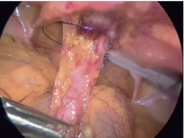
thick-ened urachal duct. We discharged the patient with an-tibiotic and antiinflammatory therapy. After two weeks we reviewed the patient. The patient was well and we repeat an ultrasound scan that confirmed the presence of an urachal cyst. A voiding cystourethrogram showed no signs of vesico-ureteral reflux and no comunication between the bladder and the cyst. The patient under-gone laparoscopic excision of the cyst. Under general anesthesia the patient was placed in a supine position and a Foley catheter was inserted and removed at the end of the surgical procedure. The peritoneal cavity was accessed using the Hasson open tecnique throught the umilicus and insufflated using CO2 with intra-ab-dominal pressures maintained at 12 mmHg. A 30° tel-escope was used. We inserted under direct vision two other operative 5 mm ports at the left and the right ab-dominal wall. We identified the median, laterals um-bilical ligaments and the cyst. Dissection of median umbilical ligament began below the urachal cyst with a laparosocopic hook and continued just above the bladder dome. We used two ligatures (endoloops) to secure the end of the median umbilical ligament and with excised the speciment that was sent for
histolog-Correspondence to:
Mario Messina
Division of Paediatric Surgery, Dept of Paediatrics, Obstetrics and Reproductive Medicine. University of Siena. Policlinico “Le Scotte”.
Viale Bracci 16, 53100 Siena. Italy. E-mail: [email protected]
U
RACHAL CYST:
AN UNSPECTED COMPLICATIONAngotti R., Giannotti G., Ferrara F., Varetti C., Bindi E., Di Maggio G., Messina M.
Division of Paediatric Surgery, Dept of Paediatrics, Obstetrics and Reproductive MedicineUniversity of Siena, Policlinico “Le Scotte”, Siena, Italy
Abstract. The urachus is the remnant of the allantois, which usually becomes obliterated shortly after birth. Urachal remnants
due to an incomplete obliteration of different portion of the urachus are rare, but they need to be treated surgically because of their potential for infectious complications and malignant degeneration. We present a case report with an unespected post-operative complication. M.E., a 10 years old boy, came to the Accident and Emergency Department for an acute abdominal pain, without other symptoms, twice in one year. The blood tests, urine sample and voiding cystourethrogram were normal. The ultrasound scan showed a thickened urachal duct. After antibiotic and anti-inflammatory therapy for two weeks, we performed laparoscopic surgery. In the second postoperative day the patient showed abdominal pain and hematuria. An ul-trasound scan and a voiding cystourethrogram showed a leak from the dome of bladder. We performed an open surgery to close the defect on the bladder’s dome. The patient was discharged in 10th postoperative day. Now he is healthy. Clinically manifest persistent urachal anomalies are rare, but they carry a risk of recurrent infection and subsequent malignant degen-eration. For these reasons the radical excision of the remnant is suggested. Today, due to the large laparoscopic experience, all the reports showed that this technique can be used safely, but we have to pay attention to all steps of the procedure. This case is a paradigmatic situation and it illustrates the importance of a meticulous technique during the excision of urachal remnant. Indeed even if laparoscopic excision could be safe and effective, it is not free of complication.
52
JOURNAL OF THeSIeNAACADeMY OFSCIeNCeS, PUbLISHeD SINCe1761 - VOL. 4 - 2012
ical evaluation. (Figure 1) In the second post-operative day the patient started to complain increased abdomi-nal pain and hematuria. The blood examination were normal and the ultrasound scan showed a lot of free fluids in the peritoneal cavity. A voiding cystourehro-gram showed a leak of contrast in the peritoneal cavity from the dome of the bladder. (Figure 2) At that point we decided to perform an open surgery. Under general anesthesia through a Pfannestiel incision we identified the bladder and we found a 4 cm long defect on the dome. (Figure 3) We closed the defect with a double layers sutures and left the vesical catheter. The patient was discharged 10 days after the second operation without any voiding problems.
DISCUSSION
Clinically manifest persistent urachal anomalies are rare, but they carry a risk of recurrent infection and subsequent malignant degeneration in adulthood (ade-nocarcinoma, urothelial carcinoma or sarcoma). [3] These are the reasons why radical excision of the rem-nant is suggested. In 1993 Trondsen et all (cutting) re-ported the first laparoscopic excision of a urachal remnant. [4] Since then there have been several reports of laparoscopic treatment of various types of urachal remnants in patients of various ages. [3-5] All the re-ports showed that the laparoscopic technique can be used safely and effectively with minimal morbidity. According to Turial et all. the minimally invasive ap-proach is superior cosmetically and visually compared to the traditional lower midline vertical or transverse mid hypogastric incision. [3] We operated with the same tecnique other 6 patients without any intraoper-ative or postoperintraoper-ative complications, but in this case we opened the bladder during the first operation with-out realizing it. Cutting et all were the first in Literature to describe a different position for the ports. [6] They operated 5 cases. In one case they had a recurrence of the problem due to inadequate removal of the residual urachal tissue. Because of that, in the fifth case, they decided to try a different tecnique, positioning all the ports in the lateral part of the abdomen. The lateral view provided by this ports positioning seemed to give them a better perspective on the full extent of the urachal remnant and the bladder. We want to underline another technical point. We removed the urinary catheter at the end of the procedure, but this seems to be premature. The urinary catheter could be essential to protect the sutures or ligatures at the bladder post-operatively and it helps to increase and decrease the filling state of the bladder intraoperatively. [4] As a matter of fact a good visualization of dome of the blad-der intraoperatively, if necessary even filling it with methylene blue, is essential to avoid a major compli-cation as we had.
Figure 1. After dissection of the urachal remnant we secure
the end of the median umbilical ligament with two ligatures (endoloops).
Figure 2. In the second postoperative day a voiding
cys-tourehrogram showed a leak of contrast in the peritoneal cav-ity from the dome of the bladder.
Figure 3. Under general anesthesia through a Pfannestiel
in-cision we identified the bladder and we found a 4 cm long defect on the dome.
53
CLINICAL
CASeS
CONCLUSION
With the description of this case we want to under-line the importance of a meticolous technique during the excision of urachal remnant. The laparoscopic ex-cision could be safe and effective, but it is not free of complication. The patient and the parents have to be awared about every options and every possible com-plications.
ReFeReNCeS
1. Nix JT, Menville JG, Albert M, Wendt DL “Congenital patent ura-chus” J Urol, 79: 264, 1958.
2. Mesrobian HG, Zacharias A, Balcom AH, Cohen RD “Ten years of experience with isolated urachal anomalies in children” J Urol, 158: 1316-1318, 1997.
3. Turial S, Hueckstaedt T, Schier F, Fahlenkamp D “Laparoscopic treatment of urachal remnants in children” J Urol, 177: 1864-1866, 2007.
4. Trondsen E, Reiertsen O, Rosseland AR “Laparoscopic excision of a urachal sinus” Eur J Surg, 159: 127-128, 1993.
5. Khurana S, Borzi PA “Laparoscopic management of complicated urachal disease in children” J Urol, 168: 1526-1528, 2002. 6. Cutting CWM, Hindley RG, Poulsen J “Laparoscopic management