Università degli Studi di Ferrara
DOTTORATO DI RICERCA IN
SCIENZE BIOMEDICHE ENDOCRINOLOGICHE E
NEUROFISIOLOGICHE
CICLO XXI
COORDINATORE Prof. Alessandro Martini
NEW INSIGHTS AND POSSIBLE THERAPEUTIC
IMPLICATIONS OF ADENOSINE ANALOGS AND
PULSED ELECTROMAGNETIC FIELDS (PEMFs) IN
OSTEOARTICULAR PATHOLOGIES
Settore Scientifico Disciplinare BIO/17
Dottorando
Tutore
Dott. Federica Francesca Masieri
Prof. Angelo Caruso
INDEX
ABBREVIATIONS
Pag. 1
I
INTRODUCTION
Pag. 2
I.1
THE JOINT STRUCTURE AND THE SYNOVIAL FIBROBLAST (SF)
ROLE IN THE HEALTHY ARTICULAR ENVIRONMENT
I.2
OSTEOARTHRITIS, A COMPLEX ARTICULAR PATHOLOGY: MAIN
CLINICAL CHARACTERISTICS, RISK FACTORS AND POSSIBLE
GENETIC IMPLICATIONS
I.3
INFLAMMATION IN OA: SFs AND CHODROCYTES ROLE
I.4
ADENOSINE: A NATURAL, PONTENT ANTIINFLAMMATORY
MEDIATOR
I.5
PULSED ELECTROMAGNETIC FIELDS (PEMFs) AS AN USEFUL
ANTIINFLAMMATORY
BIOPHYSICAL
STIMULUS
IN
OSTEOARTICULAR PATHOLOGIES
II
MATERIALS AND METHODS
Pag. 20
II.1
SF CULTURES
II.2
IMMUNOFLUORESCENCE STAINING
II.3
RT-PCR
II.4
WESTERN BLOTTING OF ADENOSINE RECEPTORS
II.5
CHARACTERISTICS OF PEMFs
II.6
SATURATION
BINDING
EXPERIMENTS
TO
ADENOSINE
II.7
MEASUREMENT OF cAMP LEVELS IN BOVINECHONDROCYTES OR
FIBROBLAST-LIKE SYNOVIOCYTES
II.8
SF TREATMENTS WITH ADENOSINE AGONISTS AND EMF
EXPOSURE
II.8.(a)
BOVINE SF TREATMENTSII.8.(b)
HUMAN SF TREATMENTSII.9
PGE-2 AND IL-6 ASSAY
II.10
MTT ASSAY
II.11
STATISTICAL ANALYSIS
III.
AIM OF THE STUDY
Pag. 31
IV
RESULTS
Pag. 33
IV.1
PHENOTYPE CHARACTERIZATION OF BOVINE AND HUMAN SFs
IV.2
WESTERN BLOTTING OF ADENOSINE RECEPTOR SUBTYPES IN
BOVINE AND HUMAN SFs
IV.3
SATURATION BINDING EXPERIMENTS IN BOVINE AND HUMAN
SFs IN THE PRESENCE AND IN THE ABSENCE OF PEMFs
IV.4
cAMP ASSAYS IN BOVINE AND HUMAN SFs IN THE PRESENCE
AND IN THE ABSENCE OF PEMFs
IV.5
FUNCTIONAL ACTIVITY OF ADENOSINE ANALOGS AND PEMFs
ON THE RELEASE OF INFLAMMATORY MEDIATORS IN BOVINE
SFs
IV.5(a)
DOSE DEPENDENT EFFECTS OF TNF-α AND IL-1β TREATMENT ON PGE-2 RELEASE IN BOVINE SFsIV.5(b)
ADENOSINE AGONISTS AND PEMF EXPOSURE INHIBIT PGE-2 RELEASE IN BOVINE TNF-α TREATED SFs IN THE PRESENCE OF ENDOGENOUS ADENOSINEIV.5(c)
DEPLETION OF ENDOGENOUS ADENOSINE WITH ADA INCREASES BASAL PGE-2 RELEASE IN SFsIV.5(d)
DEPLETION OF ENDOGENOUS ADENOSINE WITH ADA POTENTIATES ADENOSINE AGONISTS’ EFFECTS BUT LIMITS EMF-INHIBITORY EFFECTS ON PGE-2 RELEASE IN TNF-α TREATED SFsIV.5(e)
DEPLETION OF ENDOGENOUS ADENOSINE WITH ADA DID NOT MODIFY CELL VIABILITYIV.6
CHANGES IN COX-2 EXPRESSION ARE ASSOCIATED TO THE
CHANGES IN PGE-2 RELEASE INDUCED BY ADENOSINE AGONISTS
AND PEMFs EXPOSURE IN TNF-α TREATED BOVINE SFs
IV.7
cAMP ROLE IN THE MODULATION OF PGE-2 RELEASE IN TNF-α
STIMULATED BOVINE SFs
IV.8
IL-1β INDUCES A DOSE RESPONSE INCREASE ON PGE-2 AND IL-6
RELEASE IN HUMAN SFs
IV.9
PEMFs ROLE IN THE MODULATION OF PGE-2 AND IL-6 RELEASE
IN IL-1β STIMULATED hSFs
V.
DISCUSSION
Pag.56
VI
CONCLUSIONS
Pag.64
ABBREVIATIONS
Pathologies: OA: osteoarthritis RA: rheumatoid arthritis
Drugs:
NSAIDs: nonsteroidal anti-inflammatory drugs
Cell types:
SFs: Synovial fibroblasts (sometimes referred also as SF) bSFs: bovine synovial fibroblasts
hSFs: human synovial fibroblasts
Inflammation and matrix degradation related molecules or genes: IL-1β: Interleukin-1β
TNF-α: Tumor Necrosis Factor-α IL-6: Interleukin-6
PGE-2: Prostaglandin E-2 COX-2: Cyclooxygenase-2 NO: Nitric Oxide
MMP(s): Metalloproteinase(s)
Adenosine analogs and related compounds: CHA: N6-cyclohexyladenosine
NECA: 5’-N-ethylcarboxamidoadenosine
CGS 21680: 2-[p-(2-carboxyethyl)-phenetyl-amino]-5’-N-ethylcarboxamidoadenosine Cl-IB-MECA: N6-(3-iodobenzyl)2-chloroadenosine-5’-N-methyluronamide
ADA: Adenosine deaminase
Methodologies/pulsed electromagnetic fields exposure: ELISA: enzyme-linked immuno-sorbent assay
I
INTRODUCTION
I.1 THE JOINT STRUCTURE AND THE SYNOVIAL FIBROBLAST (SF) ROLE IN THE HEALTHY ARTICULAR ENVIRONMENT
The main role of the skeletal apparatus is to sustain the body, moreover this feature is accompanied to the movement ability. The joints are present where two bones take contact. The contact can be direct or mediated by fibrous tissue, cartilage or synovial fluid. Each joint in the human body is specific for a particular movement and a wide series of specialized structures such as bone surfaces, cartilages, ligaments, tendons and muscles contribute to the final movement.
The joints are functionally classified on the basis of their ability to move: immobile joints are named synarthrosis, partially mobile joints are the amphiarthrosis and a completely mobile joints is named diarthrosis.
The mobile joints, or diarthrosis, are named also synovial joints and allow a great range of movement; they are normally present at the long bones extremity. The elbow, the ankle, the knee, the rib, the wrist, the shoulder and the hip joints are typical examples of diarthrosis (Martini et al. 2004).
Normally the bone extremities present in the joint are covered by the articular cartilage. The articular cartilage main functions are to adsorb the shocks and to reduce the friction.
Figure 1: Synovial joint typical structure. As an example is reported a knee joint. The figure is from: www.nytimes.com/.../adam/19698Synovialfluid.html.
The synovial joint is surrounded by an articular capsule composed by dense connective tissue. The articular cavity is covered by a synovial membrane composed by loose connective tissue and protrudes into the articular space between the two articular cartilages, as represented in Figure 2 (A).
The synovial thin layer histological features are shown in Figure 2 (B): the epithelium is cuboidal to low cuboidal and covers a loose connective tissue containing a rich plexus of small blood vessels.
Figure 2: Synovial membrane histological representation (A). Under the cuboidal epithelium, the loose connective tissue contains numerous blood vessels (B). These images are from: neuromedia.neurobio.ucla.edu/campbell/bone/wp.html.
This specialized structure produces the synovial fluid fuelling the articular cavity. Synovial fluid has principally three functions (Martini et al. 2004):
-Lubrication: when the articular cartilage is compressed the thin layer of synovial fluid reduces the frictions between the articular surfaces. The articular cartilage acts as a sponge: when the movement is finished, the fluid return in the previous position.
-Chondrocytes nourishment: in a synovial joint 3 ml of synovial fluid are normally present. This relatively poor amount of liquid is continuously recycled to eliminate the catabolytes produced from the articular chondrocytes and enriched of nutrients. The synovial fluid circulates during the articular movement; in fact the articular cartilage compression and re-expansion pump the synovial fluid inside and outside of cartilage matrix.
-Amortization: the synovial fluid adsorbs articular trauma. To give an example, the hip, the knee and the ankle are very compressed during the walk and above all during the run. The pressure increase generated during the movement is damped thanks to the synovial fluid, which redistributes uniformly the pressure on the articular surface.
There are principally three types of synovial cells: macrophages, fibroblasts and dendritic cells (Burmester at al. 1983). Synovial fibroblasts (SFs) are the most important cells present in the synovial layer and play key roles in normal embryogenesis and mature joint functions. During development, it is the SF that forms the main element of the embryonic joint tissue and that, in response to hyaluronic acid and other signals, begins to define the joint space and capsule (Abeles et al 2006). In the healthy adult joint, the SF performs several functions. As the primary stromal cell of the joint, the SF appears responsible for the production of collagen and other connective tissue molecules that form and maintain the joint capsule. The SF is also the main cell involved in the secretion of hyaluronic acid and other molecules into the joint space itself, thus providing lubrication to the joint surface as well as signalling functions to the joint tissues. Healthy SF are also likely to secrete controlled amounts of enzymes, such as matrix metalloproteinases (MMPs), that have the ability to digest connective tissue and presumably maintain the structure and flexibility of the joint capsule through remodelling.(Mor et al. 2005).
I.2 OSTEOARTHRITIS, A COMPLEX ARTICULAR PATHOLOGY: MAIN CLINICAL CHARACTERISTICS, RISK FACTORS AND POSSIBLE GENETIC IMPLICATIONS
The healthy joint requires a fine-tuned balance between molecular signals that regulate homeostasis, damage, restoration, and remodelling of the tissues. Homeostatic processes ensure the adaptive maintenance of tissue integrity and function (Lories 2008). The balance is determined at the level of the individual cells, of the tissue architecture, and of the interactions between different tissues in the joint organ, notably articular cartilage, synovium, and bone.
Osteoarthritis (OA) is a severe progressive articular disease and represents the most widespread cause of physical morbidity and impaired quality of life of a rapidly growing number of patients all over the world. The OA disease process is representative of a seriously imbalanced conditions of the articular components: in fact OA affects not only the cartilage but also the entire joint structure, including the synovial membrane, bone, ligaments and periarticular muscles.
A majority of individuals over the age of 65 have radiographic and/or clinical evidence of osteoarthritis. The most frequently affected sites are the hands, knees, hips, and spine. Importantly, the symptoms are often associated with significant functional impairment, as well as signs and symptoms of inflammation, including pain, stiffness, and loss of mobility (Felson 2006).
By a clinical point of view, the OA cartilage lesions are divided into four different categories on the basis of the “Outerbrige parameters” (e.g. dimension, depth and erosions scores): type I-II lesions are typical of early OA stages and are normally self-replaced with firbocartilagineous tissue; on the other hand type III-IV lesions have not spontaneous healing and often drive to a complete articular surface degeneration (Buckwalter et al. 1990). Further, a typical characteristic in OA is the presence of synovial pannus, which is an aberrant synovial membrane proliferation invading the articular cavity. In a recent study, OA and rheumatoid arthritis (RA) pannus characteristics have been identified and it has been established that RA and OA pannus have similar histological and cytological features, as well as an analogous pro-inflammatory cytokine profile expression (Furuzawa-Carballeda et al. 2008).
Although the real pathophysiological events originating OA have not yet been established, several risk factors in driving the pathology progression have been identified: aging, obesity and female gender, are often referred as the main risk factors contributing to OA outcomes (Englund and Lohmander 2004).
Also structural conditions, not necessarily connected to the patient age, have been widely revised in the onset and/or in the OA progression, and in particular meniscus lesions, subchondral bone oedema, articular misalignment driving to abnormal mechanical charge on the joint and synovitis (Lane et al. 1993; Sharma et al. 1999; Felson et al. 2004; Brandt et al. 2006; Martel-Pelletier at al. 2006).
Finally, although genetic factors that may cause OA are not yet fully understood, there are several lines of evidence indicating that genetic abnormalities can result in earlier onset of OA (Valdes et al. 2006). Candidate gene studies and genome-wide linkage analyses have revealed polymorphisms or mutations in genes encoding ECM (e.g.: Type II and IX collagen, aggrecan) and signalling molecules (e.g.: IL-1 gene cluster, COX-2) that may determine susceptibility to OA (Loughlin 2005; Valdes et al. 2007).
All these possible risk factors are thought to participate in creating the most important OA features: the cartilage alterations, due to the impaired chondrocytes functions, and the general status of inflammation, driven also by the altered behaviour of synovial fibroblasts.
While many disease modifying therapies are available for the more aggressive and inflammatory arthritis syndromes, such as rheumatoid arthritis, specialists treating OA are limited to taking care of patients symptomatically and supportively, with little ability to alter its disease course; the only solution for the patient is often a total joint replacement surgery (Samuels et al.2008).
In last past two decades, several techniques for articular cartilage lesion treatment have been used: drilling, abrasions and microfracture are referred as repairing techniques; osteochondral massive transplantation, mosaicoplastic, perichondrium or periostium transplantation and autologous chondrocytes transplantation are reported as regenerating techniques (Marcacci et al. 2003).
Historically, medical therapy of OA has been directed to the treatment of signs and symptoms, mainly with the simple use of analgesics, such as acetaminophen, and the nonsteroidal anti-inflammatory drugs (NSAIDs). NSAIDs, including the COX-2 selective agents, are among the most widely prescribed drugs worldwide, but gastrointestinal, cardiovascular and renal effects have limited their use in many patients. Adjunctive therapies have included intra-articular injections of corticosteroids and hyaluronans, which provide symptomatic benefit in selected patients (Abramson
I.3 INFLAMMATION IN OA: SFs AND CHODROCYTES ROLE
As previously cited, the OA pathophysiology involves not only the breakdown of articular cartilage, but changes in the bone and synovium as well. These changes are believed to be related to a complex network of biochemical pathways, which implicate the diffusion of catabolic factors and cytokines between the different joint tissues to the cartilage. There is evidence that the molecular cross-talk between the above tissues is an integral element of the disease pathogenesis (Martel-Pelletier et al. 2006.; (Martel-Pelletier et al. 2006). Figure 3 illustrates the complex pathways involved in OA development.
Figure 3: Molecular pathogenesis of osteoarthritis. Potential biomarkers and targets for disease modification are released as a result of events in cartilage, bone, and synovium. Image adapted from: Samuels et al. Osteoarthritis: a tale of three tissues. Bull NYU Hosp Jt Dis. 2008;66: 245.
It is now thought that much of the cytokine expression is initially derived by the synovium, and predominantly from the synovial macrophages which drive the inflammatory and destructive responses in OA (Bondeson et al. 2006). These cytokines are thought to stimulate the chondrocytes and SFs to synthesize further cytokines as well as matrix degrading proteases.
The articular chondrocyte is central to the altered metabolism of the joint, by undergoing to a series of complex changes, including hypertrophy, proliferation, catabolic alterations and apoptosis.
Many of these changes are induced by cytokines, eicosanoids and reactive oxidant species produced by the chondrocytes themselves, which are key protagonists in the autodestruction of articular cartilage (Belcher et al. 1997;Pelletier et al. 2001; Aigner et al. 2002).
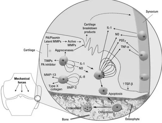
The relationship between the increased levels of catabolic enzymes and inflammatory mediators such as Prostaglandins and Nitric Oxide (NO) and the levels of IL-1β TNF-α in OA synovial fluids and joint tissue is well documented (Goldring and Berenbaum 2004). Although the mechanism by which production of inflammatory mediators by the recruited macrophages in the articular environment is initiated remains unclear; abnormal mechanical and oxidative stresses are probably involved (Goldring and Goldring 2007). Figure 4gives a schematic representation of the imbalance of cytokine network in OA, driving to the cartilage matrix degradation.
IL-1β is synthesized by chondrocytes at concentrations that are capable of inducing the expression of degrading extracellular matrix enzymes, such as metalloproteinases (MMPs) and aggrecanases (ADAMTS), and it colocalizes with TNF-α, MMP-1, MMP-3, MMP-8 and MMP-13, and type II collagen cleavage epitopes in regions of matrix depletion in OA cartilage (Tetlow et al., 2001; Wu et al., 2002). In particular the main MMP involved in OA is MMP-13 thanks to its ability to more effectively degrade type II collagen (Pelletier et al. 2007). In addition to inducing the synthesis of MMPs and other proteinases by chondrocytes, IL-1β and TNF-α increase the synthesis of prostaglandin E2 (PGE-2) by stimulating the expression or activity of cyclooxygenase-2 (COX-2); further they up-regulate the production of NO via inducible nitric oxide synthetase. IL-1β also induces other proinflammatory cytokines such as IL-6 and chemokines, including IL-8, and suppresses the expression of a number of genes associated with the differentiated chondrocyte phenotype (Goldring and Berenbaum 2004; Goldring and Goldring 2004).
Figure 4:The cytokine imbalance in osteoarthritis.
IL-1β TNF-α IL-17 IL-18 LIF OSM IL-6 IL-8 sTNF-R IL-4 IL-11 IL-10 IL-13 IL-1ra Cartilage Matrix degradation Cartilage Matrix synthesis IGF-1 BMPs PGE-2
Although there remains debate regarding the essential role of synovial inflammation in OA, synovitis involving infiltration of activated B cells and T lymphocytes and overexpression of pro-inflammatory mediators is common in early and late OA (Benito et al. 2005). Indeed chondrocytes are thought to be the main cells in OA pathogenesis, however several evidences indicate that also SFs play an important role in driving OA by an increased proliferation and the secretion of a wide range of pro-inflammatory mediators, including cytokines, growth factors, and lipid mediators of inflammation.
Similar activities are displayed by SFs in RA, a pathology in which they play a central role in driving the articular inflammation (Moulton 1996; Müller-Ladner et al. 2005; Abeles and Phillinger 2006; Christodoulou and Choy 2006).
The inflammatory activities of SFs are thought to contribute in deregulating chondrocyte functions, favouring an imbalance between catabolic and anabolic activities (Loeser 2006). Figure 5 highlights the inflammatory role of SFs and the complex cytokine interaction that contribute to OA pathogenesis.
Figure 5:Inflammatory role driven by SFs.
Neutrophils Osteoclasts Osteoblasts TNF-αααα IL-1 Osteobl Osteocl
Bone
Bone
Cartilage
Chondrocytes IL-8 PGE2 IL-6SFs
Pannus
Capsule
Endothelial
venule
Among the inflammatory factors released by SFs, the pro-inflammatory cytokine IL-1β plays a central role in OA pathophysiology and aetiology (van den Berg et al. 1999; Martel- Pelletier et al. 1999; Goldring and Goldring 2004; Martel-Pelletier et al. 2006).
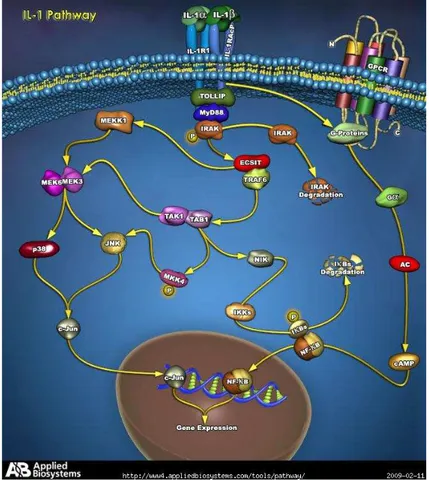
Interleukin-1β, the major isoform produced in human tissues, is synthesized as 31-kD pro-IL-1β that is devoid of signal sequence and is released as the 17.5-kD active form after cleavage by IL-1β converting enzyme (ICE, or caspase-1) (Auron 1998). IL-1β activity is mediated by its binding only to the type I IL-1 receptor (IL1-RI) belonging to the Toll-like receptor family, with the induction of multiple phosphorylation-dependent signalling pathways that regulate gene expression.
These pathways include the serine-threonine kinases of the MAP kinase family and NF-kB cascades (Pope and Tschopp 2007). In Figure 6, is represented the classical IL-1 pathway.
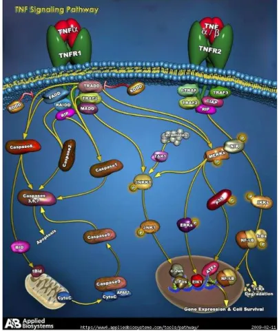
Another crucial cytokine released by SFs is TNF-α, which is synthesized as an active precursor that is cleaved by the TNF-converting enzyme (TACE), a member of the ADAM family. The proteolytic cleavage of the extracellular domains of the TNF receptors results in generation of the soluble receptors, sTNF-R55 and sTNF-R75. Both are produced spontaneously by OA SFs and chondrocytes, but sTNF-R75 is released at higher levels by these cells. Further sTNF-R75 is localized in cells at sites of focal loss of proteoglycans in OA cartilage (Goldring 1999; Martel-Pelletier 1999 and 2006). TNF-signal transduction pathways are complex and still not fully understood. Regulation of the transcription factor NF-κB is a key component of TNF-α signal transduction, but p38 MAPK and JNK are also involved (Bradley 2008), as represented in Figure 7.
Figure 7: TNF-α pathway. Image from: www.ambion.com/tools/pathway/pathway.php?path.
TNF-α contributes to the articular disease pathophysiology by stimulating its own production and inducing chondrocytes to produce additional inflammatory mediators, such as NO, and a variety of eicosanoids, in particular prostaglandin E-2 (PGE-2) and leukotriene B4 (LTB4) (van de Loo et al.
1995). Worth of note, in cultured OA SFs, TNF-α can stimulate a great series of
inflammation-related molecules (e.g.: IL-1β, IL-6, IL-8, GMCSF, IL-10, IFN-γ, PGE-2) and induce the release of some MMPs such as MMP-1 and MMP-13 (Castor et al. 1997; Goldring 2000; Fuchs et al. 2004).
In addition, TNF-α can synergize with other inflammatory molecules, such as IL-8, and potentiate TNF-α induced PGE-2 production in OA SFs (Alaaeddine at al. 1999).
Prostaglandines (PGs) are members of the eicosanoid family (oxygenated C20 fatty acids) and are produced by nearly all cells within the body (Smith 1989). Prostaglandins are lipid mediators that are not stored by cells; rather, they are synthesized from arachidonic acid via the actions of cyclooxygenase (COX) enzymes, either constitutively or in response to cell-specific trauma, stimuli, or signalling molecules (Smith 1989; Berembaum 2000; Funk 2001). The most abundant prostanoid in the human body is PGE-2 (Serhan and Levy 2003).
In Figure 8 is schematized the PGE-2 production pathway. PGE-2 is synthesized from arachidonic acid, a polyunsaturated fatty acid derived from dietary sources that resides in the cell membrane. In stimulated inflamed cells, the constitutively present in the cytosol phospholipase A2α enzyme
(cPLA2α) translocates to the nuclear membrane, where it enzymatically releases arachidonic acid.
Inflammatory stimuli also induce the transcription and protein expression of both cyclooxygenase-2 (COX-2) and microsomal prostaglandin Esynthase-1 (mPGES-1) enzymes at the nuclear membrane and endoplasmic reticulum. COX-2 transforms arachidonic acid to PGG-2 which is subsequently converted to PGH-2. mPGES-1 may then act on PGH-2 to generate PGE-2. PGE-2 may exit the cell by simple diffusion, or by active transport (Park et al. 2006).
PGE-2 is a key mediator of inflammation and pain in both OA (Hardy et al. 2002; Martel-Pelletier
et al. 2006) and RA (Bomardier et al. 1981).
Several studies in OA SFs show that PGE-2 plays a central role among the others pro-inflammatory molecules which are involved in the inflammatory pathway. In fact, in SFs cultured in the presence of IL-1β, PGE-2 acts as a modulator of related-inflammation activities such as the proliferation of inflammatory cells, and regulates the release of other cytokines, in particular IL-6 (Inoue et al. 2001 and 2002). In addition, in OA SFs cultured in the presence of TNF-α, PGE-2 release is modulated also by anti-inflammatory cytokines such as IL-10 (Alaaeddine et al. 1999). Further, in human OA synovial tissue explants, eicosanoinds and particularly PGE-2 seem to strictly regulate both IL-1β and TNF-α release (He at al. 2002).
All the complex cytokine network taking part in OA pathophysiology, and all molecules produced by SFs have an effect on the release of the same molecules or of other inflammatory-related molecules and matrix degradation enzymes by articular chondrocytes. The cross-talk between cartilage and synovium, and between these tissue and the underlying bone (Bertolini et al. 1986; Abramson and Yazici 2006) contributes to maintain and auto-induce the inflammatory status typically present in OA.
I.4 ADENOSINE: A NATURAL, PONTENT ANTIINFLAMMATORY MEDIATOR
The purine nucleoside adenosine is present in almost all tissue and cells and its formation is closely related to the energy consumption of the cell. Adenosine is produced from adenosine 5’-monophosphate (AMP) by the action of 5’nucleotidase enzyme and is metabolized by deamination or phosphorylation, via adenosine deaminase (ADA) and adenosine kinase (AK), respectively (Schulte and Fredholm 2003). The molecular structure of adenosine is represented in Figure 9. Adenosine acts as a potent endogenous inhibitor of inflammatory processes in several tissue and cells. To cite few, adenosine has been demonstrated to regulate mast cell-degranulation (Linden
1994; Marquardt 1998), to diminish TNF-α and IL-6 expression in human macrophages and rat
cardiomyocytes (Bouma et al. 1994; Wagner et al. 1999), to modulate the release of cAMP and superoxide anion products in human neutrophils and HL-60 cell line ( Varani et al. 2003-A; Gessi
et al. 2002 and 2004).
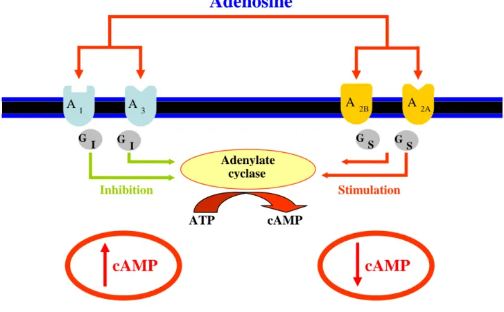
Adenosine can interact with four G-coupled receptor subtypes: A1, A2A, A2B and A3. Classically G protein are defined as etherotrimeric proteins with three subunits: α, β and γ. The first discovered and more widely studied adenosine/G protein pathway involves the adenylate cyclase modulation. Inactive α subunit of G proteins is linked to GDP. When adenosine binds to a specific receptor subtype, GDP is converted to GTP and β-γ complex dissociates from α-GTP complex which can exert its interaction with adenylate cyclase enzyme. α subunit is also able to idrolize GTP to GDP, thanks to its intrinsic GTPase activity, which induces its reunion to β-γ complex.
Figure 10 summarizes the different adenosine receptor coupling to the adenylate cyclase enzyme. A2A and A2B receptors are positively coupled to adenylate cyclase via Gs stimulatory proteins, which lead to an increase of cAMP second messenger formation. On the contrary, A1 and A3 receptor are negatively coupled to adenylate cyclase via Gi inhibitory proteins, leading to a decrease in cAMP levels (Fredholm et al. 2001).
Figure 10:Adenosine receptor coupling to the classic adenylate cyclase pathway.
Adenosine and its receptors have been recently involved in the regulation of inflammatory processes related to OA.
Specifically, in equine chondrocytes cultured in vitro and stimulated with IL-1 or lypopolisaccharide (LPS) some authors have shown that the activation of A2A adenosine receptors by specific agonists, such as NECA, can inhibit inflammatory activities. In particular both endogenous adenosine and A2 receptors have been implicated in the decrease of inflammation-induced NO release (Benton et al. 2002; Tesch et al. 2002). Further, in cartilage explants Tesch and collaborators have demonstrated that adenosine pathway can modulate cartilage homeostasis. In fact, PGE-2, NO and glicosaminoglycan release were increased in cultured explants exposed to ADA, which was used to deplete endogenous adenosine.
A 1 A 3 A 2B A 2A G I G I G S G S Adenylate cyclase
Adenosine
Inhibition Stimulation ATPcAMP
cAMP
cAMPFurther they showed that DPMA, a selective A2A receptor adenosine analog, inhibited the release of these molecules, indicating the involvement of A2A receptor in anti-inflammatory activities (Tesch et al. 2004). Similar results, in the same in vitro model, were obtained by using ITU, a specific adenosine kinase inhibitor, which inhibits adenosine degradation, in the presence of pro-inflammatory stimuli. In particular, ITU was able to inhibit pro-pro-inflammatory-induced PGE-2, NO and glicosaminoglycan release (Petrov et al. 2005).
Moreover adenosine pathway involvement has been demonstrated in several in vivo models. In a rat adjuvant-induced arthritis (AIA) model, spinal injection with CHA, a specific A1 adenosine receptor analog, significantly decreased inflammatory parameters and the impairment of cartilage and bone (Boyle et al. 2002). In a septic arthrosis model created by Staphilococcus aureus injection in rabbit knee, the treatment with ATL146e, a specific A2A adenosine receptor agonist, induced a diminished loss of cartilage, synovial inflammation and white blood cell infiltration with respect to arthrosic controls animals (Cohen et al. 2004 and 2005). More recently, Fishman and collaborators demonstrated an A3 adenosine receptor involvement in AIA rat model treated with the selective adenosine analog IB-MECA: clinical and pathological score of the disease were found to be reduced with respect to IB-MECA-untreated control animals (Fishman et al. 2006). All of these data indicate that adenosine may protect against arthritis, acting, at least in part, by modulating chondrocytes activity.
On the contrary, very few data are reported in literature about the adenosine role in SFs, although some studies suggest that adenosine may target SFs too. In fact the adenosine A2 receptor agonist NECA was found to reduce MMPs stimulation in cultured SFs in the presence of IL-1β (Boyle et al. 1996). Nakamachi and collaborators performed a study in which ADA activity was analysed in OA and RA synovial fluids. This activity was found to be increased both in OA and RA, suggesting a link among the adenosine pathway and these pathologies (Nakamachi et al. 2003). In a recent comparative study, a novel A3 adenosine receptor agonist named CF502 was found to reduce clinical manifestations in AIA rat models and regulate signalling inflammation pathway in cultured RA SFs (Ochaion et al. 2008).
On the basis of the above observations and of the role of SFs to elicit and to maintain joint inflammation, one of the purpose of this study was to characterize by a pharmacological point of view adenosine receptor subtypes in SFs and to investigate their potential anti-inflammatory activity.
I.5 PULSED ELECTROMAGNETIC FIELDS (PEMFs) AS AN USEFUL ANTIINFLAMMATORY BIOPHYSICAL STIMULUS IN OSTEOARTICULAR PATHOLOGIES
Biophysical stimulation with low-energy and low-frequency pulsed electromagnetic fields (PEMFs) has been documented displaying several positive effects in different tissue and cell types.
Some studies suggest an anti-inflammatory role of PEMF-stimulation in several animal and human cell models. In particular, PEMFs inhibit TNF-α release in peripheral blood human cells (Jonai et
al. 1996) and reduce IL-1β and IL-6 levels by inducing a simultaneous increase of
anti-inflammatory cytokines such as IL-4 and IL-2 (Chuian et al. 2005). Further PEMFs were able to down-regulate iNOS production and expression in human monocytes (Reale et al. 2006), to stimulate angiogenesis contributing to the healing wound (Tepper et al. 2004) and to inhibit the inflammatory process and reduce chemokine release, favouring cell proliferation in keratinocytes and inhibiting the inflammatory process (Vianale et al. 2008).
Several previous studies have been focused on the effects of PEMFs in articular cells, mainly chondrocytes and osteoblasts. In vivo studies in rat have shown that PEMFs can stimulate chondrogenesis and endochondral ossification (Aaron et al. 2002) and regulate chondrocyte differentiation and expression of matrix proteins (Ciombor et al. 2002; Bobacz et al. 2006). In addition, the results of in vitro studies have identified several cellular effects mediated by PEMFs. In particular, it has been reported that PEMFs can stimulate cell proliferation and DNA synthesis in human articular chondrocytes from OA patients (Pezzetti et a. 1999; De Mattei et al. 2001).
Further, PEMF effects were studied also in bovine articular cartilage explants cultured in vitro showing that, in the presence of IL-1β, PEMF stimulation induced a significant increase in proteoglycan synthesis, thus counteracting the cytokine catabolic effect activity (De Matttei et al 2003). Moreover PEMFs were able to act in concert with IGF-I in stimulating proteoglycan synthesis, mediating their effect through cell-matrix interaction (De Mattei et al. 2004). Recently, De Mattei and collaborators have also investigated the effects of PEMFs with different amplitude, frequency and length signal, identifying the fields characteristics which are able to exert the maximal chondroprotective effect (De Mattei et al. 2007).
In addition, chondroprotective activities of PEMFs have been demonstrated in vivo in Dunkin Hartley Guinea pig animal model, that bears morphological, biochemical and immunohistochemical similarities to human OA. PEMF exposure preserved cell morphology and decreased matrix degradation enzyme levels in this animal model (Ciombor et al. 2003).
Further studies on this model were performed by Fini and collaborators showing that PEMF stimulation was able to exert a chondroprotective effect also in the presence of severe cartilage lesions, improving histological and histochemical scores and slowing the progression of OA versus untreated control animals (Fini et al. 2005 and 2008). Moreover, PEMF treatment was able to favour the integration of osteochondral autograft implants in sheep. Interestingly this was associated to a decrease observed in IL-1β and TNF-α levels in synovial fluid of PEMF-treated sheep compared to untreated controls, indicating a role for PEMFs in reducing inflammatory cytokines and create a suitable articular environment for the implant grafts (Benazzo et al. 2008).
PEMF stimulation is still under investigation for use in patients with OA (Schnitzer 2002). However, even if different physical parameters and exposure times of stimulation were used, positive results were obtained in several clinical studies. In a double-blind randomized clinical trial, patients with primary knee OA were evaluated at different time points for pain level, joint motion and tenderness. The actively treated group with PEMFs showed a significant improvement for each variable at each experimental point (Trock et al. 1994). Another research group performed a randomized, double-blind, placebo-controlled study on the efficacy of PEMF stimulation in patients with symptomatic knee OA. At 6 weeks, follow-up observation in patients showed a significant improvement in the treated group with regards to pain and disability (Pipitone and Scott 2001). Also other clinical studies showed that PEMF stimulation was safe, reduced impairment activities of daily life and improved knee function in patients with chronic knee pain due to OA (Jacobson et al. 2001; Nicolakis at al. 2002). In a more recent pilot, randomized, prospective and double-blind study, PEMF effect was evaluated in patients undergoing arthroscopic treatment of knee cartilage. PEMF exposure improved significantly clinical scores in patients, also after a follow-up of 3 years and the percentage of patients who used NSAIDs between 45 and 90 day after the surgery was significantly reduced in PEMF-exposed patient group versus the unexposed control group (Zorzi et al. 2007).
Altogether results of previous in vitro and in vivo studies indicate that PEMF can stimulate anabolic activities in cartilage, suggesting that they may exert a potential anti-inflammatory role.
In addition, previous studies performed in human neutrophils have demonstrated that PEMFs can evoke a specific up-regulation of A2A and A3 adenosine receptors. This suggests that biophysical stimulation may have anti-inflammatory activities mediated through specific adenosine receptor subtypes (Varani et al. 2002 and 2003-A).
On the above observations, in this study we aimed to investigate a possible functional anti-inflammatory role of PEMFs in modulating anti-inflammatory events in SFs and to elucidate if exists a possible connection between adenosine pathway and the PEMF action way.
II
MATERIALS AND METHODS
II.1 SF CULTURES
Bovine SFs were obtained by culture of the bovine synovial fluid, aspirated from metacarpo-phalangeal joints (Figure 11) of 14-18-month-old animals (Limousine breed), as previously described (Stebulis et al. 2005). Briefly, synovial fluid was aspirated from the metacarpo-phalangeal joints by a syringe. Then, fresh synovial fluid was diluted 1:4 with complete medium: Dulbecco’s modified Eagle’s/Ham’s F12 (1:1) medium (DMEM/F12) (Gibco-Invitrogen, Paisley, UK) supplemented with 10% foetal bovine serum (FBS) and antibiotics (penicillin 100 U/ml, streptomycin 0.1 mg/ml) (Gibco-Invitrogen, Paisley, UK) . The obtained suspension was plated in 25 cm2 culture flasks (Falcon, Becton Dikinson and Company, Franklin Lakes, NJ, USA). After 3 hours, medium was removed and fresh complete medium was added to the flasks. Cells were maintained in culture and re-expanded when reaching confluence. Bovine synovial cells at the third and fourth passages were used in all experiments.
Figure 11: Metacarpo-phalangeal bovine joint. The synovial fluid was aspirated by a syringe from
Human SFs were isolated from synovial pannus derived from six OA patients who underwent total hip or knee joint arthroplasty (Figure 12). The selected patients fulfilled the American College of Rheumatology criteria for OA diagnosis. In particular were evaluated some physical parameters (e. g.: joint swelling, tenderness and pain, decreased range of motion in joint, crepitus, pattern of other eventually affected joints) and the X-ray or MRI analysis. The mean age of patients was 67± 9 years (two males and four females). All studies had ethical approval from the local ethic committee and informed consent was obtained from patients when samples were taken.
Figure 12:Synovial pannus isolation during knee arthroplasty. Blue arrow indicates the synovial
pannus.
Synovial pannus was minced with a scalpel; the pieces obtained were abundantly washed in Earle saline solution and antibiotic and then were digested in 0,25% trypsin, 0,02% EDTA solution for at least six hours. The cell suspension obtained was filtered trough a sterile gauze and centrifuged at 160g. The pellet was resuspended in complete medium. The non-adherent cells were discarded after overnight incubation, and the plated cells were rinsed in Earle saline solution and cultured in complete medium in a 5% CO237°C incubator. Human synovial cells at the third passage were used in all experiments.
II.2 IMMUNOFLUORESCENCE STAINING
Both bovine and human SFs were characterized by immunofluorescence staining with vimentin, a marker for mesenchymal cells (Upragarin at al. 2005). SFs were fixed with cold methanol, washed with phosphate-buffered saline (PBS) and incubated with the primary monoclonal antibody (mAb) for the human vimentin (Sigma-Aldrich, Italy) at 1:200 dilution for 1 hours at 37°C. Washed slides were then incubated with a secondary fluorescein isothiocyanate-conjugated goat anti-mouse antibody for 1 hours at 37°C in the dark. Nuclei were stained with the DNA dye, 4’,6-diamidino-2-phenylindole (DAPI) (0.1 mg/ml in PBS ethylene glycol tetraacetic acid (EGTA)) for 10 minutes. Both secondary antibody and DAPI were from Sigma-Aldrich S.r.l. (Milan, Italy). Fluorescence was visualized using the Nikon Eclipse TE 2000-E microscope (Nikon Instruments Spa, Italy) equipped with a digital camera (DXM 1200F).
II.3 RT-PCR
CD14 and COX-2 expression in SF cultures was assayed by RT-PCR. CD14 expression was evaluated to exclude any contamination by macrophages in SF cultures. COX-2 expression was evaluated to investigate adenosine and PEMF role, alone or combined. Total RNA extraction was performed by a commercial kit (RNeasy Kit, Qiagen, Deutschland). RNA conversion to cDNA was performed by the kit SuperscriptTM First-Strand Synthesis System (Invitrogen, USA). Oligonucleotide primers for CD14 were dp5’-CTGGAAGCCGGCG-3’; rp5’-AGCTGAGCAGGAACCTGTGC-3’ and oligonucleotides for glyceraldehydes 3-phosphate dehydrogenase (GAPDH) were dp5’-TGGCATCGTGGAGGGACTTAT-3’; rp5’-GACTTCAACAGCGACACTCAC-3’. Sequences were selected to amplify both human and bovine genome. Oligonucleotides for COX-2 were dp5’-TCCAGATCACATTTGATTGACA-3’; rp5’-TCTTTGACTGTGGGAGGATACA-3’ (Woclawek-Potocka et al. 2005). Oligonucleotides sequences were from separate exons to exclude genomic DNA contaminations. Two microliters of cDNA were amplified by the specific oligonucleotide sets; PCR reactions were performed in a total volume of 25 µl containing 1 U Taq DNA polymerase (Roche Molecular Biochemicals, Indiana, USA), 25 pmol of each primer, 200 µM deoxynucleotide triphosphates (dNTPs) in 1X PCR buffer (10 mM Tris, pH 8.3, 50 mM KCl, 1.5 mM MgCl2).
Cycling parameters were: 1 minute at 94°C; 1 minute at the specific annealing temperature (55°C for CD14, 61°C for GAPDH, 60°C for COX-2); and 1 minute at 72°C. PCR product sizes were 403 bp for CD14, 370 bp for GAPDH and 450 bp for COX-2. Primer sequences, cycling parameters and PCR product sizes are summarized in Table 1. mRNA from human macrophages was used as a positive control for CD14 expression. PCR products were analyzed on 1.5% agarose gel, stained with ethidium bromide and visualized under UV.
PRIMERS
PCR PRODUCT
SIZES
DENATURATION ANNEALING EXTENSION
CD14 dp5’-CTGGAAGCCGGCG-3’ rp5’-AGCTGAGCAGGAACCTGTGC-3’
403bp
1’ at 94°C
1’ at 55°C 1’ at 72°C
GAPDH dp5’-TGGCAT CGTGGAGGGACTTAT-3’ rp5’-GACTTCAACAGCGACACTCAC-3’370bp
1’ at 94°C
1’ at 61°C 1’ at 72°C
COX-2 dp5’-TCCAGATCACATTTGATTGACA-3’ rp5’-TCTTTGACTGTGGGAGGATACA-3’450bp
1’ at 94°C
1’ at 60°C 1’ at 72°C
Table1: PCR cycling conditions and product fragment size (expressed in base pairs: “bp”) related to oligonucleotide primers used in PCR reactions.
II.4 WESTERN BLOTTING OF ADENOSINE RECEPTORS
Bovine and human SFs were harvested and washed twice with ice-cold PBS containing 1 mM sodium orthovanadate, 104 mM 4-(2-aminoethyl)-benzenesulfonyl fluoride, 0.08 mM aprotinin, 2 mM leupeptin, 4 mM bestatin, 1.5 mM pepstatin A, and 1.4 mM E-64. Then cells were lysed in Triton lysis buffer and the protein concentration was determined using bicinchoninic acid (BCA) protein assay kit (Pierce, Illinois, USA). Aliquots of total protein sample (50 µg) were analyzed using antibodies specific for human A1, A2A, A2B and A3 adenosine receptors (1 µg/ml dilution), as previously described (Merighi et al. 2002). β-actin expression was analyzed using a specific antibody for human β-actin (Cell Signaling Technology, Pero, Italy) (1:1000 dilution).
Filters were washed and incubated for 1 hours at room temperature with peroxidase-conjugated secondary antibodies (1:2000 dilution). Specific reactions were revealed with Enhanced Chemiluminescence Western blotting detection reagent (Amersham Biosciences, New York, USA).
II.5 CHARACTERISTICS OF PEMFs
The PEMF generator system used in binding, cAMP assay and in PGE-2 and IL-6 functional assays was the same used in previous studies (I-ONE, Igea, Carpi, Italy) (De Mattei et al. 2003; 2004 and 2007; Varani et al. 2002 and 2003-A). The magnetic field was generated by a pair of circular coils of copper wire placed opposite to each other (Figure 13). The coils were powered by the generator system, which produced the input voltage of pulse.
Figure 13: PEMF generator system furnished by IGEA (Carpi, Italy).
The pulse duration of the signal was 1.1 ms and the repetition rate 75 Hz, yielding a duty cycle of 1/10. The intensity peak of the magnetic field was 1.5 mT and the induced electric field, as detected with a standard coil probe (50 turns, 0.5 cm internal diameter of the coil probe, 0.2 copper diameter), was 0.07 mV/cm. The temperature, continuously monitored by a thermoresistor within the incubator, was constant through the exposure time and exactly maintained during the binding and functional experiments.
II:6 SATURATION BINDING EXPERIMENTS TO ADENOSINE RECEPTORS
All the saturation binding experiment were performed in collaboration with the research group of the Department of Clinical and Experimental Medicine, Pharmacology Unit, University of Ferrara. For membrane preparation, the culture medium was removed, the cells were washed with PBS and scraped off T75 flasks in ice-cold hypotonic buffer (5 mM Tris-HCl, 2 mM ethylendiamine tetraacetic acid (EDTA) pH 7.4). The cell suspension was homogenized by using a Polytron and was centrifuged for 30 minutes at 100,000g. The membrane pellet was resuspended in the same buffer solution used in the binding experiments, incubated with 2 IU/ml of adenosine deaminase for 30 minutes at 37°C and centrifuged for 30 minutes at 100,000g. Finally the suspension was used in saturation and binding experiments. The protein concentration was determined according to a Bio-Rad method with bovine albumin as reference standard (Bradford 1976).
The bovine and human SF membranes were PEMF-treated for the specific incubation times related to the A1, A2A, A2B and A3 binding experiments, as previously described (Varani et al. 2002 and 2003-A). Saturation binding experiments to A1 adenosine receptors were performed according to the method described previously using [3H]-1,3-dipropyl-8-cyclopentyl-xanthine ([3H]-DPCPX, specific activity 120 Ci/mmol; NEN-Perkin Elmer Life and Analytical Sciences, USA) as radioligand (Borea at al. 1994). The membranes derived from PEMFs-treated or untreated bovine or human SFs (100 µg of protein/assay) with 8-10 concentrations of the radioligand [3H]-DPCPX (0.01-20 nM) were incubated in Tris-HCl 50 mM, pH 7.4, for 90 minutes at 4°C. Non-specific binding was determined in the presence of DPCPX 1 µM. Saturation binding experiments to A2A adenosine receptors were performed according to the method described previously using [3 H]-4-(2-[7-amino-2-(2-furyl)[1,2,4] triazolo [2,3-a] [1,3,5] triazin-5-yl-amino]-ethyl ([3H]-ZM 241385, specific activity 27.4 Ci/mmol; American Radiolabeled Chemicals Inc, Saint Louis, MO, USA) as radioligand (Varani et al. 2003-B). The membranes derived from PEMFs-treated or untreated bovine or human SFs (100 µg of protein/assay) were incubated for 60 minutes at 4°C with 8-10 concentrations of the radioligand [3H]-ZM 241385 (0.01-20 nM) and Tris-HCl 50 mM, MgCl2 10 mM, pH 7.4. Non-specific binding was determined in the presence of ZM 241385 1 µM. Saturation binding experiments to A2B adenosine receptors were performed using [3H]-N-benzo [1,3[dioxol-5-yl-2-[5- (2,6-dioxo-1,3-dipropyl-2,3,6,7-tetrahydro-1H-purin-8-yl)- 1-methyl-1H-pyrazol-3-yloxy]-acetamide ([3H]-MRE 2029F20, specific activity 123 Ci/mmol; Amersham International Chemical Laboratories, Buckinghamshire, UK) as radioligand (Gessi et al. 2005).
The membranes obtained as previously described (100 µg of protein/assay) with 8-10 concentrations of [3H]-MRE 2029F20 in the range 0.01-20 nM were incubated in Tris-HCl 50 mM, MgCl2 10 mM, EDTA 1 mM, pH 7.4 at 4°C for 60 minutes. Non-specific binding was determined in the presence of MRE 2029F20 1 µM. Saturation binding experiments to A3 adenosine receptors were performed using [3H]-5N-(4-methoxyphenylcarbamoyl) amino-8-propyl-2-(2-furyl) pyrazolo [4,3-e]-1,2,4-triazolo [1,5-c] pyrimidine ([3H]-MRE 3008F20, specific activity 67 Ci/mmol; Amersham International Chemical Laboratories, Buckinghamshire, UK) as radioligand (Varani et al. 2000). The membranes treated as above mentioned (100 µg of protein/assay) with 8-10 concentrations in the range 0.01-50 nM of [3H]-MRE 3008F20 were incubated in Tris-HCl 50 mM, MgCl2 10 mM, EDTA 1 mM, pH 7.4, at 4°C for 150 minutes. Non-specific binding was determined in the presence of MRE 3008F20 1 µM. In saturation binding experiments, at the end of the incubation time, bound and free radioactivity was separated by filtering the assay mixture through Whatman GF/B glass fibre filters by using a Brandel cell harvester. The filter bound radioactivity was counted by Scintillation Counter Packard Tri Carb 2500 TR with an efficiency of 58%.
II.7 MEASUREMENT OF cAMP LEVELS IN BOVINECHONDROCYTES OR FIBROBLAST-LIKE SYNOVIOCYTES
PEMF-treated or untreated bovine and human SFs (106 cells/ml) were resuspended in 0.5 ml incubation mixture Krebs Ringer phosphate buffer, containing 1 IU/ml adenosine deaminase and 0.5 mM 4-(3-butoxy-4-methoxybenzyl)-2-imidazolidinone (Ro 20-1724) as phosphodiesterase inhibitor and preincubated for 10 minutes in a shaking bath at 37°C. Then the effect of a typical A1 adenosine agonist was studied by using CHA at different concentrations (1 nM-1 µM) that was added to the mixture for a further 10 minutes. To evaluate the effect of a typical A2A adenosine agonist, CGS 21680 was used at different concentrations (1 nM-1 µM) that were added to the mixture for a further 5 minutes. In similar experimental conditions, the effect of N-ethylcarboxamidoadenosine (NECA) an adenosine non-selective agonist was studied. To evaluate the effect of a typical A3 adenosine agonist, forskolin 1 µM and Cl-IB-MECA at different concentrations (0.1 nM-100 nM) were added to the mixture and the incubation continued for a further 5 minutes. The effect of a selective A2A or A3 antagonist such as 7-(2-phenylethyl)-2-furyl)pyrazolo [4,3-e]-1,2,4-triazolo-[1,5-c] pyrimidine (SCH 58261) (1 µM) or MRE 3008F20 (1 µM) on CGS 21680(1 µM) or Cl-IB-MECA(100 nM) was evaluated, respectively.
The cells were also incubated with forskolin (1 µM) and/or Ro 20-1724 (0.5 mM) to evaluate the adenylyl cyclase activity.
In addition, cAMP levels were evaluated in bovine TNF-α-pre-treated and untreated bovine SFs (106 cells/ml), which were resuspended in 0.5 ml incubation mixture Krebs Ringer phosphate buffer, containing 1.0 IU/ml adenosine deaminase and 0.5 mM Ro 20-1724 as phosphodiesterase inhibitor and preincubated for 10 minutes in a shaking bath at 37°C. Then the effects of CHA (A1) and CGS 21680 (A2A) adenosine analogs were evaluated respectively in TNF-α pre-treated cells. CHA (1 nM-1 µM) was added to the mixture for a further 10 minutes. Moreover CGS 21680 (1 nM-1 µM) was added to the mixture and incubated for a further 5 minutes.
The reactions were terminated by the addition of cold 6% trichloroacetic acid (TCA). The TCA suspension was centrifuged at 2000g for 10 minutes at 4°C and the supernatant was extracted four times with water saturated diethyl ether. The final aqueous solution was tested for cAMP levels through a competition protein binding assay by using [3H]-cAMP as radioligand (specific activity 21 Ci/mmol, NEN Research Products, Boston, MA, USA) (Varani et al. 2002). Samples of cAMP standards (0-10 pmol)were added to each test tube containing trizma base 0.1 M, aminophylline 8.0 mM, mercaptoethanol 6.0 mM, pH 7.4 and [3H]-cAMP (at the final concentration of 1 nM). The binding protein, previously prepared from beef adrenals, was added to the samples and incubated at 4°C for 150 minutes. At the end of the incubation time and after the addition of charcoal the samples were centrifuged at 2000g for 10 minutes. The clear supernatant was mixed with 4 ml of Atomlight and counted in a Scintillation Counter Packard Tri Carb 2500 TR.
II.8 SF TREATMENTS WITH ADENOSINE AGONISTS AND EMF EXPOSURE
For the analysis of PGE-2 release, bovine SFs at third-fourth passage were plated at 10,000/cm2 in complete medium in multiwells (Nunc, Denmark, 1.6 cm the diameter of each well) and used after 5 days plating. Further for the analysis of PGE-2 and IL-6 release, human SFs at third passage were plated at 5000/ cm2 in complete medium in multiwells and used after 7 days plating. In preliminary experiments, increasing doses of the recombinant human TNF-α and recombinant human IL-β (both from Preprotech, USA), selected in the range of those used in previous studies, were tested (Fahmi et al. 2001; Burger et al 2003). In the following experiments, TNF-α was used at 10 ng/ml, which elicited maximal PGE-2 increase in bovine SFs in preliminary experiments. Further in human SFs IL-1β was used at 50 ng/ml, which induced maximal PGE-2 and IL-6 release in preliminary experiments. Control cells were incubated in complete medium alone.
II.8(a) BOVINE SF TREATMENTS
In a first series of experiments in bovine SFs, adenosine analogs were added to both control and TNF-α-treated cultures in the presence of endogenous adenosine. The adenosine agonists CHA (A1), CGS 21680 (A2A), 50-N-ethylcarboxamidoadenosine (NECA) (non-selective), and N6-(3-iodobenzyl)2-chloroadenosine-50-N-methyluronamide (Cl-IB-MECA) (A3) were used at 1 µM (Sigma, USA). In a second series of experiments, treatments with adenosine agonists in the presence of TNF-α were performed in complete medium containing 2 IU/ml adenosine deaminase (ADA, Fluka-Sigma-Aldrich, Switzerland) to deplete endogenously released adenosine. Different ADA concentrations (0.5-4 IU/ml) were previously tested on PGE-2 release to evaluate the effects of depleting endogenous adenosine. To investigate the effects of PEMFs on PGE-2 production, bovine SF cultures treated as described above, were exposed to PEMFs during the whole treatment period (24 hours).
In some experiments, 1 µM forskolin (Sigma, USA), a direct activator of adenylate cyclase enzyme, was added to both control and TNF-α-treated cultures, in the absence and the presence of CHA (A1) and CGS 21680 (A2A) adenosine analogs.
At each condition tested, after 24 hours of treatment, medium was removed from the well, stored at -80°C for subsequent determination of PGE-2 and the monolayer protein content was evaluated accordingly to the Lowry methodology (Lowry 1951).
II.8(b) HUMAN SF TREATMENTS
The effects of PEMF exposure were investigated on PGE-2 and IL-6 release in human IL-1β-treated and untreated SFs. The reduced availability of human specimens consented to investigate only PEMF effects on PGE-2 and IL-6 release. After 24 hours of treatment, medium was removed from the well and stored at -80°C for subsequent determination of PGE-2 and IL-6. The monolayer protein content was evaluated accordingly to the Lowry methodology (Lowry 1951).
II.9 PGE-2 AND IL-6 ASSAY
The concentration of PGE-2 was measured using a commercially available competitive enzyme immunoassay according to the manufacturer’s instructions (PGE2 Assay, R&D Systems, Inc., Minneapolis, USA). The minimum detectable dose for this assay kit ranged from 18.2 to 36.8 pg/ml of PGE-2. Samples and standards were assayed in duplicate. PGE-2 production was normalized to the total protein content and expressed as pg PGE-2/µg protein.
The concentration of IL-6 was measured using a commercially available quantitative enzyme immunoassay according to the manufacturer’s instructions (Human IL-6 ELISA kit, Diaclone, USA). The minimum detectable dose for this assay kit was 2 pg/ml of IL-6. Samples and standards were assayed in duplicate. IL-6 production was normalized to the total protein content and expressed as pg IL-6/µg protein.
Figure 14 summarizes the main steps of typical quantitative sandwich ELISA and competitive ELISA tests.
Figure 14: Schematic representation of quantitative and competitive ELISA tests. Coat well with
1st 1°Ab (capture) E E Sandwich ELISA Add Antigen Add 2nd 1° Ab (detection) Add Enzyme Labeled 2° Ab Add Substrate Measure Color Change O D pg of antigen E E E O D pg of antigen Competitive ELISA Add
Standarde & Samples
Add
Enzyme Labeled Antigen
E E E Add Substrate Measure Color Change
II.10 MTT ASSAY
The effects of ADA 2 IU/ml on cell proliferation and viability in bovine SFs treated with TNF-α and adenosine analogs, in the presence and in the absence of PEMFs were evaluated by the MTT assay (Ahmed et al. 2006; Tomita et al. 2006). Briefly, 100 µl of MTT solution (5 mg/ml in PBS) (Sigma-Aldrich, UK) were added to each well and incubated at 37°C for 3 hours. The medium was then discarded and 500 µl of isopropanol/HCl 0.04 N were added to each well for the formazan solubilization. The solution absorbance was measured at 540 nm (Cary-50, UV-Visible Spectrophotometer, Varian).
II.11 STATISTICAL ANALYSIS
A weighted nonlinear least-squares curve fitting program Ligand (Munson and Rodbard 1980) was used for computer analysis of saturation binding experiments performed by the research group of the Department of Clinical and Experimental Medicine, Pharmacology Unit, University of Ferrara. Analysis of data was done with Student’s t test (unpaired analysis). Differences were considered significant at a value of P< 0.01 (n= 4). All data are reported as mean ± S.E.M. of independent experiments.
Functional data on cAMP, PGE-2 and IL-6 were obtained from at least four independent experiments. Each experiment was performed in triplicate. Analysis of data was done with Student’s t test. Differences were considered significant at a value of P < 0.01 for cAMP data (n= 4), and at a value of P< 0.05 for PGE-2 and IL6 data (n= 6). All values are expressed as mean ± S.E. of independent experiments.
III
AIM OF THE STUDY
The aim of this study was to investigate a potential link connecting adenosine pathway and biophysical stimulation with low-frequency and low-energy pulsed electromagnetic fields (PEMFs) in synovial fibroblasts (SFs). SFs are known to play an important role in driving inflammatory activity in articular pathologies such as osteoarthritis (OA) and rheumatoid arthritis (RA): they produce a series of pro-inflammatory cytokines (e.g: IL-1β, TNF-α, IL-6), chemokines and lipid mediators of inflammation and pain, in particular PGE-2 (Castor at al. 1997; Alaaeddine et al 1999; Inoue et al. 2002; Goldring and Goldring 2004; Li et al. 2005; Martel-Pelletier 2006; Fernandes et al. 2008). The cross-talk between the inflammatory pathway promoted by SFs and the articular chondrocytes contribute to maintain and auto-induce the typical inflammatory status present in OA (Abramson and Yazici 2006).
Adenosine is known as a potent endogenous anti-inflammatory molecule in several tissues and cell types (Bouma et al. 1994, Wagner et al. 1999, Gessi et al. 2003 and 2006; Varani et al. 2006). A potential role of adenosine in modulating inflammation associated to arthritic pathologies has been previously documented. In vivo and in vitro studies have shown the efficacy of adenosine analogs to diminish articular damage in animal models of septic arthrosis and to reduce the expression of metalloproteinases in cultured SFs (Boyle 2002; Cohen et al. 2004 and 2005; Boyle et al. 1996).
Previous studies have shown that PEMFs are able to increase extracellular matrix components synthesis and to counteract the catabolic effects of the pro-inflammatory cytokine IL-1β in chondrocytes (De Mattei et al 2003; Bobacz et al. 2006). Moreover in vivo PEMFs slow the arthrosic process in animal models (Ciombor et al. 2003; Fini et al. 2005 and 2008). Interestingly, PEMF-treatment is able to significantly reduce TNF-α and IL-1β levels in the synovial fluid of sheep transplanted with osteochondral autografts, suggesting an anti-inflammatory activity for the biophysical stimulation (Benazzo et al. 2008).
It as been suggested that PEMF effects may be due, at least in part, to a modulation of adenosine pathway. This hypothesis is based on the documented PEMF effects, which are able to evoke a specific up-regulation of the A2A and A3 adenosine receptors in human neutrophils (Varani et al. 2002 and 2003-A).
On this basis the aim of the study was to characterize adenosine receptors in SFs and to investigate the potential link between adenosine pathway and PEMFs.
We articulated our study and we pointed our efforts to:
1) characterize, by a pharmacological point of view, the presence of adenosine receptors subtypes (A1, A2A, A2B and A3) in two cell models: bovine and human SFs;
2) verify the effect of PEMFs on affinity and density parameters of the adenosine receptors characterized ;
3) investigate the functionality of adenosine receptor subtypes in the presence and in the absence of PEMFs through the analysis of cAMP release;
4) investigate if adenosine receptor agonists and PEMF biophysical stimulation, alone or combined, may modulate pro-inflammatory parameters (PGE-2 and IL-6 release; COX-2 expression) in SFs treated with known inflammatory stimuli.
IV
RESULTS
IV.1: PHENOTYPE CHARACTERIZATION OF BOVINE AND HUMAN SFs
Bovine and human SFs used in our experiments showed a fibroblast-like phenotype, as we can see in Figure 15 (A, C); further cells showed the expression of vimentin, the main intermediate filament protein in mesenchymal cells and synovial fibroblast (Figure 15: B, D). In addition, results obtained by RT-PCR showed the absence of CD14 expression in both cell types. CD14 is a typical membrane biomarker and its non-appearance indicated the absence of contaminating macrophages or monocytes in our cultures (Figure 15: E).
A) B) C) D) Mac bSFs bSFs hSFs 18s 28s
403bp
E) RNA M Mac bSFs bSFs hSFsFigure 15: Bovine and human SFs in culture. (A, C) Phase contrast, 10X. (B, D) Vimentin expression by immunofluorescence (green); nuclei were counterstained in blue with DAPI. (E) CD14 mRNA expression in macrophages (MC) and in bovine and human SFs (bSFs, hSFs respectively) (E, upper panel). M is 100 bp DNA ladder marker (Biolabs). One microgram of total RNA was loaded for each lane and stained with ethidium bromide to confirm equal RNA quantity used for RT-PCR (E, lower panel).
IV.2 WESTERN BLOTTING OF ADENOSINE RECEPTOR SUBTYPES IN BOVINE AND HUMAN SFs
In the first part of the study, we investigated the expression of the four classes of adenosine receptors both in bovine and in human SFs by Western Blotting.
Figure 16 (A) shows the immunoblot signals of A1, A2A and A2B adenosine receptors in bovine SFs. The intensity of each band in immunoblot assay showed a similar expression for A2A and A2B receptor subtypes, while the expression of A1 adenosine receptors seemed to be slightly reduced in comparison to the other receptors. Unfortunately, A3 adenosine receptors in bovine SFs were undetectable by Western Blotting, probably because of the low degree of homology between bovines and humans.
On the other side, the immunoblot analysis of adenosine receptors in human SFs indicated the expression of all receptor subtypes. The band intensity showed a greater expression for A2A and A3 adenosine receptors, in comparison to A1 and A2B receptors which appeared less represented. (Figure 16: B). β-actin expression was used as a control of equal loaded protein lysates.
Bovine SFs
A1R A2AR A2BR A3R
Human SFs
β-actin
β-actin
A)
Figure 16: Western Blotting analysis of adenosine receptor subtypes in bovine (A, yellow evidenced panels) and in human (B, blue evidenced panels) SFs. β-actin was used as a control protein expression to verify equal amount of charged lysates (red evidenced panels for both human and bovine SFs). Western Blotting was performed as described in Materials and Methods
IV.3 SATURATION BINDING EXPERIMENTS IN BOVINE AND HUMAN SFs IN THE PRESENCE AND IN THE ABSENCE OF PEMFs
As Western Blotting results indicated the presence of adenosine receptors in SFs, a series of pharmacological experiments was carried out, in order to obtain a more precise characterization of adenosine receptor subtypes. We studied the binding parameters of A1, A2A, A2B and A3 adenosine receptors both in bovine and human SFs, in the absence and in the presence of PEMFs. All adenosine receptors were identified both in bovine and human SFs.
In Table 2, the affinity (KD) and density (Bmax) values of adenosine receptor subtypes in both cell types are shown .
The affinity values were similar in both bovine and human SFs and ranged from 0.67±0.01 to 2.05±0.17 nM in bovine cells and from 2.02±0.18 to 2.3±0.24 nM in human cells.
Bmax values, derived from 4 independent experiments, showed a greater density (Bmax) of all adenosine receptors in human SFs in comparison to bovine SFs. In human SFs, the presence of A2A (264f±28 fmol/mg of protein) and A3 (285±30 fmol/mg of protein) adenosine receptors on cell membrane was higher than A1(125±11 fmol/mg of protein) and A2B (134±11 fmol/mg of protein), whilst in bovine SFs there was a similar density of all receptors (76±6; 84±5 and 83±4 fmol/mg of protein are the Bmax values for A2A, A2B and A3 receptors, respectively), with a minor prevalence of A1 receptors (30±2 fmol/mg of protein).