Università degli Studi di Messina
Dipartimento di Scienze Chimiche, Biologiche,
Farmaceutiche ed Ambientali
Dottorato di Ricerca in Biologia Applicata
e Medicina Sperimentale
XXXII ciclo
Curriculum: Scienze del Farmaco
SSD Bio/14
MOLECULAR MECHANISMS INVOLVED
IN THE
IN VITRO PROTECTIVE EFFECTS
OF ANTHOCYANINS AGAINST
INTESTINAL INFLAMMATION
Tesi di dottorato di:
Romina BASHLLARI
Tutor:
~:(\CL $ 0 ç ) ~ .
Chiar.ma Prof.ssa Antonina SAIJA
~o~~S'~
Coordinatore del corso di dottorato:
Chiar.ma Prof.ssa Maria Assunta LO GULLO
INDEX
Abbreviations
Abstract
Part 1: Background
pag.1Chapter 1: PATHOGENESIS AND MOLECULAR
ASPECTS OF INFLAMMATORY BOWEL DISEASE
(IBD)
pag. 2
1.1 Anatomy and physiologyof the gut pag. 3
1.1.1 Histology pag. 3
1.1.2 The Intestinal barrier pag. 4
1.1.3 Microbiota pag. 5
1.2 Inflammatory bowel diseases pag. 6
1.2.1 Etiopathogenesis of IBD pag. 7
1.2.1.1 Genetic factors pag. 7
1.2.1.2 Immunological factors pag.9
1.2.1.3 Environmental factors pag. 10
1.3 Obesity and IBD pag. 13
1.3.1 Adipose tissue as an endocrine organ pag. 14
1.3.2 Crosstalk between adipose tissue and bowel in IBD pag. 16
1.4 Lipotoxicity pag. 19
1.5 Molecular mechanisms involved in IBD pag. 21
1.5.1 Reactive oxygen species (ROS) pag. 21
1.5.2 NF-κB pathway pag. 23
1.5.3 Nrf2 pathway pag. 26
1.5.4 Crosstalk between Nrf2 and NF-κB pag. 28
1.6 Oxidative stress and intestinal permeability pag. 29
Chapter 2: ANTHOCYANINS: BIOAVAILABILITY AND
MOLECULAR ACTIVITIES
pag. 342.1 Introduction pag. 35
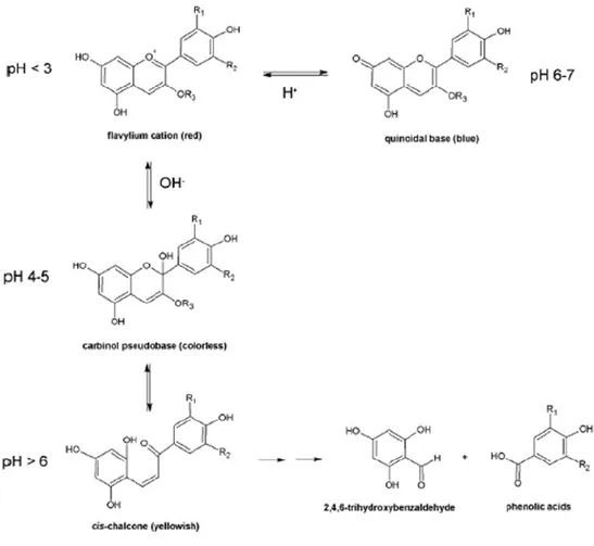
2.2 Chemistry of anthocyanins pag. 35
2.3 Sources of anthocyanins pag. 37
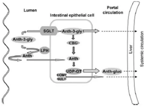
2.4 Bioavailability pag. 38
2.4.1 Systemic bioavailability pag. 44
2.4.2 Effect of food matrix on the bioavailability pag. 45
2.5 Biological activity of anthocyanins pag. 46
2.5.1 Anti-inflammatory activity pag. 47
2.5.2 Antioxidant activity pag. 48
2.6 Conclusions pag. 52
Part 2: Experimental
pag. 53Introduction pag. 54
3.
MATERIALS AND METHODS
pag. 583.1 Materials pag. 58
3.2 Cell culture and treatment pag. 59
3.2.1 Cell culture pag. 59
3.2.2 Preparation of the PA - bovine serum albumin complex
pag. 60
3.2.3 Pretreatment with C3G and exposure to PA pag. 61
3.3 Methods pag. 61
3.3.1 Western blot analysis pag. 61
3.3.1.1 Nuclear and cytoplasmic proteins extraction pag. 61
3.3.1.2 Total proteins extraction pag. 62
3.3.1.3 Determination of protein content pag. 62
3.3.1.4 Immunoblotting pag. 62
3.3.2 Evaluation of gene expression pag. 63
3.3.2.1 RNA extraction pag. 63
3.3.2.3 Preparation of cDNA pag. 64
3.3.2.4 Quantitative RT-PCR pag. 65
3.3.2.5 Post-analysis elaboration pag. 66
3.3.3 Determination of ROS pag. 67
3.3.4 Total Antioxidant Activity pag. 67
3.3.5 Fluorescein permeability pag. 68
3.4 IN VITRO GASTROINTESTINAL DIGESTION MODEL pag. 69
3.4.1 Simulated gastrointestinal digestion of an anthocyanin
extract pag. 69
3.4.2 Total anthocyanins content pag. 71
3.4.3 HPLC profile of anthocyanins pag. 72
3.4.4 Ferric reducing antioxidant power (FRAP) assay pag. 73
3.4.5 Caco-2 cells pretreatment with digested BBE and exposure to TNF-α
pag. 73
3.5 Statistical analysis pag. 74
4.
RESULTS AND DISCUSSIONS
pag. 754.1 STUDIES ON AN IN VITRO MODEL OF PA-INDUCED
INTESTINAL INFLAMMATION pag. 75
4.1.1 Effect of C3G on NF-κB pathway pag. 75
4.1.2 Effect of C3G on NF-κB transcriptional activity pag. 78
4.1.3 Effect of C3G on COX-2 activation pag. 81
4.1.4 Effect of C3G on oxidative stress pag. 82
4.1.5 Effect of C3G on Nrf2/Keap1 pathway pag. 84
4.1.6 Effect of C3G on PA-induced impaired intestinal
permeability pag. 87
4.2 STUDIES ON IN VITRO SIMULATED
GASTROINTESTINAL DIGESTION pag. 88
4.2.1 Effect of gastrointestinal digestion on anthocyanins
recovery pag. 88
4.2.2 Stability of anthocyanins after simulated
gastrointestinal digestion pag. 89
4.2.3 Effect of simulated digestion on the antioxidant
4.2.4 Pearson’s correlation test pag. 93
4.2.5 Effect of digested BBE on TNF-α induced intestinal
inflammation pag. 95
4.2.6 Effect of digested BBE on Nrf2/Keap1 pathway pag. 98
5
CONCLUSIONS
pag. 101ABBREVIATIONS
ADME Absorption Distribution Metabolism and Excretion AJ Adherent JunctionsAP Apical
AP-1 Activator Protein 1
ARE Antioxidant Response Element BBE Bilberry and Blackcurrant Extract
BBE-IP Bilberry and Blackcurrant Extract- Intestinal Phase BL Basolateral
BMI Body Mass Index CD Crohn Disease COX-2 Cyclooxygenase-2 Cul3 Cullin3
ERK Extracellular Signal-Regulated Kinase FAD Flavin Adenine Dinucleotide
FFA Free Fatty Acids
FRAP Ferric Reducing Antioxidant Power GI Gastrointestinal
GLUT Glucose Transporter HO-1 Heme Oxygenase 1
IBD Inflammatory Bowel Disease
IEC Intestinal Epithelial Cells IFN-γ Interferon gamma IKK IkB Kinase
IκB Inhibitor of kappa B IL-6 Interleukin-6 IL-8 Interleukin-8
JNK c-Jun N-terminal Kinase
Keap1 Kelch-like ECH-associated protein 1 LPS Lipopolysaccharide
MAPK Mitogen Activated Protein Kinase
NADPH Nicotinamide Adenine Dinucleotide Phosphate NF-κB Nuclear Factor-κB
NQO1 NADPH Quinone Oxidoreductase 1
NRF2 Nuclear Factor Erythroid 2-Related Factor-2 PA Palmitic Acid
PKC Protein Kinase C
PKR Protein Kinase RNA-activated RNS Reactive Nitrogen Species ROS Reactive Oxygen Species RI Recovery Index
SAT Subcutaneous Adipose Tissue SCFA Short Chain Fatty Acid SFA Saturated Fatty acid
SGLUT Sodium-Glucose Transport TAA Total Antioxidant Activity TJs Tight Junctions
TLR Toll-Like Receptor
TNF-α Tumor Necrosis Factor alfa UC Ulcerative Colitis
VAT Visceral Adipose Tissue
VEGF Vascular Endothelial Growth Factors ZO-1 Zonula Occludens 1
Abbreviation of anthocyanins and metabolites
Cya-3-Ara Cyanidin-3-Arabinoside Cya 3-Gal Cyanidin-3-Galactoside C3G / Cya-3-Glu Cyanidin-3-Glucoside
Cya-3-Rut Cyanidin-3-Rutinoside Del-3-Ara Delphinidin-3-Arabinoside Del-3 Gal Delphinidin-3-Galactoside Del 3-Glu Delphinidin-3-Glucoside Del-3-Rut Delphinidin-3-Rutinoside Mal-3-Ara Malvidin-3-Arabinoside Mal-3-Gal Malvidin-3-Galactoside Mal-3-Glu Malvidin-3-Glucoside Peo-3-Ara Peonidin-3-Arabinoside Peo-3-Gal Peonidin-3-Galactoside Peo-3-Glu Peonidin-3-Glucoside Pet-3-Ara Petunidin-3-Arabinoside Pet-3-Gal Petunidin-3-Galactoside Pet-3-Glu Petunidin-3-Glucoside PCA Protocatechuic Acid
Abstract
The inflammatory bowel disease (IBD) is a group of multifactorial pathologies with an unknown etiology, characterized by an alternation of an acute and a remission phase of the intestinal epithelium inflammation. In the last years there was a worldwide increased incidence of IBD, especially in the industrialized and growing countries. In addition, epidemiological studies reported a positive correlation between gut inflammation and obesity.
Recent in vivo and in vitro studies have supported the beneficial effects of anthocyanins, a class of flavonoid compounds widely distributed in Mediterranean diet, in various chronic inflammatory diseases, such as IBD, since they possess anti-inflammatory and antioxidant activity.
In the first part of this study, we aimed to evaluate the molecular mechanisms involved in the modulation of intestinal epithelial inflammation by using an in
vitro model consisting of Caco-2 cells exposed to high concentrations of palmitic
acid (PA), and the protective effects exerted by cyanidin-3-O-glucoside (C3G) pre-treatment. For all the experiments, fully differentiated Caco-2 were pretreated for 24h with different concentration of C3G (10 and 20µM), added on the apical side, and then exposed to PA 100µM, added on the basolateral chamber, for 6h.
The data obtained demonstrated the C3G anti-inflammatory activity through the modulation of NF-κB pathway induced by PA. In addition, C3G was able to improve the intracellular redox status through the activation of the adaptive cellular response modulated by Nrf2 pathway. Furthermore, it ameliorates the intestinal barrier by reducing the intestinal paracellular activity altered by PA.
Since the bioavailability of the anthocyanins seems to be very low mainly due to poor stability during gastrointestinal digestion, in the second part of this study an
in vitro simulated gastrointestinal digestion of a purified and standardized bilberry
and blackcurrant extract (BBE), rich in anthocyanins, was performed. We further studied the bioactivity of the BBE, after the static simulated digestion, on an in
vitro model of intestinal inflammation by using differentiated Caco-2 cells exposed
to TNF-α.
The outcomes confirmed the high instability of the anthocyanins in mild alkaline environment of the small intestine reporting a 13% of recovery index. However, although the high loss of anthocyanins, the digested BBE maintained part of its bioactivity, proved by the inhibition of the of NF-κB pathway induced by TNF-α, and by the activation of Nrf2 pathway.
These data hence confirm that anthocyanins, introduced by diet or food supplements, could represent a possible approach for the prevention of IBD.
Keywords: Inflammatory Bowel Disease, anthocyanins, inflammation, free fatty
1
2
Chapter 1
Pathogenesis and molecular aspects of
Inflammatory Bowel Disease (IBD)
3
1.1 Anatomy and physiologyof the gut
The intestine represents the last part of the digestive system and is made up of the small intestine and the large intestine. The small intestine is long 6-7 meters and extends from the pyloric sphincter to the ileocecal valve, with the main function of absorbing the products of digestion. Anatomically it is divided into three portions, duodenum, jejunum, and ileum. Whereas, the large intestine extends from the ileal valve to the anal orifice, surrounding the small intestine on three sides. With a length of about 1.5 meters and a diameter of 10 cm at the beginning, and 7 cm at the level of the rectal ampulla, it represents 1/5 of the entire gastrointestinal tract. Anatomically it is divided mainly into 3 portions: caecum, colon, and rectum. The cecum is the first part of the large intestine with a blind-bottomed sack shape and is suspended below the ileocecal valve. The colon in turn is divided into ascending colon, transverse colon, and descending colon. Its main function is to absorb water and electrolytes, to synthesize some vitamins, and to transform the undigested material (kilo) into feces that will be then eliminated through defecation (Drake et
al., 2010).
1.1.1 Histology
The large intestine wall is made by 4 concentric layers: mucosa, submucosa, muscular, and serous. The inner one (mucosa) consists of a layer of simple columnar epithelium which folds inwards forming invaginations called Lieberkühn crypts or intestinal glands. The intestinal epithelium consists of: a) enterocytes, cylindrical cells with a brush border in the apical portion that absorb water, electrolytes and vitamins produced by the bacterial flora, b) globet cells that secrete mucus, which facilitates the transport of feces and protects the intestine from the gases and acids produced by intestinal bacteria, and c) Paneth cells, which are mainly found at the base of the crypt and secrete antibacterial molecules (Clevers, 2013). The submucosa instead contains many blood and lymphatic vessels and the nervous plexus of Meissner. The muscular layer has a circular inner and an external and longitudinal layer divided into 3 bands called taeniae, these are shorter than the colon thus causing the formation of haustra, intestinal gibbosities. Moreover, in the muscular there is also the plexus of Auerbach, which together with the Meissner one form the enteric nervous system (Deakin et al., 2006).
4
1.1.2 The Intestinal barrier
The intestine represents one of the largest part of the organism that is in contact with the external environment, so in addition to the function to absorb the substance needed, it must prevent the penetration of harmful microorganisms. This last function is explicated by the intestinal barrier. The intestinal barrier is made by physical, biochemical, and immunological elements; from external to inner layer the barrier is constituted by the mucus layer, the intestinal epithelial cells (IEC), and the adaptative and innate immune cells of the lamina propria (Fig. 1). The mucus layer covers the entire surface hydrating and protecting the mucosa from the pathogens, through antimicrobial peptides (AMPs) and secretory IgA molecules (sIgA). Small intestine presents only one mucous gel layer while the colon has two, the inner separates the IEC form the microbiota, depleting of the harmful bacteria, while the outer one permits the colonization of the microbiota (Johansson
et al., 2016). The intestinal epithelium, instead, represents the real and strongest
part of the barrier. As mentioned above it is a polarized monolayer made up by enterocyte, globet cells, Paneth cells, and enteroendocrine cells, which physically separate the lumen from the lamina propria. The selective permeability is regulated by the presence of the junctional complexes; adherens junctions (AJ), tight junctions (TJ), and desmosomes (Galipeau et al., 2016). The tight junctions (TJ) consist of approximately 50 different membrane proteins, located in the apical part and in the lateral region of the IEC. They include: a) integral membrane proteins such as the adhesion molecules claudin and occludin, which extend into the intracellular space acting as a bridge between two cells, b) cytoskeletal linkers such as the proteins of the zonula occludens (ZO-1; ZO-2; ZO-3) and cingulin, c) signal proteins able to act as transcription factors, regulators of the cell cycle, and to activate various downstream cascade reactions (Cereijido et al., 2007). Below the TJs are present the adherens junctions which promote the initiation and stabilization of the cell-cell adhesion and the regulation of actin cytoskeleton. They are mostly composed by cadherins (transmembrane proteins) and catenins (Bischoff et al., 2014)
5
Fig. 1: Intestinal barrier composition (image from http://www.WeiLab.com)
1.1.3 Microbiota
The intestine is also the site of the largest bacterial ecosystem in humans. In fact, the intestinal lumen hosts more than 700 different species of non-pathogenic microorganisms. The composition and density of the microbiota varies widely from individual to individual, even of the same species, due to the diet, environmental and genetic factors. In general, the microbiota consists mainly of bacteria but also fungi and viruses are present. The 90% of the total bacteria present in the large intestine derives from 30-40 species belonging to the genera
Bacteroides, Bifidobacterium, Clostridium, Faecalibacterium, and Eubacterium
(Guarner et al., 2003; Beaugerie et al., 2004). The relationship between intestinal flora and man cannot be defined as a simple commensal relationship but more as a mutualistic relationship (Sears et al., 2005). In fact, while man provides the ideal environment for bacterial growth, these in turn perform different important duties for humans, such as facilitating chemical digestion and absorption, synthesizing vitamins and saturated short chain fatty acids, preventing the growth of pathogenic microorganisms, “training” the immune system and regulating bowel development (Sherwood et al., 2013). Without microbiota the human organism would not be
6
able to digest some food components, in fact unlike man some bacteria possess enzymes capable of digesting some polysaccharides, such as fibres, some types of starches, and oligosaccharides and sugars. They metabolize these saccharides into short chain fatty acids (SCFA) such as propionic acid, butyric acid, and acetic acid. These fatty acids can be used by man as an energy source (Gibson et al., 2004). They also increase the production of some cells of innate immunity such as exinophiles, basinophils and neutrophils (Levy et al., 2016). The intestinal microbiota also produces a large amount of vitamins, especially vitamin K, B12, therefore making a significant contribution especially when the dietary intake is very low. Finally, the flora also metabolizes some amino acids such as tryptophan and consequently produces substances (3-indolopropyl acid, 3-indolealdehyde, and indole) which exert a neuro-protective effect and help maintaining homeostasis and reactivity of the intestinal barrier (Zhang et al., 2015; Wikkoff et al., 2009). In healthy conditions the flora is well separated from the intestinal tissue thanks to a thick layer of mucus produced by IEC. A damage of the flora and the barrier leads to the infiltration of harmful microorganisms and consequently activation of different inflammatory pathways and diseases, such as the inflammatory bowel disease.
1.2 Inflammatory bowel diseases
Inflammatory bowel diseases (IBD) are a group of pathologies characterized by an alternation of an acute and a remission phase of the intestinal epithelium inflammation (Melgar et al., 2010). The two main forms of IBD are ulcerative colitis (UC) and Crohn Disease (CD), which share similar symptoms, such as diarrhea, abdominal pain, and bleeding, while they differ in the location and intensity of the inflammation. In fact, ulcerative colitis is characterized by a diffuse and interrupted inflammation of the colon with the ulcer that occurs very rarely and only in the most severe conditions of this pathology. On the contrary, ulceration is typical of Crohn's disease, which can penetrate and become transmural affecting the entire digestive tract (from the mouth to the intestine) (Melgar et al., 2010).
7
1.2.1 Etiopathogenesis of IBD
1.2.1.1 Genetic Factors
The etiopathogenesis is still not clear but it is believed it can be mainly due to: genetic susceptibility, alteration of the immune system, and environmental factors (Fig. 2). Several studies have in fact shown that IBD are the result of an anomalous and disproportionate response of the immune system to environmental factors, including bacterial flora, specific antigens, and food-derived agents in genetically susceptible individuals (Kucharzik et al., 2006). Recent genome-wide association studies (GWAS) have shown that more than 40 gene loci belonging to adaptive immunity, intestinal barrier function, and autophagic pathways are related to the development of IBD. Among the specific CD genes, particular importance is given to the NOD2/CARD15. In two different genetic studies three polymorphisms have been identified on the above-mentioned gene on chromosome 16, which increase the probability of developing CD by 20-40% (Hugot et al., 2001; Ogura et al., 2001; Cuthbert et al., 2002). The NOD1 and NOD2 genes recognize a distinct motif present in peptidoglycans and play an essential role in the signalling pathway of innate immunity response. In fact, their activation results in the activation of the caspase signalling pathway and the κB nuclear transcription factor (NF-κB) with a consequent increase in the production of pro-inflammatory cytokines. In the specific, the NOD2 gene encodes an intracellular protein, NOD2, also known as CARD15, which is expressed in Paneth cells and in antigen-presenting cells, and it can be induced in intestinal epithelial cells (Guitierrez et al., 2002; Lala et al., 2003). Once activated by its muramyl dipeptide ligand (MDP), CARD15 interacts with Rick/Rip2 and subsequently induces the activation of NF-κB and the mitogen activated protein kinase pathway (MAPK) (Guitierrez et al.,2002; Inohara et al., 2000; Lala et al., 2003). Furthermore, it is considered that NOD2 can have an effect also on the activation of the caspase cascade, however this mechanism has not been completely clarified, in fact the mutations seen in CD patients do not involve the CARD domain considered important for caspase 1 activation. NOD also regulates the expression of α-difensin in Paneth cells and therefore is believed to inhibit the invasion of bacteria present in the intestinal lumen (Kobayashi et al., 2005). Therefore, mutations at the level of this gene result in a protein that is no longer able to correctly interact with the MDP with the consequent inability of the
8
intestinal mucosa to fight bacterial infection and the initiation of the systemic inflammatory response that leads to an uncontrolled inflammation. Genes associated to colitis instead, include genes coding for matrix proteins (ECM1), interleukins (IL-2, IL-10), E-cadherins (CDH1), suggesting thus that, in contrast to CD, in the pathogenesis of UC there is a loss of intestinal barrier function (Franke
et al., 2008; Barret et al., 2009; Fisher et al., 2008; Festen et al., 2009; Silverberg et al., 2009). Finally, genes related to both IBD pathologies belong to the
interleukin 23 (IL-23) pathway, to the transducer and activator of transcription 3 (STAT 3), and to the Multi Drug Resistance 1 (MDR-1) (Duerr al., 2006; Franke et
al., 2008; Stoll et al., 2004; Kaser et al., 2008)
9
1.2.1.2 Immunological factors
The immune system constitutes a defensive barrier between microorganisms located within the lumen and the intestinal epithelium. In physiological conditions the intestinal mucosa is characterized by the abundant presence of lymphocytes both at intraepithelial and subepithelial level. Several studies have shown that a malfunction of the innate and adaptive immune system can contribute to the aberrant intestinal inflammatory response in patients with IBD. In fact, the damage at the intestinal mucosa in IBD patients is related to a high increase in effector immune cells, such as CD4+ and CD8+, intraepithelial cytolytic lymphocytes, perforins and granzymes containing T cells (Singh et al., 2001). It has been shown that the accumulation of CD4+ plays a fundamental role in the development and exacerbation of IBD, and this is probably due to the increased activation of T lymphocytes and to their increased infiltration in the intestinal wall as a response to the increase of adhesion and chemoattractant molecules in the inflamed mucosa and a concomitant decrease in the apoptotic process (Kaser et al., 2010; Ina et al., 1999; Stefulj et al., 2001). Following their activation, CD4+ and CD8+ can differentiate into the various T helper lymphocytes Th1, Th2, Th17 (Neurath et al., 2006). Crohn's disease is associated with a Th1 cell-mediated response characterized by enhanced production of interleukins (1, 2, 6, 12, IL-18), tumor necrosis factor alpha (TNF-α), and interferon gamma (IFN- γ) (Lotz et
al., 2006). TNF-α exerts its anti-inflammatory effect by stimulating the production
of IL-6 and IL-1β by intestinal macrophages (Baumann et al., 1994). Furthermore, patients with intestinal Crohn's disease have a high number of Th17 lymphocytes and other subgroups of Th cells secreting various interleukins (17A, 17F, IL-22, etc). IL-22 is a pro-inflammatory cytokine upregulated both at the serum and tissue levels of CD patients (Schmechel et al., 2008). In contrast, in ulcerative colitis the immune response is regulated by Th2-type cytokines, such as IL-4, IL-5 and IL-10 (Camoglio et al., 1998; Sawa et al., 2003). Activation of the IL-10 secreting CD4+ T lymphocytes at the intestinal level of patients with ulcerative colitis has been proposed to be due to the consequence of elevated IL-10 and IL-8 levels (Melgar et al., 2003).
10
1.2.1.3 Environmental factors
Without any doubt, environmental factors such as smoking, diet, stress and microbiome play a fundamental role in the development of IBD. In recent years, research is increasingly focusing on the study of microbiota and diet.
The bacterial flora in the intestine of mammals is a complex and dynamic system with a steady state, which can be modified by various environmental factors, such as diet, drugs, and lifestyle. As a consequence of the diet changes there is also a variation in the composition of commensal bacteria (Ley et al., 2006; Sonnerburg
et al., 2004; De Filippo et al., 2010). The consumption of so-called Western diets,
mainly consisting of processed and refined foods, red meats, sweetened beverages, and at the same time accompanied by a low consumption of fibers, fruits and vegetables, has been associated with the development of metabolic diseases associated with a systemic and mild inflammation (Wellen et al., 2005). In physiological conditions the microbiota maintains a symbiotic relationship with the intestinal mucosa offering crucial functions for metabolism, immunity and protection against pathogens. So, the abundance and diversity of the flora plays a fundamental role in providing essential functions such as, clearance of pathogens, resistance to colonization, and symbiosis (Statovci et al., 2017). An in vivo study carried out on wild-type mice fed with a diet rich in lipids and sugars showed a decrease in microbiota biodiversity and an increase in opportunistic pathogens with a consequent reduction in the function of the intestinal barrier (Zhang et al., 2012). Epidemiological studies suggested an increased risk of developing IBD related to a high consumption of red and processed meats, and lipids (saturated and unsaturated fatty acids), and low consumption of vitamin D (de Silva et al., 2014; Wiese et al., 2014) (Fig. 3). While a recent study by the European Prospective Investigation in Cancer (EPIC) found no correlation between the body mass index and the development of IBD, suggesting therefore that a high-calorie diet is not enough to trigger intestinal inflammation (Chan et al., 2013).
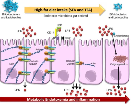
Saturated fatty acids (SFA) occur naturally in derivatives of plant origin, mainly short-chain ones, while long-chain ones, such as palmitic acid, stearic acid and myristic acid, are found in products of animal origin (lard, butter, pork and beef, etc.) (Vannice et al., 2013). In vitro studies have shown that SFA, with a mechanism very similar to LPS, can act as mediators of pro-inflammatory
11
cytokines by binding and activating the toll like receptor 4 (TRL4) and consequently up-regulating the expression of NF-κB (Lee et al., 2003). The last one plays an important role in the activation of many pro-inflammatory mediators, such as COX-2, TNF-α, IL-1β, IL-6, CXCL8, IL-12, and IFN-γ (Lee et al., 2003) Furthermore, it was demonstrated that palmitic acid (PA) and stearic acid can induce the degradation of the factor κB inhibitor (IκB), the phosphorylation of C-Jun N-terminal kinase (MAPK), and the kinase regulated by extracellular signal (ERK ) on macrophages (Lyons et al., 2016). A high intake of SFA also induces a modification of the microbiota by increasing the number of Gram-negative bacteria and therefore the natural ligand for TRL4, LPS, inducing an increase in intestinal permeability, thus leading to a state of endotoxemia (Moreira et al., 2012). The intake of SFA increases the formation of low-density lipoprotein (LDL) and decreases their turnover with the consequent formation of oxidized LDL, which are a well-known molecular pattern associated with damage and recognized by TRL4, inducing therefore an inflammatory response (Stewart et al., 2010). In contrast, monounsaturated acids contained in olive oil, avocado, macadamia nuts and lard, have been shown to reduce cholesterol and increase levels of high-density lipoprotein (HDL) (Jekins et al., 2010). Furthermore, the treatment of pro-inflammatory macrophages (M1) with palmitoleic acid induces an increase in M2 anti-inflammatory profile (Chan et al., 2010). In an in vivo study it was shown that the dietary intake of oleic acid reduced the risks of developing intestinal inflammation (de Silva et al., 2014). However, the role of monounsaturated acids in the development of IBD is partly unknown. Polyunsaturated fatty acids (PUFAs), since they contain more than a double bond, are more prone to oxidation. They are divided into two groups: omega 3 (alpha linoleic acid [ALA], docoesanoic acid [DHA]) and omega 6 (linoleic acid [LA], arachidonic acid [ARA]). The latter is the main n-6 PUFA found in inflammatory cells and is involved in the synthesis of eicosanoids such as leukotrienes and prostaglandins, which are known inflammatory mediators (Schmitz et al., 2007). While, omega 3 showed an anti-inflammatory activity, in fact, unlike saturated fatty acids, they inhibit the TRL4 signal and consequently the transcription of pro-inflammatory genes. DHA can improve the function of the intestinal barrier by increasing the expression of tight junctions (Liu et al., 2012). Therefore, in order to maintain an intestinal homeostasis it is important that the omega 6/omega 3 ratio is very low.
12
Unfortunately, in recent years this ratio has increased dramatically due to processed foods, low consumption of dietary fibers and fish products. In line with this, in a recent study a high ARA:eicosapentaenoic acid (EPA) ratio was found at the level of the inflamed intestinal mucosa of patients with UC (Pearl et al., 2013).
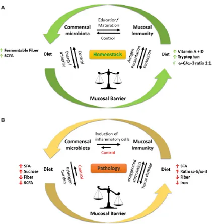
Fig. 3: Effect of high fatty diet on the intestinal barrier. A diet rich in fibres and vitamin D helps
in the homeostasis maintenance (A). In fact, the fibres introduced by the diet are converted by microbiota into SCFA which increase the mucosal barrier integrity. Furthermore, microbiota induces the maturation and education of immune system cells, this in turn control the microbial activity preventing intestinal damage. On the contrary a high fatty diet (B) and low in fibres promotes the pathogenic proliferation and the intestinal barrier disruption. In addition, a microbiota made mainly by non-beneficial bacteria induces the activation of inflammatory cytokines, leading to further activation of inflammatory process and tissue damage. The tissue damage in turn is not anymore able to control the microbiota composition, creating thus a negative and dangerous loop. (from Statovci et al., 2017)
13
1.3 Obesity and IBD
In recent years the percentage of overweight people has increased by 28% in developed countries and by around 60% in developing countries, and no country has reported a decrease (Ng et al., 2014). In the United States more than a third of the adult population is obese. In parallel with this increase, the incidence of developing IBD is also increasing globally. In an epidemiological study conducted in Scotland on 489 patients with IBD, 18% were obese and 38% were overweight, whereas only 3% of patients with DC and 0.5% of those with UC were underweight (Steed et al., 2009). Similar results were found in cohort studies of infant patients, in fact 9-10% of children with Crohn's disease and 20-34% of children with ulcerative colitis had a body mass index (BMI) for age over 85 percentiles (Kugathasan et al., 2007). Despite the high prevalence of obesity in patients with IBD, mild obesity has been associated with a risk of developing Crohn's disease but not ulcerative colitis (Singh et al., 2017). Furthermore, the effect of obesity on the development of IBD may be age-dependent, in fact, the condition of obesity in juvenile and adolescent age is associated with a greater risk compared to obesity in old age (Singh et al., 2017). The condition of obesity in addition to be a risk factor for CD etiopathogenesis can also alter the course and outcome of the disease. Indeed, it has been shown that patients with Crohn's disease and overweight are more prone to a more active inflammation and also show an inflammation of the anorectal tract, therefore the rate of hospitalization for these patients is greater than in normal weight subjects (Mendall et al., 2011; Blain
et al., 2002; Nascimento et al., 2012; Hass et al., 2006). The response to therapy
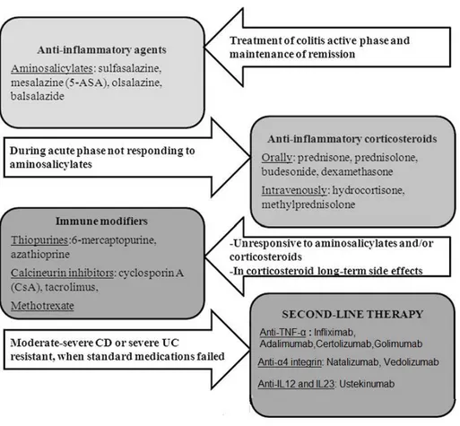
also appears to be different between obese/overweight and normal weight patients. Bhalme et al. (2013) observed an increased risk of loss of response to adalimumab, a recombinant anti-TNF-α monoclonal antibody, but not of infliximab, another monoclonal anti-TNF-α antibody, in obese patients. This effect could be due to the increased amount of body fat that modifies the pharmacokinetic properties of the drugs. In addition to this also the high concentration of circulating pro-inflammatory mediators alter the pharmacological effect (Bhalme et al., 2013; Kredel and Siegmund, 2014)
14
1.3.1 Adipose tissue as an endocrine organ
Adipose tissue has always been considered as a passive form of connective tissue with the only function of storing energy in the form of triglycerides and releasing energy in the form of free fatty acids (FFA). However, due to the wide range of hormones, proteins, cytokines, and enzymes secreted by adipocytes, it is now considered an endocrine organ that performs different functions (Kredel and Siegmund, 2014). It consists of a group of adipocytes, preadipocytes, macrophages, endothelial cells and fibroblasts, and is subdivided into visceral and subcutaneous adipose tissue (Rocha et al., 2007; Gregor et al., 2011). The two types of tissue show a very different immunological and metabolic profile. Subcutaneous tissue (SAT) is approximately 80% of body fat and has a greater number of preadipocytes than visceral tissue. The adipocytes of the SAT have a high sensitivity to insulin and have a greater ability to accumulate FFA and triglycerides. In contrast, visceral adipose tissue (VAT) accounts for 10-20% in men and 5-10% in women of total body fat. It is highly vascularized and has a high content of M1 pro-inflammatory macrophages which mainly secrete TNF-α and IL-1. Furthermore, VAT is characterized by a high expression of pro-inflammatory cytokines, such as interleukins (IL-6, IL-8) and chemokines (MCP-1), adiponectin and cells of innate immunity. Additionally, an increase in visceral adipose tissue is associated with a condition of insulin resistance and metabolic syndrome (Kredel and Siegmund, 2014). In physiological conditions, adipokines regulate lipid metabolism, glucose metabolism, and the effect of insulin through a set of interactions with other cytokines (Balistieri et al., 2010). The condition of obesity is instead characterized by an increase in the entire adipose tissue with different modifications of the humoral, cellular, and stromal components (Deiuliis et al., 2011; Fain et al., 2004; Kintscher et al., 2008). In vivo studies carried out on animals and obese human patients have shown how in these subjects there is an increase of cytokines, which modify the expression of different pro-inflammatory mediators and can activate an innate immune response, thus triggering a mild chronic inflammation typical of obesity (Olefsky et al., 2010). Chronic inflammation leads to the development of secondary diseases and further can affect the progress of other diseases, such as for example IBD (Barbarroja et al., 2010; Scaffler et al., 2006). Visceral fat has been recognized to be the most metabolically
15
active fraction of all body fat compartments and therefore could be a more indicative and specific factor for the risk of developing IBD than the total obesity determined by BMI (Kredel and Siegmund, 2014; Uko et al., 2014). Fat accumulation can also be locally limited, in fact patients suffering from Crohn's disease show a peculiar hyperplasia, independent of body mass, of mesenteric VAT, the so-called “creeping fat”. Creeping fat (CF) is an extension of visceral tissue that extends from the roots of the mesentery and envelops the inflamed part of the large intestine, covering more than 50% of its surface, and consequently acting as a bridge between the accumulation of fat and inflammatory activity (Peyrin-Biroulet et al., 2007; Sheehan et al., 1992; Fink et al., 2012). While in a condition of obesity tissue enlargement is generally due to a cellular hypertrophy and very rarely to a hyperplasia, the creeping fat instead is characterized by a hyperplasia of adipose tissue. In fact, the adipocytes of creeping fat are significantly smaller and their number is about 4 times greater than in the normal mesenteric adipose tissue (Peyrin-Biroulet et al., 2007). In the adipose tissue of obese patients there are up-regulated especially genes with pro-inflammatory activity, in CF instead there is an increase in gene expression of both the pro and anti-inflammatory cytokines (Zulian et al., 2010). Due to their small size, the adipocytes of creeping fat produce less pro-inflammatory cytokines and are less responsive to stimulation (Skurk et al., 2007; Kopp et al., 2010). However, despite this, they are the major active producers of different leptin mediators, adiponectin, and resistin (Paul et al., 2006; Batra et al., 2009) (Fig. 4). It is worthwhile to be aware that while creeping fat is a distinctive feature of Crohn's disease, it is generally absent in individuals with ulcerative colitis. However, there are some observations of edematous adipose tissue in patients with UC (Edling et al., 1963; Eklof et al., 1970). Furthermore, due to the different molecular profile of adipocytes and tissue morphology, the visceral fat of patients with UC has inflammatory characteristics very different from those of subjects with CD. However, also UC is associated with an obesity condition with an increase in adipose tissue mass (John et al., 2006; Levy et al., 2005).
16
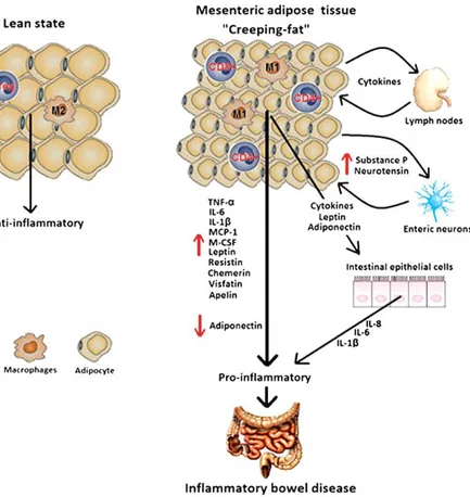
Fig. 4: Morphological and functional differences between the adipose tissue in healthy state and in patient with CD. In healthy condition the adipose tissue is characterized by “normal size”
adipocytes secreting mainly anti-inflammatory mediators. The creeping fat, instead, is characterized by smaller adipocytes and a high population of macrophages secreting different pro-inflammatory cytokines (TNF-α, IL-6), adipokines (adiponectin, leptin) that induce intestinal inflammation (from Goncalves et al., 2015).
1.3.2 Crosstalk between adipose tissue and bowel in IBD
- adipokines
In the last years, in order to better understand the mechanism underlying this crosstalk between adipose tissue and intestinal inflammation, the research was focused on the role of molecules secreted by adipocytes on the initiation and progression of IBD. It has been reported that adipocytes and preadipocytes secrete more than 50 adipokines (Trayhurm et al., 2004). As mentioned above the adipocytes secrete several pro-inflammatory cytokines, chemokines and adipokines, showing thus to possess a strong pro-inflammatory action and to induce an innate immune response.
VAT can be considered a crucial source of cytokines, produced both by adipocytes as well as by macrophages and lymphocytes, liable for the inflammatory process of
17
IBD (Bertin et al., 2010; Desreumaux et al., 1999; Karmiris et al., 2006; Fantuzzi
et al., 2008; Schsffler et al., 2008). In patients with IBD and in patients with mild
obesity was observed a high plasma concentration, directly proportional to body mass, of TNF-α and IL-6 (Bertin et al., 2010). Moreover, it has been reported that the adipose tissue of patients with IBD secrete a higher amount of TNF-α compared to the one of healthy patients, and in addition high levels of this cytokine were also found in the bloodstream, mucosa and faeces, suggesting the involvement of a systemic effect (Gambero et al., 2007; Murch et al., 1991; Breese
et al., 1994; MacDonald et al., 1990). Another cytokine that is over-expressed in
VAT of people with IBD is IL-6, and several studies have suggested that around 30% of the circulating IL-6 derives from visceral adipose tissue (Park et al., 2005). In addition, since Crohn's disease is associated with the Th1 mediated cell response, which is characterized by an increased production of IL-6, IFN-γ and TNF-α, it can be concluded that creeping fat contributes to the typical Th1 response of UC (Zulian et al., 2012; Goncalves et al., 2015). Furthermore, macrophages and T lymphocytes of mesenteric adipose tissue in active CD patients release more cytokines (IL-6, IL-4, and IL-13) than those of non-active and healthy patients. Therefore, the aberrant expression of cytokines in the creeping fat of CD subjects is due in part to macrophages and T lymphocytes of adipose tissue (Jung
et al., 2013). The mesenteric adipose tissue of patients with IBD, in addition to an
over-expression of cytokines, is characterized also by an overproduction of adipokines, such as adiponectin, resistin, and leptin (Maconi et al., 2008; Barbier
et al., 2003). The latter was initially thought to perform only the activity of appetite
suppressant, but now several studies have shown its involvement in inflammatory processes. Higher levels of leptin have been found in the intestinal lumen of patients with IBD compared to healthy one, and both its gene and protein expression are overexpressed in patients with Crohn's disease (Bertin et al., 2010; Desreumaux et al., 1999; Sitaraman et al., 2004; Paul et al., 2006). Leptin stimulates and promotes the proliferation of mononuclear cells and plays a crucial role in the inflammatory response by increasing the secretion of inflammatory cytokines using the STAT3 pathway (Matarese et al., 2002; Procaccini et al., 2012; La Cava et al., 2004; Williams et al., 2004). It also stimulates differentiation of T cells in the Th1 phenotype rather than in the Th2 (Lord et al., 2002).
18
Resistin is mainly secreted by macrophages, mononuclear cells, and stem cells (Karmiris et al., 2008). It has been associated with inflammatory processes since its expression in adipose tissue is induced by pro-inflammatory cytokines, such as IL-1, IL-6, and TNF -α (Kaser et al., 2003). In addition, in vitro studies on murine and human macrophages cell lines have reported that the bacterial lipopolysaccharide activates the expression of resistin through a pro-inflammatory cytokine cascade (Lehrke et al., 2004). In turn, resistin once activated induces the activation of IL-12, TNF-α and adhesion molecules in mononuclear cells, human endothelial cells and macrophages (Bokarewa et al., 2005; Pang et al., 2006). In fact, it has been shown that in human mononuclear cells resistin induces, and is also induced by, IL-6 and TNF-α through the NF-κB pathway (Bokarewa et al., 2005; Pang et al., 2006).
Whereas the role of adiponectin in the pathogenesis of IBD is still partially unknown (Bertin et al., 2010). Adiponectin, secreted mainly by mature adipocytes, has anti-inflammatory effects (Karmiris et al., 2008). In fact, in vitro studies on different cell lines have shown that it is able to negatively modulate inflammatory pathways through the inhibition of NF-κB, and to interfere with the function of macrophages (Ouchi et al., 1999; Yokota et al., 2000). In addition, adiponectin suppresses TNF-α production by activating the MAPK signalling pathway in murine macrophages, and it further blocks the TRL-induced NF-κB activation in human macrophages (Wolf et al., 2004; Yamaguchi et al., 2005; Zhao et al., 2005). It up-regulates expression of anti-inflammatory cytokines, such as IL-10 and IL-1Ra in dendritic cells, monocytes and macrophages (Wolf et al., 2004), suggesting thus that its anti-inflammatory effect is mediated principally by the cells of the immune system. Low levels of adiponectin have been reported in patients with active CD and its expression in adipose tissue is inversely proportional to the degree of intestinal inflammation (Rodrigues et al., 2012). However, an increase in both the levels of gene expression and proteins in VAT have also been reported in subjects with Chron's disease (Yakamoto et al., 2005). Also with regard to the serum levels of this adipokine, conflicting results have been reported (Zhao et al., 2005; Ohashi et al., 2010), suggesting thus that the physiological role of adiponectin in the formation and progression of IBD still remains a question mark.
19
In the last period the possible involvement of neuropeptides in the pathogenesis of IBD has been demonstrated. Enteric neurons are characterized by a high amount of substance P (SP), neurotensin (NT) and vasoactive intestinal polypeptides (Shepard et al., 1987; Bishop et al., 1980). In vitro studies on pre-adipocytes showed an increase in IL-6 and IL-8 expression, via the NF-κB pathway, when treated with NT and SP, respectively. This pro-inflammatory effect could create a cascade reaction that leads to the recruitment of immune cells and the formation of creeping fat (Karagiannides et al., 2008). Also Gross and co-workers have shown that substance P can influence the size of fat depots by acting on the replication and apoptosis of pre-adipocytes. Specifically, the treatment of adipocytes with SP induces an increase in the proliferative activity and at the same time reduces apoptosis (Gross et al., 2009). Therefore, the neuropeptides could act as a bridge between the adipose tissue and the intestinal response during the IBD condition acting on two different fronts: the formation of the creeping fat and the induction of an inflammatory response at the level of the adipocytes (Fink et al., 2013).
1.4 Lipotoxicity
The lipids introduced through the diet are certainly essential in promoting inflammation. Lipids are a heterogeneous class of molecules including fatty acids, sterols, phospholipids and triglycerides. In addition, to be an efficient source of energy, they also act as important components of the cell membrane and molecules that regulate metabolic homeostasis. However, a wrong lifestyle, and genetic and epigenetic factors, can alter the balance between their metabolism and their composition becoming thus harmful to the health with consequent organelle dysfunction, cell death, dysfunction in energy metabolism, and chronic inflammation (Ertnunc and Hotamisligil, 2016). All these negative effects together induce a pathological condition which is called lipotoxicity. A condition of lipotoxicity in addition to causing an alteration of lipid metabolism, converges with the immune and stress response, contributing so to the development of various diseases (Fu et al., 2012). A diet rich in lipids induces an increase in free fatty acids, which enter the mitochondria where they are oxidized or esterified into triglycerides through the β-oxidation process (Pessayre et al., 2001). During the
20
process of β-oxidation of fatty acids, which depends on the redox reactions of the co-factors NAD+/NADH and FAD/FADH2, electrons are released and transferred
to the mitochondrial electron transport chain, here they can be bound to oxygen forming radicals superoxide anions and other reactive oxygen species (ROS). In turn, ROS and other radical species oxidize the polyunsaturated lipids of fat deposits, thus triggering the process of lipid peroxidation. The intermediates of this metabolic process also react with oxygen to form different ROS, causing so an imbalance in the redox state with the consequent formation of a condition of oxidative stress (Pessaye et al., 2002; Matsuzawa-Nagata et al., 2008). Furthermore, lipid peroxidation and ROS reduce the levels of endogenous antioxidants and vitamins, leading to a further increase in intracellular ROS levels with a consequent increase in ROS-dependent cell damage (Pessaye et al., 2002). The ROS-mediated state of inflammation induces the activation of different inflammatory signal pathways including the NF-κB pathway, one of the main transcription factors related to inflammatory processes, and consequently of other downstream pro-inflammatory mediators of NF-κB such as TNF-α, IFN-γ, and inducible synthetase of nitric oxide (iNOS) (Weisberg et al., 2008). Furthermore, lipids influence the cellular function by activating membrane receptors, in fact, palmitic acid directly activates the inflammatory pathways by increasing the expression of TRL4 on cell membranes or by stimulating the protein kinase R (PKR) (Nakamura et al., 2010). Following its activation by harmful lipids, such as palmitic acid or oxidized cholesterol, PKR induces the activation of the JNK pathway. JNK downstream mediators include genes involved in the inflammatory processes, apoptosis, modulation of inflammasome and of the transcription factor activating protein 1 (AP-1) (Takada et al., 2010; Kang et al., 2012; Peng et al., 2015) (Fig. 5). Some of the principal pro-inflammatory mediators involved in the initiation and progression of IBD induced by a condition of lipotoxicity will be described in more detail below.
21
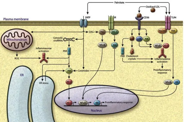
Fig. 5: FFA-induced lipotoxicity. Increased amount of toxic lipids causes failure of metabolic
regulation, converging on inflammatory and stress pathways. Palmitate can activate TLR4 signalling, leading to activation of inflammasomes and induction of inflammatory gene transcription factors such as NF-κB, AP-1 and interferon regulatory factor (IRF). Palmitic acid directly contributes to the synthesis of diacylglycerols and ceramides, which activate stress kinases, PKCs, the NF-κB and JNK pathways. It further can be transported inside the cell by the FATPs, and through PKR can induce ER stress. Additionally, lipotoxicity can also induce the ROS production from mitochondria, which is linked to inflammasome activation (from Ertnunc and Hotamisligil, 2016).
1.5 MOLECULAR MECHANISMS INVOLVED IN IBD 1.5.1 Reactive oxygen species (ROS)
Chronic intestinal inflammations are multifactorial pathologies characterized by a massive infiltration of granulocytes and macrophages in the intestine, which in addition to inducing the activation of a broad spectrum of pro-inflammatory cytokines also produce a huge quantity of ROS and reactive nitrogen species (NOS) (Biasi et al., 2013). ROS are small molecules including oxygen radicals [superoxy (O2*), hydroxyl radical (*OH)], non-radical species [oxygen singlet (O2)
and hydrogen peroxide (H2O2)] (Speciale et al., 2019). These molecules are highly
reactive and are able to cause damage to proteins, lipids and nucleic acids, generating hence oxidized macromolecules which are considered as triggers for
22
various pathologies (Tian et al., 2017). Although they have always been considered as harmful substances, in reality at low and moderate concentrations ROS are essential for cellular homeostasis and are beneficial for many biological processes. In fact, at the level of the intestinal epithelium, they are involved in the defence mechanism against pathogens by inducing the respiratory burst in the phagocytes (Biasi et al., 2013). In physiological conditions the intracellular levels of ROS are kept stable and balanced by different cellular processes. Endogenous ROS are produced in intracellular organelles such as endoplasmic reticulum, mitochondria, peroxisomes, nucleus, cytosol, and extracellular matrix. Among these, mitochondria are considered the main organelles in which ROS production takes place, they are in fact the site of the electron transport chain, the main source of ROS, as well the site of the catabolism of fatty acids (Novak and Mollen 2015; Poyton et al., 2009). At the same time these organelles are also the most damaged and the primary targets of oxidative stress (Tian et al., 2017). A high ROS production, in fact, is able to suppress the electron transport chain with a consequent decrease in ATP production and damage at the mitochondrial DNA level. When this situation continues over time, the mitochondrial homeostasis is affected and subsequently cell death pathway is activated (Chen et al., 2008; Scherz-Shouval et al., 2007). Several enzymes, such as lipoxygenases, cyclooxygenases, (involved in lipid metabolism), oxidase, peroxidase, through the catalysis of chemical reactions, are considered responsible for the production of endogenous ROS (Kulkarni et al., 2007; Swindle et al., 2007). In addition, external and environmental factors, such as smoking, alcohol, drug use and chemotherapy also participate in the production of ROS (Goyette et al., 2007). Under physiological conditions, eukaryotic cells in order to counteract and avoid over-expression of intracellular ROS, induce the over-expression of genes coding for antioxidant proteins, thus regulating the intracellular redox state. However, the excessive production of ROS associated with a decrease in antioxidant activity is related to an increase in intestinal permeability, impaired immune response, damage at the DNA level, lipid peroxidation, oxidation of the amino acid lateral chains, apoptosis, and carcinogenesis (Ridnour et al., 2005; Valko et al., 2001; Valko et al., 2007). This condition is called oxidative stress and is due to an imbalance between antioxidants and radical species.
23
The endogenous defence system against oxidizing substances consists mainly of a) intracellular enzymatic antioxidants: superoxide dismutase (SOD), glutathione peroxidase (GPX), and catalase (CAT), b) non-enzymatic intracellular antioxidants: glutathione (GSH), and c) extracellular antioxidants, uric acid, vitamins, minerals (Tian et al., 2017). The first endogenous antioxidant system evaluated in patients with IBD was GSH/GSSG/GPX. GSH is a water-soluble antioxidant molecule containing a thiol group derived from cysteine and expressed in cytosol, nucleus, and mitochondria. The maintenance of its homeostasis is provided by synthesis starting from cysteine or from the regeneration of oxidized glutathione (GSSG) and uptake of GSH by dependent sodium transport (Vučetić et
al., 2017). Together with enzymes glutathione S-transferase (GST) glutathione
reductase (GSR) and GPX, it forms an important antioxidant barrier in the intestinal mucosa. The GPX enzyme allows the reduction of hydrogen peroxide in water or of lipidic hydroperoxides (ROOH) in stable alcohols, catalysing the oxidation of GSH in disulphide oxidized glutathione. Currently 8 GPX isoforms have been detected, among these the isoform 2 (GPX2) is specifically expressed in the gastro-intestinal epithelium (Dayer et al., 2008). In vivo studies on CD and UC mouse models have shown a pivotal role of GPX2 in the defence against oxidative stress and inflammation in the intestinal mucosa (Dayer et al., 2008; Te Velde et
al., 2008). It has also been shown to be induced under pathogenic conditions such
as IBD and gastric cancer. One of the mechanisms by which the condition of oxidative stress starts and propagates intestinal inflammation is mediated by the NF-κB signalling pathway, which is responsible for the regulation of several pro-inflammatory mediators.
1.5.2 NF-κB pathway
NF-κB is an inducible redox-sensitive nuclear transcription factor responsible for several biological processes, such as inflammation, immune response, cell growth, and apoptosis (Hayden et al., 2012). Its activation in fact induces the transcription of several pro-inflammatory mediators. It consists of homodimers and heterodimers of proteins of the NF-κB family, which includes 5 monomers, RelA
24
(p65), RelB, cRel, NF-κB 1(p50), and NF-κB 2. All these proteins share a N-terminal region, the Rel Homology Region (RHR) responsible for dimerization, nuclear translocation, DNA binding, and binding to the κB (IκB) inhibitor (Baldwin, 2001). Furthermore the RelA, RelB, cRel subunits also contain an essential domain for the transcriptional activity, the transcriptional activation domain (TAD), while p50 and p52 are lacking of it, so they act as inhibitors through a mechanism of competition with the TAD-containing dimers for κB site binding (Huxford et al., 2009; Franzoso et al., 1992). In most cells NF-κB is a heterodimer composed of the RelA (p65) and NF-κB (p50) subunits, which also represents the most active and stable form of this family (Speciale et al., 2019). In healthy conditions NF-κB is located in the cytosol bound to the κB inhibitor, which by binding to the RHR domain of the dimer masks the nuclear localization signal, preventing thus its translocation to the nucleus (Huxford et al., 1998). Following NF-κB activation, it translocates to the nucleus where it binds to specific DNA sites and regulates the expression of many genes involved in inflammatory processes, innate immunity, cell proliferation, and apoptosis (Speciale et al., 2019). The activation and therefore the translocation of this factor can occur through two different pathways (Fig. 6): the classical or canonical pathway and the non-canonical pathway. Activation by the non-canonical pathway involves the IκB kinase (IKK) consisting of two subunits (IKKα and IKKβ) and the regulatory protein NEMO (NF-κB essential modulator). External stimuli from pro-inflammatory mediators (TNF-α), associated pathogenic molecular pattern (PAMPs), and associated damage molecular patterns (DAMPs), induce IKK phosphorylation which in turn phosphorylases IκB causing its dependent proteasome degradation and allowing the nuclear translocation of NF-κB (Bonizzi et al., 2004). While stimuli involved in cell differentiation and development, such as the beta lympho-toxin receptor (LTβR), CD40L, TNF receptors etc., activate instead the non-canonical pathway. This pathway is mediated by the NF-κB inducing kinase (NIK) and by IKKα. NIK activation leads to phosphorylation and subsequent proteasomal processing of the NF-κB2 (p100) precursor in p52 through an IKK1/IKKα-dependent manner. The formed p52 mature subunit is free to dimerize with the RelB subunit and consequently to translocate to the nucleus and activate the gene transcription of different mediators (Sun, 2012). At intestinal level it is involved in IEC homeostasis and in the permeability modulation of the intestinal barrier
25
(Pasparakis et al., 2008). In fact, an abnormal activation of this factor is a typical feature of IBD (Neurath et al., 1996; Rogler et al., 1998). Administration of NEMO-binding drugs that block the link between the NEMO and IKK subunits has been shown to reduce the degree of inflammation in different murine models of colitis and to inhibit IKK activation (Shimozawa et al., 2004). Furthermore, high levels of the p65 subunit in fibroblasts, endothelial cells, and macrophages of patients with IBD have been reported (Gelbmann et al., 2003; Rogler et al., 1998). Excessive NF-κB activation is thought to be responsible for the induction of transcription of several genes having a central role in the pathogenesis of IBD which can be divided into. These genes play a fundamental role in inducing damage to the intestinal barrier by stimulating the production of other NF-κB-dependent cytokines (Seidelin et al., 2005; Clevers et al., 2011)
- genes regulating cell proliferation and apoptosis: cyclic D1 and D3, proteins of the Bcl family, endothelial growth factor (EGF) (Biasi et al., 2013)
- genes regulating intestinal permeability and angiogenesis: claudin, myosin light chain kinase (MLCK), vascular endothelial growth factor (VEGF). For example, TNF-α induces a partial increase in intestinal permeability by activating MLCK in a dependent NF-κB manner (Ye and Ma, 2008)
- metalloproteinases which degrade the extracellular matrix and the cells of the intestinal mucosa (Ravi et al., 2007)
- enzymes inducing eicosanoid, ROS and RNS: COX-2, lipooxygenases, iNOS, which participating in ROS metabolism can induce NF-κB activation and therefore increase cell permeability (Andersen et al., 2005).
26
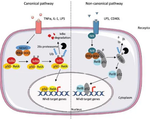
Fig. 6: Canonical and non-canonical pathway leading to the activation of NF-κB. The
canonical pathway is triggered by cytokines such as TNFα and Interleukin-1 (IL-1). These induce the phosphorylation of IκBα by the IKK complex, leading to its degradation by the 26S proteasome. The RelA/p50 complex is free to translocate to the nucleus and to activate the transcription of target genes. In the non-canonical pathway TNF cytokine family induce the activation of IKKα through the NF-κB-inducing kinase (NIK) leading to the phosphorylation of the p100 subunit and processing in p52. P52 can dimerize with RelB and translocate to the nucleus (from Viennois et al., 2013).
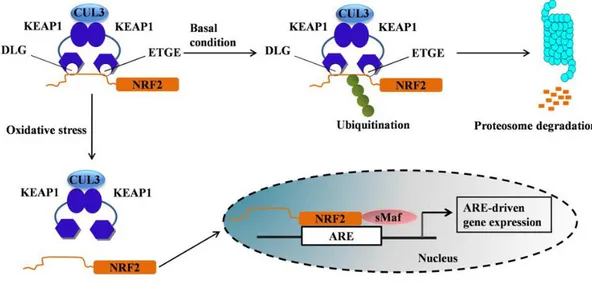
1.5.3 Nrf2 pathway
The Nuclear factor-erythroid 2 related factor 2 (Nrf-2), is a key transcription factor in the maintenance of redox homeostasis at the level of the intestinal mucosa. In fact, this factor regulates the expression of detoxifying and antioxidant enzymes presenting the Antioxidant Responsive Element (ARE) sequence on their promoter. It is expressed at the level of different tissues such as liver, kidneys (organs involved in the detoxification processes), as well as at the level of the gastrointestinal tract, skin, and lungs (organs more in contact with external agents) (Speciale et al., 2013).
27
Under physiological conditions, Nrf2 is bound to the cytosolic proteins Keap-1 and Cullin 3 (Cul3), with the last one inhibiting the transcriptional activity of Nrf2 by ubiquitination and proteasomal degradation, while Keap-1 is a substrate that facilitates this reaction. A condition of oxidative stress or other oxidative stimuli induces a burst of the Keap- Cull-3 inhibition system allowing Nrf2 to translocate to the nucleus where it will bind to the ARE sequence and may induce the transcription of genes coding for antioxidant enzymes (Cullian et al., 2004; Kobayashi et al., 2004; Zhang et al. 2004) (Fig. 7).
Nrf2 modulates the transcription of about 250 genes that can be divided into: - antioxidant enzymes: heme-oxygenase 1 (HO-1), glutathione peroxidase 2 (GPX2), glutamate-cysteine ligase catalytic subunit (GCLC), glutamate-cysteine ligase-modifying subunit (GCLM), sulfiredoxin 1 (SRXN1) and tioredoxin (TRX) - enzymes of phase 1 metabolism: NAD(P)H quinone oxidoredutase (NQO1), enzyme responsible for the reduction of quinones, highly reactive species which can cause stress.
- phase 2 detoxifying enzymes: glutathione S microsomal transferase 1 (GTST1) (Hayes et al., 2000; Speciale et al., 2019)
Nrf2 plays an essential role in the pathogenesis of IBD. In vivo studies on a Dextran sulfate sodium (DSS)-induced colitis model on Nrf-2 knock out (KO) and wild type (WT) mice showed that KO mice had more severe symptoms associated with a greater expression of pro-inflammatory genes and a decrease in antioxidant genes compared to WT ones (Khor et al., 2006; Khor et al., 2008).
28
Fig. 7: Schematic mechanism of Nrf2 activation. Under basal conditions, Nrf2 binds to its
repressors Keap1 and Cul3 which lead to ubiquitination followed by proteasome degradation. Oxidative stress stimuli induce a burst of the Keap-1/Cul-3 inhibition system, and Nrf2 is free to translocate to the nucleus where it binds to ARE genes and can induce transcription of several gene coding for antioxidant and detoxifying enzymes (from Ahmed et al., 2017).
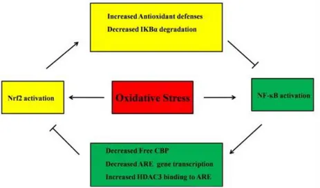
1.5.4 Crosstalk between Nrf2 and NF-κB
In the last few years a possible crosstalk between Nrf2 and NF-κB has been suggested, where the two pathways reciprocally inhibit the transcription and/or activity of downstream proteins (Fig. 8). This assumption is based on the fact that different activation and inhibition mechanisms have been demonstrated between the two pathways. In fact, it has been shown that the activation of Nrf2 and HO-1, following the exposure to antioxidants, is able to inhibit the nuclear translocation of NF-κB in prostate cancer cells (Bellezza et al., 2012) In addition, NF-κB nuclear levels obtained from the lungs of Nrf2 KO mice exposed to LPS were much higher than those from WT mice. In line with this, Lee and coworkers (2009) reported that Keap1 is able to induce IKKβ degradation by ubiquitation, consequently inhibiting p65 nuclear translocation.
On the other hand, NF-κB can in turn directly inhibit Nrf2 activity. In fact, since both factors bind to the same region of the transcription co-activator CREB binding protein (CBP), an increase in p65 levels prevents the binding of Nrf2 with CBP, repressing hence its activity (Liu et al., 2008). Furthermore, Yu et al. (2011)
29
demonstrated that p65 inhibits Nrf2 pathway through the nuclear translocation of Keap1.
It is believed that the levels of oxidative stress can move the balance needle towards the activation of one pathway or to the other one. A low degree of oxidative stress induces the activation of the Nrf2 pathway, and therefore an antioxidant response, while an intermediate level activates the NF-κB pathway and the inflammatory response. High levels are instead responsible for the induction of cell death processes (Gloire et al., 2006)
However, many aspects of this crosstalk are still unknown, so further studies are necessary in order to better understand this interaction.
Fig. 8: Crosstalk between Nrf2 and NF-κB pathways (modified from Ahmed et
al., 2017).
1.6 Oxidative stress and intestinal permeability
Intestinal epithelial cells (IEC) form a barrier over the entire GI tract, preventing the entry of pathogenic microorganisms and toxic molecules, thus maintaining intestinal homeostasis. They are the first cells that come into contact with the external environment and therefore can be considered as a first defence mechanism against possible harmful stimuli. The IECs can recognize pathogenic organisms, and secrete cytokines and chemokines activating thus the immune response