1 SCUOLA NORMALE SUPERIORE
Pisa
CLASSE DI SCIENZE MATEMATICHE, FISICHE E NATURALI
CORSO DI PERFEZIONAMENTO IN NEUROBIOLOGIA
Triennio 2008-2010 Tesi di perfezionamento
ENVIRONMENTAL STIMULATION AND ENVIROMIMETICS:
IMPACT ON A MURINE MODEL OF
DOWN SYNDROME
Candidata: Tatjana Begenisic
2 INDEX
CHAPTER 1: DOWN SYNDROME – INSIGHTS FROM MURINE MODELS 6
1.1. The human trisomy 21 7
1.2. Modeling Down syndrome in mice 10
1.3. Central nervous system dysfunction in Down syndrome – comparison between
affected humans and Ts65Dn mouse model 13
1.3.1. Cognitive and behavioral impairments 13
1.3.2. Motor and sensory abnormalities 19
1.3.3. Neuroanatomical correlates of neurophysiological impairments 22 1.3.4. Disruption of neurotransmitter systems and neurotrophic factors 30
1.4. Altered synaptic plasticity and disruption of excitatory-inhibitory balance as a central mechanism of neurophysiological impairments in Down syndrome 35
CHAPTER 2: EFFECTS OF ENVIRONMENTAL ENRICHMENT AND
ENVIROMIMETICS ON BRAIN IN HEALTH AND DISEASE 42
2.1. Neural consequences of environmental enrichment 43
2.2. Influence of environmental enrichment on brain development 45
2.3. Maternal care, tactile stimulation and visual system development in environmental
enrichment conditions 48
3 2.5. Beneficial effects of environmental enrichment on functional outcome in models of
brain disorders 53
2.6. Enviromimetics – a novel class of therapeutics 57
2.7. Antidepressant fluoxetine as a putative enviromimetic 58
CHAPTER 3: AIM OF THE THESIS AND EXPERIMENTAL DESIGN 62
CHAPTER 4: MATERIALS AND METHODS 66
4.1. Animals 66
4.2. DNA extraction 67
4.3. Genotyping 67
4.4. Screening for retinal degeneration 68
4.5. Rearing environments 69
4.6. Pharmacological treatment 70
4.7. Maternal care observations 71
4.8. Eye-opening observation 72
4.9. Morris water maze 72
4.10. Novel place recognition test 73
4 4.12. In vivo electrophysiology: assessment of visual acuity and binocularity 75
4.13. Analysis of GABA release in hippocampal and visual cortex synaptosomes 76
4.14. Statistics 78
CHAPTER 5: RESULTS 79
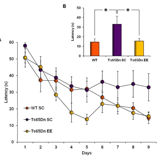
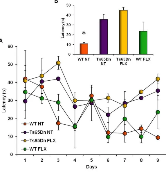
5.1. Effects of adult-onset environmental enrichment and fluoxetine treatment on Down syndrome related phenotypes displayed by Ts65Dn mice 79 5.1.1. Environmental enrichment promotes spatial learning and memory in
Ts65Dn mice 79
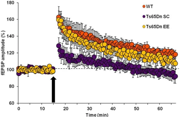
5.1.2. Recovery of LTP at medial perforant path–granule cell synapses in Ts65Dn
mice reared in environmental enrichment 81
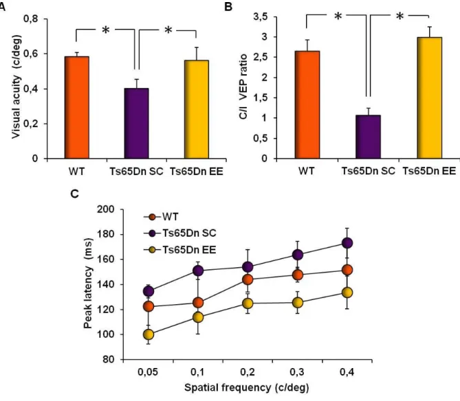
5.1.3. Restoration of visual functions in Ts65Dn mice by environmental enrichment 83 5.1.4. Environmental enrichment normalizes excessive GABA release in the
hippocampus and visual cortex of Ts65Dn mice 85
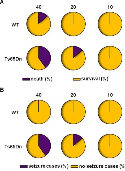
5.1.5. Evaluation of fluoxetine pro-convulsive side effects in Ts65Dn mice 86 5.1.7. Chronic treatment with fluoxetine improves short-term spatial memory
impairment in Ts65Dn mice 91
5.1.8. Effects of chronic fluoxetine administration on hippocampal synaptic plasticity
at the CA3-CA1 synapse 92
5.2. Impact of environmental enrichment on development in Ts65Dn mice 96 5.2.1. Maternal effects of environmental enrichment in Ts65Dn mice 96 5.2.3. Assessment of visual acuity development in Ts65Dn mice 101
5 6.1. Environmental enrichment and enviromimetics: amelioration of neurological phenotypes associated with Down syndrome in adulthood 103 6. 1. 1. Effects of environmental enrichment on adult Ts65Dn mice 103 6. 1. 2. Effects of fluoxetine treatment on adult Ts65Dn mice 108
6.2. Developmental impact of environmental enrichment in Ts65Dn mice 113 6.2.1. Maternal effects of environmental enrichment in Ts65Dn mice 113 6.2.2. Influence of pre-weaning onset of environmental enrichment on visual system
development in Ts65Dn mice 116
6.3. Conclusions 119
6
CHAPTER 1
DOWN SYNDROME – INSIGHTS FROM MURINE MODELS
Down syndrome (DS) is a genetic pathology caused by triplication of chromosome 21 (Hsa21) and is characterized by a number of physical and mental abnormalities, with intellectual disability (ID) as the most serious health problem. Complexity of DS arises from the substantial genetic basis of syndrome, the dysfunction of multiple systems and the high phenotypic variability of its clinical manifestations among affected individuals (Antonarakis and Epstein, 2006). Virtually all people living with DS have a triplication of at least one segment of Hsa21 being responsible for inappropriate development and resulting in structural and functional anomalies that persist throughout the entire lifespan. Beside the disruption of development, trisomic gene expression affects also the properties of differentiated adult cells, compromising further their already altered functions.
Sequencing of Hsa21 and identification of its mouse ortholog genes have led to generation of transgenic mice that have enabled remarkable advances in better understanding of the DS nature. The mouse is the organism of choice in modern biomedical research as it allows a vast degree of genetic alteration, high reproducibility of experiments due to the 1breeding simplicity and use of intrusive studies. Respect to human genome, orthologs in mouse differ minimally in the structures of conserved genes (Gardiner and Davisson, 2000), although differences in regulation of gene expression may be expected (Gharib and Robinson-Rechavi, 2011). Importantly, developmental programs and basic mechanisms of mature functions are conserved across mammals. In line with this is the occurrence of DS like phenotype in mice trisomic for Hsa21 orthologs (Das and Reeves, 2011).
7 recapitulation of disease aspects, but also a predictive ability, i.e. certain phenotypic abnormalities for the first time observed in trisomic mice have been confirmed later in DS patients (Belichenko et al., 2004). Moreover, systematic investigation of these models enables a more precise definition of genotype-phenotype interaction, better understanding of altered molecular pathways and identification of potential therapeutic targets, and finally design of preclinical studies with treatments likely to be tested in clinical trials.
1.1. The human trisomy 21
DS is a complex clinical entity caused by trisomy of Hsa21 (T21) and characterized by numerous features affecting multiple systems, with cognitive impairment as the most prominent and deleterious symptom. With the prevalence of 1 in 850-1000 infants (Shin et al., 2009) DS represents the leading cause of ID of genetic origin and an important public health issue due to inability of affected individuals to live an independent life. DS was described for the first time in 1866 by John Langdon Down, but the link of its numerous features with T21 was revealed only later in 1959 by the cytological profiling study performed by Jerome Lejeune (Megarbane et al., 2009). In the majority of cases DS is caused by the full T21 (95%), but the occurrence of two other genetic variations including translocation T21 (4%) and mosaic T21 (1%) also gives rise to DS phenotype (Parker et al., 2010). The full T21 is due to a meiotic nondisjunction of Hsa21 that is related to an advanced age of mother in about 75% of DS cases, while in remaining 25% it has paternal origin (Turleau and Vekemans, 2010). This nondisjunction seems to be associated with errors in recombination and an age-dependent loss of meiotic chromosome cohesion (Hassold and Hunt, 2001, Hodges and Wallace, 2005), but why these recombinant mistakes take place and how their consequential burden of DS could be avoided it is still far away from being understood.
8 The presence of an additional copy of Hsa21 implicates a quantitative rather than a qualitative nature for DS. However, genes situated in Hsa21 account for a less than 2% of total genomic material (Gardiner and Davisson, 2000), leading to the intriguing question of how can such a relatively modest quantitative genomic alteration give rise to such serious pathological consequences. According to the ‘‘gene dosage effect hypothesis’’, particular phenotypic traits associated with T21 are direct consequences of the imbalance of individual genes located on the triplicate chromosome (Pritchard and Kola, 1999, Antonarakis et al., 2001, Antonarakis et al., 2004). In agreement with this hypothesis is the identification of several “dosage-sensitive” regions, including genes and noncoding conserved elements, across the length of Hsa21, that have been shown to be sufficient for induction of typical features of DS individuals (Korbel et al., 2009, Lyle et al., 2009). On the contrary, the “amplified developmental instability hypothesis” postulates that triplication of a relative small number of genes disrupts global gene expression and regulation of intracellular signaling pathways, leading to deleterious effects on development and function (Hall, 1965, Shapiro, 1983, Pritchard and Kola, 1999, Moldrich et al., 2007). The main strength of this hypothesis is in the interpretation of DS as a “genomic disorder”, which suggests overlapping underlying mechanisms between DS and other trisomies. A synthesis of these opposite approaches proposes that some dosage sensitive genes, whose triplication per se would have only modest effects, may account for the numerous DS phenotypes only in combination with other triplicate genes (Olson et al., 2004, Roper and Reeves, 2006).
Similar to other conditions caused by chromosome imbalance, DS afflicts structure and function of multiple systems, producing detrimental effects on physical and mental health. Additionally, the clinical manifestations of DS change across the lifespan, contributing further to the complexity of syndrome (Antonarakis and Epstein, 2006). The first line of health problems are congenital malformations that include craniofacial and skeletal
9 dysmorphic features with more cosmetic than functional effects (Frostad et al., 1971, Fischer-Brandies, 1988), and significant cardiovascular (Ferencz et al., 1989) and gastrointestinal (Levy, 1991) anomalies that, if not treated properly, could lead to serious morbidity. Presence of cancer phenotype is controversial, as at the birth there seems to exist an increased risk for myieloproliferative disorders, while in the adulthood prevalence of many malignant tumors is decreased (Wechsler et al., 2002). The second line of symptoms develops progressively during life and, beside growth retardation, obesity, thyroid dysfunction and male sterility (Antonarakis and Epstein, 2006), it refers mostly to central nervous system (CNS) dysfunctions. CNS symptomatology of DS starts with central hypotonia at birth and continues with delayed cognitive development in infancy and childhood, leading to mild to moderate mental retardation that aggravates in adulthood, with an additional loss of cognitive abilities due to precoocious development of Alzheimer disease (AD). Generally, the lifetime of DS individuals is shorter with respect to the normal population and its duration is strongly influenced by the constellation of clinical signs that vary enormously in prevalence and severity across DS population.
Despite the numerous health problems mentioned above, life quality of people living with DS has been improved significantly due to enhanced medical and social care, which also resulted in an increased life expectancy from 12 years in 1940s to the current 60 years and over (Glasson et al., 2002, Roizen and Patterson, 2003, Bittles and Glasson, 2004, Bittles et al., 2007). However, CNS abnormalities are still orphan of an effective therapy, with ID being the most significant obstacle to an independent life for DS individuals. The availability of animal models in the field of DS basic research has started to reveal underlying mechanisms of T21 that contribute to the etiology of CNS dysfunction and cognitive impairment, providing the basis for designing of clinical trials aiming at the amelioration of mental abilities (Reeves and Garner, 2007, Wetmore and Garner, 2010, Rissman and Mobley,
10 2011, Haydar and Reeves, 2012)
1.2. Modeling Down syndrome in mice
The basis for modeling DS in mice is a conserved synteny between the long arm of Hsa21 and mouse chromosomes 16 (Mmu16), 17 (Mmu17) and 10 (Mmu10) (Pletcher et al., 2001). The long arm of Hsa21 is approximately 33.7 Mb in length and contains ~ 430 protein-coding genes, of which ~175 have a homolog in the mouse genome. The distal end of Mmu16 carries the largest homolog region containing 37Mb. This region spans from Rbm11 to Znf295 and is composed of ~115 orthologous genes with a subset of them, such as Ncam2,
App, Grik, Sod, Synj1, Olig1, Olig2, Dyrk1a, Girk2, Bace2, being involved in brain
development and functioning (Haydar and Reeves, 2012). The two remaining homolog regions are identified on Mmu17 and Mmu10 and are much smaller, containing 1.1 Mb and 2.3 Mb and consisting of 19 and 41 orthologous genes, respectively (Das and Reeves, 2011). The spontaneous Robertsonian translocations of Mmu16 naturally occurs in mice (Gropp et al., 1975) and this trisomy for Mmu16 (Ts16) has been considered the first model of DS. However, Ts16 model has a limited experimental use due to its very low postnatal viability, probably caused by trisomy of portions of Mmu16 that show sinteny with genes encoded by Hsa3, 8, 12, 6 and 22, which are not involved in etiology of DS (Moore and Roper, 2007). Genetic engineering of different segments of Hsa21-orthologues genes resulted in the availability of a comprehensive set of transgenic strains carrying those genes that show synteny with Hsa21 (Sérégaza et al., 2006). Importantly, these transgenic strains outlive Ts16 mice and may be studied at any developmental stage and across the entire lifespan.
The most important group of transgenic DS models encompasses mouse lines with triplication of various portions of the distal end of Mmu16. The most widely used and characterized model is Ts65Dn, which carries segmental trisomy in a form of small
11 chromosome produced by a Robertsonian translocation of Mmu16 to Mmu17. Trisomy in this model expands from Mrpl39 to Znf295 (Kahlem et al., 2004) and results in a dosage imbalance for ~104 genes conserved on Mmu16. Introduction of this model has been fundamental as it demonstrated for the first time that trisomy for syntenic genes of Hsa21 gives raise to DS-related outcomes in mice (Reeves et al., 1995). A detailed phenotypic characterization of Ts65Dn mice revealed a number of abnormalities in cognitive performance and brain morphology similar to those observed in people with DS. These findings have encouraged the use of Ts65Dn mouse as a standard to compare the incidence and severity of trisomic phenotypes reported in mouse models that came afterward (Holtzman et al., 1996, Baxter et al., 2000, Richtsmeier et al., 2000, Cooper et al., 2001, Rueda et al., 2005, Lorenzi and Reeves, 2006, Moore, 2006, Roper and Reeves, 2006).
Additionally developed partial trisomies carry smaller set of genes compared to Ts65Dn mice. In the case of the Ts1Cje model, the trisomic segment spans from Sod1 to
Znf295 and counts for ~81 orthologous (Sago et al., 1998), while the Ts1Rhr model has a
triplication for even a smaller region composed of ~33 genes expanding from Cbr1 to Orf9 (Olson et al., 2004). Ts1Cje and Ts1Rhr mice also show cognitive impairments, but the severity of their deficits is generally attenuated in comparison with Ts65Dn (Sago et al., 1998, Olson et al., 2007, Belichenko et al., 2009a). More complex models can be created when a third copy of a gene or chromosomal segment is subtracted from existing segmental trisomies by crossing them to a strain that carries a null allele/s for the gene/segment of interest thereby decreasing dosage of targeted gene/segment to euploid levels in the context of remaining trisomy (Salehi et al., 2006, Sussan et al., 2008). For instance, both Ms1Cje/Ts65Dn and Ms1Rhr/Ts65Dn were produced by breeding the corresponding monosomy of Ts1Cje and Ts1Rhr trisomies to the Ts65Dn mouse (Sago et al., 2000, Olson et al., 2004). These models have been developed in an attempt to understand the contribution of
12 specific orthologous genes to particular features of DS. However, the phenotypic analysis of these models revealed that the putative DS critical region (DSCR), believed to contain all essential genes responsible for the typical characteristics of DS, is not critically involved in the learning and memory deficit (Olson et al., 2007). These findings challenged the concept of specific dosage-sensitive genes as a primary cause of an abnormal phenotype of DS and rather supported the hypothesis that the over-expression of a large number of triplicated genes generally disrupts genomic homeostasis, leading to impaired regulation of developmental processes (Shapiro, 1997).
The models that are trisomic for the two remaining syntenic regions of Hsa21 mapped on Mmu17 and Mmu10 have been used to provide additional insights into the genetic basis of DS. The Ts1Yah mouse carries three copies of 12 genes, spanning from U2af1 to Abcg1, of the Mmu17 syntenic to the sub-telomeric region of Hsa21 (Pereira et al., 2009). This model has been created in order to understand how the telomeric region of Hsa21 contributes to DS phenotype. The effects of trisomy on learning and memory in Ts1Yah model are contradictory as some aspects of cognitive performance are impaired, while other are even improved with respect to euploid controls, emphasizing the concept that a complex interaction between genes underlies behavioral alterations in trisomic mice (Pereira et al., 2009). Interestingly, the Ts2Yey model, that contains triplication for 41 genes of the Mmu10 region which is homologue to the Hsa21 telomeric end, does not show any cognitive impairment (Yu et al., 2010). Recently, a ‘triple trisomy’ model, the Ts1Yey;Ts2Yey;Ts3Yey mouse, has been developed. The Ts1Yey;Ts2Yey;Ts3Yey mouse is the first model with a dosage imbalance for all the Hsa21 orthologous found in the mouse genome and it carries in triplicate the syntenic segments from all three mouse chromosomes, Mmu16, Mmu17 and Mmu10 (Yu et al., 2010). Nevertheless, the revealed brain phenotype of this model is very close to the Ts65Dn justifying the use of the Ts65Dn mouse as the referent murine model for
13 DS (Yu et al., 2010). Beside DS models based on synteny between human and mouse genome, a transchromosomic model, Tc1, has been also introduced. Tc1 mice have a transferred copy of Hsa21, but the human chromosome is lost in a subset of cells from all tissues (O'Doherty et al., 2005). The mosaicism for Hsa21 reported in this model could account for the mild cognitive deficits display by Tc1 mice (O'Doherty et al., 2005).
Given that the cognitive performance is the most compromised function in DS, the behavioral phenotypes of DS models have been thoroughly assessed to establish their relevance for the human condition. Particularly, impairments of spatial and recognition learning and memory have been studied in depth by use of a battery of behavioral tests that have helped to identify the hippocampus as the brain region specifically affected by trisomy in mice (Das and Reeves, 2011). In addition to behavioral evaluations, hippocampal morphological and electrophysiological correlates of cognitive deficits have been studied in DS models revealing a clear relationship between the number of trisomic genes that are orthologous to Hsa21 and the level of expression of functional and structural abnormalities.
1.3. Central nervous system dysfunction in Down syndrome – comparison
between affected humans and Ts65Dn mouse model
1.3.1. Cognitive and behavioral impairments
HumansDifficulties in intellectual functioning are the most striking feature of DS. The major obstacle to an independent life in DS patients are limitations in cognitive performance that are additionally complicated by functional difficulties coming from various areas, such as linguistic, social, motor, and sensory. However, similarly to other genetic disorders associated with ID, different domains of intellectual processing in DS are not affected to the
14 same level by T21, leading to a complex cognitive profile characterized by specific weaknesses and strengths (Fidler et al., 2006).
Cognitive impairments in DS could be classified into those that are a product of disrupted neural development resulting in retardation of learning and memory from birth, and those that occur in adulthood as a consequence of accelerated neurodegenerative processes (Contestabile et al., 2010). DS individuals during the course of childhood and adolescence develop mild to severe ID that, when expressed with the Intelligence Quotient (IQ), falls into the range from 30 to 70 (Vicari et al., 2000, Vicari et al., 2004, Vicari et al., 2005). Cognitive development of DS infants begins relatively typically and the first delays are observed at the age of two years which may be associated with the myelination lag at this developmental stage (Koo et al., 1992). One of the key features of the overall slowdown in maturation of learning abilities for children with DS are difficulties in the maintenance of acquired skills and a persistent use of counterproductive strategies for novel problem solving tasks (Wishart, 1993).
Learning deficits in DS children refer to both short-term and long-term memory (Carlesimo et al., 1997, Vicari et al., 2000, Brown et al., 2003, Vicari et al., 2005). When assessed with tasks requiring low processing levels, visuospatial components of short-term and working memory tend to be speared in contrast to verbal processing, which instead demonstrates significant deficits respect to age-matched controls (Vicari et al., 1995, Jarrold et al., 1999, Vicari et al., 2006). As the task becomes more demanding, certain impairments emerge also in the visuospatial skills, and those reported for verbal processing worsen (Lanfranchi et al., 2004, Visu-Petra et al., 2007). Strengths and weaknesses are also present in domain of long-term memories. Explicit memory is significantly compromised in DS children due to poor information encoding, impaired retrieval abilities and attention deficits (Carlesimo et al., 1997, Brown et al., 2003, Krinsky-McHale et al., 2008), whereas learning
15 abilities for tasks requiring implicit memory processing seem to be preserved (Vicari et al., 2000). This is in agreement with a different mechanism underlying this two types of memory formation, as implicit memory is a more automatic process based on low levels of attention, while explicit memory requires a high degree of attention to carry on intentional learning and develop successful retrieval strategies.
Impairment of long-term memory in DS has been associated with hippocampal and prefrontal lobe dysfunctions. Namely, deficit in spatial long-term memory reported in pre-school DS children assessed by delayed recall of place learning task (Pennington et al., 2003) pointed to altered hippocampal function, whereas impairment of non-verbal reasoning ability and attention has indicated a specific executive control defect due to abnormal prefrontal functioning, supporting the concept that ID in DS is differentially affected by the degree of required control (Rowe et al., 2006). A high prevalence of behavioral disinhibition, illustrated by hyperactivity, aggression, stubbornness, disobedience, impulsivity and already mentioned attention deficits (Cuskelly and Dadds, 1992, Dykens et al., 2002), is also indicative of prefrontal dysfunctions in DS. Recognition memory, another type of explicit memory partially dependent on hippocampus (Yonelinas et al., 2005) and frontal lobe (Neufang et al., 2006) integrity, is also affected in DS. DS children perform normally in the task of matching photographs of simultaneously presented faces, but perform significantly worse in the more demanding task of matching faces to non-present people (Wishart and Pitcairn, 2000). Thus, similarly to described working memory deficits, the level of task difficulty determines the quality of recognition memory performance, indicating that in DS cognitive deficits may be generally more pronounced in challenging situations.
In addition to developmental ID, DS patients may undergo additional cognitive decline with aging and develop precocious AD (Nieuwenhuis-Mark, 2009). Although at the neuropathological level virtually all subjects with DS develop typical hallmarks of AD during
16 adulthood (Ball and Nuttall, 1980, Hof et al., 1995, Folin et al., 2003, Nadel, 2003, Lott and Head, 2005), only a portion of patients demonstrate clinical signs of dementia (Devenny et al., 1996, Devenny et al., 2005). Clinical progression of AD in DS has some similarities with dementia onset in the general population, with confusion, forgetfulness, impairment of recent memories and a relative preservation of distant memories at early stages of disease (Deb et al., 2007). On the other hand, the signs of frontal lobe dysfunction, such as indifference, apathy, depression, socially-deficient communication and impaired adaptive functioning (Zigman et al., 1996, Lott and Head, 2001, Ball et al., 2006), may be expressed during initial phase of AD in some cases of DS (Deb et al., 2007, Ball et al., 2008), while among the general population these clinical manifestation typically occur only in a late phase of AD.
As discussed above, frontal lobe abnormalities and defective executive functions have been also reported in young DS subjects (Rowe et al., 2006), emphasizing the diagnostic difficulties in detecting cognitive decline caused by dementia in the context of ID (Nieuwenhuis-Mark, 2009). Moreover, in DS the risk of developing some forms of mental disease increases over time (Dykens et al., 2002), representing an additional confounding factor in the correct diagnosis of the cognitive deficit. Depression is the most common psychiatric problem among young adults affected by DS (Myers and Pueschel, 1991, Collacott et al., 1992) and its progression may lead to additional decline in adaptive behavior and cognitive performance during mid and late adulthood (Burt et al., 1992).
Ts65Dn
Similarly to human DS subjects, disruption of neural development processes leads to early cognitive impairments also in Ts65Dn mice (Bianchi et al., 2010b), with a further worsening occurring in adulthood due to neurodegenerative processes (Hyde and Crnic, 2001, Faizi et al., 2011).
17 Short-term working memory has been assessed by spontaneous alternation tasks, revealing a reduced performance in Ts65Dn mice (Fernandez et al., 2007, Belichenko et al., 2009a) and resembling short-term memory deficits observed in DS individuals (Lanfranchi et al., 2004, Visu-Petra et al., 2007). Furthermore, Ts65Dn mice, as well as persons with DS, are characterized by a functional dissociation between implicit and explicit long-term memory. Namely, Ts65Dn mice perform similarly to wild type (WT) mice in implicit memory tasks, as indicated by normal acquisition and maintenance of skills in the rotarod performance test (Hyde and Crnic, 2001, Fernandez et al., 2007).
On the other hand, Ts65Dn mice demonstrate selective impairments in explicit learning and memory tasks. Two prototypical domains of explicit memory that can be delineated in rodents through a variety of behavioral tasks are spatial and recognition learning and memory. These experimental paradigms depend on the functional integrity of the medial temporal lobe, composed of the hippocampus and the parahippocampal region, the same areas associated with deficits in declarative memory displayed by patients with DS (Nelson et al., 2005). A particular vulnerability of the trisomic hippocampus is well illustrated by the outcomes of two different forms of fear conditioning. In an acoustic fear conditioning, a behavioral test based on the amygdale (Anagnostaras et al., 1999), Ts65Dn mice do not show any significant defect (Salehi et al., 2009, Faizi et al., 2011), while there is a clear evidence of impaired performance in a context fear conditioning (Costa et al., 2008, Salehi et al., 2009, Bianchi et al., 2010b, Faizi et al., 2011), a paradigm mainly dependent on the hippocampus (Kim and Fanselow, 1992).
The most frequently used test to assess spatial long-term learning and memory in Ts65Dn mice is the Morris water maze (MWM). This paradigm evaluates visuospatial integration, as mice are trained to find a platform hidden in a tank filled with an opaque water, using a complex spatial mapping strategy based on extra- and intra-maze cues
18 (Morris, 1984, Redish and Touretzky, 1998). It has been repeatedly shown that Ts65Dn mice perform similarly to euploid mice in a cued task that does not involve visuospatial integration because the hidden platform is marked with visible cues such as a flag (Escorihuela et al., 1998, Sago et al., 2000, Bimonte-Nelson et al., 2003, Rueda et al., 2008). This indicates that motivation is not affected in Ts65Dn mice which have sufficient motor and visual skills to reach a visible platform. However, in the hidden-platform task Ts65Dn mice display significant navigational impairments and a general inability to create successful strategies to find and remember the position of the platform (Escorihuela et al., 1995, Reeves et al., 1995, Demas et al., 1996, Holtzman et al., 1996). It is important to note that deterioration in the performance of Ts65Dn mice in MWM may be partially due to a stressful nature of the test as a certain degree of aversiveness is an integral part of MWM (Stasko and Costa, 2004). This indicates that challenging situations are more prone to reveal deficits not evident in stress-free circumstances. This is also supported by the recent finding that Ts65Dn mice show identical outcomes as WT mice in place preference and place avoidance learning tasks when tested in the home cage environment, which can be considered a stress-free condition (Faizi et al., 2011). However, recognition memory evaluated by non aversive object recognition tests is also impaired in Ts65Dn mice, illustrating the persistence of some cognitive deficits even in low stress circumstances. Ts65Dn mice exhibit significant deficits in the simple version of the novel object recognition task (a simple protocol involving a pair of identical objects during the familiarization phase), but a complete inability to distinguish novel and familiar objects in the complex version of the test (a more challenging protocol involving a pair of different objects during the familiarization phase) (Fernandez et al., 2007). Importantly, the profile of this impairment is in accordance with previously described neuropsychological studies in human DS, reporting more pronounced difficulties in intellectual processing in DS patients with increasing task demands (Lanfranchi et al., 2004,
19 Visu-Petra et al., 2007).
The majority of the above mentioned behavioral tests refers to young adult Ts65Dn mice at the age of 3-4 months, but for some tasks longer follow up studies have been performed, allowing to uncover an additional amount of cognitive performance decline with age (Hyde and Crnic, 2001, Faizi et al., 2011). This is reminiscent of the cognitive deterioration due to an early onset of AD among DS individuals. It has been proposed that hyperactivity, frequently observed in Ts65Dn mice (Davisson et al., 1993, Reeves et al., 1995, Coussons-Read and Crnic, 1996, Stewart et al., 2007) may be an early sign of AD in this model. Namely, it has been shown that prefrontal cortex lesions leads to hyperactivity in rodents (Kolb, 1974, Takakusaki, 2008), and this type of disinhibited behavior in Ts65Dn mice could be driven by an altered prefrontal information processing, as it has been shown to be the case in the human DS (Rowe et al., 2006).
1.3.2. Motor and sensory abnormalities
HumansTogether with a poor cognitive performance, various motor and sensory deficits are associated with DS. Acquisition of motor milestones generally follows the same sequences found in typically developing children, but is characterized by significant delays that tend to be more pronounced for the latest milestones (Palisano et al., 2001, Vicari et al., 2006). Two typical characteristics of DS that significantly influence motor performance are low muscle tone and lack of control of muscle stiffness (Davis and Sinning, 1987, Vicari et al., 2006). Impairments are evident in all motor domains, as illustrated by a poor performance in tasks assessing fine and gross motor skills and motor planning (Jobling, 2001, Mon-Williams et al., 2001). However, performance in some specific skills, like running speed, agility and visual-motor control, reaches the level of age-matched controls (Jobling, 2001, Mon-Williams et al.,
20 2001) indicating that some qualities of motor function are preserved in DS.
The development and function of auditory and visual pathways are also affected by T21 (Folsom et al., 1983, Diaz and Zuron, 1995). Hearing loss occurs in approximately two thirds of DS children and is one of their most common sensory disabilities (Roizen et al., 1993). During infancy there is a high incidence of conductive hearing loss due to congenital malformation of the ear anatomy and repetitive ear infections, while progressive sensorineural loss is frequently found from middle childhood to adult life, as demonstrated by difficulties to evoke potentials by auditory stimulus in the brainstem (Jiang et al., 1990, Chen and Fang, 2005).
Although less frequent then auditory defects, visual system impairments are also an integral component of DS symptomatology. Visual acuity (VA), the ability to see fine details of objects, and contrast sensitivity, the ability to discriminate between different brightness levels, are reduced in infants and children with DS when compared with their normally developing peers (Courage et al., 1994, Woodhouse et al., 1996). Ophthalmic anomalies, such as refractive errors (Woodhouse et al., 1997), accommodative inaccuracy (Cregg et al., 2001), strabismus (Haugen and Hovding, 2001), and nystagmus (Wagner et al., 1990) are typical of DS patients, but cannot entirely account for their reduced vision, because children without such anomalies still show poor VA and contrast sensitivity (John et al., 2004). Abnormalities in cortical visual evoked potentials (VEPs) in DS patient rather point to a functional deficit of central visual pathways (Ellingson, 1986). The basis of this dysfunctional visual processing may be structural dendritic abnormalities characterized by progressive loss of dendritic branching and total length in the visual cortex, during the period from the first postnatal months to the two years of age (Becker et al., 1986). In adulthood, some DS patients show AD-like visual deficits (Rocco et al., 1997), as impaired color discrimination and stereoacuity, the ability to detect differences in distance, indicating that
21 both congenital abnormalities and age-related neuropathology within the visual pathways could contribute to sight defects.
Overall, the importance of motor and sensory deficits in DS is not only in the primary loss of their functions, but also in the capability to negatively influence the intellectual functions already compromised by trisomy.
Ts65Dn
Assessment of motor and sensory function in Ts65Dn mice has demonstrated that acquisition of developmental milestones in trisomic pups recapitulates the abnormal aqusition of motor and sensory skills of DS infants (Holtzman et al., 1996, Palisano et al., 2001, Vicari et al., 2006). Motor function has been evaluated in detail and it has been reported that young adult Ts65Dn mice display an altered motor performance in several paradigms including grip force, running and swimming speed and motor coordination in the rotarod test (Costa et al., 1999). The motor coordination deficit and its relevance for gait has been additionally investigated by motorized treadmills and the CatWalk automatic gait analysis system, revealing abnormalities in walking patterns and dynamics (Hampton et al., 2004, Faizi et al., 2011). The main deficits reported in Ts65Ds mice correspond to gait abnormalities observed also among DS children (Shumway-Cook and Woollacott, 1985, Parker et al., 1986, Galli et al., 2008).
While well studied in DS subjects, sensory deficits have been reported only recently in trisomic mice. Ts65Dn mice have elevated thresholds for auditory-evoked brainstem responses (ABR) compared to WT controls (Han et al., 2009). This finding faithfully recapitulates human deficits as an earlier clinical study found that ABR thresholds of DS infants are higher than those found in typically developing children (Werner et al., 1996). Similarly to the conductive nature of hearing loss among young DS population (Buchanan,
22 1990, Hassmann et al., 1998), auditory deficits in Ts65Dn mice have been correlated with a high prevalence of otitis media, thought to be provoked by altered craniofacial dimensions and middle ear anatomy (Hill et al., 2007) and depressed immune function (Han et al., 2009). However, while hearing loss aggravates with age in patients with DS due to additional sensorineural complications (Chen and Fang, 2005), ABR time-course experiments show a time-stable hearing loss in Ts65Dn mice (Han et al., 2009).
Loss of vision has been also recently reported for the first time in Ts65Dn mice. Similarly to persons with DS (John et al., 2004), recordings of VEPs revealed that Ts65Dn mice have lower spatial resolution, the electrophysiological correlate of VA, and higher contrast threshold, the electrophysiological inverse correlate of contrast sensitivity, than WT mice (Scott-McKean et al., 2010). Electroretinography assessments showed no significant deficits in retinal physiology in Ts65Dn mice in comparison with WT control mice (Scott-McKean et al., 2010), supporting the assumption based on clinical studies that a dysfunction of central components of visual processing may be involved in the visual deficits associated with DS (John et al., 2004).
1.3.3. Neuroanatomical correlates of neurophysiological impairments
HumansMorphological alterations characteristic of the DS brain arise from a disruption of neural development leading to abnormal anatomy and connectivity of CNS, evident from the earliest evolutionary stages, and from a progressive atrophy believed to result from AD-like neurodegeneration, occurring during adulthood and superimposing on the existing neural changes.
Brain volume is smaller in DS subjects compared to age-matched healthy controls, even after normalizing for body size that is generally reduced in DS (Yoshimura et al., 1990,
23 Raz et al., 1995). Macroscopic analysis of postmortem brain tissue and ultrasonographical data have shown that the reduction of the overall volume of DS brain emerges during prenatal development (Winter et al., 2000, Guihard-Costa et al., 2006) and progresses further during the gestational period (Schmidt-Sidor et al., 1990, Golden and Hyman, 1994, Engidawork and Lubec, 2003). Singular brain areas are also modified by trisomy as illustrated by small cerebellum, frontal and temporal lobes, reduced number and depth of the cerebral sulci and a narrow superior temporal gyrus (Coyle et al., 1986, Wisniewski, 1990, Becker et al., 1991). Magnetic resonance imaging (MRI) studies have confirmed these regional brain abnormalities, emphasizing a disproportional reduction of cerebellar and hippocampal volumes in young DS subjects (Raz et al., 1995, Pearlson et al., 1998, Aylward et al., 1999, Pinter et al., 2001).
In agreement with the reduced volumes, lesser amount of neurons has been calculated in the hippocampus, the parahippocampal gyrus, cerebellum and neocortex of DS fetuses (Baxter et al., 2000, Guidi et al., 2008, Larsen et al., 2008) and in the cortex of DS children (Wisniewski, 1990), indicating that morphological alterations observed among young DS individuals may arise from decreased output of newborn neurons during development. Direct evidence that T21 disrupts neurogenesis at early developmental stages has come from the study showing that cell proliferation is impaired in the dentate gyrus (DG) and ventricular germinal matrix in the brains of DS fetuses at 17-21 weeks of gestation (Contestabile et al., 2007). Further analyses revealed that the G2 phase of neuronal cell cycle is prolonged in DS, probably accounting for slower proliferative rate during neurogenesis (Contestabile et al., 2007) and fewer numbers of differentiated neurons in the DS developing brain (Guidi et al., 2008). An increased apoptotic rate has been also reported in the hippocampus of DS fetuses contributing to the overall lack of neural cells in this brain structure (Guidi et al., 2008). Moreover, T21 significantly modifies hippocampal cell phenotypes producing more cells
24 with a glial phenotype and less cells expressing neuronal markers (Guidi et al., 2008). Significant neurogenesis impairments were also found in the cerebellum of fetuses with DS, while the rate of apoptotic cell death was similar to controls (Guidi et al., 2011), pointing to different mechanisms underlying reduced cell numbers in the hippocampus and cerebellum.
Numerous brain structures of DS individuals brain undergo further atrophic changes with age. Similarly to patients with sporadic AD, old DS subjects are characterized by extensive cerebral atrophy, extracellular deposition of the amyloid β peptide (Aβ) in the brain parenchyma and blood vessel walls (amyloid angiopathy) and accumulation of intraneuronal neurofibrillary tangles (NFT) composed of a hyperphosphorilated form of tau protein (Mann, 1988, Jellinger and Bancher, 1998). Neuropathological changes in sporadic AD typically occur over 65 years of age and follow a specific sequence, as in the preclinical stadium they affect transentorhinal and entorhinal cortex, while in more advanced stages of the disease they extend to the hippocampus and neocortical regions (Braak and Braak, 1997). On the contrary, almost all DS patients demonstrate AD pathology by the age of 30, with the earliest pathological markers occurring in the medial temporal lobe and spreading to the prefrontal cortex, basal ganglia, thalamus, hypothalamus and midbrain (Wisniewski et al., 1985). The pathological alterations of these regions are believed to underlie the age-dependent cognitive decline in DS patients, asindicated by a positive correlation between the number of plaques or tangles and the degree of dementia severity (Blessed et al., 1968, Ropper and Williams, 1980, Terry and Davies, 1980, Wilcock and Esiri, 1982, Ulrich, 1985, Wisniewski et al., 1985). Importantly, MRI studies have revealed that in some particularly vulnerable brain regions, such as the hippocampus and the amygdala, atrophic changes precede the clinical onset of dementia, emphasizing that evaluation of regional atrophy may help to identify people with DS in the prodromal stages of AD (Krasuski et al., 2002).
25 is an increased Aβ burden due to an additional copy of the amyloid precursor protein (APP) gene mapped on Hsa21 (Rumble et al., 1989). Aβ is the major constituent of extracellular insoluble aggregates that are considered to be key morphological markers for AD. Increasing evidence indicates that overproduction of Aβ, besides its role in the formation of fibrillar aggregates, interferes with physiological mechanisms underlying learning and memory, triggering an early onset of dementia in DS (Conti and Cattaneo, 2005, Gasparini and Dityatev, 2008). It has been demonstrated that Aβ oligomers, small soluble clusters of Aβ, alter synaptic plasticity of excitatory synapses in rodents (Lambert et al., 1998, Wang et al., 2002, Townsend et al., 2006, Shankar et al., 2008, Li et al., 2009), and clinical data illustrate that Aβ oligomers in AD patients are even better correlated with clinical symptoms then Aβ plaques (Mc Donald et al., 2010).
Deterioration of cognitive performance in old DS subjects has been also related to degeneration of basal forebrain cholinergic neurons (BFCNs). Deficits in the cholinergic system are very similar to those occurring in AD (Mufson et al., 2008, Schliebs and Arendt, 2011) and loss of BFCN and decreased activity of choline acetyltransferase (ChAT) have been observed in both DS and AD (Whitehouse et al., 1982, Yates et al., 1983, Casanova et al., 1985, Mann, 1988, Mufson et al., 1993). The function of basal forebrain cholinergic system is apparently normal in DS fetuses and infants (Kish et al., 1989, Lubec et al., 2001) and the number of neurons and ChAT activity start to decrease later during adolescence and adulthood (Yates et al., 1983, Casanova et al., 1985, Godridge et al., 1987, Mann, 1988, Mufson et al., 1993, Schneider et al., 1997), further supporting the view that degenerative processes occur in DS subjects during aging (Contestabile et al., 2010).
The aberrant developmental and degenerative mechanisms in DS act together at the cellular level to alter the morphological properties of dendrites. The receptive function of dendrites relies on the integrity of dendritic spines that are critically involved in brain
26 connectivity and plasticity (Sorra and Harris, 2000, Kasai et al., 2003, Newpher and Ehlers, 2009). Abnormalities in density and morphology of dendritic spines and reduced dendritic branching have been associated not only with DS but also with other forms of ID (Huttenlocher, 1990, Kaufmann and Moser, 2000, Dierssen and Ramakers, 2006). Spine density and length and branching of dendrites are reduced in hippocampus and cortex of DS brain (Suetsugu and Mehraein, 1980, Takashima et al., 1981, Becker et al., 1986, Takashima et al., 1989, Ferrer and Gullotta, 1990, Schulz and Scholz, 1992, Takashima et al., 1994). Interestingly, in the fetal period and during the earliest postnatal months, normal, or even increased, dendritic branching has been observed in the visual and prefrontal cortex of DS brain (Takashima et al., 1981, Becker et al., 1986, Vuksic et al., 2002) indicating that DS neurons reach the same structural complexity typical of age-matched controls. However, the period of early postnatal maturation is characterized by a greater dendritic retraction of DS neurons respect to typically developing infants, resulting in the gradual appearance of dendritic abnormalities during the first months of age (Takashima et al., 1981). Dendritic abnormalities emerging in childhood progress with age, as illustrated by additional reductions of spine density and dentridic length and branching in aged adults with DS (Takashima et al., 1989). This age-dependent deterioration of dendritic phenotype is consistent with the early onset of AD among DS patients as dendritic pathology accompanies also initial stages of AD and correlates significantly with the progressive decline of mental faculties (Baloyannis, 2009).
Ts65Dn
Differentially from human DS, the overall size of postnatal Ts65Dn brain is not altered (Reeves et al., 1995). On the contrary, anatomical changes have been detected in specific brain regions, such as cerebellum and hippocampus, indicating that cognitive deficits
27 displayed by this model are based on selective vulnerability of specific neural circuits rather than on gross anatomical changes.
A reduction of cerebellar volume has been found in trisomic pups (Roper and Reeves, 2006) indicating that, similarly to human DS, the neuroanatomical modifications of Ts65Dn mice emerge at early stage of neural development. Morphological alterations of cerebellum have been analyzed in depth, reveling that both the molecular layer and the internal granular layer of Ts65Dn mice are thinner respect to WT controls due to decreased densities of Purkinje neurons and granular cells (Baxter et al., 2000). These findings have a predictive value as similar deficits in cerebellum structure were found afterwards also among people with DS (Baxter et al., 2000). Interestingly, despite the structural deficit, Ts65Dn mice perform as well as control mice on tasks designed to evaluate typical cerebellar functions, such as balance and motor coordination (Baxter et al., 2000, Hyde and Crnic, 2001). Since, however, increasing findings suggest an involvement of the cerebellum in various learning and memory paradigms (Rondi-Reig and Burguiere, 2005, Burguiere et al., 2010), the contribution of described cerebellar developmental deficits to some aspects of cognitive dysfunction in DS should not be ruled out.
Structural abnormalities have been observed also in the hippocampus and they are in agreement with a poor performance in hippocampus-dependent cognitive tasks displayed by Ts65Dn mice. The overall volume of hippocampus does not seem altered in Ts65Dn mice up to 7 months of age (Insausti et al., 1998, Lorenzi and Reeves, 2006, Olson et al., 2007), but significant structural alterations are evident in the hippocampal subfields from early life (Lorenzi and Reeves, 2006, Costa et al., 2008, Salehi et al., 2009, Bianchi et al., 2010b, Faizi et al., 2011). In the DG of Ts65Dn mice the number of granule cells and the volume of hilus and the granule cell layer are slightly reduced already at postnatal day 6 (P6) indicating that this early developmental hypocellularity may contribute to the specific behavioral
28 impairments reported in young trisomic mice (Lorenzi and Reeves, 2006).
Similarly to human DS, there is a growing body of evidence suggesting that the major determinant of morphological brain alteration may be a defective neurogenesis process during early developmental stages. Substantial delays in prenatal growth of the Ts65Dn cerebral cortex and hippocampus have been linked to longer cell cycle duration and reduced neurogenesis from the ventricular zone (Chakrabarti et al., 2007), while the deficit in cerebellar formation has been correlated to defective response of granule cell precursors to the signaling pathway mediated by the sonic hedgehog (SHH) growth factor (Roper et al., 2006). Emerging evidence indicates that adult neurogenesis in both the DG and the subventricular zone is also altered in trisomic mice and it has been postulated that this deficit could be involved in ID, as therapies aimed at rescuing postnatal neurogenesis have been able to ameliorate some aspects of cognitive performance (Clark et al., 2006, Bianchi et al., 2010a, Bianchi et al., 2010b, Chakrabarti et al., 2011).
Additional morphological alterations associated with aging occur also in Ts65Dn mice. Elevated levels of APP (Seo and Isacson, 2005) and Aβ (Netzer et al., 2010) have been reported in the cortex and hippocampus of Ts65Dn mice, but this model does not develop typical pathological hallmarks of AD (Reeves et al., 1995, Kurt et al., 2004). The occurrence of age-related cognitive decline in the absence of plaques and tangles and the recent demonstration that lowering Aβ levels rescues learning and memory in Ts65Dn mice (Netzer et al., 2010) support the hypothesis that soluble Aβ forms may be crucially involved in the pathophysiology of AD. Similarly to human pathology, BFCN are also affected by trisomy in Ts65Dn mice. It has been reported that an elevated expression of APP in Ts65Dn mice provokes an enlargement of early endosomes employed in axonal trafficking and an impairment of retrograde transport of nerve growth factor (NGF) from the hippocampus to the basal forebrain, leading to degeneration of BFCNs (Cataldo et al., 2003, Salehi et al.,
29 2006, Chang and Gold, 2008). NGF is a neurotrophic factor that enhances the survival, differentiation and function of specific neurons of the peripheral and central nervous systems, including BFCNs (Sofroniew et al., 2001). A critical involvement of APP in the loss of BFCNs is illustrated by the reversion of the degenerative phenotype after infusion of NGF (Cooper et al., 2001). The degeneration of BFCN in Ts65Dn mice occurs from 6-10 up to 20 months of age (Holtzman et al., 1996, Granholm et al., 2000, Hunter et al., 2004, Lockrow et al., 2009) and may account for age-related impairments in learning and memory (Hyde and Crnic, 2001, Hunter et al., 2003). Given that BFCNs provide strong modulatory inputs to the hippocampus, the cholinergic deafferentation may in turn underlie age-related volume reductions of hippocampal subfields (Kurt et al., 2004). However, differentially from observations in humans, the activity of ChAT appears to be increased in the cortex and hippocampus of 10 months old Ts65Dn mice and remains stabile even in older mice (Cooper et al., 2001, Seo and Isacson, 2005, Contestabile et al., 2006, Contestabile et al., 2008, Chen et al., 2009) indicating a possible activation of some compensatory synaptic mechanisms that may attenuate the impact of cholinergic loss on cognitive decline in this model.
Similarly to human DS, significantly reduced spine density and dendritic complexity of the cortical pyramidal neurons and the hippocampal pyramidal and granular neurons have been observed in adult Ts65Dn mice compared to control littermates (Dierssen et al., 2003, Belichenko et al., 2004, Belichenko et al., 2007). Electron microscopy has revealed that these dendritic abnormalities are accompanied by a size increment of presynaptic boutons, postsynaptic spines and average length of synaptic cleft (Belichenko et al., 2004). Dendritic alterations are already present at P21 indicating that, similarly to human DS, trisomy alters spine dynamic in Ts65Dn mice from early stages of development. Importantly, it has been revealed that various types of synapses are differentially affected by the trisomic condition. In the dentate granule cells of the hippocampus, there is a shift of inhibitory synaptic
30 connections away from the dendritic shafts and onto the dendritic necks, which would be expected to facilitate inhibitory synaptic transmission given the significantly reduced volume of the spine neck compared to the shaft (Belichenko et al., 2004). Moreover, inhibitory synapses have greater apposition lengths in Ts65Dn mice while excitatory synapses are unaltered, indicating a shift towards excessive inhibition in these mice (Belichenko et al., 2009b). The altered spine morphology and shift towards excessive inhibition characterizing DS may directly affect information processing by suppressing not only excitatory synaptic transmission, but also plasticity related signaling cascades that frequently rely on depolarization-mediated calcium influx into the postsynaptic structural domains (Cramer and Galdzicki, 2012).
1.3.4. Disruption of neurotransmitter systems and neurotrophic factors
HumansIn addition to the above discussed alterations in the cholinergic system, the glutamatergic, serotonergic, noradrenergic and γ-aminobutyric acid (GABA)-ergic systems are also profoundly disrupted in DS individuals. Reduced levels of serotonin (5-HT), GABA and dopamine (DA) have been reported in the frontal cortex of trisomic fetuses in comparison to euploid fetuses (Whittle et al., 2007). The imbalance in these neurotransmitter systems that emerges from the prenatal period may be involved in the disruption of numerous aspects of DS brain development. It has been shown that 5-HT plays an important role in neurogenesis, neuronal differentiation, dendritic development, axon myelination and synaptogenesis (Whitaker-Azmitia, 2001). Additionally, there is a defect in a dynamic of 5-HT receptors illustrated by an earlier peak of serotonin 5-HT1A receptors in developing DS brains (Bar-Peled et al., 1991). Reduction of GABA levels is also expected to impact the formation of DS brain as during development GABA acts as an epigenetic factor that controls cell
31 proliferation, neuroblast migration and dendritic maturation (Represa and Ben-Ari, 2005). The expression of three GABAA receptor subunits is also altered in the early prenatal life in DS, with up-regulation of subunit α2 and down-regulation of α5 and β3 subunits (Bhattacharyya et al., 2009). In the brain of adults with DS, 5-TH is below the levels of age-matched control group and is accompanied with a reduction in the levels of glutamate, aspartate and noradrenaline (NA) (Godridge et al., 1987, Risser et al., 1997). Reduced levels of 5-HT and NA in aged DS individuals are probable due to a degenerative loss of neurons from the dorsal raphe nucleus and locus coeruleus (Mann et al., 1985, Coyle et al., 1986), which send the serotonergic and noradrenergic inputs to the forebrain, respectively.
Disruption of neural development and accelerated neurodegenerative mechanisms in DS may be also affected by alterations of neurotrophin systems. Neurotrophins represent an important family of small proteins secreted in the vertebrates nervous system, that includes NGF, brain derived neurotrophic factor (BDNF), neurotrophin-3 (NT-3) and neurotrophin-4 (NT-4) (Meakin and Shooter, 1992, Huang and Reichardt, 2001). During development, neurotrophins regulate cell survival and differentiation, axon growth, dendrite pruning, the innervation pattering and the expression of neurotransmitters and ion channels necessary for normal neural functions. In the mature nervous system, neurotrophins continue to modulate neural survival and also take part in the control of synaptic function and plasticity (Huang and Reichardt, 2001, Sofroniew et al., 2001). Neurotrophins exert their action throughout two different classes of receptors. While all trophic factors bind to the p75 neurotrophin receptor, each of them also binds to specific members of the surface tyrosine receptor kinases (Trks): NGF to TrkA, BDNF and NT-4 to TrkB, and NT-3 to TrkC and to TrkA (Meakin and Shooter, 1992, Meakin et al., 1992, Chao and Hempstead, 1995). So far, clinical studies concerning DS have been focused on NGF and BDNF expression.Reduced serum levels of NGF have been found in children with DS (Calamandrei et al., 2000). Similarly, in the DS
32 fetal brain samples it has been found a lower expression of BDNF and TrkB isoforms (Toiber et al., 2010). On the other hand, opposite results were reported from adult individuals with DS, as illustrated by a significant increase of their BDNF blood plasma level with respect to age-matched controls (Dogliotti et al., 2010). New studies are necessary to better understand this apparent discrepancy in BDNF levels (lower expression in fetuses and higher expression in adults) between developing and aging DS brain.
Ts65Dn
Neurotransmitter levels and receptor expressions have been also investigated in Ts65Dn mice, indicating an alteration of multiple systems. However, some data show discrepancies with respect to human findings. For example, there are no changes of 5-HT levels in the hippocampus of Ts65Dn mice (Bianchi et al., 2010b) as well as no alterations in the number of serotonergic neurons of the dorsal and medial nuclei (Megias et al., 1997). On the contrary, reduction has been found in the expression of 5-HT1A receptors in hippocampal neurospheres and the hippocampus of newborn Ts65Dn mice (Bianchi et al., 2010b). As this receptors have been implicated in the regulation of neurogenesis (Malberg et al., 2000), their lower expression may contribute to the impairments of neural proliferation found in trisomic mice. This is supported by the rescue of early postnatal neurogenesis in Ts65Dn mice after treatment with the selective serotonin reuptake inhibitor (SSRI) fluoxetine that increases the expression of the 5-HT1A receptors up to normal levels (Bianchi et al., 2010b). Analyses of inhibitory and excitatory neurotransmitter systems in Ts65Dn mice have been particularly focused on the alterations in receptor subunit composition. The hippocampal region-specific distributions of immunoreactivity for α-amino-3-hydroxy-5-methyl-4-isoxazolepropionic acid receptor (AMPA) receptor subunits GluR1 and GluR2, GABAA receptor subunits α1 and β2/3, and GABAB receptor subunits R1 or R2 were investigated at 3 and 8 months of age,
33 revealing significant alterations in the expression of AMPA GluR2, GABAA β2/3 and GABAB R1 subunits respect to euploid animals (Belichenko et al., 2009b). Namely, in young Ts65Dn mice an overall decrease for AMPA GluR2 and GABAA β2/3 subunits was reported, while in older Ts65Dn mice it was found a reduction of AMPA GluR2, GABAB R1 and GABAA β2/3. The most interesting result is probably the age-dependent diminution in the expression of GluR2 and GABAB receptor subunit R1, in contrast with an increase in the expression of GABAA receptor subunit β2/3. The composition of N-methyl-D-aspartate receptor (NMDA) receptors appears normal in neonatal and young adult brains of Ts65Dn mice (Pollonini et al., 2008, Fernandez et al., 2009), but there is a reduced brain expression of the subunits NR2A and NR2B at 10 months of age (Vink et al., 2009). Although the physiological consequences of these alterations in the expression of AMPA, GABA and NMDA receptors are still unclear, a contribution to the additional cognitive decline found in older Ts65Dn mice may be expected. The noradrenergic system is also affected in Ts65Dn mice. Although the number of β-adrenergic receptors in the cerebral cortex and hippocampus of Ts65Dn mice is similar to that of euploid mice, their function, as assessed through analysis of 3',5'-cyclic AMP (cAMP) formation, is impaired and there is a reduced basal and stimulated production of cAMP (Dierssen et al., 1997).
Expression of neurotrophic factors is also defective in Ts65Dn mice. Reduction of BDNF levels in Ts65Dn mice have been observed in the hippocampus at 15 days and 3 months of age (Bianchi et al., 2010b, Fukuda et al., 2010) and in the frontal cortex at 6 months of age (Bimonte-Nelson et al., 2003). Importantly, normalization of BDNF expression induced by treatment with fluoxetine was able to restore survival of newborn cells, differentiation, and granule cell number in Ts65Dn mice (Bianchi et al., 2010b). Recent evidence has established an important link between chromosome 21-derived microRNA mirna21 and above mentioned deficits in expression of 5-HT1A receptor and BDNF in
34 trisomic brains. It has been demonstrated that mirna21 are overexpressed in the DS brain and, consequentially, a downregulation in the expression of the transcriptor factor methyl-CpG-binding protein (MeCP2) does occur (Kuhn et al., 2010). Numerous mutations in the MeCP2 gene have been associated with the neurodevelopmental disorder Rett syndrome (RS) (Amir et al., 1999), indicating a possible involvement of this gene also in DS brain pathology. MeCP2 activates the expression of numerous genes, including 5-HT1A receptor and BDNF (Chahrour et al., 2008), suggesting the possibility that reduced levels of these two proteins in Ts65Dn brain may be caused by silencing of MeCP2 gene. Given that fluoxetine administration increases MeCP2 expression in the DG (Cassel et al., 2006), it is possible to hypothesize that epigenetic modifications might also contribute to the restoration of a normal expression of 5-HT1A receptor and BDNF in Ts65Dn mice after treatment with fluoxetine (Bianchi et al., 2010b).
The impact of abnormal retrograde transport of NGF on progressive age-dependent degeneration of BFCNs in adult Ts65Dn mice have been discussed in the previous sections. However, it seems that there is a deficit in the hippocampal NGF expression also in the early postnatal period (Bianchi et al., 2010b), indicating that abnormal NGF dynamics should be also investigated in the context of DS neurodevelopmental pathology. Finally, increased levels of NT-3 have been found in total brain extracts of neonate Ts65Dn mice compared to their littermates (Pollonini et al., 2008), suggesting a possible attempt of the nervous system to protect itself from the neuronal loss that occurs in DS throughout development. Enhanced secretion of NT-3 has been also found in the hippocampus of adult Ts65Dn mice (Pollonini et al., 2008). As NT-3 expression by cortical neurons serves to attract basal forebrain cholinergic projections (Robertson et al., 2006), an increase in NT-3 may be a compensatory response to the age-dependent BFCNs loss of DS (Cooper et al., 2001, Chang and Gold, 2008).
35 Overall, substantial morphological and molecular evidence demonstrates that trisomy disrupts CNS development and accelerates age-related neurodegeneration of individuals with DS and Ts65Dn mice in a very similar manner, resulting in specific cognitive, behavioral, motor and sensory deficits. This clearly illustrates that the Ts65Dn mouse is an excellent model for better understanding of pathological processes underlying DS and preclinical evaluation of new therapeutic strategies.
1.4. Altered synaptic plasticity and disruption of excitatory-inhibitory
balance as a central mechanism of neurophysiological impairments in
Down syndrome
Learning and memory, key cognitive processes affected by DS, have been strongly associated with synaptic plasticity mechanisms (Bliss and Collingridge, 1993, Hofer and Bonhoeffer, 2010). Synaptic plasticity refers to the physiological ability of synapses to alter their structure, composition or function in response to changes in neural activity. Depending on the timing and strength of pre- and postsynaptic activity, synapses can either be strengthened or weakened, providing a potential mechanism for memory formation and storage (Dan and Poo, 2006). The connection between synaptic plasticity and learning and memory is typically studied within the hippocampus, as this brain region is critically involved in acquisition, storage and recall of spatial information (Nadel and Bohbot, 2001, Kesner, 2007, Moser et al., 2008). Consequentially, any structural or functional abnormalities in hippocampus that limit the capability of synapses to undergo plastic changes would be expected to compromise spatial cognition. The described morphological and behavioral alterations associated with trisomic hippocampus, like dendritic pathology (Becker et al., 1986, Takashima et al., 1989) and impairments in spatial learning and memory (Pennington et al., 2003, Edgin et al., 2010),