UNIVERSITA’ DELLA CALABRIA
Dipartimento di Fisica
CNR- Institute of Nanotechnology (NANOTEC)
Dottorato di Ricerca in
SCIENZE E TECNOLOGIE FISICHE, CHIMICHE E DEI MATERIALI
CICLO
XXIX
TITOLO TESI
Combined use of X-ray Fluorescence Microscopy, X-ray Phase Contrast Imaging, Atomic Force Microscopy and X-ray Nanotomography for high resolution quantitative Fe mapping in inflamed
cells.
Settore Scientifico Disciplinare FIS/03 (FISICA DELLA MATERIA) Coordinatore: Ch.mo Prof. Vincenzo Carbone
Firma
Supervisore/Tutor: Dr. Stefano Lagomarsino
Firma
Ch.mo Prof. Roberto Bartolino
Firma
Dottoranda: Dott.ssa Chiara Gramaccioni
Acknowledgements
First of all thanks to my supervisor Dr. Stefano Lagomarsino to believe in me and in my own capacity, He was a real scientific guide.
I would like to thank my two tutors, Dr. Stefano Lagomarsino and Ch.mo Prof. Roberto Bartolino, for such an interesting and ambitious topic, as well as for opportunities given to me and for introducing me in such vast and interesting scientific areas.
I also thank, Dr. Emil Malucelli, for his patient teachings, the answers to all my doubts, and the aids in many instances. To him in particular way I am debtor, but I also thank the Department of Pharmacy and biotechnology of the University of Bologna (also for kindness and hospitality in my days in Bologna); particularly Ch.mo. Prof. Stefano Iotti for his support and for his forward-looking advice.
I must also thank Dr.ssa Giovanna Farruggia for supporting and help me for the preparation of the frozen hydrated cells used in this thesis.
Thanks to my colleague and friend Dr.ssa Alessandra Procopio whose company has undoubtedly made this experience more enjoyable over the last few months; thanks for her support and friendship.
I would like to thank Dr.ssa Michela Fratini and Dr. Andrea Notargiacomo for having advised me throughout the years of the PhD, for the articles and all the scientific suggestions they gave me, Thanks!
Thanks also to Dr.ssa Inna Bukreeva for her help in the data analysis, and in particular for the discussion of the tomographic results
I also thank the Department of Public Health and Infectious Diseases of the Univerity of Sapienza of Roma, particularly Ch.ma. Prof. Piera Valenti, Ch.ma. Prof. Francesca Berlutti for support, and precious and timely advices that allowed me to accomplish this work with completeness and precision; thanks also to Dr. Luigi di Rosa for his support.
I would like to thank the European Synchrotron Radiation Facility for providing the Beamtime, especially the whole staff of the beamline ID16A: in particular Peter Cloetens, responsible of the beamtime, but also Yang Yang, Alexandra Pacureanu, , Sylvain Bohic and Julio Cesar da Silva for their continuos support, and their help during the experimental work and data analysis.
I thank Dr.ssa Alessia Cedola of the CNR-Nanotec of Roma for the precious and always timely advice.
I am debtor also with all the people who in one way or another have come into contact with this work and have endured my questions: Florin Fus and Yin Cheng.
Thanks to Ch.ma Prof Silvia Gaudenzi and Fra Andrea who accompanied me, with theirs cordiality, during the most significant stages of the activity of the research and without which this adventure would not have begun.
Thanks to Cost 1207 for making it possible, at many moments, to realize this work hrough more than one STSM.
Special thanks go to my family for their unconditioned support during this long working period. Thanks to God who gave me courage, strength and help for my work.
Finally, I thank Mickaël, who was always close to me in this last year. He always managed to understand my sacrifices.
I hope I did not bother anyone, and I hope to continue in the future to work on areas of the thesis and collaborate with the same people that I had the pleasure and the honor to work with for this thesis.
Werner von Braun
“Research is what I’m doing when I don’t know what I’m doing.” La pura ricerca è quando faccio quello che non so fare. Blaise Pascal
“Par l’espace, l’univers me comprend et m’engloutit comme un point; par la pensée, je le comprends.”
Attraverso lo spazio, l'universo mi comprende e mi inghiotte come un punto; attraverso il pensiero, io lo comprendo.
Abstract
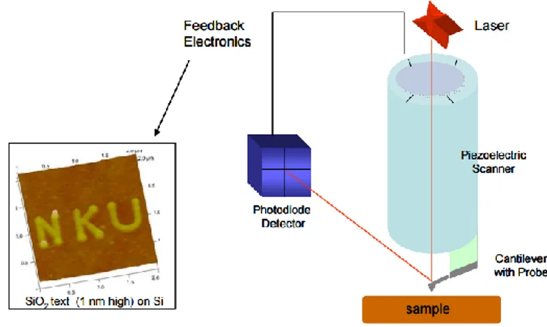
The PhD project is based on the applications of several x-ray microscopy techniques for compositional and morphological studies at nanoscale spatial resolution to a biological problem, i.e. the quantitative determination of morphological and compositional properties of epithelial cells. Three x-ray microscopy techniques were exploited in this work: X-ray fluorescence microscopy, X-ray phase contrast imaging and nanotomography, which were made at the ID16NI beamline of the European Synchrotron Radiation Facility in Grenoble, France. In addition to synchrotron-based techniques, also Atomic Force Microscopy was performed. The latter was used for morphology characterization, and forcalibration and comparison purpose. The main aim of this study was to quantitatively determine the map of iron concentration at nanoscale spatial resolution of epithelial cells infected by bacterial pathogens in the presence or absence of lactoferrin (Lf), an iron-chelating glycoprotein of natural immunity. Two experiments have been carried out at ESRF, one on freeze dried cells, and one on frozen hydrated cells this last using the cryo stage foreseen in the Id16 NI beamline, in order to examine cells as close as possible to their native state, and to avoid radiation damage. The measurement and data analysis protocols have been carefully studied for optimal combination of all the techniques, to give quantitative results. Iron concentration and mass fraction maps have been obtained, which give an insight about the modification of iron spatial distribution under the influence of lactoferrin. Moreover, for the first time it has been demonstrated the possibility to obtain quantitative element concentration in cells through the combination of x-ray nanotomography in phase contrast and x-ray fluorescence microscopy.
Key words
Synchrotron, X-ray imaging, Fuorescence, Phase Contrast, sub-cellular level, Imaging, Nanotomography, Atomic Force Microscopy, Freeze-dried, Frozen hydrated cells, Culture Cells,
Introduction
It was the 8th November of 1895, 120 years ago, when it was accidentally discovered the 'Existence of X-Ray, by the German physicist Wilhelm Roentgen. This fact in few months would revolutionize the field of medicine. In fact, next year, in the UK was built the First Department of radiology in a hospital and in a short time; the X-Rays began to be used in the world to obtain images of the fractures of bones. First radiology departments started to appear, like the one at the Glasgow Royal Infirmery. A number of remarkable X-ray images were made there: an X-ray of a kidney stone etc. New possibilities were discovered, not only to image the body parts but also to treat cancers or skin diseases. According to the most widespread history, Röntgen was doing the experiments with a fluorescent tube, an evacuated glass capsule through which was passed an electric current. For his experiment, Röntgen had covered the tube with the thick black cardboard sheets: whatever was illuminating the screen was at the same time practically invisible to the naked eye and able to penetrate the thick layer of paper, which covered the tube. Roentgen repeated the experiment many times to make sure to have not made a mistake. Then he tried to block the mysterious beam using a number of different objects, and found that only lead was good for this purpose. Finally, he replaced the screen with a photographic film and asked his wife to put his hand between the tube and the film. Röntgen in this way obtained the first X-Ray of the story: an image of the bones of the hand of his wife and his wedding ring (Figure1.1). He decided to provisionally call the mysterious rays "X" as the mathematical sign indicating an unknown quantity.
Figure 1.1: Hand with Rings: print of Wilhelm Roentgen’s first “medical” X-Ray of his wife’s hand.
However, X-rays are not only medical radiography. Immediately after their discovery, other extraordinary properties of X-rays have been studied. Among them, the possibility to obtain different X-ray fluorescence spectra for different elements, giving therefore the opportunity to accurate elemental analysis. Soon has been realized that X-rays are part of the electromagnetic spectrum, with wavelength close to the interatomic distances of crystals. Thus, the X-ray diffraction offered the opportunity to disclose the crystal structure, with countless fields of applications.
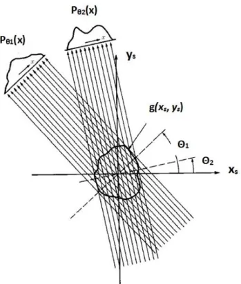
X-ray phase contrast imaging is a recent technique. Compared with absorption X-ray imaging, it requires specific properties of the X-ray beams used. These properties include strong spatial coherence, monochromaticity and high flux. As a result, most X-ray phase contrast imaging techniques have been developed using synchrotron sources. This currently limits the availability of these techniques, but there are ongoing developments to adapt it to conventional X-ray sources, such as interferometry based on diffraction networks (Pfeiffer et al., 2006).
For more than half a century X-rays have been generated by tubes similar in conception to that used by Roentgen. Only in the second half of XXth century new kind of sources, with unprecented
properties, have been developed, i.e. the synchrotron radiation sources, based on the radiating properties of charged particles circulating in a ring. ESRF is one of the most powerful synchrotron source in the world. It offers a brightness, energy range and resolution unachievable with laboratory radiation sources. This makes possible a large range of scientific applications otherwise impossible to exploit with traditional sources. A considerable part of research is dedicated to biomedical studies. Within this particular field, X-ray microscopy is a very important subject, in continuous development. One of the new beamline, build in the frame of the ESRF upgrade project, is ID16A, where the two experiments and the data analysis of this Ph.D. project have been performed. The ID16A nano-imaging end-station is dedicated to hard X-ray microanalysis nanoimaging and consists of the combination of X-ray fluorescence and 2D/3D X-ray phase imaging techniques. While X-ray fluorescence (XRF) reveals the chemical composition of the sample, the phase imaging completes the view by adding relevant information about the structure of the sample. Both techniques are exploited in this Ph.D. project in order to deal with questions related to the role of iron in inflamed cells. One of them is to quantitatively determine the intracellular map of iron concentration at nanoscale in cells infected by bacterial pathogens in the presence or absence of lactoferrin (Lf), an iron-chelating glycoprotein of natural immunity. The importance of iron for all eukaryotes, and particularly for humans, is widely recognized. Iron is essential for transport, storage and activation of oxygen, for electron transfer and many other metabolic processes. Iron homeostasis is also tightly linked to inflammation, immunity, and to response to pathogens. Macrophages are specialized cells that play a key role in these processes. Indeed, macrophages scavenge senescent and damaged erythrocytes, recycling iron recovered from hemoglobin. Many studies have addressed the relationships between iron management by macrophages, the ability of these cells to cope with invading pathogens and inflammation, and the effect of different molecules that can modulate the expression of proteins involved in iron homeostasis in macrophages. On the other hand, if not properly controlled, this situation can be dangerous leading to the anemia of inflammation (Paesano et al., 2012). Indeed, the expression of many proteins involved in cellular
iron management by macrophages varies in response to inflammatory stimuli as lipopolysaccharide (LPS) or to bacterial pathogens. Ferroportin, the sole protein able to export iron from the cells is down regulated in macrophages treated with LPS thus inhibiting iron export, increasing intracellular iron and leading to the anemia of inflammation (Paesano et al., 2012). Lactoferrin has been shown to possess potent antibacterial and anti-inflammatory properties (Valenti&Antonini, 2005; Berlutti et al., 2011). In particular, it has been recently showed that Lf counteract the LPS-induced decrease of ferroportin protein levels in adherent THP-1 macrophages challenged with LPS, a condition that mimics exposure to pathogens or inflammation. Restoring ferroportin at the plasma membrane will affect the iron export ability of these cells with possible beneficial effects, contrasting the anemia or the inflammation (Cutone et al., 2014). Therefore, it is of interest to evaluate the relationship between intracellular iron and bacterial infection in adherent macrophages. These studies at nano scale resolution could have a significant impact in defining new therapeutic interventions based on targeting intracellular iron.
In a first experiment we used freeze dried cells and we succeeded in recording fluorescence maps at 100 nm spatial resolution, together with 2D phase contrast images at 50 nm spatial resolution. Nanotomography at 50 and 25 nm resolution was also carried out on selected cells. Before the experiment session at ESRF, AFM was carried out on the same cells, in order to obtain independent information about morphology and to allow normalization of fluorescence intensity with illuminated volume, and consequently quantitative determination of concentration map. In a second experiment, we instead studied frozen hydrated cells, using the cryo stage installed in the Id16NI beamline. The frozen hydrated cells allow to examine cells as close as possible to their native state, and to avoid radiation damage. In this case, the illuminated volume cannot be measured with AFM, and therefore we used nanotomography for the purpose. The final aim of the study is combining all the techniques in order to obtain a quantitative mapping of iron concentration, mass fraction, density and volume of the cells, and to contribute to a better comprehension of the role of iron in the
inflammation process. Information on radiation damage can also be obtained by the combination of the different techniques.
The Ph.D. work is built as follows:
Chapter 1 describes the main characteristics of synchrotron radiation.
Chapter 2 is dedicated to a short description of the different X-ray microscopy techniques. Chapter 3 describes the Atomic Force Microscopy technique.
Chapter 4 gives a brief description of the beamline ID16-A where the experiments have been done.
Chapter 5 describes the procedure of sample preparation
Chapter 6 illustrates the data analysis procedure and the strategy to obtain quantitative information from the combination of all the techniques
Chapter 7 describes the results obtained on freeze-dried cells. Chapter 8 illustrates the results on frozen hydrated cells.
Chapter 9 gives the main conclusions, pointing out the problems encountered in the work, and illustrates future perspectives.
The original results, presented in this dissertation thesis, have been already published in part, and in part will be published in other manuscripts, currently in preparation (see List of publications).
My main contributions in this thesis work are presented in Chapters 5-6-7-8. In details:
Samples were cultured at the Department for of Public Health and Infectiuos Diseases, of the University Sapienza at Rome. I collaborated on the cell preparation to allow for measurements. In particular, I took part to the delicate process of fast freezing, and prepared both freeze-dried and frozen hydrated cells (see chapter V).
ii. I participated in all the synchrotron experiments described in the thesis: one on freeze dried (ls2362) and two on frozen hydrated cells (ls2433-ls2551), both carried out at the beamline ID16A of the european synchrotron radiation facility (ESRF) of Grenoble (see chapter IV). Moreover, I participated to some shifts in the frame of in-house beamline research.
iii. I carried out the data analysis on all the experiment results, in particular:
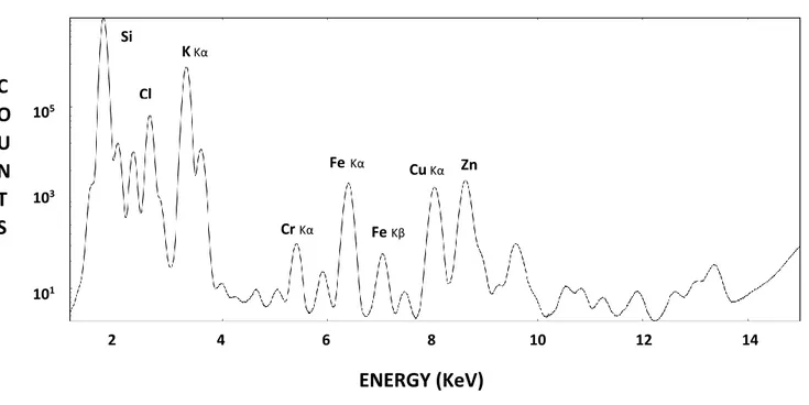
- the fluorescence analysis using the PyMCA program, obtaining the fluorescence intensity maps of Iron, Potassium and Phosphorus
- The analysis of the Phase Contrast images, using the programs developed by the beamline staff.
- The elaboration, using the MATLAB platform, of the concentration and of the mass fraction maps, combining the information obtained by the fluorescence, the Atomic Force microscopy and the Phase Contrast images.
- The analysis of the holotomography experiment, and the elaboration of the cell thickness map using segmentation, and of the concentration map combining fluorescence and holotomography.
- The elaboration of the average values of Iron, Potassium and Phosphorus in the cells, differentiating between the nucleus and the cytoplasm, presented in Annex II.
Gramaccioni C., Procopio A., Farruggia G., Malucelli E., Iotti S., Notargiacomo A., Fratini M.,
Yang Y., Pacureanu A., Cloetens P., Bohic S., Massimi L., Cutone A., Valenti P., Rosa L., Berlutti F. and Lagomarsino S. “Combined use of X-ray fluorescence microscopy, phase contrast imaging for high resolution quantitative iron mapping in inflamed cells.” Journal of Physics: Conference Series, Volume 849, conference 1.
Moreover, one work is to be submitted to Applied Phys. Letters:
Gramaccioni C., Yang Y., Pacureanu A., Cloetens P., Bohic S., Malucelli E., Iotti S., Procopio A.,
Bukreeva I., Notargiacomo A., Fratini M., Valenti P., Rosa L., Berlutti F. and Lagomarsino S.
“Nanoscale quantitative determination of intracellular element concentration combining X-ray fluorescence microscopy with holotomography.”
Several others are in preparation. Furthermore, the results have been presented in several symposium:
Poster presentation at the conference: BIOPHYSICS CNR, Area della Ricerca di Tor Vergata: “Quantitative high-resolution mapping of Fe concentration in inflamed cells, combining X-ray
Oral presentation at the conference: 23rd SILS Conference Trento: “Combined use of X-ray Fluorescence Microscopy, Phase Contrast Imaging and Atomic Force Microscopy for high resolution quantitative Fe mapping in inflamed cells”.
Oral presentation at the conference: Meeting WG3: X-ray coherent and incoherent imaging diagnostics on Advanced X-ray metrology for theranostic, Amsterdam.
Flash talk and poster presentation at the international conference X-Ray Microscopy XRM2016 OXFORD: “Combined use of X-Ray Fluorescence Microscopy, Phase Contrast Imaging and Nanotomography for high resolution quantitative Fe mapping in inflamed cells”.
Oral presentation at the conference: XTOP 2016 – 13th Biennial Conference on High-Resolution X-Ray Diffraction and Imaging –Brno, Czech Republic.
Oral presentation at the conference: COST Action: MP1203 MC meeting co-located with X-ray optic metrology meeting, Athens Greece.
Oral presentation at the conference: SILS 2016 Conference Bari: “Combined use of X-Ray fluorescence microscopy, phase contrast imaging and nanotomography for high resolution quantitative Fe mapping in inflamed cells.”
Oral presentation International Workshop on Process and Biomedical Tomography, Warsaw.
Oral presentation Microsymposiyum 2 (UDM2) ESRF user meeting 2017 Quantitative coherent X-ray diffraction imaging.
Oral presentation during the Seminar: Synchrotron radiation for Life Sciences and Biology Quantitative coherent X-ray diffraction imaging. Department of Public Health and Infectious Diseases at Sapienza University of Rome.
Poster presentation at the conference: Biophysics@rome 2017 CNR: “Nanotomography and X-Ray fluorescence microscopy for quantitative Iron concentration map in inflamed cells”.
Introduzione in Italiano
L’8 novembre del 1895, 120 anni fa, il fisico tedesco Wilhelm Röntgen scoprì per caso l’esistenza dei raggi-X, novità che nel giro di pochi mesi avrebbe rivoluzionato la medicina: l’anno successivo nel Regno Unito era già in funzione il primo dipartimento di radiologia all’interno di un ospedale e nel giro di poco tempo i raggi-X cominciarono ad essere usati in tutto il mondo per ottenere immagini delle fratture di ossa. Secondo la storia più diffusa, quel giorno Röntgen stava facendo degli esperimenti con un tubo fluorescente, una capsula di vetro sottovuoto attraverso cui veniva fatta passare una corrente elettrica. Dopo aver iniziato il suo esperimento, Röntgen si rese conto che qualcosa di strano stava accadendo. Mentre si preparava a passare alla fase successiva, distolse lo sguardo dall’apparecchio e con la coda dell’occhio notò che uno schermo cosparso di una sostanza fluorescente, che aveva sistemato a poca distanza dal tubo, stava brillando fiocamente. La luce era visibile solo con la coda dell’occhio, dove è situata una parte particolarmente sensibile della retina. Guardando fisso lo schermo, invece, Röntgen non riusciva a vedere nulla. Per il suo esperimento, Röntgen aveva coperto il tubo con degli spessi fogli di cartoncino nero: qualunque cosa stesse illuminando lo schermo era allo stesso tempo praticamente invisibile a occhio nudo e in grado di penetrare lo spesso strato di carta che copriva il tubo. Röntgen ripeté l’esperimento più volte per accertarsi di non aver commesso un errore. Poi cercò di bloccare il misterioso raggio utilizzando una serie di oggetti diversi e scoprì che soltanto il piombo riusciva nel compito. Infine sostituì lo schermo con una pellicola fotografica e chiese a sua moglie di mettere la mano tra il tubo e la pellicola. In questo modo Röntgen ottenne la prima radiografia della storia: un’immagine delle ossa della mano di sua moglie e del suo anello matrimoniale. Decise di chiamare provvisoriamente i misteriosi raggi “X”, come il segno matematico che indica una quantità sconosciuta. Nei primi anni del 1900 i raggi X incominciarono ad essere utilizzati per trattare clinicamente malattie importanti come anche il cancro. Tuttavia, all'inizio non era ovvio che pur essendo ricoprendo un ruolo fondamentale nella cura delle malattie ci si sarebbe anche dovuti proteggere bene da essi in quanto le esposizioni sono nocive. Come ci troviamo oggi, dopo più di 100 anni da una scoperta straordinaria? Che conoscono le persone dei raggi X? Normalmente quando si pronuncia questa parola viene subito in mente l’idea di ospedale o malattia. Infatti, chi di noi almeno una volta nella vita non ha dovuto fare una radiografia o una tac per una visita medica? Ma non riduciamo raggi X solamente a questo, sarebbe troppo riduttivo! Se le persone hanno la possibilità di visitare una sorgente di sincrotrone, come ad esempio European Synchrotron Radiation Facility (ESRF) di
Grenoble, il loro parere in merito ai raggi X avrebbe la possibilità di arricchirsi. ESRF è la più potente sorgente di sincrotrone in Europa; capace di raggiungere un intervallo di energia ed una risoluzione irraggiungibile con le sorgenti di laboratorio. C’è una vasta gamma di possibili applicazioni scientifiche che utilizzano le proprietà dei raggi X della radiazione da sincrotrone. A ESRF, ad esempio ci sono diverse beamline a seconda del tipo di ricerca che si vuole effettuare. I fisici lavorano insieme con i chimici, ingegneri, biologi, medici, paleontologi ecc... Anche l’industria trova la sorgente di sincrotrone utile per le sue indagini, come ad esempio la “L'Oreal" che fa i propri esperimenti a ESRF. I raggi X ad esempio sono usati per studiare la struttura, i processi di crescita e la morfologia delle superfici. Essi sono utilizzati per studiare i comportamenti dei materiali ad altissime pressioni, per rivelarne la struttura cristallina. Infine essi sono utilizzati per applicazioni biomediche ad altissima risoluzione (50 nm). L’ imaging a contrasto di fase a raggi X è un arte del tutto recente. Al contrario dell’imaging per assorbimento, richiede specifiche proprietà del fascio di raggi X. Ad esempio una forte coerenza spaziale, una monocromaticità del facsio ecc.. Pertanto, la maggior parte delle tecniche di imaging X a contrasto si sono sviluppate a seguito delle sorgenti di sincrotrone di sincrotrone. Gli esperimenti effettuati e la loro relativa dati, presentati in questa tesi di di dottorato sono stati svolti presso la beamlineID16NI a ESRF. In particolare questa beamline ID16NI è dedicato a raggi X duri e la microanalisi consiste nella combinazione di raggi X di fluorescenza con tecniche a raggi X di imaging di fase sia 2D che 3D. Mentre i raggi X di fluorescenza (XRF) ci rivelano la composizione chimica del campione, l’imaging a contrasto di fase completa la nostra analisi aggiungendo informazioni relative alla struttura del campione. Entrambe queste tecniche sono state sfruttate in questa ricerca di dottorato al fine di affrontare alcune questioni fondamentali. Uno di questi è di determinare quantitativamente la concentrazione di ferro con una spaziale nanometrica in cellule infettate da batteri i in presenza o assenza di lattoferrina, una glicoproteina naturale del sistema immunitario.
L’importanza del ferro per tutti gli organismi viventi e in modo particolare per gli esseri umani è universalmente riconosciuta. Il ferro è un elemento essenziale per il trasporto dell’ossigeno per il trasferimento degli elettroni ed è responsabile di molti altri pocessi del metabolismo.
L’omeostasi del Ferro è anche strettamente legata ai processi di infiammazione, immunità, e alla risposta agli agenti patogeni. I macrofagi sono cellule specializzate che svolgono un ruolo chiave in questi processi. In effetti, i macrofagi riciclano il ferro recuperato dall’ emoglobina. Molti studi hanno affrontato proprio la relazione tra il ferro e i macrofagi, che sono cellule in grado di reagire agli agenti patogeni e ai processi infiammatori, e si sono anche fatti degli studi riguardanti l'effetto di diverse molecole in grado di modulare l'espressione delle proteine coinvolte nell'omeostasi del ferro nei macrofagi stessi. D'altra parte, se non adeguatamente controllata la concentrazione del
ferro, questa situazione può essere pericolosa portare a processi di infiammazione anemica (Paesano et al. 2012).
La lactoferrina (Lf) è stato dimostrato che ha forte proprietà antibatteriche e antinfiammatorie (Valenti&Antonini, 2005; Berlutti et al., 2011). In particolare recentemente si è dimostrato che Lf contrasta la riduzione di ferroportina causata da LPS. Restaurando la ferroportina nella membrana del plasma, influenza dunque la capacità di esportazione del ferro da parte di queste cellule con effetti benefici che contrastano dunque l’infiammazione anemica (Cutone et al., 2014). Ciononostante sarebbe molto interessante essere in grado di poter valutare la relazione tra la concentrazione del ferro intracellulare e le infiammazioni batteriche in particolar modo come diminuisce a livello quantitativo la concentrazione del ferro in queste condizioni. Questi studi ad una risoluzione spaziale nanometrica possono avere un ruolo significativo per definire nuovi interventi terapeutici mirati alla concentrazione del ferro intracellulare.
Nel primo esperimento, ls2362, abbiamo usato cellule liofilizzate “freeze-dried”, e siamo riusciti a registrare mappe di fluorescenza con una risoluzione spaziale di 100 nm e delle immagini a contrasto di fase 2D con una risoluzione spaziale di 50 nm. È stata anche fatta tomografia a contrasto di fase con risoluzione spaziale di 50 e 25 nm sulle stesse cellule. Inoltre, prima dell'esperimento abbiamo misurato, sulle stesse cellule, la loro topografia con la tecnica dell’Atomic Force Microscopy (AFM), in modo da poter normalizzare le intensità di fluorescenza con il volume illuminato, ed essere quindi in grado di determinare quantitativamente le mappe di concentrazione. Tuttavia, dopo le misure di fluorescenza abbiamo notato un po’ di danni da radiazioni. Nel secondo esperimento, ls2433, abbiamo utilizzato cellule idratate-congelate “frozen hydrated”, utilizzando il criostato nella beamline ID16A, per esaminare le cellule il più vicino possibile al loro stato nativo. In questo caso il volume illuminato non può essere misurato con la tecnica dell’AFM, e pertanto si utilizzerà la tomografia per tale scopo. Inoltre, nel secondo esperimento si sono utilizzate invece di cellule murine delle cellule umane THP-1 macrofagi, al fine di acquisire informazioni utili per la salute umana. In questa tesi abbiamo analizzato anche i dati tomografici, delle cellule ad alta risoluzione per avere informazioni strutturali tridimensionali, cosa che non è mai stata fatta prima. Infine combinando queste tecniche (fluorescenza a raggi X, imaging a contrasto di fase 2D, tomografia a contrasto di fase e AFM), è stato possibile studiare la radiazione e gli effetti sui campioni cellulari; infatti non c’è alcun dubbio che durante l’esposizione del campione al fascio dei raggi X si sono verificati alcuni danni da radiazione.
La tesi di Dottorato è impostata nella seguente maniera:
Capitolo 1 descrizione delle caratteristiche della radiazione da sincrotrone.
Capitolo 2 descrizione delle tecniche di Imaging e di fluorescenza basate sulla radiazione da sincrotrone.
Capitolo 3 descrizione della teoria alla base della tecnica AFM.
Capitolo 4 descrizione della beamline ID16-A ESRF a Grenoble, dove sono stati svolti gli esperimenti oggetto di studio.
Capitolo 5 in questo capitolo sono descritti i campioni biologici. Murrin cells linea J774A.1 (ATCC® TIB-67™), trattate con lipopolisaccaride (LPS) e con lattoferrina (Lf).
Capitolo 6 trattazione dei dati analizzati e della metodologia adotatta per ottenere delle informazioni quantitative attraverso l’utilizzo di tutte le tecniche descritte nel precedente capitolo 2.
Capitolo 7 descrizione dei risultati ottenuti sulle cellule macrofaghe freezedried. Capitolo 8 descrizione dei risultati ottenuti sulle cellule macrofaghe frozen hydrated. Capitolo 9 riassunto della ricerca e prospettive.
Gli originali risultati, presentati in questa tesi di dottorato sono pubblicati parzialmente e in parte saranno oggetto di altre pubblicazioni attualmente in preparazione (vedi Elenco delle pubblicazioni).
I miei contributi principali in questo lavoro di tesi sono presentati nei Capitoli 5-6-7-8. Nel dettaglio:
i. Ho partecipato alla preparazione dei campioni. Le cellule sono state coltivate presso il Dipartimento per la sanità pubblica e malattie
infettive, dell'Università della Sapienza di Roma. Ho collaborato alla preparazione delle cellule per consentirne poi le misure. In particolare ho partecipato al veloce e delicato
processo di congelamento, e alla preparazione sia delle cellule freeze-dried che delle frozen hydrated (si veda il capitolo V).
ii. Ho partecipato a tutti gli esperimenti di sincrotrone descritti nella tesi: uno su cellule liofilizzate (ls2362) e due su cellule idratate congelate (ls2433-ls2551), tutti eseguiti nella beamline ID16A appartenente al sincrotrone europeo (ESRF) di Grenoble (si veda il capitolo IV). Inoltre, ho avuto a disposizione anche qualche shift aggiuntivo per la mia ricerca all’interno dell’in-house della beamline.
iii. Ho effettuato l'analisi dei dati su tutti i risultati acquisiti nel corso dei vari esperimenti, in particolare:
– Dall’analisi della fluorescenza con l’utilizzo del programma PyMCA, ho ottenuto le mappe di intensità di fluorescenza del Ferro, del Potassio e del Fosforo.
– Ho analizzato le immagini a Contrasto di Fase, utilizzando i programmi sviluppati dal personale della beamline.
– Ho elaborato, combinando le informazioni ottenute dalla fluorescenza, dalla microscopia a Forza Atomica e dalle immagini a contrasto di fase, utilizzando la piattaforma MATLAB, le mappe di concentrazione e di mass fraction.
– Ho analizzato i dati acquisiti di tomografia a contrasto di fase, ho elaborato utilizzando la segmentazione una mappa dello spessore della cellula e una mappa di concentrazione combinando fluorescenza e tomografia a contrasto di fase.
– Ho elaborato nelle cellule i valori medi di Ferro, Potassio e Fosforo, differenziando tra il nucleo e il citoplasma (si veda quanto riportato nell'allegato II).
Questa ricerca di tesi ha prodotto unalavoro già pubblicato:
Gramaccioni C., Procopio A., Farruggia G., Malucelli E., Iotti S., Notargiacomo A., Fratini M.,
Yang Y., Pacureanu A., Cloetens P., Bohic S., Massimi L., Cutone A., Valenti P., Rosa L., Berlutti F. and Lagomarsino S. “Combined use of X-ray fluorescence microscopy, phase contrast imaging for high resolution quantitative iron mapping in inflamed cells.” Journal of Physics: Conference Series, Volume 849, conference 1.
Un lavoro è in via di sottomissione ad Appl. Phys. Lett.:
Gramaccioni C., Yang Y., Pacureanu A., Cloetens P., Bohic S., Malucelli E., Iotti S., Procopio A.,
Bukreeva I., Notargiacomo A., Fratini M., Valenti P., Rosa L., Berlutti F. and Lagomarsino S.
“Nanoscale quantitative determination of intracellular element concentration combining X-ray fluorescence microscopy with holotomography.”
Altri lavori sono in via di elaborazione. Oltre alle pubblicazioni, il lavoro è stato presentato nel corso di diversi convegni:
Poster presentation at the conference: BIOPHYSICS CNR, Area della Ricerca di Tor Vergata: “Quantitative high-resolution mapping of Fe concentration in inflamed cells, combining X-ray
Oral presentation at the conference: 23 th SILS Conference Trento: “Combined use of X-ray fluorescence microscopy, phase contrast Imaging and atomic force microscopy for high resolution quantitative Fe mapping in inflamed cells.”
Oral presentation at the conference: Meeting WG3: X-ray coherent and incoherent imaging diagnostics on Advanced X-ray metrology for theranostic, Amsterdam.
Flash talk and poster presentation at the international conference X-Ray Microscopy XRM2016 OXFORD: “Combined use of X-Ray fluorescence microscopy, phase contrast imaging and nanotomography for high resolution quantitative Fe mapping in inflamed cells”.
Oral presentation at the conference: XTOP 2016 – 13th Biennial Conference on High-Resolution X-Ray Diffraction and Imaging –Brno, Czech Republic.
Oral presentation at the conference: COST Action: MP1203 MC meeting co-located with X-ray optic metrology meeting, Athens Greece.
Oral presentation at the conference: SILS 2016 Conference Bari: “Combined use of X-Ray fluorescence microscopy, phase contrast imaging and nanotomography for high resolution quantitative Fe mapping in inflamed cells.”
Oral presentation International Workshop on Process and Biomedical Tomography, Warsaw.
Oral presentation Microsymposiyum 2 (UDM2) ESRF user meeting 2017 Quantitative coherent X-ray diffraction imaging.
Oral presentation during the Seminar: Synchrotron radiation for Life Sciences and Biology Quantitative coherent X-ray diffraction imaging. Department of Public Health and Infectious Diseases at Sapienza University of Rome.
Poster presentation at the conference: Biophysics@rome 2017 CNR: “Nanotomography and X-Ray fluorescence microscopy for quantitative Iron concentration map in inflamed cells.”
Contents
Introduction/ Introduzione in italiano
CHAPTER 1
Synchrotron radiation
... 1 1 Characteristics of synchrotron radiation ... 1 1.1 Brightness, brilliance and flux ... 3CHAPTER 2
X-Ray microscopy techniques
... 5 2.1 Interaction of X-rays with matter ... 5 2.1.1 Absorption ... 5 2.1.2 Refraction and total reflection ... 6 2.1.3 Diffraction ... 7 2.2 Optics for X-Ray microscopy ... 8 2.2.1 Diffractive optics: zones plates ... 8 2.2.2 Total reflection optics: Kirkpatrick-Baez mirrors ... 11 2.3 X-Ray Fluorescence Microscopy ... 13 2.4 X-Ray Phase Contrast Imaging ... 15 2.5 X-Ray Nanotomography... 21 2.5.1 Principle of tomography……….22 2.5.2 Fourier slice theorem………. ... 23 2.5.3 Fourier reconstruction………. 24 2.5.4 Filtered back-projection ……….….24 2.5.5 Phase contrast tomography ………..2518
CHAPTER 3
Atomic Force microscopy technique
... 27 3.1 Principles ... 27CHAPTER 4
Beamline Layout
... 30 4.1 ID16A Beamline layout ... 30CHAPTER 5
Sample preparation
... 35 5.1 Methods ... 35 5.1.1 Sample preparation ... 35 5.1.2 Chemical Fixation ... 37 5.1.3 Cryofixation ... 37 5.1.4 Frozen-hydrated: preparation of cells ... 38CHAPTER 6
Data analysis and strategy for element quantification
... 40 6.1 Combination of different techniques for quantitative evaluation ... 40 6.1.1 Detection and processing of the X-ray fluorescence signa ... 42 6.1.2 Atomic Force Microscopy measurements ... 47 6.1.3 Phase contrast imaging measurements ... 48 6.1.4 Magnified Holotomography ... 53CHAPTER 7
Description of experiment on freeze-dried cells and results
... 57 7.1 Principles... 577.1.1 Results: Fluorescence maps, Phase reconstruction map, Atomic Force Microscopy map, Density map, Iron Mass Fraction map, Iron Concentration map for all the cells. ... 58 7.1.2 Results: Nanotomography and Iron concentrations map with volume informations
19
CHAPTER 8
Description of experiment on frozen hydrated cells and results
... 73 8.1 Results: Fluorescence maps, Phase reconstruction map, Iron Mass Fraction map, Iron for all the cells .. ……….73 8.2 Discussion ... 91CHAPTER 9
Main conclusions and future perspectives
... 101 9.1 Conclusion ... 101 47Riassunto in Italiano
... 103ANNEX
... 106 Annex 1 ... 107 Annex 2 ... 109Bibliography
... 112List of Publication
... 1151
CHAPTER 1
SYNCHROTRON RADIATION
1. Characteristics of synchrotron radiation
When electrons or positrons moving at relativistic speed, i.e., close to the velocity of light, if they are subjected to a magnetic field, their trajectory follows a circular orbit and synchrotron radiation (SR) is emitted in the tangential direction. The radiated energy is proportional to the fourth power of the particle speed and inversely proportional to the square of the radius of the path. The beam is concentrated into a forward narrow cone with half angle of typically 0.1 to 1 mrad. Synchrotron radiation facilities typically consist of an injection system, a storage ring and beamlines. In the injection system, electrons are generated, pre-accelerated, and sometimes a second accelerator further accelerates these electrons to few GeV before injection into the storage ring. At ESRF, the electrons are accelerated to a nominal energy of 6 GeV.
In the ring, bunches of electrons periodically circulate at relativistic speed for periods of up to many hours. The storage ring consists of radio frequency (RF) cavities, bending magnets, insertion devices and different control systems mainly to control the orbit of the electrons (Fig.1.1). The RF cavity system restores energy, which the electrons lose because of the emission of SR, and stabilizes the bunch of electrons. The high-energy electrons are maintained in a planar orbit by using bending magnet. Any accelerated charge radiates an e.m. field which, if the particle has a speed v << c (c light speed), is isotropic around the acceleration. In a synchrotron (or storage ring), the electrons have velocity v c, therefore relativistic effects dominate and the radiated e.m. field is sharply peaked in the direction of motion of the particles. The cone aperture is 1/ , where is the ratio between the particle energy and its rest mass. For electrons or positrons of energy E expressed in GeV = 1957 * E. Thus for 6 GeV electrons the aperture is about 80 rad.
2
Figure 1.1:
Schematic picture of the synchrotron ring of the ESRF (right) and external view of the building (left)Electrons are first accelerated in the linear accelerator (linac) and in a booster synchrotron, and then they are injected into a storage ring.
The emission spectrum is continous from infrared radiation up to a critical wavelength c which depends on and . The critical wavelength is defined as the value for which half of the total power is emitted at wavelengths shorter than the critical one; c is given by c = (4/3) /3. In order to improve the intensity of emission, insertion devices (I.D.) have been conceived where the charged particles pass through alternating magnetic (Figure 1.2) poles and are therefore compelled to have a zig-zag trajectory. At each wiggle (Figure 1.3), they emit radiation, which is therefore enhanced, of a 2N factor where N is the number of poles. These devices are called wigglers or undulators and the distinction is essentially due to the relation between the angular deviation at each wiggle and the aperture 1/. Values of >> 1/ identify wigglers, while 1/ identify undulators. The radiation emitted by a wiggler is the incoherent sum of the radiation fields emitted by each individual magnet. The spectrum is continuous but shifted at higher energies with respect to that of a bending magnet. Instead, in the undulator regime, the amplitudes of the field radiated at each period of the particle trajectory may interfere resulting in a periodic radiation field. The spectrum is thus not continuous, but resonances occur at given frequencies (the fundamental and the harmonics).
BENDING MAGNET BEAMLINE
3
Figure 1.2:Schematic picture of Bending Magnet. Figure 1.3:Schematic picture of Wiggler.
1.1. Brightness, brilliance and flux
What is important in S.R. is not only flux, which is defined as the number of photons per unit time in a given band-pass energy: F = ph/s/(/). For / is conventionally taken the value of 10 -3. Taking into account also the angular aperture (i.e. the collimation) we can speak of brightness which is the flux per solid angle: Brightness = ph/s/mrad2/(/) [1]. In fact also the size source is of importance and therefore the brilliance becomes the parameter of interest: Brilliance = Brightness/xy = ph/s/mrad2/xy/(/) where x and y are the transverse source sizes (horizontal and vertical). The emittance of the source is defined as: = h * v where the horizontal and vertical emittances h and v are defined as h = x'x and v = y'y, where x and y are the horizontal and vertical source size respectively and 'x and 'y are the divergence of the beam in the horizontal and vertical directions respectively . The figure of merit of a synchrotron is brilliance and emittance. The first should be as high as possible and the last should be as low as possible. Just to give an idea about numbers, if an x-ray tube has a brilliance of the order of 107, brilliance at an undulator beamline of third generation S.R. can be as high as 1020 - 1021. Horizontal and vertical emittances at ESRF reached values as low as 4 and 0.025 nm respectively.
Time structure is also important, at least for time resolved experiments. S.R. is not continuous, but is emitted in bunches corresponding to the electron bunches in the storage ring. Duration of a single bunch is, for ex. at the European Synchrotron Radiation Facility (ESRF) in Grenoble, of 20 picoseconds. Time between bunches depend on the filling mode of the storage ring. For single bunch mode (the one generally used for time resolved experiments) is about 3 s at ESRF.
One of the important characteristics of the photon beam is the coherence. There are two kinds of coherence: transverse coherence and longitudinal coherence. The transverse coherence refers to the coherence of the electromagnetic disturbances at two points perpendicular to the propagation
4
direction. Consider two points P1 and P2 that lie on the same wave front of an e.m. wave at time t=0. Let E1(t) and E2(t) be the corresponding electric fields at these points. By definition, the difference between the phases of the two fields at t = 0 is zero. If this difference remains zero at any time t > 0, there is a perfect coherence between the two points. If this occurs for any two points of the e.m. wave front, the wave is perfectly coherent (the degree of spatial coherence is 1). Usually, for any point P1, the point P2 must lie within some finite area (coherence area or length) around P1 to maintain the phase correlation.
From the Van Cittert-Zernike theorem [2] it comes out that, the radiation field from a primary incoherent source gains coherence during the propagation. Furthermore, the larger is the distance from the source and the smaller is the source size; the larger is the coherence length. The coherence length l can therefore be approximately defined as:
l = L/s where is the wavelength, L the distance source-sample and s the transversal source size. Synchrotron radiation is inherently incoherent, but due the large distance from the source, the beam at the sample position has a large coherence.
The longitudinal coherence refers to the case of two points along the propagation direction and is related to the monochromaticity of the beam.
The longitudinal coherence is characterized by the longitudinal coherence length lc, which is related to the bandwidth δλ/λ by:
5
CHAPTER 2
X-RAY MICROSCOPY TECHNIQUES
2.1 Interaction of X-rays with matter
X-rays are part of the electromagnetic (e.m) spectrum, extending in energy from about 250 eV to 50 KeV. Due to the simple relation E = h = hc/, where E is the energy, the wave frequency, the wavelength, c the light speed and h the Planck constant, the same range can be expressed in wavelength between 50 nm and 0.025 nm. The part at lower energy (up to about 2 KeV) is called “soft”, the more energetic “hard”. X-rays interact with matter essentially via electrons. An isolated electron subjected to interaction with an X-ray plane wave is instantly accelerated and then radiates as a dipole antenna.
The strength of the interaction has been established by Thomson and the ratio between the incident amplitude and the scattered one is proportional to the Thomson scattering length (called also classical electron radius) r0 = e2/mc2 = 2.82 x 10⁻¹³ cm. If the electron is bound to an atom a form factor, (atomic scattering factor) must be considered which takes into account the distribution of electrons in the atom. It can be useful to describe from a phenomenological point of view three phenomena that take place when X-rays interact with matter: absorption, refraction and diffraction.
2.1.1 Absorption
When an X-ray photon beam having an intensity I0 impinges on a target of thickness t, the intensity I after the target is simply given by: I = I0 e-t where is called the linear absorption coefficient. The physical phenomenon at the basis of absorption is the photoelectric effect. When an X-ray photon has an energy larger than the binding energy of a given electron shell, the photon is absorbed and the electrons are ejected with a kinetic energy given in first approximation by the difference between the photon energy and the binding energy. This implies that the absorption coefficient as a function of photon energy is not continuous, but has sudden jumps in
6
correspondence of the electron binding energies of the target elements. The jumps are called absorption edges and the absorption coefficient before and after the absorption edge are significantly different (6 to 8 times). Absorption edge of the first shell of electrons is called K, the second L, the third M, etc. When an electron is ejected, a hole in the corresponding shell is created which electrons of the next shell immediately fill. This gives rise in turn to emission of a photon (x-ray fluorescence) or to other electrons (Auger electron). We will describe the X-(x-ray fluorescence in detail in par. 2.3. In the energy region intermediate between two absorption edges, the absorption coefficient is approximately proportional to 3. This implies that is very different in the soft and hard regions. Just to give an example, photons of 25 KeV can pass 1 mm of Al with low losses, but are completely absorbed by an Al layer 1 micron thick if their energy is 410 eV.
2.1.2 Refraction and total reflection
The refraction index r of a material can be expressed by: r = n - i = 1 - - i. We recall here that n = (rr)1/2 where r and r are respectively the relative (to the vacuum) dielectric constant and the relative magnetic permeability of the material under consideration.
If an X-ray beam traveling in a medium with refraction index n1 impinges, at an angle 1, on the flat surface of a material having refraction index n2, it will be refracted following the simple Snell formula: cos1 /cos 2 = n2/n1
Using quantum mechanical theory and for a monatomic material in the X-ray region can be expressed respectively as:
= 2 (N0/A)m r0(Z+f ‘) and = (/4) (2.1)
where N0 is the Avogadros’ number, A the atomic mass, m the density, r0 the classical electron radius, Z the atomic number of the element and f’ a correction factor related to the rapid variation of the atomic scattering factor for X-rays in proximity of the absorption edges. is the linear absorption coefficient. is always positive and this implies that for X-rays all the materials have a refraction coefficient less than 1. Therefore if medium1 is vacuum (or air) n2 < n1 and the radiation will travel in the material with an angle 2 < 1 As a consequence, there will be an incident angle 1 = c for which 2 =0. In this case, the incident beam is totally reflected and only an evanescent wave can enter into the material. c is called the critical angle for total reflection and is not difficult
7
to see that c (2)1/2. Therefore, c is proportional to and to the square root of Z and m. This property is the basis for optics based on total reflection and for refractive x-ray optics.
2.1.3 Diffraction
In refraction and reflection the atoms do not absorb the x-ray photons, but once excited by them, they are-irradiate X-ray beam of the same energy (in the elastic approximation). The same happens in diffraction. Diffraction is a common phenomenon of interaction between a wave and an object having dimensions of the same order of magnitude of the wavelength into consideration. Every point of the object, following the principle of Huygens, becomes source of spherical waves of the same wavelength of the incoming radiation. We can distinguish between Fresnel and Fraunhofer diffraction [3]. If we take a scale factor defined as d2/2, where d is the object dimension and the wavelength, we can roughly speak of Fresnel diffraction when at least one of the relevant distances source–object (D1)or object-detector (D2) are smaller or of the same order of d2/2. Fraunhofer diffraction will take place when both the relevant distances D >> d2/2 and therefore plane wave approximation is valid. Bragg diffraction is a special case of Fraunhofer diffraction: if many objects interact with the incoming wave-front (that we suppose a plane wave) the resulting field is the superposition of the spherical wavelets emitted by all the objects. If they are disposed in an ordered array, like in a crystal, the superposition will give rise to constructive interference only for given directions where the wavelets are in phase, otherwise destructive interference will take place. This is the basis of Bragg diffraction. The simple formula which express the condition for constructive interference has been expressed by W.L. Bragg in 1913 [4] who considered the crystal as composed of sets of parallel planes of ions. The Bragg law is then:
= 2 d sin (2.2)
Where is the wavelength, d the interplanar distance and the angle of incidence of the incoming (monochromatic) radiation onto the lattice plane.
8 z P z+ /2 d
2.2 Optics for X-Ray microscopy
Many types of X-ray optics have been developed to improve the flux density (flux per unit area) woth the purpose to make possible different kind of X-ray microscopy (X-ray imaging, X-ray fluorescence microscopy, X-ray diffraction microscopy, etc.). Here we will briefly describe just two types of X-ray optics: diffractive optics (zone plates) and reflection optics (mirrors), because they are the most commonly used for X-ray microscopy.
2.2.1 Diffractive optics: zone plates
The zone plate is so called because it is constructed following the Fresnel-zone law:
A plane opaque screen with a circular aperture of radius d illuminated by coherent radiation (i.e. a plane wave) produces, at a generic distance z from it (Figure 2.1), the usual diffraction pattern of lighter and darker rings on a screen normal to the z-axis.
Figure 2.1: Opaque screen with a circular aperture of radius d. The points on the aperture distant z+/2 from P define the radius of the first Fresnel zone for the point P.
This diffraction pattern is not of interest here and the analysis will be focused on the diffraction along the z-axis alone. The field amplitude W(z) along the z-axis oscillates depending on the d2/z ratio according to the following relation [3]:
2 sin 2 1 2 F N kz i N i ikz N iAe e Ae z W F F (2.3a) where: z d NF 2 (2. 3b)9
is the Fresnel number. Different regions of the aperture emit radiation, which interferes constructively or destructively depending on the distance z. For any given point P on the z axis, it is convenient to classify the regions on the z = 0 plane (where the aperture is located) depending on the constructive or destructive interference produced in P. These regions are named Fresnel zones.
The first Fresnel zone for the point P distant z from the screen is the circle of radius r1
z z z r 2 2 2 1 2
It identifies the points on the z=0 plane at a distance smaller than z+/2 from P. The radiation emitted from these points interferes constructively in P since the optical path lengths differ by less than /2. The field generated by this circle in P is easily obtained from (2.3a) by taking d=r1 and therefore NF=1:
ikz i ikz Ae e Ae W1 1 2 (2. 4)So that its amplitude is twice larger than the field amplitude in the absence of the screen. In other words, if the aperture had a radius d=r1, then the whole radiation passing through it would interfere constructively in P thus explaining the increased intensity.
The second Fresnel zone is a ring of internal radius r1 and external radius given by:
z
z zr22 2 2 2
The field produced at P by the points in this second Fresnel zone is obtained by setting d= r2 in (2.3b) and subtracting the field produced by the points in the first zone:
ikz i i ikz Ae e e Ae W2 1 2 1 2 (2.5)As it is apparent from the negative sign, if the aperture had a radius d=r2, the radiation from the first Fresnel zone would interfere destructively with the radiation coming from the second one and the intensity in P would vanish.
The radius of the generic nth zone is:
rn nz n=1,2.. (2.6)
It is important to notice that the Fresnel zones are independent on the size of the aperture and they only depend on the position of P and on the wavelength.
For a point at distance z the number NF in (2.3b) is the number of Fresnel zones contained in the aperture. In particular, if this number is an even integer the interference of the radiation from the
10
various zones is destructive and the intensity in P is zero. If NF is an odd integer, the radiation from one whole zone is unbalanced and the intensity is maximal.
The Fresnel zone plate (FZP) is a screen of alternately transparent and opaque zones equal in size to the Fresnel zones. The efficiency is quite poor, because a relevant part of the incoming radiation is simply lost. An advancement is constituted by the phase zone plate. In the phase zone plate the opaque zone is substituted with a material which introduces a chane of pahse of . In this way both the odd and the even zones contribute to the total intensity, thus improving the ZP efficiency.
Illuminating the screen with a plane wave, for m0 the zone plate produces spherical waves with r1 the radius of the innermost zone, n the zone number and m the number of diffracted order, the focal length fm of a zone plate being approximately:
m r f r m f k m m 2 1 2 1 2 (2.7)
It is important to notice that the position of the focal point depends on the wavelength. The width of the outermost zone of a zone plate can approximately be written as:
n r dr n n 2 (2.8)
One of important characteristic of the zone plate is the resolution which can be expressed, according to the Rayleigh criterion [5,6], as:
m drn 22 . 1 (2.9)
where drn is the width of the outermost zone of the ZP. This represents the best possible resolution of the zone plate, but other factor can influence the effective spot size, such as a limited distance between source and ZP, and the non perfect monochromaticity of the beam.
The zone plates are mainly used for imaging. In this case, a condenser ZP lens concentrate the flux on a limited area of the sample, and an objective ZP lens gives a sample magnified view on the detector. The ZP lenses are mostly used in the soft X-ray region, where X-ray microscopy, especially in the water window, is able to reach resolution of the order of few tens of nanometers. In the hard X-ray region they are not very efficient, because the opaque zone should have a too large
11
aspect ration (ratio between thickness and width), to block, or even to introduce a change of phase, if high spatial resolution is needed.
2.2.2 Total reflection optics: Kirkpatrick-Baez mirrors
An interesting optics to focus hard X-rays is given by grazing incidence total-reflection mirrors. A pair of elliptical mirrors arranged in a crossed mirror geometry (Kirkpatrick-Baez configuration) is able to focus X-ray to sub-micron size (Figure 2.2).
p2
q2
p1
q1
Figure 2.2: Schematic diagram of the optical system of two crossed Kirkpatrick-Baez curved mirrors to form a demagnified image of an X-ray source.
The X-ray beam passing through a Si (111) symmetric double crystal monochromator (CM) is focused by the elliptical mirrors. The radiation incident on the first mirror (M1) is vertically focused. The reflected beam from M1 is incident on the second mirror (M2) which horizontally focuses the beam.
To achieve a focused image it is necessary to satisfy the focus equations at the centre of the first and second mirrors:
sin 2 1 1 ; sin 2 1 1 2 2 2 2 1 1 1 1 q R p q R p
12
where p1,q1 , p2,q2 are clear from Figure 2.3, R1 and R2 are the mirror radius of curvature and 1 and 2 are the angles of glancing incidence for the first and second mirror respectively. For the simple case of cylindrical mirrors, the following conditions are fulfilled:
s q s
p
p2 1 q2 1
where s is the distance between mirror centres.
When multilayer mirrors are used, it is additionally necessary to satisfy the Bragg equation for each mirror: d m d1sin 1 2 2sin 2 2
where d1 and d2 are the periods of the multilayers and m is the reflection order. Combining the equations given above one arrives to the following expression:
1 1 1 1 1 2 2 1 1 K p s K p s R d R d
where K1 is the magnification of the first mirror (K1= q1/p1). It is important to note that for high de-magnification of the source (i.e. small beam size), p1 and p2 must be much larger than q1 and q2, respectively. In general the magnification of the second mirror K2=q2/p2 is different from that of the first one and for the special case of identical radii and periods one can find:
2 1
1
K K
Kirkpatrick and Baez (KB) developed this crossed mirror geometry to eliminate astigmatism of a single spherical (or cylindrical) mirror used at glancing incidence. The spatial resolution of the KB system was limited mainly by spherical aberration. Therefore, the mirrors with elliptical cylinder shapes instead of spherical or cylindrical are used to remove spherical aberration.
The advantages with respect to other focusing optics are that optical system with small aberration can be designed, the focusing properties do not depend on the incident X-ray energy, some types of mirror figure can be manufactured with high accuracy and the working distance from the focusing element to the sample can be relatively long. Nanometer beam size can be achieved using high quality KB mirror systems.
In the beamline ID16A, where the experiments have been carried out, the distance of the end station from the source is very high (185 m). This long distance is necessary to have a high de-magnification factor, and to reach in this way nanometer beam size. The beamline is optimised for high-resolution quantitative 3D imaging techniques with a specific focus on X-ray fluorescence and projection microscopy and is mainly dedicated to problems in biology, biomedicine and
13
nanotechnology. It is optimised for hard X-ray focusing of the beam (< 20 nm) at specific energies (17 and 33.6 keV, ΔE/E ~10-2). The beamline uses fixed curvature multilayer coated KB optics. A detailed description of the beamline is done in chapter V.
2.3 X-ray Fluorescence Microscopy
As seen before, one of the basic interaction of X-rays with matter is the photoelectric absorption In this process a photon is absorbed by an atom creating a hole in the atom with the ejection of an electron. The energy of this photo-electron is the difference between that of the incident photon and the binding energy of the electron. The electron vacancies created by the incoming photon are filled by electrons cascading in from outer electron shells. These have higher energy states than inner shell electrons, and the rearrangement of electrons results in emission of Auger electrons and of X-ray photons characteristic of the given atom (Figure 2.3).
Figure 2.3: Schematic picture of the X-ray fluorescence. The inner shell electron hit by the energetic photon is expelled from the atom. The vacancy is filled by other electrons and characteristic X-ray radiation is emitted.
This emission of photons is called X-ray fluorescence and it is emitted isotropically. The X-ray fluorescence lines are historically termed by the letter K, L, or M (Figure 2.4), indicating which shell had the original vacancy, and by a subscript alpha (α) or beta (β), which indicates the higher shell from which electrons fell to fill the vacancy and produce the X-ray. For example, a Kα line is produced by a vacancy in the K shell filled by an L shell electron, whereas a Kβ line is produced by a vacancy K shell filled by an M shell electron. The K lines are by far the most intense; moreover, the Kα transition is on average 6 to 7 times more intense than the Kβ ones. Therefore, the Kα line is in general the choice for quantification purposes. Obviously, to generate K lines the incoming photons must have enough energy to excite the innermost electrons.
14
Figure 2.4: Nomenclature of X-ray transitions.
Since the X-ray fluorescence spectrum is characteristic and different for each elements, X-ray fluorescence spectroscopy has been used since the discovery of X-rays for elemental analysis. However, only with the advent of third generation synchrotron radiation sources, the X-ray Fluorescence Microscopy (XRFM) could become a technique able to give information at micrometer and sub-micrometer scale. This because the generation of X-ray fluorescence photons is very inefficient, and only with SR it is possible to obtain an intense finely focused X-ray beam able to get measurable fluorescence intensity from very small volumes. Furthermore, the monochromatic beam and the polarization of the synchrotron beam improves the Signal/background ration, which is one of the most important figure of merit in fluorescence analysis. Anyway, in relatively few years XRFM has opened up new applications, such as trace element analysis, surface analysis, chemical state analysis and microanalysis [7]. XRFM works in scanning mode. Digital images of the sample at micrometer or nanometer spatial resolution are built, pixel by pixel, by scanning the sample through the beam. The resulting X-ray fluorescence spectrum is characteristic of the chemical elements at that pixel. Mathematical deconvolution of the fluorescence spectrum reveals the chemical composition, from which quantitative elemental images of the sample are assembled. One of the most important application is in the biomedical field. XRFM provides an excellent trace element sensitivity; and, due to the large penetration depth of hard X-rays, an opportunity to image whole cells and quantify elements on a per cell basis. Moreover, because specimens prepared for XRFM do not require sectioning, they can be investigated close to their natural, hydrated state with cryogenic approaches [8], [9].