UNIVERSITÀ DI PISA
D
IPARTIMENTO DIS
CIENZEA
GRARIE,
A
LIMENTARI EA
GRO-I
NDUSTRIALIC
ORSO DIL
AUREAM
AGISTRALE INB
IOTECNOLOGIEV
EGETALI EM
ICROBICHEExpression levels of human lysosomal enzymes in Oryza sativa
under the control of different regulatory elements
Relatori Candidata
Prof. Pierdomenico Perata Sara Buti
Dott. Lorenzo Guglielminetti
This thesis has been developed at the company “Transactiva” (Udine) in
collaboration with the “Università degli Studi di Udine”
i
1
Introduction ... 1
1.1 Plant molecular farming ... 1
1.1.1 Introduction ... 1
1.1.2 Plant-derived recombinant proteins ... 4
Plant-derived vaccine antigens ... 4
Plant-derived antibodies ... 4
Therapeutic and nutraceutical proteins ... 5
Non-pharmaceutical plant-derived proteins ... 5
1.1.3 Post-translational modification ... 6
1.1.4 Rice seed as an Expression Host ... 10
1.1.5 Codon Usage ... 13
1.1.6 Agrobacterium tumefaciens plant transformation ... 15
1.2 Lysosomal enzymes used in this study ... 22
1.2.1 Anderson-Fabry disease ... 22
α-Galactosidase ... 24
Enzyme Replacement Therapy (ERT) ... 25
1.2.2 Gaucher disease ... 27
β-Glucocerebrosidase ... 29
Enzyme Replacement Therapy (ERT) ... 30
1.3 Factors affecting protein expression ... 32
1.3.1 Promoters ... 33
Seed-specific promoters ... 33
Synthetic promoters ... 36
In silico analysis ... 38
1.3.2 Untranslated regions ... 39
1.3.3 Protein accumulation and storage ... 40
ii
3
Materials and Methods ... 44
3.1 In silico design of GBA and GLA genes optimised for rice expression ... 44
3.2 Analysis of sequences ... 44
3.3 Cloning and plant transformation ... 45
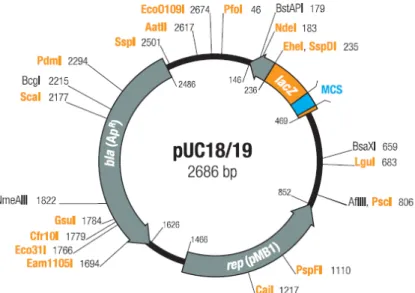
3.4 Vectors ... 46
pUC18 ... 46
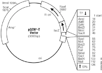
pGEM®-T ... 47
pCAMBIA ... 47
3.5 Expression vectors ... 49
GCase expression vectors ... 49
GLA expression vectors ... 50
3.6 Oryza sativa transformation mediated by Agrobacterium tumefaciens ... 53
Oryza sativa CR W3 ... 53
3.6.1 Development of embryogenic calli from rice scutellar tissue ... 54
3.6.2 Co-culture of calli with A. tumefaciens ... 55
3.6.3 Calli selection based on PMI ... 55
3.6.4 Regeneration of rice seedlings from transformed calli ... 56
3.7 Protein analysis ... 59
3.7.1 Extraction of total seed proteins from seeds containing the recombinant enzymes ... 59
3.7.2 Immunoassay DAS-ELISA ... 60
3.7.3 Western blotting ... 62
Protein extraction ... 62
Bradford assay ... 62
Preparation of the sample ... 63
Gel electrophoresis ... 63
Transfer ... 65
Blocking and detection ... 65
4
Results ... 67
4.1 In silico design of GBA and GLA genes optimised for rice expression ... 67
4.1.1 GBA gene ... 68
4.1.2 GLA gene ... 74
4.2 Analysis of the sequences ... 79
iii
4.2.2 GLA gene ... 79
4.3 Oryza sativa transformation mediated by Agrobacterium tumefaciens ... 80
4.4 Protein analysis ... 82
GCase analysis ... 82
GLA analysis ... 86
5
Discussion ... 93
5.1 Introduction ... 93
5.2 Oryza sativa CR W3 as the expression host ... 93
5.3 Signal peptide ... 96
5.4 Expression vector ... 96
5.5 GCase and GLA expression in Oryza sativa ... 99
6
Conclusions ... 106
1
1.1 Plant molecular farming
1.1.1 Introduction
Plant molecular farming (PMF) is a new branch of plant biotechnology, where plants are engineered to produce recombinant pharmaceutical and industrial proteins in large quantities. As an emerging subdivision of the biopharmaceutical industry, PMF is still trying to gain comparable social acceptance as the already established production systems that produce these high valued proteins in microbial, yeast, or mammalian expression systems.
PMF refers to the production of recombinant proteins (including industrial proteins/enzymes, therapeutic proteins) and other secondary metabolites, in plants. This involves the growing, harvesting, transport, storage, and downstream processing of extraction and purification of the protein (De Wilde et al., 2002). This technology hinges on the genetic transformability of plants, which was first demonstrated in the 1980s (Bevan et al., 1983). The first recombinant plant-derived pharmaceutical protein (the human growth hormone) and the first recombinant antibody were produced in transgenic plants in 1986 and 1989, respectively (Barta et al., 1986; Hiatt et al., 1989).
Plants possess exceptional biosynthetic capacity, including the ability to use the sun (photosynthesis) and/or very simple media to support significant biomass and protein accumulation. Their potential for low-cost production of high quality and bioactive recombinant protein is well documented (Obembe et al., 2011). Plants successfully perform the majority of post-translational modifications important for many complex eukaryotic proteins and provide tremendous flexibility in bioproduction platforms that differentially address production scale, cost, safety, and regulatory issues. Because plants cannot harbour the human and animal pathogens-of-issue for mammalian cell-based production system, they bring significant advantage in increased safety for patients (Pogue et al., 2010; Xu et al., 2011). These biosafety advantages also impact commercial aspects: they reduce purification costs and minimize risks associated with potential production shut-downs, facility decontamination, and supply limitations leading to unmet patient/customer demand.
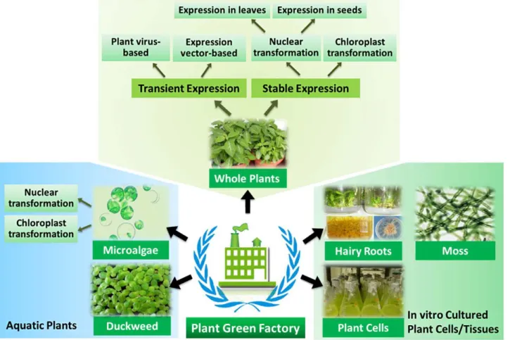
In contrast to other expression system such as yeast, bacterial and mammalian cells, plant expression system encompasses diverse forms including whole-plants, suspension cells, hairy roots,
2
moss, duckweed, microalgae, etc. (Fig. 1.1). Each of the platforms has its own strengths and weaknesses and is often best suited for certain classes of recombinant proteins based on the market, scale, cost, and upstream and downstream processing constraints of the particular protein product.
The products that are currently being produced in plants include bioactive peptides, vaccine antigens, antibodies, diagnostic proteins, nutritional supplements, enzymes and biodegradable plastics. Apart from many advantages, there are also some problems associated with plants for their use as bioreactors: these include differences in glycosylation patterns in plants and humans, inefficient expression and environmental contamination.
The factors to be investigated before attempting accumulation of recombinant proteins are: to assess the nature of the foreign protein and to determine its possible effect on the host plant; to examine the post-translational modifications required; to select a suitable host tissue and sub-cellular location for accumulation; and to determine the degree of protein purification required. Depending on these variables, there are several options for expression:
1. Choice of host plant (dicot or monocot, food or non-food); 2. Type of transformation method:
• Biological (viral, bacterial)
• Physical (biolistic, electroporation)
3. Expression parameters (stable or transient, constitutive or tissue-specific) 4. Intracellular location (cytoplasm, organelle, apoplast, plastid).
The constitutional steps involved in the whole process of production of recombinant proteins from plants include:
1. Choice of host species and optimization of coding sequence of the target gene in relation to the host;
2. Selection of expression cassette and creation of the expression vector;
3. Integration of the gene construct into the plant genome and regeneration of plants expressing the desired protein;
4. Identification and stabilization of the plant line for commercial production; 5. Purification and characterization of the recombinant protein.
3
Fig. 1.1: Various plant cell expression platforms for the production of recombinant proteins (Xu et al., 2012).
4 1.1.2 Plant-derived recombinant proteins
Plant-derived vaccine antigens
Several vaccines have been expressed in plants, since the first plant-derived vaccine-relevant protein was reported 20 years ago (Rybicki et al., 2009; Tiwari et al., 2009). These include the hepatitis B surface antigen, which has been expressed in transgenic potatoes, in tomato, in banana and in tobacco cell suspension culture (Richter et al., 2000; He et al., 2008; Kumar et al., 2005). The heat labile enterotoxin B subunit (LTB) of Escherichia coli has been expressed in potato tubers, in maize seed, in tobacco and in soybean (Lauterslager et al., 2001; Chikwamba et al., 2002; Rosales-Mendoza et al. 2009; Moravec et al., 2007). The cholera toxin B subunit (CTB) of Vibrio cholera has been expressed in several crops (including tobacco, tomato and rice), and several plant-made vaccines for veterinary purposes have been expressed in plant (Lentz et al., 2010; Ling et al., 2010). Other plant-made vaccines include the L1 protein of human papillomavirus types 11 and 16 (Giorgi et al., 2010), the Norwalk virus capsid protein, the Hemagglutinin protein from measles virus and the H5N1 pandemic vaccine candidate (D’Aoust et al., 2010), all of which have been expressed in one or two of the following plants: tobacco, potato and carrots.
There are several plant-produced vaccine candidates, which are at different stages of the clinical trials. As such, plant-based production processes are able to compete with conventional methods, breaking the limits of current standard production technologies and reaching new frontiers for the plant-based production of pharmaceutical-grade proteins.
Plant-derived antibodies
Recombinant antibodies have been found to provide passive immunization against pathogens and are considered as promising alternatives to fight infectious disease, especially in spite of the increasing microbial resistance to antibiotics and the emergence of new pathogens (Casadevall, 1998). Although there is increasing market demand, the prevailing high cost of production prevents the successful of plant-derived antibodies introduction into the health market as a therapy for infectious diseases. Plants do not only provide cheaper production platforms, as plant-derived antibodies would cost just 0.1-1% of the production cost of mammalian culture and 2-10% of microbial systems (Chen et al., 2005), but they can also assemble complex multimeric antibodies (Conrad and Fielder, 1994). Since the first recombinant antibodies were expressed in plants in 1989 (Hiatt et al., 1989), different moieties ranging from single chain Fv fragments (scFvs, which contain the variable regions of the heavy and light chains joined by a flexible peptide linker) to Fab
5
fragments (assembled light chains and shorted heavy chains), small immune proteins (SIP), IgGs, chimeric secretory IgA and single-domain antibodies have been expressed as well (Ismaili et al., 2007; Xu et al., 2007). The first plant-made scFv monoclonal antibody, used in the production of a recombinant hepatitis B virus vaccine, has been commercialised in Cuba (Pujol et al., 2005).
Therapeutic and nutraceutical proteins
The first therapeutic human protein to be expressed in plants was a human growth hormone (Barta et al., 1986). In 1990 human serum albumin, which is normally isolated from blood, was produced in transgenic tobacco and potato for the first time (Sijmons et al., 1990). Since then, several human proteins have been expressed in plants. These include epidermal growth factor ( Wirth et al., 2004; Bai et al., 2007), α-, β- and γ-interferons, which are used in treating hepatitis B and C (Leelavathi and Reddy, 2003; Zhu et al.,1994; Arlen et al., 2007); erythropoietin, which promote red blood cell production (Musa et al., 2009); interleukin, which is used in treating Crohn’s disease (Fujiwara et al., 2010); insulin, which is used for treating diabetes (Nykiforuk et al., 2006); human glucocerebrosidase, which is used for the treatment of Gaucher’s disease in genetically engineered carrot cells (Shaaltiel et al., 2007) and several others.
Antimicrobial nutraceutics, such as human lactoferrin and lysozymes, have been successfully produced in several crops (Huang et al., 2008; Stefanova et al., 2008) and they are now commercially available as fine chemicals.
Non-pharmaceutical plant-derived proteins
The non-pharmaceutical plant-derived proteins or industrial proteins are now on the market. Most of them are enzymes, such as avidin, trypsin, aprotinin, β-glucuronidase, peroxidise, laccase, cellulose and others. The molecular farming of cell-wall deconstructing enzymes, such as cellulases, hemicellulases, xylanases and ligninanes, holds great promise for the biofuel industry with respect to the production of cellulosic ethanol (Sticklen, 2008; Mei et al., 2009; Chatterjee et al., 2010), which was estimated to have the potential of reducing greenhouse gas emissions by 100% compared to gasoline (Fulton et al., 2004). Other potential non-pharmaceutical plant-derived technical proteins that are being explored and optimised for production include biodegradable plastic-like compounds such as polyhydroxyalkanoate (PHA) copolymers, poly-3-hydroxybutyrate (PHB) and cyanophycin (Conrad, 2005; Matsumoto et al., 2009). It should be noted that thus far only few plant-derived pharmaceuticals have been approved, and fewer still are commercially
6
available, mainly because of biosafety concerns and stringent governmental regulations with respect to field trials, good manufacturing practice (GMP) standards and pre-clinical toxicity testing.
1.1.3 Post-translational modification
Recombinant DNA technology has enabled the production of heterologous recombinant proteins in host systems. The majority of the early work was directed toward the expression of recombinant therapeutic proteins in prokaryote hosts, mainly in Escherichia coli. The advantages of prokaryotes as a production system are the ease with which they can be manipulated genetically, their rapid growth, the high expression level of recombinant proteins and the possibility of a large-scale fermentation. However several post-translational modifications (PTMs), including signal peptide cleavage, propeptide processing, protein folding, disulfide bond formation and glycosylation, might not be carried out in prokaryotes. As a result, complex therapeutic proteins that are produced in prokaryotes are not always properly folded or processed to provide the desired degree of biological activity. Consequently, microbial expression systems have generally been used for the expression of relatively simple therapeutic proteins, such as insulin, interferon or human growth hormone, which do not require folding or extensive post-translational processing to be biologically active.
As an expression system for recombinant proteins, plants are gaining increasing acceptance alongside traditional systems such as bacteria, yeast, baculoviruses and mammalian cell culture, particularly where eukaryotic-like post-translational modifications are required (Jacobs and Callewaert, 2009). Glycosylation is the most extensively studied PTM of plant-made recombinant proteins. However, other types of protein processing and modification that are important for the production of high quality recombinant protein also occur, co- and post-translationally.
After translation, the majority of plant proteins undergo additional covalent modifications that shape their tertiary and quaternary structures. These PTMs have been shown to affect almost every aspect of protein activity, including function, localization, stability, and dynamic interactions with other molecules. These modifications range from very simple chemical changes, such as the addition of phosphate or acetate functional groups, to modifications that are highly intricate or enormous in size, sometimes even larger than the protein itself (e.g., proteoglycans) (Stulemeejer and Joosten, 2008). Over 300 types of modifications have been identified and they can be broadly classified into four groups: addition of functional groups; addition of proteins or peptides; structural changes to proteins; changes to the chemical nature of an amino acid (Ytterberg and Jensen, 2010). Proteins may undergo a single type of modification or various combinations of PTMs. Some PTMs
7
are fixed for the life of the protein, such as cleavage of a signal peptide or glycosylation, while other changes are rapid and reversible, such as phosphorylation. In evolutionary terms the complex and diverse nature of PTMs represents an efficient and cost-effective mechanism for the exponential diversification of the genome. However, in the context of recombinant protein production, heterogeneity of PTMs can present both challenges and opportunities. Although many PTMs are evolutionarily conserved, there are also important plant-specific modifications which should be considered when expressing recombinant proteins. In so far as a recombinant protein is produced for pharmaceutical application, plant specific PTMs have the potential to substantially improve the stability of recombinant proteins, and enhance the immunogenicity and/or uptake of vaccine antigens (Bosch and Schots, 2010; Singh et al., 2009; Xu et al., 2010). However, plant-specific PTMs may also become the target of undesirable responses, such as IgE-based allergic reactions (Bosch and Schots, 2010).
Glycosylation is the most common PTM in eukaryotic cells and one of the most diverse modifications. At least 50% of human proteins are glycosylated with some estimates being as high as 70% (Apweiler et al., 1999; Lauc et al., 2010). Many of the most clinically useful proteins are glycosylated, including over 40% of the currently approved protein therapeutics, and many more glycosylated biopharmaceuticals are under development (Higgins, 2010; Walsh, 2010).
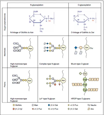
N-linked glycoproteins contain complex oligosaccharide chains (glycans) covalently attached to the amide nitrogen on the side chain of Asn residues (Gomord et al., 2010).
Yeast, baculovirus and plant expression systems are able to glycosylate proteins. However, each system exhibits significant differences in the complex processing of the glycan sidechains, including unique host-specific modifications that do not occur in humans (Fig. 1.2) (Jacobs and Callewaert, 2009).
The N-linked glycosylation pathway in plants is relatively well characterized and shares a high degree of homology with other eukaryotic organisms, including site occupancy, frequency of glycosylation and the structure and composition of the core high-mannose type glycan added in the ER. In brief, the core glycan is assembled in the ER as a mannose-rich lipid-linked precursor (generally Glc3Man9GlcNAc2) which is transferred ‘en-bloc’ by the oligosaccharyltransferase (OST) complex to an Asn residue in the context of either Asn-X-Ser or Asn-X-Thr, where X is any amino acid except proline (Gomord et al., 2010). Once attached to the protein, the core glycan undergoes cycles of trimming and reglycosylation in the ER. The correctly folded proteins are then transported to the Golgi apparatus, where the formation of complex-type glycans is undertaken
8
(Saint-Jore-Dupas et al., 2007). The degree and type of modifications undertaken in the formation of complex glycans vary between plants and mammals. Firstly, the addition of α1,3 fucose and/or β1,2 xylose by α1,3 fucosyltransferase (FucT) and β1,2 xylosyltransferase (XylT), respectively, results in the formation of plant-specific N-glycans (Gomord et al., 2010). It has been suggested that the presence of α1,3 fucose and/or β1,2 xylose on plant-made biopharmaceuticals could lead to the induction of undesirable immune and/or allergy responses (Bosch and Schots, 2010). Injection of a plant-made glycoprotein is able to elicit the production of antibodies specific for α1,3 fucose and β1,2 xylose containing glyco-epitopes (Jin et al., 2008). Mammalian-specific modifications on N-glycans include the addition of β1,4 galactose, which has not been reported to occur in plants (Bakker et al., 2001). Plant also lack homologs of mammalian N-acetylglucosaminyltransferase (GnT) III, -IV and -V which are involved in the addition of GlcNAc residues to create branched N-glycans (Nagels et al., 2011). This means that plant N-N-glycans carry only two antenna structures, while mammalian N-glycans can contain multi-antennary glycans with two or more terminal branches. These multi-antennary N-glycan structures can increase serum half-life of injected proteins by increasing the size of the molecule sufficiently to avoid rapid renal clearance in the same way as the chemical conjugation to poly-ethylene glycol (a process known as PEGylation) (Harris and Chess, 2003). Plant glycoproteins also lack sialic (neuraminic) acid (Neu5Ac) on the termini of complex N-glycans. The addition of sialic acid to mammalian glycans is common, and has been shown to be important in preventing clearance of recombinant therapeutic proteins (Egrie and Browne, 2001).
Finally, following synthesis and maturation in the ER and Golgi additional modifications may occur during transport of glycoproteins to their final destination. This involves the trimming (or removal by degradation) of terminal sugars from complex glycans leaving the core glycans with α1,3 fucose and/or β1,2 xylose additions only. These truncated glycans, termed paucimannose-type N-glycans, are commonly found in the vacuole and seeds (Floss et al. 2009, Gomord et al., 2010).
9
Fig. 1.2: Addition and processing of N-linked glycans in the ER and Golgi apparatus in plants and mammals. A precursor oligosaccharide assembled onto a lipid carrier is transferred on specific Asn residues of the nascent growing polypeptide. The N-glycan is then trimmed off with the removal of glucosyl and most mannosyl residues. Differences in the processing of plant and mammalian complex N-glycans occur during late Golgi maturation events (Gomord and Faye, 2004).
There are significant differences in O-glycosylation between plants and animals, including the sites of glycan addition, and the structure and composition of the glycans. Mammalian proteins are most commonly O-glycosylated at Ser and Thr residues with sugars such as fucose, galactose and N-acetylgalactosamine (GalNAc). The most abundant class of O-glycosylation are the mucin-type glycoproteins. O-linked glycosylation by plants is a common PTM which plays a key role in growth and development, wound healing and plant-microbe interactions (Stulemeijer and Joosten, 2008). Glycans are typically attached to Ser residues and Hyp residues. The most abundant class of O-linked plant glycoproteins are known as Hyp-rich glycoproteins (HRGPs) (Fig. 1.3) (Gomord et al., 2010). Addition of O-glycans to the hydroxyl group of Hyp is unique to plants. The process is initiated by the enzymatic addition of a single sugar, generally a galactose or arabinose, which is then built on to create linear or branched oligosaccharide chains (Saint-Jore-Dupas et al., 2007).
10
Although O-glycosylation can occur in the ER, the majority of O-glycosylation reactions occur in the Golgi apparatus. Contiguous sequences of Hyp result in the addition of short unbranched arabinooligosaccharides, for example the Ser-Hyp4 pentapeptide motif of extensions (Shpak et al.,
2001; Xu et al., 2007). Only a limited number of studies have investigated the presence (or absence) of O-glycans on recombinant plant-made proteins. There is much yet to learn about O-glycosylation in plants and, more specifically, the recognition of O-glycosylation sites in recombinant mammalian proteins.
Fig. 1.3: Types of glycan structures and linkages commonly found on plant and mammalian glycoproteins (Gomord and Faye, 2004).
1.1.4 Rice seed as an Expression Host
Plant seed has emerged as one of the ideal organs for the expression of recombinant proteins in plants (Stoger et al., 2005). Naturally, seed is the organ for protein synthesis and storage, which has a high protein content, low protease activities, and low water content (Müntz, 1998). In the context of molecular farming, these factors could translate into yield gains and could be convenient for storage and transport. Antibodies, vaccine antigens, and other recombinant proteins have been
11
shown to accumulate at high levels in seeds and to remain stable and functional for years at ambient temperature (Nochi et al., 2007). Rice seeds are composed of 7-8% protein and 92-93% starch. It has been shown that throughout the dormancy period of the rice seed, its storage proteins remain intact and functional also thanks to the advantage of encapsulation that provides resistance against degradation (Boothe et al., 2010). This means that the seed should be a suitable area for the stable deposition of recombinant proteins, which are also stable for a long period at room temperature. Rice crops are self-pollinating: this characteristic ensures that no genetic material is gained or lost and that the gene coding for the protein of interest remains present in each new generation. Rice is a stable food for the vast majority of the world’s population, it is cultivated in over 100 countries on more than 150 million hectares of land and it represents a model species for monocotyledonous and cereal plants (Yang et al., 2008). The familiarity with the agronomy and nutritional values of rice, along with the GRAS (Generally Recognized As Safe) designation by the Food and Drug Administration, makes it a strong candidate for large-scale production of biopharmaceuticals. Rice seed has many advantages over other cereal crops in terms of storage and processing and furthermore, it is produced in greater biomass (about 6000 Kg/ha) (Takaiwa et al., 2008). The complete genomic sequence of one variety of rice and the partial sequences of several other varieties are now available.
Rice caryopsis is developed from the fertilised pistil. Fig. 1.4 shows the internal structure of a rice grain. Next to the pericarp are two layers of cells named tegmen or seed coat. The embryo lies on the ventral side of the spikelet next to the lemma. The remaining part of the caryopsis is the endosperm, which provides nourishment to the germinating embryo. The embryo contains a plumule (embryonic leaves) and radicle (embryonic root), which are joined by a very short stem (mesocotyl). The portion tied to the endosperm forms the scutellum. The endosperm is wrapped by the aleurone layer below the testa (seed coat), and it has the starch storage parenchyma inside (Chang and Bardenas, 1965; Matsuo and Hoshikawa, 1993).
12 Fig. 1.4: Structure of a rice caryopsis (Chen et al.)
The endosperm is the main storage compartment for rice and accounts for over 80% of the total seed weight, thus it is the most attractive site for protein accumulation. Indeed, the seed storage proteins (SSPs) are predominantly synthesised and stably accumulated in maturing endosperm tissue. Cereal SSPs are traditionally classified into albumin, globulin, prolamin and glutelin according to their physical properties based on solubility (Shewry and Casey, 1999). Such classification is well known as ‘Osborne fractions’. Briefly, albumins are soluble in water, globulins are soluble in saline solutions, prolamins are soluble in aqueous alcohol (such as 60-70% ethanol or 50-55% propanol), and glutelins are extractable in alkali. Although this basic nomenclature is traditionally accepted in part, characterisation based on DNA sequences of isolated SSP genes by gene cloning and protein sequencing has revealed finer details of SSP structure.
The protein composition of rice endosperm is composed of 60-70% glutelins, 25-30% prolamins, 5-10% globulins, and 0-5% others. Rice glutelins is synthesized as a 57 kDa precursor and then cleaved into two major polypeptide subunits classified as α, or acidic, and β, or basic, subunits with apparent molecular weights (MWs) of 30-39 and 19-25 kDa respectively (Yamagata et al., 1982). Encoded by about 15 genes per haploid genome, glutelins genes can be classified into four subfamilies – GluA, GluB, GluC and GluD – according to the degree of nucleotide sequence similarity. GluA contains four members and GluB has the highest number of members with eight of theme. Thus far, only two members of GluC and one member of the GluD subfamily have been identified (Katsube-Tanaka et al., 2004; Kawakatsu et al., 2008)
13
As the second abundant protein in rice endosperm, the prolamins consist of three polypeptide subunits with apparent MWs of 10, 13 and 16 kDa. The name ‘prolamin’ comes from the high content of proline and glutamine found in this class of SSPs. Although originally prolamins were defined by their solubility in aqueous alcohol, many prolamins are soluble in aqueous alcohol only when reduced (Shewy and Casey, 1999). This insolubility in aqueous alcohol is because of their polymeric states via intermolecular disulphide bonds. Prolamin genes in rice genome were estimated to be more than 100 copies (Kim and Okita, 1988); however, the complete genome sequence and systematic genome-wide analysis revealed that there are 34 prolamin genes in the rice genome (Xu and Messing, 2009).
Rice globulins consist of α-, β-, γ- and δ-globulins with apparent MWs of 25.5, 15, 200 kDa and higher, respectively. Rice albumins have a wide range of MWs, with major components with apparent MWs of 18-20 kDa.
The most obvious destinations for protein accumulation in seeds are the protein storage organelles (i.e. the protein bodies and protein storage vacuoles), as these have developed to facilitate stable protein accumulation (Müntz, 1998). Seed storage proteins pass through the endomembrane system, which is generally well developed in storage cells and thus suitable for chaperone-assisted folding, assembly and post-translational modification even if the protein is complex. In most seed crops, storage proteins are sequestered in protein storage vacuoles, which are post-Golgi compartments. Cereals are unique in harbouring an additional class of protein bodies that are directly derived from the endoplasmic reticulum (ER). In rice endosperm, the two types of protein bodies co-exist as separate entities in the same cell and contain the two major classes of storage proteins, prolamines and glutelins (Krishnan et al., 1986). Prolamines accumulate in protein bodies inside the rough ER. By contrast, glutelins accumulate in protein storage vacuoles, and are conveyed to these organelles by transport vesicles budding from the Golgi apparatus (Müntz, 1998).
1.1.5 Codon Usage
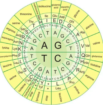
In biological systems, nucleic acids contain information which is used by a living cell to construct specific proteins. The sequence of nucleobases on a nucleic acid strand is translated by cell machinery into a sequence of amino acids making up a protein strand. Each group of three bases, called “codon”, corresponds to a single amino acid, and there is a specific genetic code by which each possible combination of three bases corresponds to a specific amino acid. All amino acids except Met and Trp are coded by two to six codons (Fig. 1.5).
14
DNA sequence data from diverse organisms clearly shows that synonymous codons for any amino acid are not used with equal frequency even though choices among the codons should be equivalent in terms of protein structures. Studies in E. coli and yeast have demonstrated that rare codons and level of tRNA can affect translation time. The observation that preferred codons are recognised by tRNAs in greatest abundance and rare codons are recognised by tRNAs in lowest abundance has led to the suggestion that tRNA abundance and codon usage have co-evolved (Higgs and Ran, 2008).
Non-random use of synonymous codons universally exists both within and between organisms. The results of numerous studies have demonstrated that there is a species-specific pattern of codon usage. In particular, closely related organisms always share similarities in codon frequency (Sharp et al., 1988). However, it has been observed that there are big differences in codon usage among genes within one species. For example, correspondence analysis has identified at least two classes of genes in Arabidopsis thaliana according to codon usage, in which one group of genes was highly biased to G/C and the other group had a weaker preference for A/T biased codons (Chiapello et al., 1998). Analysis of codon usage data has both theoretical and practical importance in understanding the basics of molecular biology. In Escherichia coli, Saccharomyces cerevisiae, Caenorhabditis elegans, Drosophila melanogaster, Arabidopsis thaliana and Oryza sativa, there was a strongly significant correlation between gene expression level and codon usage bias (Ikemura, 1981; Sharp and Li, 1986; Duret and Mouchiroud, 1999). Highly expressed genes displayed much more significant variation in codon usage than genes expressed at lower levels, suggesting that codon usage patterns had a functional significance. Using an experimental approach, it has been demonstrated that there is a positive relationship between codon usage bias and gene expression level by transforming plants with vectors expressing genes with modified codon usage (Iannacone at al., 1997; Rouwendal et al., 1997). This suggests stronger natural selection constrains on highly expressed genes, with respect to lower expressed genes, to optimise translation efficiency and accuracy by the use of optimal codons (Bulmer, 1988).
Many studies have indicated that, in all life forms, it appeares that codon usage bias is determined by diverse factors, such as expression levels, gene length, protein secondary structure, etc. (Duret and Mouchiroud, 1999; Gupta et al., 2000).
One of the approaches used to increase the translation efficiency in a given host is to optimise the codon usage by changing the nucleotide sequence without changing the amino acid sequence to suit the respective host (Gustafssion et al., 2004). By using this strategy, expression of the Bacillus
15
thuringiensis cryIA (b) protein in transgenic tobacco and tomato increases up to 100-fold (Perlak et al., 1991). Experiments with tobacco-expressed green fluorescent protein have demonstrated the benefit of codon optimization in plants (Rouwendal et al., 1997). Preferred codon usage differs between monocots and dicots, and it is greatly different even between nucleus and plastid of the same plants. Engineering the required sequence according to the codon usage can greatly increase the protein production rate and decrease the overall cost of the protein production (Liu and Xue, 2005).
Fig. 1.5: Genetic code.
1.1.6 Agrobacterium tumefaciens plant transformation
Thirty-five years ago, the concept of using Agrobacterium tumefaciens as a vector to create transgenic plants was viewed as a prospect and a “wish.” Today, many agronomically and horticulturally important species are routinely transformed using this bacterium, and the list of species that is susceptible to Agrobacterium-mediated transformation seems to be growing daily. However there are still many challenges for genotype-independent transformation of many economically important crop species and, in addition, predictable and stable expression of transgenes remains problematic.
16
Agrobacterium tumefaciens is a gram-negative rod-shaped bacterium closely related to nitrogen-fixing bacteria which dwell at root nodules in legumes. Unlike most other soil-dwelling bacteria, it infects the roots of plants to cause Crown Gall Disease (Fig. 1.6) (Cubero et al., 2006). In the wild, A. tumefaciens targets dicots and causes economic damage to plants with agricultural importance, such as walnuts, tomatoes and roses. However, scientists have exploited the ability of this bacterium to put DNA into its host to create transgenic plants. A. tumefaciens has emerged as an important molecular tool for manipulating plants and creating genetically modified crops for research and agriculture. Because of its importance in the laboratory, a complete genome of A. tumefaciens strain C58 was published in 2001 (Goodner et al., 2001).
The genus Agrobacterium has been divided into a number of species. However, this division has reflected, for the most part, disease symptomology and host range. Thus, Agrobacterium radiobacter is an “avirulent” species, Agrobacterium tumefaciens causes crown gall disease, Agrobacterium rhizogenes causes hairy root disease, and Agrobacterium rubi causes cane gall disease. More recently, a new species has been proposed, Agrobacterium vitis, which causes galls on grape and a few other plant species (Otten et al., 1984).
Fig. 1.6: Crown gall caused by Agrobacterium tumefaciens
(www.delange.org/Vegetable_Garden_Disease_Arizona/Vegetable_Garden_Disease_Arizona.htm).
Agrobacterium tumefaciens is an unusual bacterium because it is one of the few bacteria that has both a linear and a circular chromosome. Its genome has a total of 5.7 million base-pairs, with 2.8 million residing on its circular chromosome and 2.1 million residing on its linear chromosome (Goodner et al., 2001). Most of the genes essential for its survival are located on the circular chromosome, although through evolution some essential genes have migrated to the linear chromosome. Based on sequence analysis, it was determined that the linear chromosome was derived from a plasmid that was transformed into the bacteria a long time ago (Goodner et al.,
17
2001). A. tumefaciens contains flagella, which are important in its life cycle as they help it to swim through the soil to find its plant hosts. Mutations in flagella genes reduced virulence of A. tumefaciens in the laboratory. A. tumefaciens can use a variety of substrates for energy and carbon, but it is especially evolved to use a class of chemicals called opines, which are amino acid-like compounds that are intermediates of metabolism in most organisms. A. tumefaciens forces the plants that it infects to produce opines, a molecule that the bacteria use as a source of energy and carbon (Moore et al., 1997). There are many types of opines which it can use, such as nopaline, agropine, mannopine (which are common), and chrysopine, deoxy-fructosyl-oxo-proline (which are uncommon) (Moore et al., 1997). It is believed that each strain of A. tumefaciens can only metabolise one type of opine, and contains genes for its synthesis (usually in its T-DNA which it transfers to its plant host) and catabolism, although this is not strictly true. Due to its ability to integrate DNA into its plant hosts, A. tumefaciens has been used to make transgenic plants since the 1970's. Even though other methods, such as biolistic, have been developed to put genes inside plants, A. tumefaciens has remained popular for genetic manipulation of plants due to the low copy number of genes in plants created and to stability of the transgene (Li et al., 2000). The key strength is that A. tumefaciens injects whatever DNA is flanked by a specific 25 bp border repeat sequence (Li et al., 2000). In normal A. tumefaciens this DNA is the T-DNA, so the first step is to create an artificial plasmid with the T-DNA excised, the following step is to insert the desired gene construct with the flanking border repeats into the plasmid. The gene construct usually contains a selection marker (Li et al., 2000) (usually kanamycin resistance) along with the desired gene(s) to be expressed in the plant. This artificial plasmid is then transformed into A. tumefaciens, presumably one that lacks a normal Ti plasmid. Plant tissue is then infected with this engineered A. tumefaciens, placed on a selection media (such as one containing kanamycin). Using plant tissue culture methods, the selection media kills the cells that have not been transformed and the shoots, which have successfully grown, contains the desired gene which has been inserted. However, these selection systems always employ antibiotics like kanamycin for neomycin phosphotransferase II (NptII) gene and hygromycin for hygromycin B phosphotransferase (Hpt) gene, or herbicide for bialaphos resistance gene (bar) or protoporphyrinogen oxidase (PPO) gene as selectable agents to allow the exclusive growth of transformed cells by killing other cells. These genes are used to select out the transformed cells and to let them grow into whole plants. However, the presence of the antibiotic resistance genes is undesirable for commercial applications and government policies are discouraging the use on these specific marker genes, even if there is a history of safe use in plants. To address this, various measures – such as site-specific recombination, co-transformation,
18
transposon-mediated repositioning system and recombinase – were taken to eliminate those selectable genes in plants when they were transplanted to field, but these strategies were laborious and hard to control, and none of these strategies has been successfully utilised in commercial production until now (Srivastava and Ow, 2004; Matthews et al. 2001; Cotsaftis et al., 2002; Luo et al., 2007). Besides that, an alternative to obtain transgenic plants for commercial requirement and for a reduced public concern is to adopt positive selection by using non-toxic substances as the selectable agent, such as xylose for xylose isomerase gene (xylA) (Haldrup et al., 1998), ribitol for ribitol operon (rtl) from Escherichia coli (Lafayette and Parrott, 2001) and mannose for phosphomannose isomerase (pmi) gene (Joersbo and Okkels, 1996). pmi was first isolated from E. coli and sequenced by Miles and Guest (1984). By making use of it, plants are enabled to convert mannose, in the form of mannose-6-phosphate that most plants cannot metabolise, to fructose-6-phosphate: plants expressing this gene can thus use mannose as the sole carbon source. Unlike negative selection used to kill the non-transformed cells, this selection system allows them to stay dormantly and those expressing pmi gene grow normally by using mannose as carbon source, making pmi gene work efficiently in transformation. Therefore, it has been attempted to use the pmi gene as a selectable marker for the transformation of various plant families, including maize, pearl millet, bentgrass, sorghum, wheat and sugarcane of Poaceae; Arabidopsis and cabbage of Brassicaceae; apple, papaya and almond of Rosaceae; cassava of Euphorbiaceae; tomato of Solanaceae; sugar beet of Chenopodiaceae; onion of Liliaceae; cucumber of Cucurbitaceae.
Modern methods for creating transgenic plants using A. tumefaciens are usually a variation of the method described above. In order to simplify the process of plasmid construction, a binary vector is sometimes used (Li et al., 2000). A binary vector is simply a strain of A. tumefaciens with a Ti plasmid that contains all the genes necessary to inject DNA into the plant but that contains no T-DNA with border repeats (Li et al., 2000). Another plasmid containing the gene that is to be inserted into the plant is then put in. Since the T-DNA does not have to be on the Ti plasmid in order for it to be integrated into the host genome, it can be put on a separate plasmid as long as it has the flanking 25-bp border repeats. A previous limitation of using A. tumefaciens to create transgenic plants is that A. tumefaciens only infects dicots and gymnosperms in nature (Li et al., 2000). However, additional methods have been discovered which extend A. tumefaciens host range to monocots, so this method is now useful for creating transgenic plants for all flowering plants. Moreover, even non plant species can be transformed by Agrobacterium under laboratory conditions (Lacroix et al., 2006), including yeast (Piers et al., 1996), various fungi (Michielse et al., 2005), and cultured human cells (Kunik et al., 2001).
19
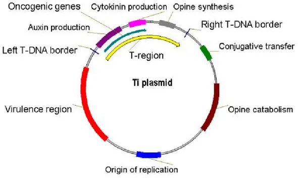
The molecular basis of genetic transformation of plant cells by Agrobacterium is transferred from the bacterium and integration into the plant nuclear genome of a region of a large tumour-inducing (Ti) or rhizogenic (Ri) plasmid resident in Agrobacterium. Ti plasmids are on the order of 200 to 800 kbp in size (Fig. 1.7) (Suzuki et al., 2000). The transferred DNA (T-DNA) is referred to as the T-region when located on the Ti or Ri plasmid. T-regions on native Ti and Ri plasmids are approximately 10 to 30 kbp in size. Thus, T-regions generally represent less than 10% of the Ti plasmid. Some Ti plasmids contain one T-region, whereas others contain multiple T-regions (Suzuki et al., 2000). T-regions are defined by T-DNA border sequences. These borders that are 25 bp in length are highly homologous in sequence (Jouanin et al., 1989). They flank the T-region in a directly repeated orientation. In general, the T-DNA borders delimit the T-DNA, because these sequences are the target of the VirD1/VirD2 border-specific endonuclease that processes the T-DNA from the Ti plasmid (Peralta and Ream, 1985).
Fig. 1.7: The structure of the Ti plasmid
(http://www.gophoto.it/view.php?i=http://molbioandbiotech.files.wordpress.com/2007/08/ti_plasmi d1.jpg).
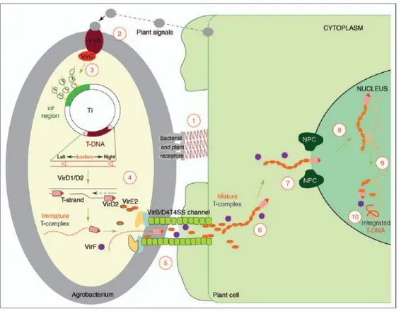
The principal steps and factors involved in Agrobacterium-mediated plant transformation are comparatively well-understood (Fig. 1.8) (Gelvin, 2010). Briefly, agrobacteria sense phenolic substances that are secreted by wounded plant tissue. Reception of these signals drives the expression of bacterial virulence (vir) genes. Subsequently, Vir proteins are produced and single-stranded T-DNA molecules are synthesized from the Ti plasmid. The T-complex, i.e. T-DNA
20
associated with certain Vir proteins, is injected into the host cytoplasm. A sophisticated network of bacterial and plant factors mediates translocation of the T-DNA to its final destination, the host cell´s nucleus. Agrobacterium inserts substrates (T-DNA and virulence proteins including VirD2, VirE2, VirE3, VirD5 and VirF) into the host cell (Cascales and Christie, 2003). Remarkably, under laboratory conditions Agrobacterium can genetically transform virtually any type of eukaryote, ranging from yeast (Bundock et al., 1995) to human cells (Kunik et al., 2001). The T44 complex – consisting of T-DNA, bacterial virulence proteins (VirE2, VirD2) and the host factor VIP1 (VirE2-interacting protein 1) – is imported into the nucleus. Subsequently, the proteinaceous components are stripped off and they release the T-DNA from the T-complex. This step relies on degradation of VirE2, VirD2 and VIP1 by the plant SCF proteasomal machinery. The bacterial F-box protein VirF, which is contained in and confers substrate specificity to the SCF complex, participates in this degradation. If the T-complex disintegrates before it is in contact with the host’s chromatin, the delivered transgenes are expressed for only a few days. The loss of transgenic activity at later stages is likely to result from the T-DNA being degraded by host nucleases (Gelvin, 2003). In contrast, if the T-DNA is shielded until the T-complex is in contact with chromatin, stable transformants can be obtained. Due to its affinity for histones, VIP1 most probably guides the T-DNA to its target destination, the chromatin (Lacroix et al., 2008).
Fig. 1.8: A model for the Agrobacterium-mediated genetic transformation (Tzfira and Citovsky, 2006)
21
Since the discovery of the gene transfer mechanism (Schell and Van Montagu, 1997), Agrobacterium strains have been converted (“disarmed”) into efficient delivery systems for the genetic manipulation of plants. While transient expression approaches can provide rapid answers on e.g. subcellular localization, protein-protein interaction and promoter/effector relationships (Pitzschke, 2013), genetic engineering requires the transgene(s) to be stably integrated in the host genome. The employed and so-called disarmed/non-oncogenic A. tumefaciens strains are deprived of their tumour-inducing properties, and the T-DNA region is used as a vehicle for the introduction of tailor-made DNA sequences. Any DNA sequence placed between T-DNA "border sequences" (Ti-plasmid-derived 25-bp direct repeats) can be transferred (Gelvin, 2012). Disarmed strains, therefore, facilitate transformation, but do not provoke callus growth or other abnormalities caused by oncogenic strains. Consequently, phenotypic abnormalities that may be exhibited by transformed plants are primarily due to the particular transgene being expressed.
22
1.2 Lysosomal enzymes used in this study
Lysosomal storage disorders are a group of more than 40 diseases caused by a deficiency of enzymes, membrane transporters, and other proteins involved in various aspects of lysosomal biology.
The lysosomal enzymes used in this study are the human α-galactosidase and the human β-glucocerebrosidase enzymes. In human, mutations in the genes coding these two proteins cause, respectively, the Anderson-Fabry disease and the Guacher disease. The chosen expression system concerning the above mentioned enzymes is the plant model mocotyledon Oryza sativa.
1.2.1 Anderson-Fabry disease
Anderson-Fabry disease (AFD) is caused by mutations of the GLA gene located on the X chromosome (Xq22.1) (Bishop et al., 1988) that results in deficiency of the enzyme α-galactosidase. According to “The Human Gene Mutation Database” at the Institute of Medical Genetics in Cardiff (www.hgmd.cf.ac.uk/ac/index.php), there are currently 431 mutations described. Of those, 295 are missense/nonsense type mutations, 66 are small deletions, 12 are large deletions, 21 are splice defects, 3 are complex rearrangements and one is large insertion. The frequency of de novo mutations is uncertain but it may be as high as 10% of cases (Schaefer et al., 2005). The cause of this large number of different mutations in the GLA gene is not known. One might speculate that having the Fabry trait presents a selective advantage such as resistance to certain types of bacterial infections, in particular those that express the Escherichia coli shiga-like toxin verotoxin (Cilmi et al., 2006).
Mutations retaining residual α-galactosidase activity are generally associated with a phenotype which is milder than the one present in mutations that result in complete loss of function. Mutations affecting functionally important residues, such as those in the hydrophilic core of the enzyme involved in determining its tertiary structure and in the active site, tend to cause more severe disease. Patients with the classic most severe form of Fabry disease almost always have a mutation that causes a total absence of α-galactosidase activity; whereas patients with missense mutations often have some residual enzyme activity ranging from 2% to 25% (Desnick et al., 2001).
Since Fabry disease is an X-linked disorder and most cases result from inherited mutations rather than new mutations, most patients have blood relatives who are either affected males or carrier females. Identification of affected males is relatively easy and it can be performed by using a
23
combination of pedigree analysis and measurement of α-galactosidase activity in plasma or leukocytes. The identification of carrier females is more difficult because many have normal levels of α-galactosidase. The only way to make a definitive diagnosis is to show that the female carries the same GLA gene mutation as her affected male relative.
The incidence of AFD is reported to range from 1/117,000 to 1/40,000, but these figures are likely to underestimate the burden of disease because the protean manifestations of the disease often lead to misdiagnosis and underreporting (Metha et al., 2004). The distribution is panethnic, with increased incidence in certain populations in Nova Scotia (Canada) and West Virginia (United States) because of founder effects (Zarate and Hopkin, 2008).
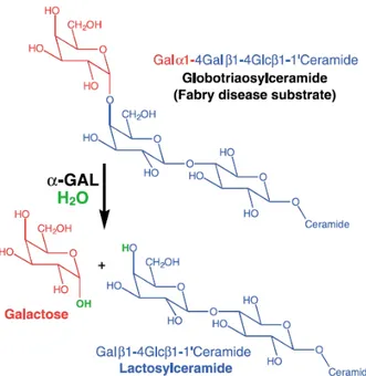
The lysosomal enzyme α-galactosidase catalyses the removal of galactose from oligosaccharides, glycoproteins, and glycolipids that have been internalised in lysosomes via endocytosis. Globotriaosylceramide, which is normally cleaved by α-galactosidase to form lactosylceramide, is the main enzyme substrate that accumulates in AFD. A-galactosidase also degrades blood group substances B and P1, but these are not thought to play a role in the pathogenesis of AFD (Garman and Garboczi, 2004). In the absence of the functional enzyme, the globotriaosylceramide accumulates in multiple cell types and it leads to a progressive organ failure (Fig. 1.9). Although most symptoms begin in childhood, clinical diagnosis is frequently delayed. In males, symptoms typically begin in the first decade of life with acroparesthesia and pain, febrile crises, hypohidrosis, heat intolerance, gastrointestinal disturbance, and cutaneous angiokeratomas. From the second decade onwards, patients develop proteinuria and neurologic manifestations. Cardiac involvement is present early in life but is not usually manifested clinically until the third or fourth decade. Thereafter, heart involvement contributes to substantial morbidity and premature death from heart failure, arrhythmia, and stroke (Linhart and Elliot, 2007; Zarate and Hopkin, 2008).Although death from Fabry disease-related complications before adulthood is probably very rare, most affected males die by the end of the sixth decade of life (Branton et al., 2002).
24
Fig. 1.9: The reaction catalysed by α-galactosidase (Garman and Garboczi, 2004). α-Galactosidase
Lysosomal α-galactosidase is a relatively heat-labile, homodimeric glycoprotein consisting of 2 identical 49 kDa subunits (Bishop and Desnick, 1981). The structure of α-galactosidase was determined by X-ray crystallographic methods. The X-ray structure reveals α-galactosidase as a homodimeric glycoprotein with each monomer composed of two domains, a (β/α)8 domain
containing the active site and a C-terminal domain containing eight antiparallel β strands on two sheets in a β sandwich (Fig. 1.10) (Garman and Garboczi, 2004).
Fig. 1.10: The α-galactosidase monomer. The monomer is coloured from N (blue) to C terminus (red). Domain 1 contains the active site at the centre of the β strands in the (β/α)8 barrel, while
25
The enzyme exists in several forms, which differ in the amount of sialic acid in the carbohydrate chains. Activity is easily measured with the use of such synthetic substrates as 4-methylumbelliferyl-α-D-galactopyranoside; optimum pH is 4.6. The GLA gene is approximately 12 kb and contains 7 exons that are associated with extensive 5’ regulatory and 3’ flanking sequences. The processed message is 1.45 kb and encodes a 49 kDa precursor polypeptide of 429 amino acids (Korneich et al., 1989). The primary polypeptide gene product undergoes cotranslational glycosylation in the endoplasmic reticulum, with downstream trimming of the polypeptide and modification of the oligosaccharide (including 6-Ophosphorylation of mannose residues) required for localization in lysosomes (Mach, 2002). A proportion of the phosphorylated enzyme is secreted from the cell and is taken up by receptor-mediated endocytosis through mannose-6-phosphate receptors in the plasma membrane (Fig. 1.11) (Ghosh et al., 2003).
The secretion and reuptake of α-galactosidase provides the rationale for enzyme replacement therapy (Brady, 2006).
Fig. 1.11: Sequence of events in the biosynthesis and trafficking of galactosidase. Nascent α-galactosidase molecules are shown as solid circles. P = phosphorylation of mannose residues; RER = rough endoplasmic reticulum (Clarke, 2007).
Enzyme Replacement Therapy (ERT)
Recombinant human α-galactosidase has the ability to restore enzyme function in patients (Schiffman et al., 2001), and enzyme replacement therapy using α-galactosidase was approved in
26
Europe in 2001 and in the United States in 2003 as a treatment for Fabry disease. A-galactosidase became the second recombinant protein approved for the treatment of a lysosomal storage disorder, after β-glucosidase, a treatment for Gaucher disease (Beutler and Grabowski, 2001). A-galactosidase represents one of a small number of human recombinant proteins approved for the treatment of any disease. A second treatment for Fabry disease (specific for the cardiac variant of the disease) uses galactose infusion, which presumably helps stabilize the mutant α-galactosidase protein (Frustaci et al., 2001). In addition to enzyme replacement therapy and galactose infusion, gene replacement therapy using the GLA gene shows potential as a treatment for Fabry disease (Park et al., 2003).
Two forms of α-galactosidase for ERT exist. These are agalsidase-α (Replagal®, Shire Human Genetic Therapies, Cambridge, MA, 0.2 mg/kg per infusion) and agalsidase-β (Fabrazyme®, Genzyme Corporation, Cambridge, MA, 1 mg/kg per infusion). Both of them are approved in Europe and many other countries, but in the US the FDA approved only agalsidase-β (Eng et al., 2001). Both forms of the enzyme are usually administered every two weeks. These two glycoproteins have identical amino acid sequences but are produced in different cell lines: Replagal® is produced in a genetically engineered human cell line, whilst Fabrazyme® is produced in a Chinese hamster ovary (CHO) cell line, resulting in different glycosylation at the N-linked carbohydrate attachment sites. Compared with agalsidase-α, agalsidase-β contains a higher proportion of the mannose-6-phosphate residues that are required for cellular uptake of exogenously administered enzyme and is taken up more readily by cultured skin fibroblasts. Replagal® is produced in a genetically engineered human cell line, while Fabrazyme® is produced in a CHO cell line. Replagal® contains a greater amount of complex carbohydrate while Fabrazyme® contains a higher fraction of sialylated and phosphorylated carbohydrate (Lee et al., 2003). Because the polypeptide sequence of the two glycoproteins is identical, these differences in carbohydrate composition are solely responsible for the differences in tissue distribution and dose response of the two enzyme replacement therapies.Enzyme replacement therapy with either drug is very expensive, costing approximately €210,000 per year for the average adult with the disease.
Because of its utility in the treatment of Fabry disease, much effort has been put into the expression and purification of large amounts of human α-galactosidase. The endogenous enzyme has been purified from human placenta, liver cells, spleen cells, plasma, and fibroblasts; recombinant enzyme has been produced in Escherichia coli bacterial cells, COS monkey cells, CHO cells, baculovirus-infected Sf9 insect cells, Pichia pastoris yeast cells, and continuously
27
cultured genetically engineered human fibroblasts.The transient-expression of this protein has been also obtained in Nicotiana benthamiana through the use of viral vectors.
1.2.2 Gaucher disease
Gaucher disease is a prevalent lysosomal storage disease in which affected individuals inherit mutations in the gene GBA encoding GCase. The human GBA gene, encoding acid β-glucocerebrosidase (GCase), is 7.5 kb long and consists of eleven exons and ten introns (GenBank No. J03059). It is located on the longer arm of chromosome one at position twenty-one (1q21). There is a highly homologous pseudogene sequence located 16 kb downstream (GenBank No. J03060) (Horowitzet al., 1989). Both the gene and pseudogene are in the same orientation and share 96% exonic homology. Importantly, some mutations or groups of mutations appear to originate from the pseudogene sequence. More than 200 different mutations have been identified in patients with Gaucher disease (Montford et al., 2004). They are distributed throughout the gene, with the majority being missense mutations, but frame-shift, splice-site, insertion and deletion mutations and recombinant alleles carrying multiple mutations have also been described.
Lysosomal enzymes are synthesised in the endoplasmic reticulum and tagged for lysosomes in the Golgi apparatus. This tag comes in the form of mannose-6-phosphate labels.
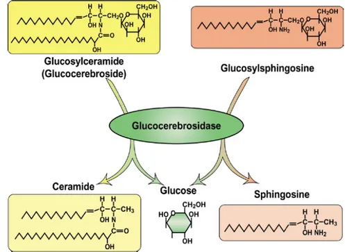
The disease results from the inherited autosomal recessive deficiency of the lysosomal enzyme β-glucocerebrosidase (EC.3.2.1.45), which cleaves the glycolipid glucocerebroside into glucose and ceramide (Fig. 1.12), and it leads to accumulation of glucocerebroside in the body, predominantly in the liver, spleen, and bone narrow. This disease is the most common of the sphingolipidoses and the most frequently inherited disorder among Ashkenazi Jews, with an incidence of about 1:60,000 in the general population, increasing to 1:1,000 in the Ashkenazi Jews (Meikle et al., 2007).
Although the enzyme deficiency exists in all cells of the body, accumulation of glucocerebroside within the lysosomes occurs only in macrophages, called Gaucher cells. Residual enzyme activity ranges from 5% to 25%. In a few cases, Gaucher disease is due to a mutation affecting the protein saposine C, whose presence is required to achieve optimal β-glucocerebrosidase activity. Accumulation of the substrate within macrophages leads to elevations in serum levels of IL-1 β, IL6, TNFα, IL10, and M-CSF (Guggenbuhl et al., 2008).
There are three forms of the disease, types I, II, and III, described as the non-neurophatic, acute neurophatic, and chronic neuropathic forms respectively (Ali et al., 2011). Type I is the most
28
common type and results in the aforementioned symptoms, with the enlargement of the spleen and liver occurring most often. Other symptoms include osteolytic lesions, anemia, and hepatic fibrosis. While type I Gaucher disease only affects 45,000-60,000 people worldwide, 1 in 850 Ashkenazy Jews are afflicted, and an approximated 1 in every 15 Ashkenazy Jews is a carrier of the disease. Type II is the rarest form of the disease and is characterised by rapid neurological deterioration. It usually has a very early onset and the afflicted person most often dies by the age of two. Type III Gaucher disease also results in neurological problems, but these problems tend to progress more slowly and more mildly than type II. Symptoms for type III are onset at varying points in the life of those afflicted (Bohra and Nair, 2011).
Gaucher disease was first observed in 1882 by Phillippe Gaucher, a 28 year old French doctor after whom the disease is named. Observing large cells during a splenic aspirate in a spleen, Gaucher thought it was a splenic neoplasm (Gaucher, 1882). Almost forty years later, in 1924, Epstein recognised the storage of glucocerebrosides (Epstein, 1924) and, later, in 1965, Dr. Roscoe Brady and his team described that this storage was due to a lack of the GCase enzyme (Brady et al., 1965).
Fig. 1.12: Glucocerebrosidase is a β-glucosidase, hydrolyzing its primary substrate, glucocerebroside, into glucose and ceramide. An alternative substrate, glucosylsphingosine, is also degraded into glucose and sphingosine (Sidransky, 2004).
29 β-Glucocerebrosidase
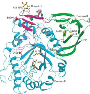
β-Glucocerebrosidase (also called acid β-glucosidase, D-glucosyl-N-acylsphingosine glucohydrolase, or GCase) is an enzyme with glucosylceramidase activity that is needed to cleave, by hydrolysis, the beta-glucosidic linkage of the chemical glucocerebroside, an intermediate in glycolipid metabolism. It is localised in the lysosome, it has a molecular weight of 59.7 kDa and the sequence is composed by 497 amino acid residues. The structure of β-Glucocerebrosidase was determined by X-ray crystallographic methods. The X-ray structure contains two β-Glucocerebrosidase molecules per asymmetric unit. Its overall fold comprises three domains (Fig. 1.13). Domain I (residues 1-27 and 383-414) consists of one main three-stranded, anti-parallel β-sheet that is flanked by a perpendicular amino-terminal strand and a loop. It contains two disulphide bridges (residues 4-16 and 18-23), which may be required for correct folding.
Glycosylation, which is essential for catalytic activity in vivo, is seen in the crystal structure at residue N19. Domain II (residues 30–75 and 431–497) consists of two closely associated β-sheets that form an independent domain, which looks like an immunoglobulin (Ig) fold. Domain III (residues 76–381 and 416–430) is a (β/α)8 TIM barrel, which contains the catalytic site. It contains
three free cysteines (at positions 126, 248 and 342). Domains II and III seem to be connected by a flexible hinge, whereas domain I tightly interacts with domain III.
Site-directed mutagenesis and homology modelling of β-Glucocerebrosidase suggest that E235 is the acid/base catalyst, and tandem mass spectrometry identified E340 as the nucleophile. These two residues are located near the carboxyl termini of strands 4 and 7 in domain III. Of the ~200 known β-Glucocerebrosidase mutations, many are rare and restricted to a few individuals. Most mutations either partially or completely abolish catalytic activity or are thought to reduce β-Glucocerebrosidase stability. The most common mutation, N370S, accounts for 70% of mutant alleles in Ashkenazi Jews and 25% in non-Jewish patients. N370S causes predisposition to type-1 disease and precludes neurological involvement, suggesting that it causes relatively minor changes in β-Glucocerebrosidase structure and, therefore, catalytic activity. Consistent with this is the localization of N370 to the longest α-helix (helix 7) in β-Glucocerebrosidase, which is located at the interface of domains II and III, but too far from the active site to participate directly in catalysis. Interestingly, several other mutations are found in this helix, all of which seem to point into the TIM barrel (Dvir et al., 2003).
30
Fig. 1.13: β-Glucocerebrosidase three-dimensional structure determined by X-ray crystallographic methods (Dvir et al., 2003).
Enzyme Replacement Therapy (ERT)
Gaucher disease has no cure, but treatments of the symptoms for types I and III of the disease have been proven to be successful. Type I Gaucher disease was the first lysosomal storage disorder for which ERT became available. In a clinical trial performed by Barton et al., 12 patients with the non-neuronopathic form (type I) of Gaucher disease received β-glucocerebrosidase that was derived from human placenta and treated with specific glycosidases to expose mannose residues in the oligosaccharide chains (in fact, this enzyme has to be taken up by macrophages via the mannose receptor, and not by the mannose-6-phosphate receptor) (Barton et al., 1991). Based on this trial, the enzyme preparation was approved for treatment of patients with Gaucher disease. Some years later, the placenta-derived β-glucocerebrosidase (alglucerase, Ceredase®, Genzyme, Cambridge, MA) was replaced by a recombinant form produced in CHO cells. Also, this enzyme preparation (imiglucerase, Cerezyme®, Genzyme, Cambridge, MA) needed to be modified for targeting mannose receptor sites on macrophages. In the last 10 years, countless publications and reports have confirmed the positive effects of imiglucerase and, because of its safety and efficacy profile, ERT has become the standard of care for type I Gaucher patients (Weinreb, 2008). Other enzymes utilized in ERT are taliglucerase alfa (Elelyso®, Protalix Biotherapeutics, Carmiel, Israel) or velaglucerase (VPRIV®, ShireHGT, CambridgeMA); all of them are administered intravenously. Treatment dose varies between 15 units/kg to 60 units/kg body weight (Pastores, 2010).
31
Another form of treatment is substrate reduction therapy (SRT), in which the production of glucocerebrosides is slowed by the substrate, reducing the production and accumulation of glycolipids (Zimran and Elstein, 2003). At present, the only licensed agent is miglustat (Zavesca®, Actelion Pharmaceuticals US, San Francisco, United States), but this option is generally used as a second-line agent in patients who are unsuitable for ERT, or because of individual patient preference. The drug, an iminosugar, has the advantage of being an oral agent. However, it has a higher frequency of unwanted effects, and may be less effective for several aspects of Gaucher disease than the available ERT.
The recombinant form of GCase supplied by Genzyme Co. is expensive, costing approximately €340,000 per year for an average adult of 70 kg.
This high cost limits the number of patients able to receive treatment. Recombinant GCase is also a difficult enzyme to synthesise biochemically, and human cells synthesize small amounts of the protein. Increased levels of production are inhibited by a protein called TCP80. Unfortunately, GCase and TCP80 analogues are found in all mammals (Xu et al., 1999). Alternative sources for this recombinant protein have already begun to be explored. One such alternative is protein expression plants. The production of human proteins in transgenic plants offers many economic and qualitative benefits over current forms of production. One of the advantages is the reduced health risk, as plants are unable to serve as hosts for human pathogens (Ni, 1997). Another advantage of plants is that proteins stored in seed can be stored for longer time and more easily than proteins stored in animal cells can. Transgenic CHO cells, for example, must be processed soon after harvest to prevent significant enzyme loss (Reggi et al., 2005). On the other hand, transgenic seeds have been shown that they can be stored for weeks (and months or even years at colder temperatures) without experiencing significant enzyme activity loss (Kusnadi et al., 1998). Besides that, TCP80 analogues have not been found in plants, which could enable higher protein yields due to the lack of GCase inhibition (Reggi et al., 2005). Creating recombinant forms of GCase in plants is an active field of research. A patent as recent as May 2011 was granted by the United Stated Patent and Trademark Office (USPTO) for the production of recombinant GCase. This patent (Nr. 7,951,557) by Shaaltiel et al. claims the production of a recombinant GCase in carrot cells. Other similarly based patents lay claims on recombinant forms of other therapeutic lysosomal enzymes, also in plants (Shaaltiel et al., 2012).